
Transperineal total mesorectal excision for rectal cancer ...
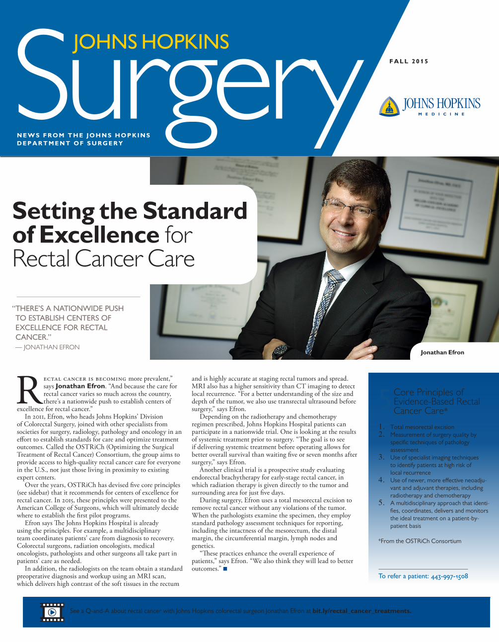
Rectal cancer is becoming more prevalent,” says Jonathan Efron. “And because the care for rectal cancer varies so much across the country, there’s a nationwide push to establish centers of
excellence for rectal cancer.”In 2011, Efron, who heads Johns Hopkins’ Division
of Colorectal Surgery, joined with other specialists from societies for surgery, radiology, pathology and oncology in an effort to establish standards for care and optimize treatment outcomes. Called the OSTRiCh (Optimizing the Surgical Treatment of Rectal Cancer) Consortium, the group aims to provide access to high-quality rectal cancer care for everyone in the U.S., not just those living in proximity to existing expert centers.
Over the years, OSTRiCh has devised five core principles (see sidebar) that it recommends for centers of excellence for rectal cancer. In 2015, these principles were presented to the American College of Surgeons, which will ultimately decide where to establish the first pilot programs.
Efron says The Johns Hopkins Hospital is already using the principles. For example, a multidisciplinary team coordinates patients’ care from diagnosis to recovery. Colorectal surgeons, radiation oncologists, medical oncologists, pathologists and other surgeons all take part in patients’ care as needed.
In addition, the radiologists on the team obtain a standard preoperative diagnosis and workup using an MRI scan, which delivers high contrast of the soft tissues in the rectum
and is highly accurate at staging rectal tumors and spread. MRI also has a higher sensitivity than CT imaging to detect local recurrence. “For a better understanding of the size and depth of the tumor, we also use transrectal ultrasound before surgery,” says Efron.
Depending on the radiotherapy and chemotherapy regimen prescribed, Johns Hopkins Hospital patients can participate in a nationwide trial. One is looking at the results of systemic treatment prior to surgery. “The goal is to see if delivering systemic treatment before operating allows for better overall survival than waiting five or seven months after surgery,” says Efron.
Another clinical trial is a prospective study evaluating endorectal brachytherapy for early-stage rectal cancer, in which radiation therapy is given directly to the tumor and surrounding area for just five days.
During surgery, Efron uses a total mesorectal excision to remove rectal cancer without any violations of the tumor. When the pathologists examine the specimen, they employ standard pathology assessment techniques for reporting, including the intactness of the mesorectum, the distal margin, the circumferential margin, lymph nodes and genetics.
“These practices enhance the overall experience of patients,” says Efron. “We also think they will lead to better outcomes.” n
F A L L 2 0 1 5
N E W S F R O M T H E J O H N S H O P K I N S D E P A R T M E N T O F S U R G E R Y
SurgeryJOHNS HOPKINS
“ THERE’S A NATIONWIDE PUSH TO ESTABLISH CENTERS OF EXCELLENCE FOR RECTAL CANCER.” — JONATHAN EFRON
See a Q-and-A about rectal cancer with Johns Hopkins colorectal surgeon Jonathan Efron at bit.ly/rectal_cancer_treatments.
Setting the Standard of Excellence for Rectal Cancer Care
Jonathan Efron
Core Principles of Evidence-Based Rectal Cancer Care*
1. Total mesorectal excision2. Measurement of surgery quality by
specific techniques of pathology assessment
3. Use of specialist imaging techniques to identify patients at high risk of local recurrence
4. Use of newer, more effective neoadju-vant and adjuvant therapies, including radiotherapy and chemotherapy
5. A multidisciplinary approach that identi-fies, coordinates, delivers and monitors the ideal treatment on a patient-by-patient basis
*From the OSTRiCh Consortium
To refer a patient: 443-997-1508
5
B R E A S T S U R G I C A L O N C O L O G Y
Managing the Axilla to Prevent Lymphedema
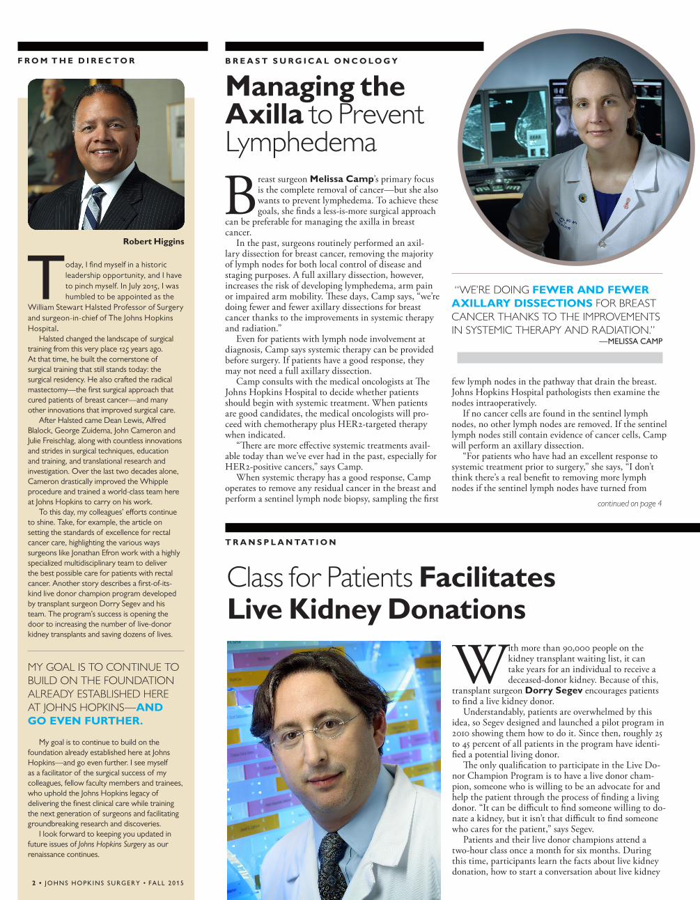
Breast surgeon Melissa Camp’s primary focus is the complete removal of cancer—but she also wants to prevent lymphedema. To achieve these goals, she finds a less-is-more surgical approach
can be preferable for managing the axilla in breast cancer.
In the past, surgeons routinely performed an axil-lary dissection for breast cancer, removing the majority of lymph nodes for both local control of disease and staging purposes. A full axillary dissection, however, increases the risk of developing lymphedema, arm pain or impaired arm mobility. These days, Camp says, “we’re doing fewer and fewer axillary dissections for breast cancer thanks to the improvements in systemic therapy and radiation.”
Even for patients with lymph node involvement at diagnosis, Camp says systemic therapy can be provided before surgery. If patients have a good response, they may not need a full axillary dissection.
Camp consults with the medical oncologists at The Johns Hopkins Hospital to decide whether patients should begin with systemic treatment. When patients are good candidates, the medical oncologists will pro-ceed with chemotherapy plus HER2-targeted therapy when indicated.
“There are more effective systemic treatments avail-able today than we’ve ever had in the past, especially for HER2-positive cancers,” says Camp.
When systemic therapy has a good response, Camp operates to remove any residual cancer in the breast and perform a sentinel lymph node biopsy, sampling the first
few lymph nodes in the pathway that drain the breast. Johns Hopkins Hospital pathologists then examine the nodes intraoperatively.
If no cancer cells are found in the sentinel lymph nodes, no other lymph nodes are removed. If the sentinel lymph nodes still contain evidence of cancer cells, Camp will perform an axillary dissection.
“For patients who have had an excellent response to systemic treatment prior to surgery,” she says, “I don’t think there’s a real benefit to removing more lymph nodes if the sentinel lymph nodes have turned from
With more than 90,000 people on the kidney transplant waiting list, it can take years for an individual to receive a deceased-donor kidney. Because of this,
transplant surgeon Dorry Segev encourages patients to find a live kidney donor.
Understandably, patients are overwhelmed by this idea, so Segev designed and launched a pilot program in 2010 showing them how to do it. Since then, roughly 25 to 45 percent of all patients in the program have identi-fied a potential living donor.
The only qualification to participate in the Live Do-nor Champion Program is to have a live donor cham-pion, someone who is willing to be an advocate for and help the patient through the process of finding a living donor. “It can be difficult to find someone willing to do-nate a kidney, but it isn’t that difficult to find someone who cares for the patient,” says Segev.
Patients and their live donor champions attend a two-hour class once a month for six months. During this time, participants learn the facts about live kidney donation, how to start a conversation about live kidney
T R A N S P L A N TAT I O N
“ THIS GROUP DOESN’T DO AS WELL WITH THE SAME PROCEDURES WE OFFER EVERYONE FOR HERNIAS. EVEN THOUGH THEY MAKE SCAR TISSUE THAT LOOKS GOOD, IT DOESN’T HOLD VERY WELL.”
—DAVID EFRON
F R O M T H E D I R E C TO R
Class for Patients Facilitates Live Kidney Donations
continued on page 4
“WE’RE DOING FEWER AND FEWER AXILLARY DISSECTIONS FOR BREAST CANCER THANKS TO THE IMPROVEMENTS IN SYSTEMIC THERAPY AND RADIATION.”
—MELISSA CAMP
2 • JOHNS HOPKINS SURGERY • FALL 2015
Today, I find myself in a historic leadership opportunity, and I have to pinch myself. In July 2015, I was humbled to be appointed as the
William Stewart Halsted Professor of Surgery and surgeon-in-chief of The Johns Hopkins Hospital.
Halsted changed the landscape of surgical training from this very place 125 years ago. At that time, he built the cornerstone of surgical training that still stands today: the surgical residency. He also crafted the radical mastectomy—the first surgical approach that cured patients of breast cancer—and many other innovations that improved surgical care.
After Halsted came Dean Lewis, Alfred Blalock, George Zuidema, John Cameron and Julie Freischlag, along with countless innovations and strides in surgical techniques, education and training, and translational research and investigation. Over the last two decades alone, Cameron drastically improved the Whipple procedure and trained a world-class team here at Johns Hopkins to carry on his work.
To this day, my colleagues’ efforts continue to shine. Take, for example, the article on setting the standards of excellence for rectal cancer care, highlighting the various ways surgeons like Jonathan Efron work with a highly specialized multidisciplinary team to deliver the best possible care for patients with rectal cancer. Another story describes a first-of-its-kind live donor champion program developed by transplant surgeon Dorry Segev and his team. The program’s success is opening the door to increasing the number of live-donor kidney transplants and saving dozens of lives.
MY GOAL IS TO CONTINUE TO BUILD ON THE FOUNDATION ALREADY ESTABLISHED HERE AT JOHNS HOPKINS—AND GO EVEN FURTHER.
My goal is to continue to build on the foundation already established here at Johns Hopkins—and go even further. I see myself as a facilitator of the surgical success of my colleagues, fellow faculty members and trainees, who uphold the Johns Hopkins legacy of delivering the finest clinical care while training the next generation of surgeons and facilitating groundbreaking research and discoveries.
I look forward to keeping you updated in future issues of Johns Hopkins Surgery as our renaissance continues.
Robert Higgins
E N D O C R I N E S U R G E R Y
T R A N S P L A N TAT I O N
ROUGHLY 25 TO 45 PERCENT OF ALL PATIENTS IN THE PROGRAM HAVE IDENTIFIED A POTENTIAL LIVING DONOR.
donation, and how to identify and access their social networks.
Betsy King, an assistant resident in surgery and man-ager of the classes, says the champions are ready to go out and start a conversation with people about live donation as soon as the second class is over. “It’s amazing,” she says. “They feel confident to go forth and start talking right away.”
King also says participants are often surprised when they discover all of the people in their social network. “They all brainstorm out loud, listing the groups they know in their community, like their church and volunteer organizations,” she says. “They get ideas from one another and find that their network is larger than they think.”
Segev and King hope to adapt the program for liver transplant patients and expand the program to other transplant centers around the country. n
To refer a patient: 410-955-5045
An App for That One of the most valuable tools par-ticipants get from the Live Donor Champion Program is their own per-sonal stories. Patients write about themselves, how their life has been affected and why they want a trans-plant. Their champions can then print the stories as handouts, email the stories or make a video of the patient reading the story.
To make the process of writing their stories easier, Johns Hopkins provides a Facebook app to the Live Donor Champion Program participants. Created with liver transplant surgeon Andrew Cameron, the app asks for answers to certain questions and then generates stories in a format that can be posted on Facebook, with links to information about live donation.
Twenty years ago, physicians discovered neuroendocrine tumors when there was a telltale symptom of hormone secretion, like ulcers in Zollinger-Ellison syndrome
or low blood sugar from insulinomas. Today, most patients diagnosed with neuroendocrine tumors are asymptomatic; their tumors are discovered incidentally through CT imaging for something unrelated.
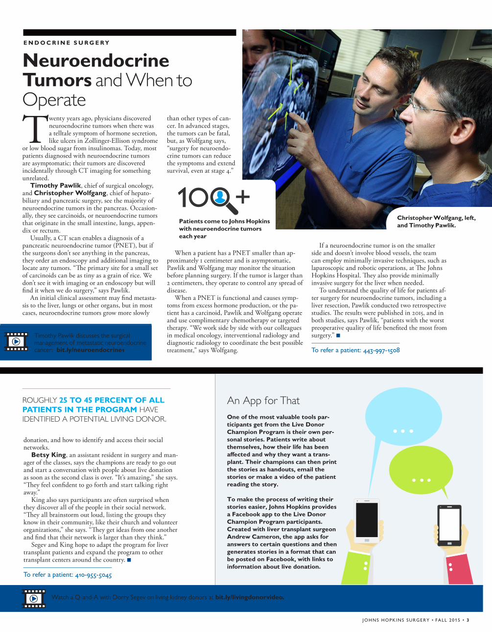
Timothy Pawlik, chief of surgical oncology, and Christopher Wolfgang, chief of hepato-biliary and pancreatic surgery, see the majority of neuroendocrine tumors in the pancreas. Occasion-ally, they see carcinoids, or neuroendocrine tumors that originate in the small intestine, lungs, appen-dix or rectum.
Usually, a CT scan enables a diagnosis of a pancreatic neuroendocrine tumor (PNET), but if the surgeons don’t see anything in the pancreas, they order an endoscopy and additional imaging to locate any tumors. “The primary site for a small set of carcinoids can be as tiny as a grain of rice. We don’t see it with imaging or an endoscopy but will find it when we do surgery,” says Pawlik.
An initial clinical assessment may find metasta-sis to the liver, lungs or other organs, but in most cases, neuroendocrine tumors grow more slowly
than other types of can-cer. In advanced stages, the tumors can be fatal, but, as Wolfgang says, “surgery for neuroendo-crine tumors can reduce the symptoms and extend survival, even at stage 4.”
When a patient has a PNET smaller than ap-proximately 1 centimeter and is asymptomatic, Pawlik and Wolfgang may monitor the situation before planning surgery. If the tumor is larger than 2 centimeters, they operate to control any spread of disease.
When a PNET is functional and causes symp-toms from excess hormone production, or the pa-tient has a carcinoid, Pawlik and Wolfgang operate and use complimentary chemotherapy or targeted therapy. “We work side by side with our colleagues in medical oncology, interventional radiology and diagnostic radiology to coordinate the best possible treatment,” says Wolfgang.
If a neuroendocrine tumor is on the smaller side and doesn’t involve blood vessels, the team can employ minimally invasive techniques, such as laparoscopic and robotic operations, at The Johns Hopkins Hospital. They also provide minimally invasive surgery for the liver when needed.
To understand the quality of life for patients af-ter surgery for neuroendocrine tumors, including a liver resection, Pawlik conducted two retrospective studies. The results were published in 2015, and in both studies, says Pawlik, “patients with the worst preoperative quality of life benefited the most from surgery.” n
To refer a patient: 443-997-1508
Neuroendocrine Tumors and When to Operate
JOHNS HOPKINS SURGERY • FALL 2015 • 3
Watch a Q-and-A with Dorry Segev on living kidney donors at bit.ly/livingdonorvideo.
Timothy Pawlik discusses the surgical management of metastatic neuroendocrine cancer: bit.ly/neuroendocrine1
Patients come to Johns Hopkins with neuroendocrine tumors each year
1 +Christopher Wolfgang, left, and Timothy Pawlik.
positive to negative. If there’s still residual disease present, then the patient should have additional nodes removed.”
After surgery, the final pathology results help Camp decide if the patient will need additional treat-ment with radiation. If so, she consults with radia-tion oncologists to create a plan. In select patients, radiation to the axilla can be as effective for local control as axillary dissection but with less risk of lymphedema.
“Management of the axilla in breast cancer is moving away from a surgery-focused approach of axillary dissection in all patients with lymph node involvement and toward a multidisciplinary approach involving a combination of systemic therapy, surgery and radiation,” she says. “In appropriately selected patients, this approach does not compromise local control and results in a lower risk of lymphedema.” n
To refer a patient: 443-997-8282
Managing the Axilla to Prevent Lymphedema (continued from page 2)
ICD-10 Is Here
When referring patients to Johns Hopkins Medicine, please be sure to include ICD-10 codes.
For more information, visit www.cms.gov/Medicare/Coding/ICD10/.
Surgery
Johns Hopkins Medicine901 S. Bond St., Suite 550Baltimore, Maryland 21231
This newsletter is published for the Department of Surgery by Johns Hopkins Medicine Marketing and Communications.
Department of SurgeryRobert Higgins, director of the Department of Surgery
Marketing and CommunicationsDalal Haldeman, Ph.D., M.B.A., senior vice presidentLisa Rademakers, writer and editorKristen Caudill, designerKeith Weller, photographer
For questions or comments, [email protected] or 443-287-2527.
© 2015 The Johns Hopkins University and The Johns Hopkins Health System Corporation.
For referrals or appointments to the Department of Surgery, call 443-997-1508.
Surgery
2Setting the Standard of Excellence for Rectal Cancer Care
1
Insid
e
N E W S F R O M T H E J O H N S H O P K I N S D E P A R T M E N T O F S U R G E R Y
JOHNS HOPKINS
JOHNS HOPKINS
Non-Profit Org.U.S. Postage
PAIDPermit No. 5415Baltimore, MD
Managing the Axilla to Prevent Lymphedema
Johns Hopkins Surgery Referral Line Call 443-997-1508 to refer a patient.Easy access and quick appointments for your patients at these convenient locations: Green Spring StationWhite MarshJohns Hopkins Bayview Medical CenterThe Johns Hopkins Hospital Our schedulers are available to assist you Monday through Friday, 8 a.m. to 5 p.m. For after-hours or emergency transfers, please use the Hopkins Access Line at 410-955-9444.
3
F A L L 2 0 1 5
Neuroendocrine Tumors and When to Operate
Explore Our New Online Resource for Physicians: Clinical Connection
Connect with Johns Hopkins health care professionals about the latest clinical innovations and advances in patient
care. Access videos, articles, news, clinical trials and much more.
Scan the QR code or visit www.hopkinsmedicine.org/clinicalconnection.


