SURGERY FOR LUNG CANCER - ERS-education
85
Transcript of SURGERY FOR LUNG CANCER - ERS-education


SURGERY FOR LUNG CANCER
Prof. Lorenzo Spaggiari
University of Milan
European Institute of Oncology
FACULTY DISCLOSURE
No financial interest in, or arrangement with, a company whose
products or services are discussed in the lecture;
No other financial connections, direct or indirect, or other situations that
might raise the question of bias in the work reported or the
conclusions, implications, or opinions stated – including pertinent
commercial or other sources of funding for the individual Speaker or
for the associated department or organization, personal
relationships, or direct academic competition.

INTRODUCTION
• Lung cancer remains as the n° 1 cancer killer in Europe and United States
• Survival is directly related to stage at diagnosis
• Even patients with early-stage lung cancer have recurrence rates about
30%-40%, with a 5-year survival ranging dramatically from 50% to 90%,
due to occult disease and inadequate nodal staging;
• Node-positive disease lowers 5-year survival to 27%.
Pathologic nodal stage is the strongest predictor
of long-term survival in surgical NSCLC
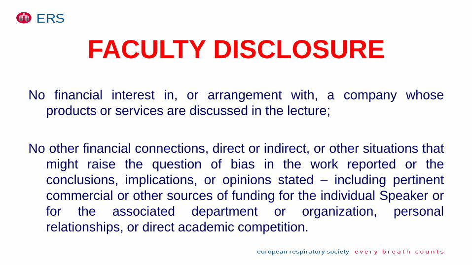
Imaging CT scan and PET FDG
LYMPH NODE STAGING PATHWAYS
Diagnosis and Management of Lung Cancer, ACCP guidelines 2013
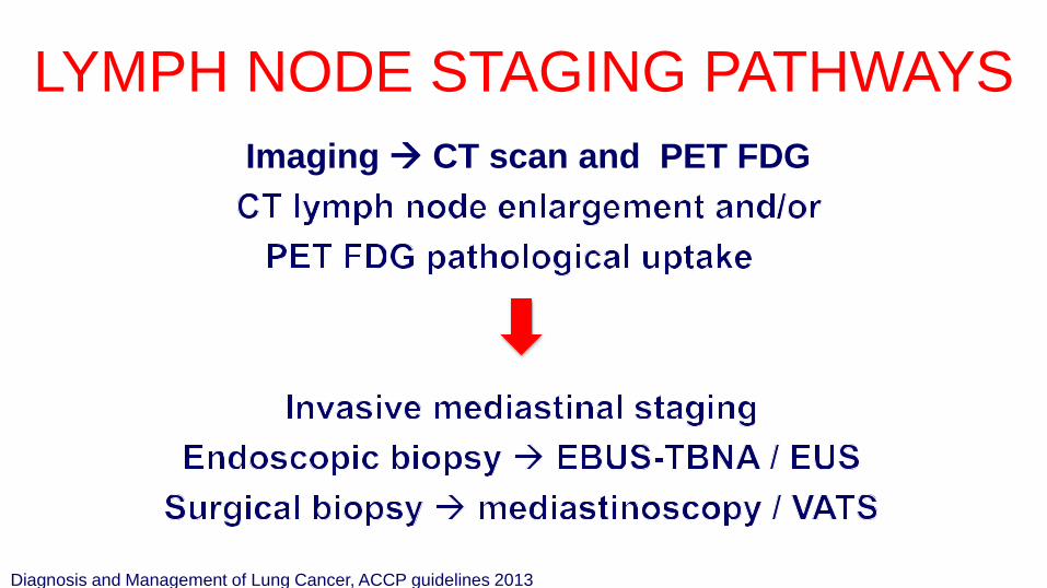
INTRODUCTIONESTS guidelines, 2014
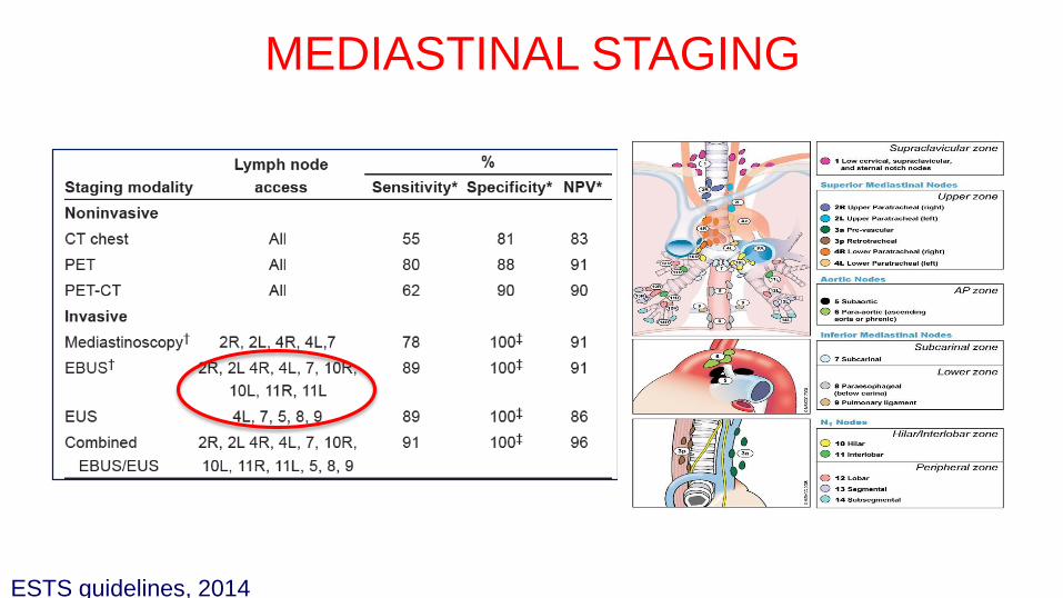
MEDIASTINAL STAGING
ESTS guidelines, 2014

58 6883
6346
10373
5778
6085 77
6179
20 12 14 78
256
323
376393
0
50
100
150
200
250
300
350
400
450
1998 1999 2000 2001 2002 2003 2004 2005 2006 2007 2008 2009 2010 2011 2012 2013 2014 2015
MED vs EBUS at European Institue of Oncology
Med EBUS
MEDIASTINAL STAGING

EBUS - TECHNIQUE
Outpatient setting
Moderate sedation
Extended hilar stations
«All in ONE» procedure
No complications
High diagnostic rate
First choice for
invasive
mediastinal
staging!

ENDOBRONCHIAL ULTRASOUND
Clinical Review. Kinsey et al, AJRCCM 2014

IEO EXPERIENCE
1407EBUS-TBNA From 2011 to 2015
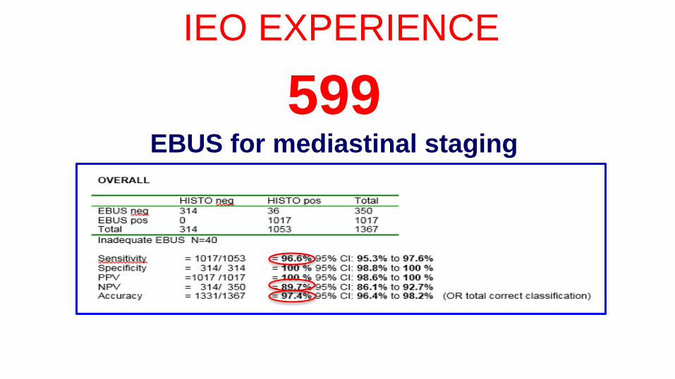
IEO EXPERIENCE
1407EBUS-TBNA From 2011 to 2015
599EBUS for mediastinal staging

JTO 2014
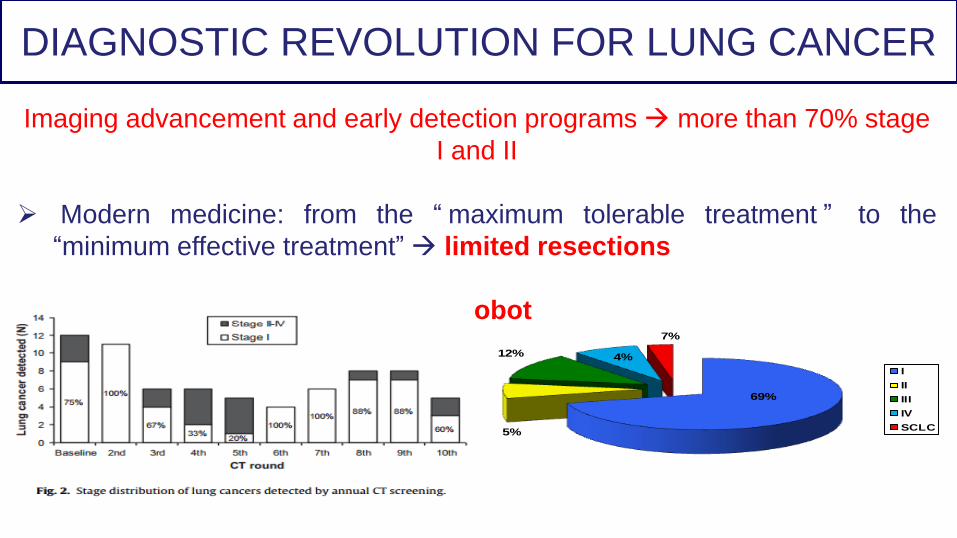
DIAGNOSTIC REVOLUTION FOR LUNG CANCER
Imaging advancement and early detection programs more than 70% stage
I and II
Modern medicine: from the “ maximum tolerable treatment ” to the
“minimum effective treatment” limited resections
Less invasive treatment VATS/Robot
69%
5%
12% 4%
7%
I
II
III
IV
SCLC
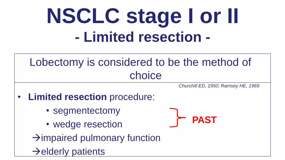
Lobectomy is considered to be the method of
choiceChurchill ED, 1950; Ramsey HE, 1969
• Limited resection procedure:
• segmentectomy
• wedge resection
impaired pulmonary function
elderly patients
NSCLC stage I or II- Limited resection -
PAST

cT1N0 peripheral NSCLC ≤ 3 cm
1982-1988
Locoregional recurrence rate -> threefold increase with wedge
(17% vs 6%) -> p=0.008
Overall death rate -> 30% increase in limited resection group -> p=0.088
statistically significant survival benefit to lobectomy (p = 0.088)
PitfallsNo modern pre-operative staging, tumor size up to 3 cm, lymph node sampling
Ann Thorac Surg 1995
Wedge/Segmentectomy
n°122Lobectomy
n°125
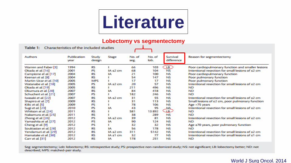
LiteratureLobectomy vs segmentectomy
World J Surg Oncol. 2014

347 patients who underwent lobectomy (n=294) or sublobar resection (n=53)
for non–small cell lung cancer manifesting as a solid nodule, from 1993 to 2011
J Thorac Cardiovasc Surg 2013
nodule diameter of ≤20 mm
Propensity scoring was performed
using the same covariates

Fox N and Bauer T. Oncology Issue, 2008
INCLUSION CRITERIA EXCLUSION CRITERIA
Single tumor ≤2 cm Double cancers (<5yrs)
Suspected NSCLC cN0 Prior CT/RT
Peripheral Locally advanced or
metastatic disease
Performance status 0 -2 Age: < 18 yrs old
1297 pts
AIM: to evaluate the “non inferiority” in overall survival of segmentectomy
compared to lobectomy in peripheral Stage IA NSCLC ≤ 2 cm
LobectomyWedge/
segmentectomy
RANDOMIZATION
Frozen section -> pNSCLC
N1 and N2 sampling
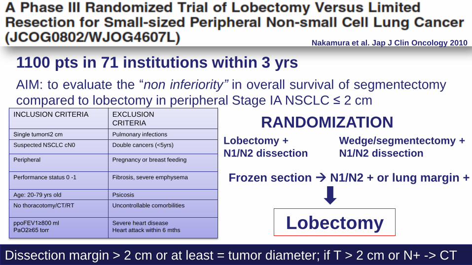
Nakamura et al. Jap J Clin Oncology 2010
AIM: to evaluate the “non inferiority” in overall survival of segmentectomy
compared to lobectomy in peripheral Stage IA NSCLC ≤ 2 cmINCLUSION CRITERIA EXCLUSION
CRITERIA
Single tumor≤2 cm Pulmonary infections
Suspected NSCLC cN0 Double cancers (<5yrs)
Peripheral Pregnancy or breast feeding
Performance status 0 -1 Fibrosis, severe emphysema
Age: 20-79 yrs old Psicosis
No thoracotomy/CT/RT Uncontrollable comorbilities
ppoFEV1≥800 ml
PaO2≥65 torr
Severe heart disease
Heart attack within 6 mths
1100 pts in 71 institutions within 3 yrs
Lobectomy +
N1/N2 dissection
Wedge/segmentectomy +
N1/N2 dissection
Dissection margin > 2 cm or at least = tumor diameter; if T > 2 cm or N+ -> CT
RANDOMIZATION
Frozen section N1/N2 + or lung margin +
Lobectomy

INDICATIONS
Benign disease not amenable of wedge
Malignant diseaseNSCLC Stage I less 2 cmcN0Limited respiratory functionelderly
CONTRAINDICATIONSLocally advancedDouble tumorsTechnical DifficultiesPreop CT and/or RTNo one-lung ventilation
CONCLUSION- Limited resection -

Posterolateral
Thoracotomy
Lateral Muscle Sparing
Thoracotomy
NSCLC stage I or II- Minimally invasive approach-
VATS ROBOT

“We define VATS pulmonary resection as a video assisted,
minimally access approach in which the surgeon operates
primarily by watching the television monitor and uses
no rib spreading throughout the entire procedure” Yim AP. Pearson, 2008
Full Endoscopic Procedure (Monitor-based)
Individual Dissection & Stapling of Hilar Structures
No Rib Spreading
VATS pulmonary resection

Benefits of VATS:
- Reduce in postoperative pain
- Rate of postoperative complications
- Better preserved respiratory functions
- Reduction of lenght of in-hospital stay
- Fastern return to previous activity level
VATS = Standard approach for early stage lung
cancer in USA

In a matched analysis of 1195 patients in each treatment category, no
statistical differences in 3 year overall survival, DFS, or cancer specific
survival (OS: 70.6% v 68.1%, P=0.55;DFS: 86.2% v 85.4%, P=0.46; cancer
specific survival: 92% v 89.5%, P=0.05).
BMJ 2014
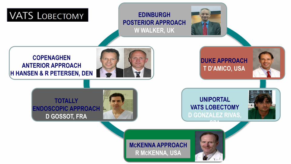
EDINBURGH
POSTERIOR APPROACH
W WALKER, UK
TOTALLY
ENDOSCOPIC APPROACH
D GOSSOT, FRA
MCKENNA APPROACH
R MCKENNA, USA
UNIPORTAL
VATS LOBECTOMY
D GONZALEZ RIVAS,
SPA
DUKE APPROACH
T D’AMICO, USA
COPENAGHEN
ANTERIOR APPROACH
H HANSEN & R PETERSEN, DEN
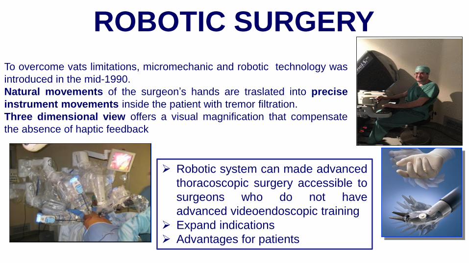
Robotic system can made advanced
thoracoscopic surgery accessible to
surgeons who do not have
advanced videoendoscopic training
Expand indications
Advantages for patients
To overcome vats limitations, micromechanic and robotic technology was
introduced in the mid-1990.
Natural movements of the surgeon’s hands are traslated into precise
instrument movements inside the patient with tremor filtration.
Three dimensional view offers a visual magnification that compensate
the absence of haptic feedback
ROBOTIC SURGERY

CPRL – Complete port robotic lobectomy
CPRS – Complete port robotic segmentectomy
RAL – Robotic assisted lobectomy
ROBOTIC LOBECTOMIES- Literature-
Lead Author Year Pts OT LOS Compl. Mortality Conversion
(min) (Days) (%) (%) (%)
RAL
Melfi 2004 107 220 5 na 1 na
Park 2006 30 218 4.5 26 0 12
Gharagozloo 2009 100 216 4 21 3 13
IEO 2010 54 224 4.5 20 0 9.4
Park, IEO, Pisa 2011 325 210 5 25 na 8
IEO 2012 91 213 5 20 0 10
CPRL / CPRS
Dylewski 2011 165 / 35 90 3 26 0 1.5
Cerfolio 2011 106 /16 132 2 27 0 10

ROBOTIC LOBECTOMY
- IEO tecnique -
• Lateral position• Robot at the head posteriorly• Four incisions including a
small utility incision• Camera arm: VII space mid
axillary line• No rib spreading • Individual ligation of hilar
elements
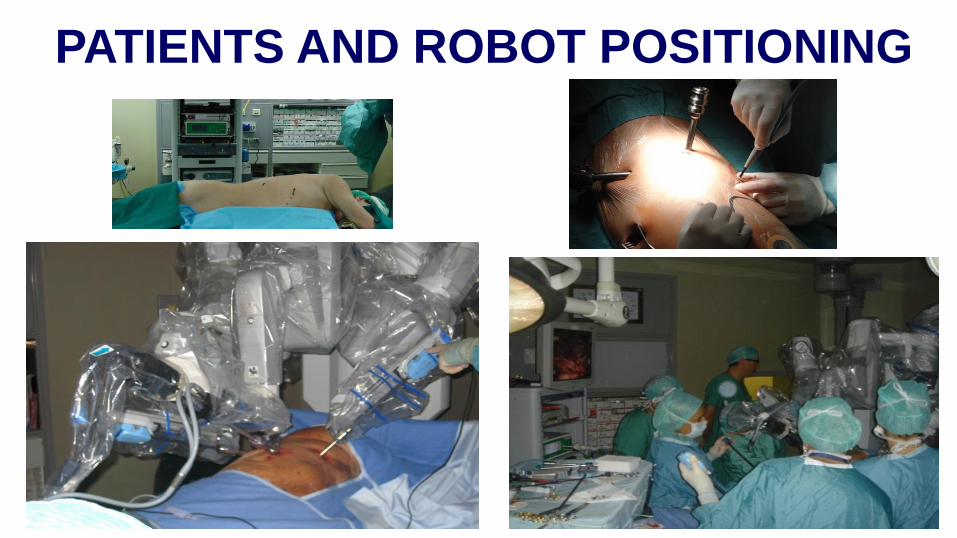
PATIENTS AND ROBOT POSITIONING
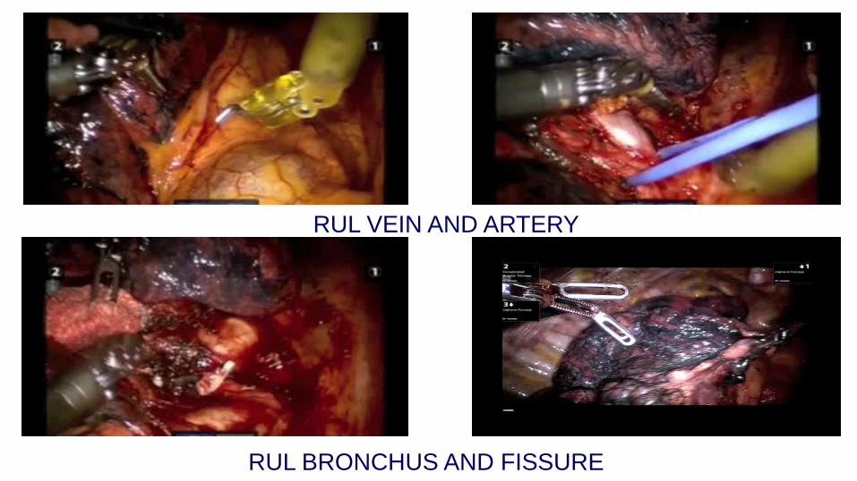
RUL VEIN AND ARTERY
RUL BRONCHUS AND FISSURE

LYMPHADENECTOMY
right left

JTCVS 2010
LEARNING CURVE - SAFETY - RADICALITY
1) Learning curve include 18 pts, complications, postoperative days and operative time
declines with experience
2) Postoperative stay was SHORTEN after robotic than open procedures
3) Complications and N° lymph nodes removed were comparable in open and robotic
lobectomies
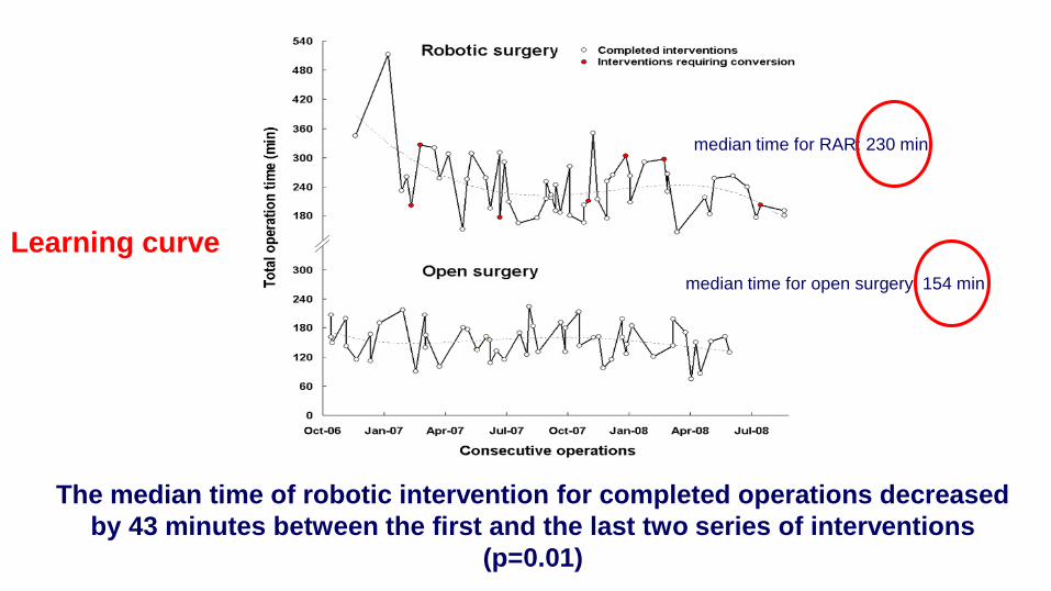
The median time of robotic intervention for completed operations decreased
by 43 minutes between the first and the last two series of interventions
(p=0.01)
median time for RAR: 230 min
median time for open surgery: 154 min
Learning curve

No difference in lymph node dissection
J Thorac Cardiovasc Surg, 2011
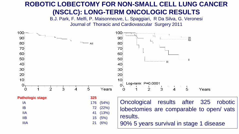
ROBOTIC LOBECTOMY FOR NON-SMALL CELL LUNG CANCER
(NSCLC): LONG-TERM ONCOLOGIC RESULTSB.J. Park, F. Melfi, P. Maisonneuve, L. Spaggiari, R Da Silva, G. Veronesi
Journal of Thoracic and Cardiovascular Surgery 2011
Pathologic stage
IA
IB
IIA
IIB
IIIA
325
176
72
41
15
21
(54%)
(22%)
(13%)
(5%)
(6%)
Oncological results after 325 robotic
lobectomies are comparable to open/ vats
results.
90% 5 years survival in stage 1 disease

ROBOT vs VATS
ADVANTAGES
1. Intuitive movements
2. Tremor filtration
3. Increased degrees of freedom
4. Motion scaling
5. Stereoscopic vision
6. Stable camera platform
7. Equivalence between the dominant and
non-dominant hands
8. Motion analysis
9. Eye-hand-target alignment
10. Possibly shorter learning curve
DISADVANTAGES
1. Costs
2. Loss of tactile feedback
3. Limited instrumentation available
4. Significant system set-up time
5. Need of at least one experienced
assistant
6. Possible delayed response by the
surgeon in case of catastrophic event

257 patients with early stagesprimary lungmalignancies
2007-2013
172 Robotics
26 cases/yy (single surgeon)
2010-2014
85 Vats
22 cases/yy (single surgeon)
Two surgeons :
- Same age
- Similar experience in standard-open thoracic surgery
ROBOT vs VATS- IEO experience -

4 ARMS ROBOTIC ASSISTED LOBECTOMY
(PARK-MELFI MODIFIED TECHNIQUE)
3
2
Camera
1
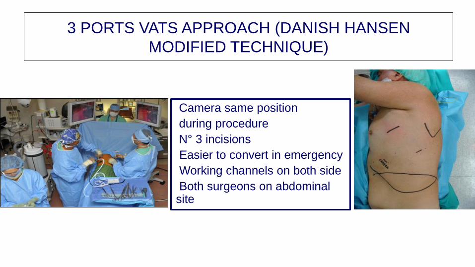
VATS APPROACH
Camera same position
during procedure
N° 3 incisions
Easier to convert in emergency
Working channels on both side
Both surgeons on abdominal site
3 PORTS VATS APPROACH (DANISH HANSEN
MODIFIED TECHNIQUE)

PATIENTS CHARACTERISTICSCharacteristics Robotic surgery VATS Pvalue
Total 172 85
Age
<60 58 17
60-69 85 34
70+ 29 34 0.0002
Median age, (range) 64 (39-79) 67 (41-82) 0.001
FEV (%)
Median (range) 93 (49-149) 98 (51-147) 0.53
Side
Left 73 26
Right 98 59 0.08
Lobe
Superior 103 55
Medial 12 8
Inferior 56 22 0.48
Diameter
<10mm 36 2
10-19mm 76 29
20-29mm 34 21
≥30mm 25 31 <0.0001
Median, mm (range) 15 (2-80) 25 (4-75) <0.0001
pT
pT0-1 112 31
pT2 48 42
pT3-4 8 6 0.0002
pN
pN0 142 64
pN1 13 13
pN2 13 8 0.14

Robotic surgery
-
60
120
180
240
300
360
420
480
Nov-
06
May-
07
Nov-
07
May-
08
Nov-
08
May-
09
Nov-
09
May-
10
Nov-
10
May-
11
Nov-
11
May-
12
Nov-
12
May-
13
Nov-
13
To
tal
op
erati
ng
tim
e (
min
)
VATS
-
60
120
180
240
300
360
420
Nov-
06
May-
07
Nov-
07
May-
08
Nov-
08
May-
09
Nov-
09
May-
10
Nov-
10
May-
11
Nov-
11
May-
12
Nov-
12
May-
13
Nov-
13
To
tal
op
erati
ng
tim
e (
min
)
DURATION OF SURGERY: VATS VERSUS
ROBOTICS AND CONVERSIONS (IN RED)
Mean time: 199 min Mean time: 181 min
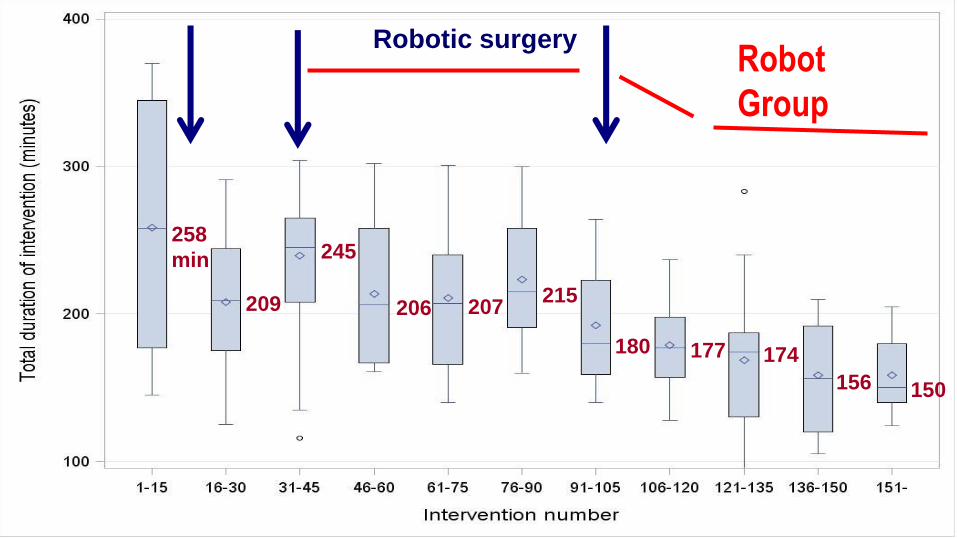
Robotic surgery
258
min
209
245
206 207215
180 177 174
156 150
Robot
Group
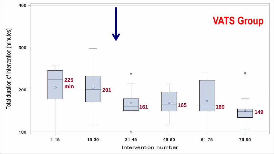
VATS
225
min201
161 165 160149
VATS Group
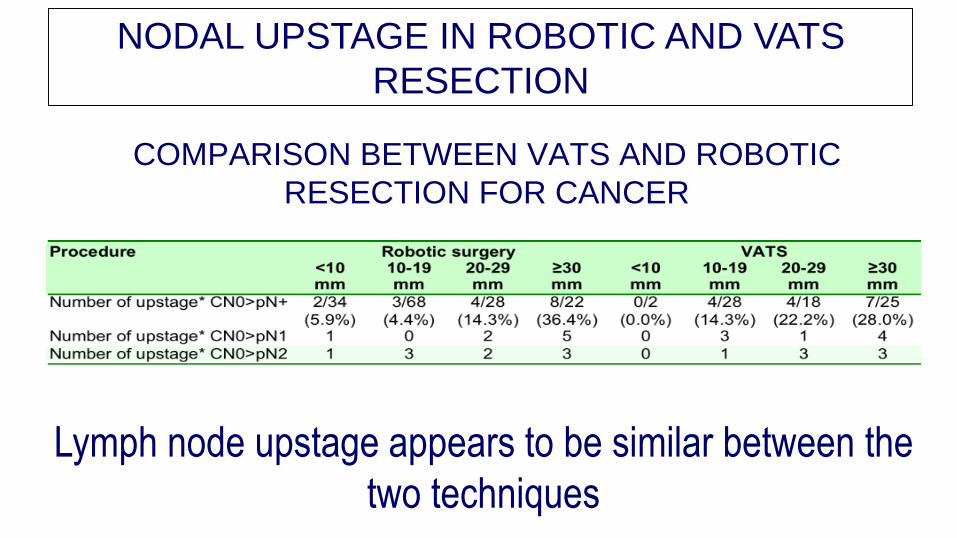
COMPARISON BETWEEN VATS AND ROBOTIC
RESECTION FOR CANCER
Lymph node upstage appears to be similar between the
two techniques
NODAL UPSTAGE IN ROBOTIC AND VATS
RESECTION
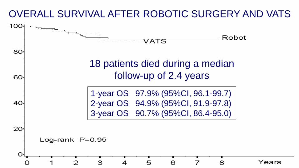
1-year OS 97.9% (95%CI, 96.1-99.7)
2-year OS 94.9% (95%CI, 91.9-97.8)
3-year OS 90.7% (95%CI, 86.4-95.0)
18 patients died during a median
follow-up of 2.4 years
OVERALL SURVIVAL AFTER ROBOTIC SURGERY AND VATS
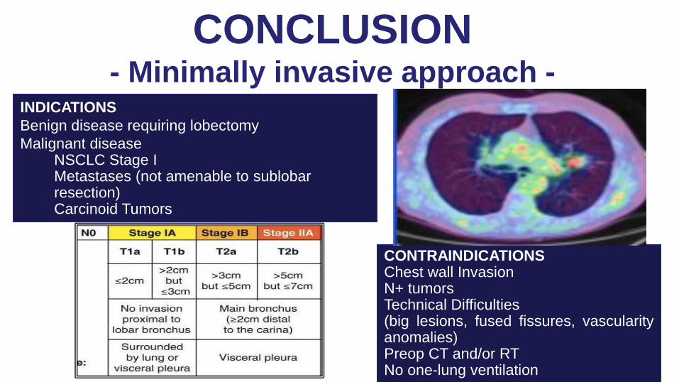
INDICATIONS
Benign disease requiring lobectomy
Malignant diseaseNSCLC Stage I Metastases (not amenable to sublobarresection)Carcinoid Tumors
CONTRAINDICATIONSChest wall InvasionN+ tumorsTechnical Difficulties(big lesions, fused fissures, vascularityanomalies)Preop CT and/or RTNo one-lung ventilation
CONCLUSION- Minimally invasive approach -

IEO ALGORITHM
SCREENING PROGRAM
Limited resection
Minimallyinvasive surgery
Ebus TBNApN0

WHAT IS THE FUTURE?
116 patients with histologically proven clinical stage I NSCLC who were treated with sublobar
resection (SLR; n = 42), radiofrequency ablation (RFA; n = 25) or radiotherapy (RT; n =49)
between 2009 and 2013
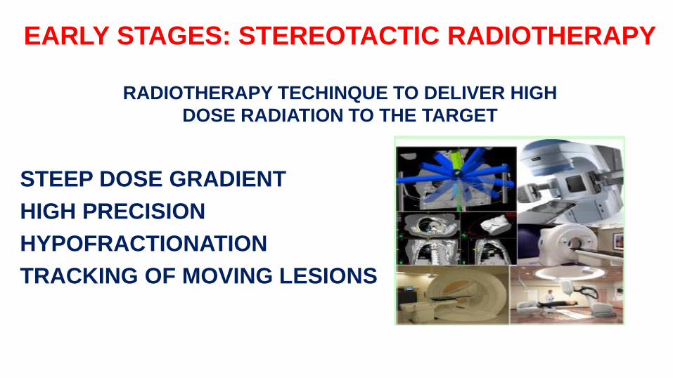
EARLY STAGES: STEREOTACTIC RADIOTHERAPY
STEEP DOSE GRADIENT
HIGH PRECISION
HYPOFRACTIONATION
TRACKING OF MOVING LESIONS
RADIOTHERAPY TECHINQUE TO DELIVER HIGH
DOSE RADIATION TO THE TARGET

Local Control: 80-98% 2yOS: 50-80%
MEDIAN FUP: 10-50 m
Nagata IJROBP 2011
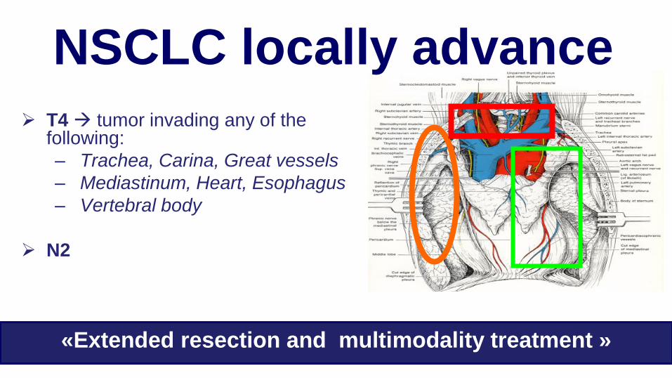
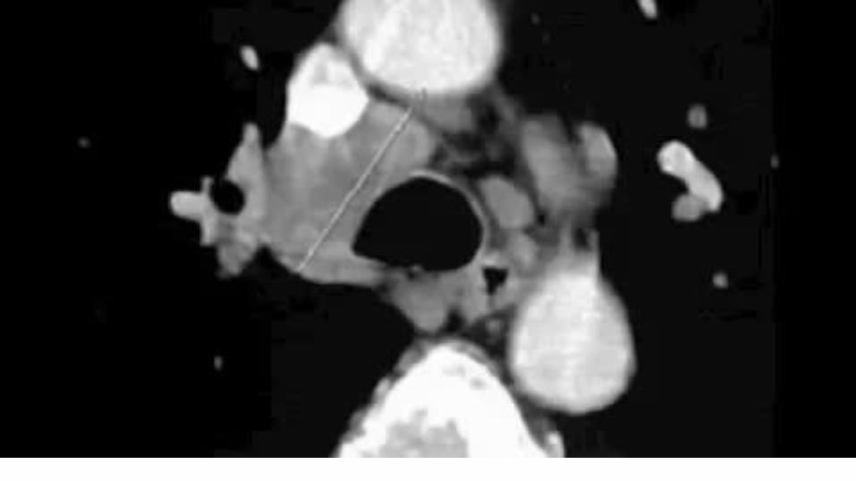
T4 tumor invading any of the following:
– Trachea, Carina, Great vessels
– Mediastinum, Heart, Esophagus
– Vertebral body
N2
NSCLC locally advance
«Extended resection and multimodality treatment »
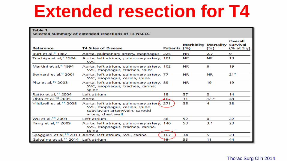
Extended resection for T4
Thorac Surg Clin 2014
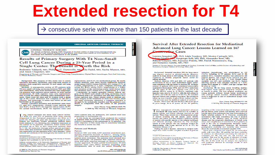
Extended resection for T4 consecutive serie with more than 150 patients in the last decade
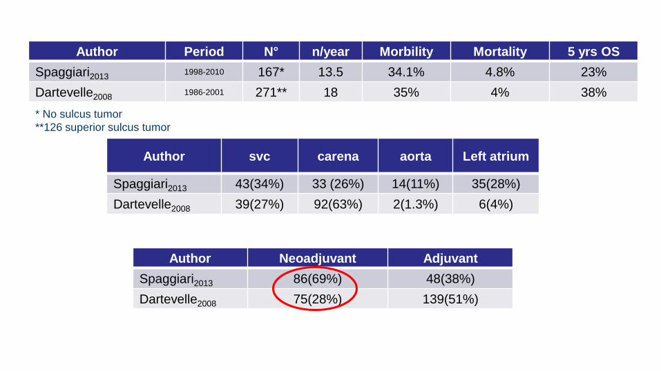
Author Period N° n/year Morbility Mortality 5 yrs OS
Spaggiari20131998-2010 167* 13.5 34.1% 4.8% 23%
Dartevelle20081986-2001 271** 18 35% 4% 38%
* No sulcus tumor
**126 superior sulcus tumor
Author svc carena aorta Left atrium
Spaggiari2013 43(34%) 33 (26%) 14(11%) 35(28%)
Dartevelle2008 39(27%) 92(63%) 2(1.3%) 6(4%)
Author Neoadjuvant Adjuvant
Spaggiari2013 86(69%) 48(38%)
Dartevelle2008 75(28%) 139(51%)

Access
Lateral thoracotomy 108 (86.4%)
Hemiclamshell 11 (8.8%)
Posterolateral thoracothomy 3 (2.4%)
Anterolateral 3 (2.4%)
Type of T4
Atrio 35 28.0%
SVC 43 34.4%
SVC+carena 18 14.4%
Carena 15 12.0%
Aorta 14 11.2%
42 explorative thoracotomies
Ann Thorac Surg 2013
Between 1998 and 2010, 167 patients with involvement of one or more
mediastinal organs underwent operations with the intent to perform ER

Complete resection
Lymph node statusDown staging
Type of resection

T4N0-1 TUMORS
heart, great vessels, mediastinum, esophagus, spine, or trachea Once considered unresectable
Resection generally limited to patients N0 \ N1
Technically challenging with increased morbidity
T4N2 tumors have poor 5-year survival rates and operative mortality
exceeds 5-year survival, and surgery is generally discouraged.
Cancer J 2013

But…Many patients experience postoperative
complications after extended resections
about 50%
Only few patients complete adjuvant
chemotherapy protocols

• Low compliance of adjuvant chemotherapy
• High rate of systemic recurrences after extended resection
• Small number of extended resection for a disease too much
advanced
• High rate of positive margins after resection
Induction therapyWHY?
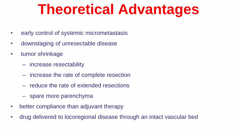
• early control of systemic micrometastasis
• downstaging of unresectable disease
• tumor shrinkage
– increase resectability
– increase the rate of complete resection
– reduce the rate of extended resections
– spare more parenchyma
• better compliance than adjuvant therapy
• drug delivered to locoregional disease through an intact vascular bed
Theoretical Advantages

Theoretical Disadvantages
• delay in local control
– Local progression
– Unresectable disease
• increase of surgical difficulties
• increase morbidity / mortality
– Bronchial fistulae
– Respiratory complications

Induction treatment for T4 extended resection
IEO experience
Type of resection Induction CT
Superior vena cava 69%
Aorta 72.2%
Left atrium 67%
Vertebrae 52.6%

20%
15% 17%
Induction CT
NSCLC stage IIIA-N2• Patients with stage IIIA-N2 tumors represent a heterogeneous group with
different clinical presentation, and both prognosis and treatment strategies
based on the extension of the disease to the mediastinum
• Chemotherapy followed by surgery in highly selected patients with or
without postoperative radiotherapy suggested an improvement in
resectability and in long term survival up to 54% at 5-year over single-
modality therapy
Numerous non-randomized phase II and phase III trials using
induction chemotherapy have been reported in the literature
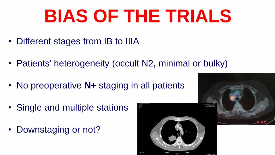
BIAS OF THE TRIALS
• Different stages from IB to IIIA
• Patients’ heterogeneity (occult N2, minimal or bulky)
• No preoperative N+ staging in all patients
• Single and multiple stations
• Downstaging or not?
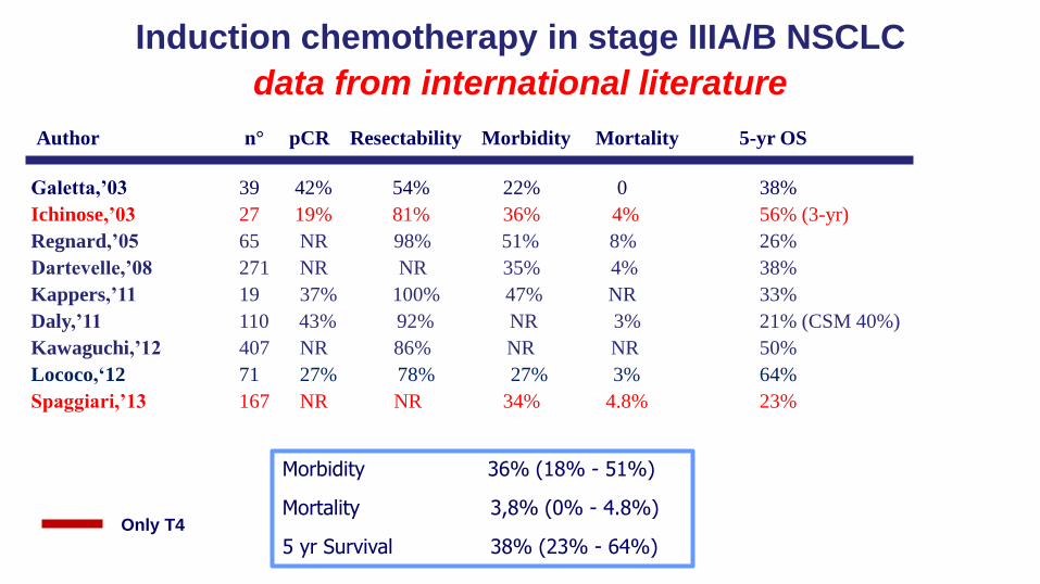
Author n° pCR Resectability Morbidity Mortality 5-yr OS
Galetta,’03 39 42% 54% 22% 0 38%
Ichinose,’03 27 19% 81% 36% 4% 56% (3-yr)
Regnard,’05 65 NR 98% 51% 8% 26%
Dartevelle,’08 271 NR NR 35% 4% 38%
Kappers,’11 19 37% 100% 47% NR 33%
Daly,’11 110 43% 92% NR 3% 21% (CSM 40%)
Kawaguchi,’12 407 NR 86% NR NR 50%
Lococo,‘12 71 27% 78% 27% 3% 64%
Spaggiari,’13 167 NR NR 34% 4.8% 23%
Induction chemotherapy in stage IIIA/B NSCLC
data from international literature
Morbidity 36% (18% - 51%)
Mortality 3,8% (0% - 4.8%)
5 yr Survival 38% (23% - 64%)Only T4
IEO experience
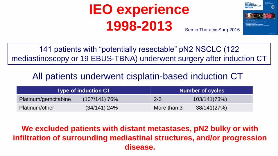
1998-2013
141 patients with “potentially resectable” pN2 NSCLC (122
mediastinoscopy or 19 EBUS-TBNA) underwent surgery after induction CT
All patients underwent cisplatin-based induction CT
We excluded patients with distant metastases, pN2 bulky or with
infiltration of surrounding mediastinal structures, and/or progression
disease.
Type of induction CT Number of cycles
Platinum/gemcitabine (107/141) 76% 2-3 103/141(73%)
Platinum/other (34/141) 24% More than 3 38/141(27%)
Semin Thoracic Surg 2016
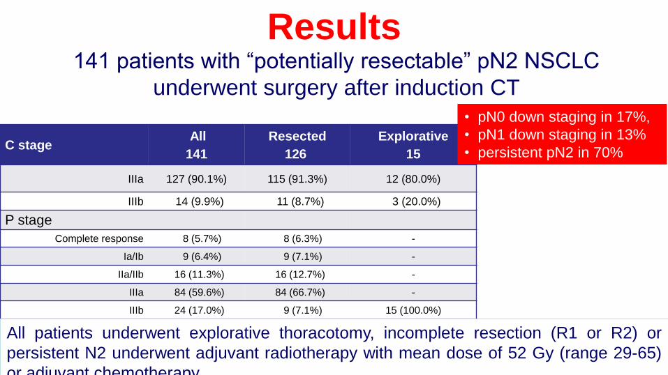
Results
C stageAll
141
Resected
126
Explorative
15
IIIa 127 (90.1%) 115 (91.3%) 12 (80.0%)
IIIb 14 (9.9%) 11 (8.7%) 3 (20.0%)
P stage
Complete response 8 (5.7%) 8 (6.3%) -
Ia/Ib 9 (6.4%) 9 (7.1%) -
IIa/IIb 16 (11.3%) 16 (12.7%) -
IIIa 84 (59.6%) 84 (66.7%) -
IIIb 24 (17.0%) 9 (7.1%) 15 (100.0%)
141 patients with “potentially resectable” pN2 NSCLC
underwent surgery after induction CT
• pN0 down staging in 17%,
• pN1 down staging in 13%
• persistent pN2 in 70%
All patients underwent explorative thoracotomy, incomplete resection (R1 or R2) or
persistent N2 underwent adjuvant radiotherapy with mean dose of 52 Gy (range 29-65)
or adjuvant chemotherapySemin Thoracic Surg 2016
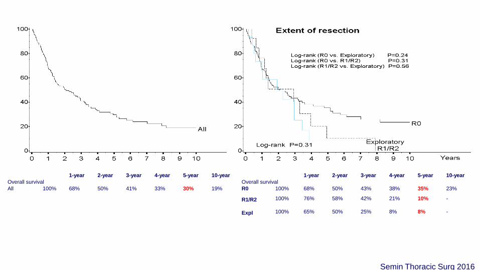
1-year 2-year 3-year 4-year 5-year 10-year 1-year 2-year 3-year 4-year 5-year 10-year
Overall survival Overall survival
All 100% 68% 50% 41% 33% 30% 19% R0 100% 68% 50% 43% 38% 35% 23%
R1/R2 100% 76% 58% 42% 21% 10% -
Expl 100% 65% 50% 25% 8% 8% -
Semin Thoracic Surg 2016

Baseline 1-year 2-year 3-year 4-year 5-year 10-year
Overall survival
pN0 100% 81% 75% 64% 59% 46% 37%
pN1 100% 61% 47% 38% 27% 27% 27%
pN2 100% 65% 44% 38% 34% 30% 22%
Semin Thoracic Surg 2016

Baseline 1-year 2-year 3-year 4-year 5-year 10-year Baseline 1-year 2-year 3-year 4-year 5-year 10-year
Overall survival Overall survival
2-3 100% 63% 45% 37% 32% 28% 15% pN0 2-3 100% 73% 64% 46% 46% 46% 24%
4-5 100% 82% 64% 64% 58% 58% 45% pN0 4-5 100% 90% 90% 90% 76% 76% 61%
pN+ 2-3 100% 61% 42% 35% 29% 25% 15%
pN+ 4-5 100% 78% 51% 51% 51% 51% 38%
Semin Thoracic Surg 2016
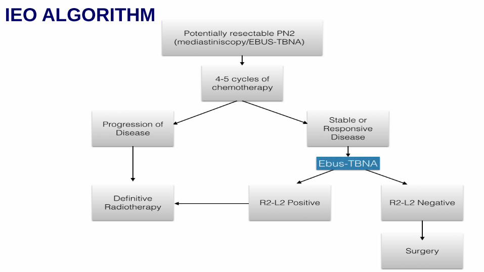
IEO ALGORITHM
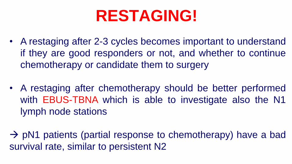
• A restaging after 2-3 cycles becomes important to understand
if they are good responders or not, and whether to continue
chemotherapy or candidate them to surgery
• A restaging after chemotherapy should be better performed
with EBUS-TBNA which is able to investigate also the N1
lymph node stations
pN1 patients (partial response to chemotherapy) have a bad
survival rate, similar to persistent N2
RESTAGING!

• T4 resections are feasible but selection of the candidate is
paramount
• The factors that were found to possibly affect survival were
the completeness of resection, the lymph node status
• Induction therapy may improve patient’s selection avoiding
unnecessary surgery in more than 20% of the cases
CONCLUSION- Locally advanced stage -

• In patients stage IIIA-N2 chemotherapy played an essential role in the sterilization of
lymph node metastasis resulting in a significant increase in survival when compared with
patients in whom the nodal down staging was not the case N+ (46% vs. 28% at 5 years)
• Number of cycles of chemotherapy were strictly related to a better survival. In patients
with “potentially resectable” pN2 disease we reach up to 76% survival at 5 yrs by using 4-
5 cycles of third-generation induction chemotherapy, with an acceptable morbidity and
mortality
it will be essential to investigate the group of best survivors in term of genetic and
molecular target such as MiRNA identifing possible “pretreatment prognostic
factor” as predictive signature of chemotherapy efficacy (ongoing study).
CONCLUSION- Locally advanced stage -

Median OS
CSR vs CR
P<0.001
WHAT ABOUT STAGE IIIB?

• ‘oligometastases’ = diagnosed with oligometastaticdisease
• ‘oligorecurrence’ = relapsed oligometastatic disease
• ‘oligoprogression’ = status after cytoreductive therapy
cohorts probably have different prognoses
therapy
Oligometastatic NSCLC distinct cohorts
Courtesy of J. Vansteenkiste (ELCC 2016)
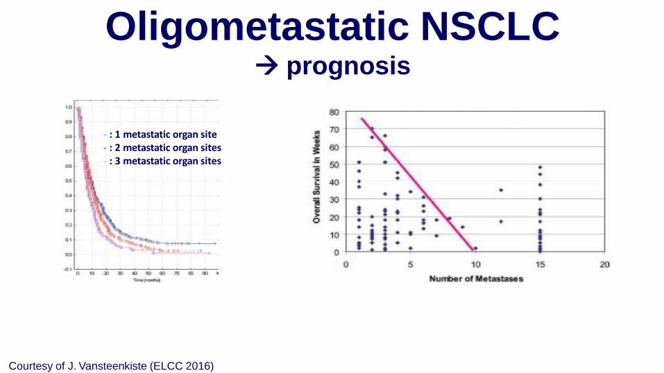
- : 1 metastatic organ site- : 2 metastatic organ sites- : 3 metastatic organ sites
Oligometastatic NSCLC prognosis
Courtesy of J. Vansteenkiste (ELCC 2016)
Adeno
Squam
Large
Small
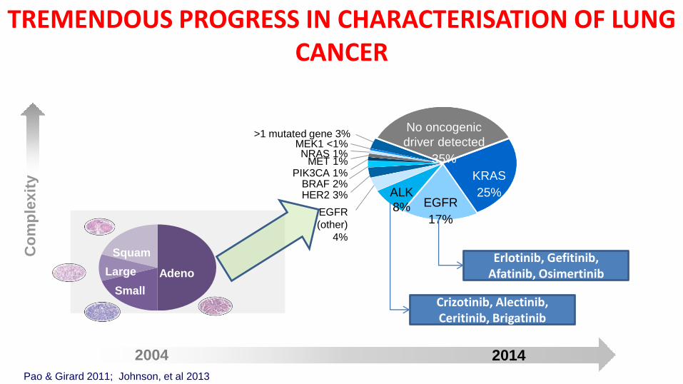
TREMENDOUS PROGRESS IN CHARACTERISATION OF LUNG CANCER
>1 mutated gene 3%MEK1 <1%NRAS 1%
MET 1%PIK3CA 1%
BRAF 2%HER2 3%
EGFR
(other)
4%
ALK
8% EGFR
17%
KRAS
25%
No oncogenic
driver detected
35%
Co
mp
lexit
y
20142004
Pao & Girard 2011; Johnson, et al 2013
Erlotinib, Gefitinib, Afatinib, Osimertinib
Crizotinib, Alectinib, Ceritinib, Brigatinib
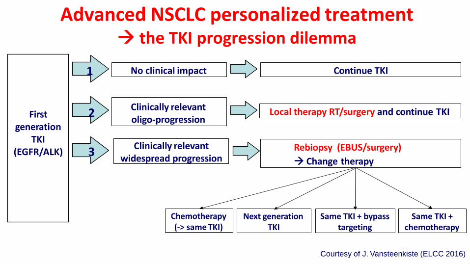
First generation
TKI(EGFR/ALK)
Local therapy RT/surgery and continue TKI
Continue TKI
Clinically relevantoligo-progression
2
3
1
Advanced NSCLC personalized treatment the TKI progression dilemma
No clinical impact
Clinically relevantwidespread progression
Rebiopsy (EBUS/surgery)
Change therapy
Chemotherapy (-> same TKI)
Next generation TKI
Same TKI + bypass targeting
Same TKI + chemotherapy
Courtesy of J. Vansteenkiste (ELCC 2016)

M Kris et al, JAMA 2014
Driver, NO TT= 2.4 ys
NO driver= 2.1 ys
Driver, TT= 3.5 ys

CONCLUSIONS
Surgery is:
• Gold standard for early stage lung cancer
• Part of a multimodality treatment for locally
advanced NSCLC
• Diagnostic and palliative tools for not surgical or
metastatic patients

Thank you!!!