Surgery and Prosthetic Rehabilitation Overdenture by Digital Smile … · 2019-01-09 ·...
9
International Journal of Science and Research (IJSR) ISSN: 2319-7064 Index Copernicus Value (2016): 79.57 | Impact Factor (2017): 7.296 Volume 7 Issue 12, December 2018 www.ijsr.net Licensed Under Creative Commons Attribution CC BY Fully Digital Workflow for an Implant Retained Overdenture by Digital Smile Project to Guided Surgery and Prosthetic Rehabilitation Luca Lavorgna, DDS 1 , Tommaso Vitali, DDS 2 , Ilaria Caviggioli DDS 3 , Luca Ortensi, DDS. 4 1 Private Practice, Telese, Italy 2 Private Practice, Castiglione del Lago, Italy 3 Private Practice, Galliate, Italy 4 Adjunct Professor, Department of Prosthodontics, University of Catania, Catania, Italy Corrisponding Author: Dr. Luca Ortensi, Via De Gombruti, 5, 40123, Bologna, Italy Abstract: The development of digital technologies in dentistry has changed the therapeutic approach in edentulous patients both in the preliminary stages of clinical case studies and when supporting the actual surgical and manufacture phases. The goals of the digital workflow are to reduce the number of patient appointments and to improve the predictability of treatment outcome. A thorough description of a complex clinical case analyzes how these new digital technologies are used in every step of the surgical and prosthetic therapy to perform the reconstruction of a bar retained overdenture. 1. Introduction In recent years, modern prosthetic dentistry has been making use of digital technologies to support both the diagnostic and therapeutic stages of patient rehabilitation. 1-4 Traditional removable and implant-supported dentureshave also benefited from these innovations and have been further digitally developed both in the virtual planning of clinical cases and as support during the actual manufacture phase. 5,6 The purpose of digital manufacture of implant retained overdentures is to reduce the number of patient appointments and to improve the predictability of treatment outcome. 7,8 In a previous article the authors described the digital flow procedure for making removable dentures using a specific new software called the Digital Smile System R (DSS)consisting of tools and a data library of natural and artificial teeth used in interdisciplinary dentistry to improve diagnostic vision, develop communication/ information, and to enhance predictability throughout the course of treatment (restorative, surgical, and prosthetic). 9 In the procedure described, the implant positioning, although prosthetically guided, involves “free hand” surgery. The use of properly planned guided surgery, based on the anatomical situation and of the chosen prothetic, it is a now consolidated operative criteria in clinical practive. 10,11 The aim of this clinical report is to describe the application of these digital technology advancements in overdenture implant rehabilitation through the description of a complex clinical case, where guided-implant surgery becomes an integral part of the fully digital workflow. 2. Clinical Report A 67-year-old female patient came to the dental office complaining of reduced chewing ability and loss of retention of her upper removable complete denture in the oral cavity. She furthermore asked to improve the appearance of her smile and face, complaining that she was dissatisfied with the color and visibility of her teeth which, even when facial mimicry was more accentuated, were barely noticeable and featured unnatural slanted planes. The medical history showed no disease incompatible with dental therapy and an overall good state of health: the patient was classified as ASA1. The esthetic analysis of the facial appearance showed a reduction of the vertical dimension of occlusion with an increase in perilabial wrinkles. Furthermore, the musculature of the cheeks was unsupported by the old prosthesis, and the entire face had visibly lost tone (Fig.1). The patient exhibited an unattractive, slanted smile with an unbalanced look: the occlusal plan appears crooked and the interincisive line does not correspond with midline of the face. The clinical intraoral analysis showed that the upper denture was unsatisfactory because of an inadequate extension of the prosthetic bodies, incorrect occlusal contacts, and an inadequate intermaxillary relationship. In the lower arch, there was a recently-made fixed prosthesis. During the visit, the patient underwent a lateral radiographic exam. The radiographic examination made it possible to study the hard and soft tissues of the patient’s face, and safely allows recognition of high occlusal risk brachyfacial patients, providing prognostic information for treatment planning purposes. 12 Facial and intraoral photographs were taken as an essential aid for completing the treatment plan. In general, facial photos enable further diagnostic assessments regarding the overall esthetics of the face and the physiognomic traits, which are then addressed in the prosthetic therapy. 13 At the end of the first visit, different treatment options, with their respective associated risks and benefits, were presented and discussed with the patient. The patient accepted the permanent treatment plan of an overdenture maxillary dental prosthesis implant, supported with four dental implants. Paper ID: ART20194005 10.21275/ART20194005 1534
Transcript of Surgery and Prosthetic Rehabilitation Overdenture by Digital Smile … · 2019-01-09 ·...
International Journal of Science and Research (IJSR) ISSN: 2319-7064
Index Copernicus Value (2016): 79.57 | Impact Factor (2017): 7.296
Volume 7 Issue 12, December 2018
www.ijsr.net Licensed Under Creative Commons Attribution CC BY
Fully Digital Workflow for an Implant Retained
Overdenture by Digital Smile Project to Guided
Surgery and Prosthetic Rehabilitation
Luca Lavorgna, DDS1, Tommaso Vitali, DDS
2, Ilaria Caviggioli DDS
3, Luca Ortensi, DDS.
4
1Private Practice, Telese, Italy
2Private Practice, Castiglione del Lago, Italy
3Private Practice, Galliate, Italy
4Adjunct Professor, Department of Prosthodontics, University of Catania, Catania, Italy
Corrisponding Author: Dr. Luca Ortensi, Via De Gombruti, 5, 40123, Bologna, Italy
Abstract: The development of digital technologies in dentistry has changed the therapeutic approach in edentulous patients both in the
preliminary stages of clinical case studies and when supporting the actual surgical and manufacture phases. The goals of the digital
workflow are to reduce the number of patient appointments and to improve the predictability of treatment outcome. A thorough
description of a complex clinical case analyzes how these new digital technologies are used in every step of the surgical and prosthetic
therapy to perform the reconstruction of a bar retained overdenture.
1. Introduction
In recent years, modern prosthetic dentistry has been making
use of digital technologies to support both the diagnostic and
therapeutic stages of patient rehabilitation.1-4
Traditional
removable and implant-supported dentureshave also
benefited from these innovations and have been further
digitally developed both in the virtual planning of clinical
cases and as support during the actual manufacture phase.5,6
The purpose of digital manufacture of implant retained
overdentures is to reduce the number of patient
appointments and to improve the predictability of treatment
outcome.7,8
In a previous article the authors described the
digital flow procedure for making removable dentures using
a specific new software called the Digital Smile
SystemR(DSS)consisting of tools and a data library of
natural and artificial teeth used in interdisciplinary dentistry
to improve diagnostic vision, develop communication/
information, and to enhance predictability throughout the
course of treatment (restorative, surgical, and prosthetic).9In
the procedure described, the implant positioning, although
prosthetically guided, involves “free hand” surgery. The use
of properly planned guided surgery, based on the anatomical
situation and of the chosen prothetic, it is a now
consolidated operative criteria in clinical practive.10,11
The
aim of this clinical report is to describe the application of
these digital technology advancements in overdenture
implant rehabilitation through the description of a complex
clinical case, where guided-implant surgery becomes an
integral part of the fully digital workflow.
2. Clinical Report
A 67-year-old female patient came to the dental office
complaining of reduced chewing ability and loss of retention
of her upper removable complete denture in the oral cavity.
She furthermore asked to improve the appearance of her
smile and face, complaining that she was dissatisfied with
the color and visibility of her teeth which, even when facial
mimicry was more accentuated, were barely noticeable and
featured unnatural slanted planes. The medical history
showed no disease incompatible with dental therapy and an
overall good state of health: the patient was classified as
ASA1.
The esthetic analysis of the facial appearance showed a
reduction of the vertical dimension of occlusion with an
increase in perilabial wrinkles. Furthermore, the musculature
of the cheeks was unsupported by the old prosthesis, and the
entire face had visibly lost tone (Fig.1). The patient
exhibited an unattractive, slanted smile with an unbalanced
look: the occlusal plan appears crooked and the interincisive
line does not correspond with midline of the face.
The clinical intraoral analysis showed that the upper denture
was unsatisfactory because of an inadequate extension of the
prosthetic bodies, incorrect occlusal contacts, and an
inadequate intermaxillary relationship. In the lower arch,
there was a recently-made fixed prosthesis.
During the visit, the patient underwent a lateral radiographic
exam. The radiographic examination made it possible to
study the hard and soft tissues of the patient’s face, and
safely allows recognition of high occlusal risk brachyfacial
patients, providing prognostic information for treatment
planning purposes.12
Facial and intraoral photographs were
taken as an essential aid for completing the treatment plan.
In general, facial photos enable further diagnostic
assessments regarding the overall esthetics of the face and
the physiognomic traits, which are then addressed in the
prosthetic therapy.13
At the end of the first visit, different treatment options, with
their respective associated risks and benefits, were presented
and discussed with the patient. The patient accepted the
permanent treatment plan of an overdenture maxillary dental
prosthesis implant, supported with four dental implants.
Paper ID: ART20194005 10.21275/ART20194005 1534
International Journal of Science and Research (IJSR) ISSN: 2319-7064
Index Copernicus Value (2016): 79.57 | Impact Factor (2017): 7.296
Volume 7 Issue 12, December 2018
www.ijsr.net Licensed Under Creative Commons Attribution CC BY
The first clinical stage of the new prosthetic therapy entails
making the preliminary impressions. This is in no way a
marginal step in the process as it is essential to fully record
the anatomy of the endoral structures of the edentulous
upper jaw. A digital impression of the edentulous upper arch
(3Shape Trios, Denmark), of the pre-existing dentures and
of the opposite arch were made (Fig 2).14,15.
At this point two photos were taken of the patient’s face
according to a coded technique for the DSS software.16
It is
important to take photos of the face keeping the patient in a
position that is stable and repeatable over time, trying not to
change the enlargement ratio between shots. For this
purpose the patient was invited to sit comfortably keeping
her back straight while the operator used a camera set on a
tripod to stabilize its position in relation to the patient being
photographed. The subject had to be positioned so that her
Frankfurt Plane (the line that joins the Porion and the Orbital
Point) was parallel to the horizon.
The patient may wear dedicated glasses used to calibrate the
digital pre-rendering software (DSS). The glasses represent
a true measuring system that differentiates this software
from other similar systems.17
Thanks to their shape and the presence of calibration
markers used as a reference, the glasses facilitate
maintenance of the perpendicular position of the patient and
the camera.
The first facial photo was taken asking the patient to smile
and show as many teeth as possible. The second facial
photograph was taken with cheek retractors (Fig.3). The
photo made it possible to correctly assess the parallelism
between the bipupillary plane and the occlusal plane, as well
as the consistency between the median and interincisive line.
This made it possible to import the photos (JPEG format)
into the DSS and to proceed with the esthetic pre-rendering
of the future prosthetic therapy.
Digital pre-rendering with the DSS program consisted of
creating a digital teeth arrangement with virtual artificial
teeth contained in the software library. The teeth were
chosen according to esthetic and functional parameters18
and
could be replaced with others of different shapes or color. If
necessary the anterior and posterior teeth were positioned
using the old denture as a guide. In this way the patient
could see the possible esthetic end result and participate in
the therapeutic project together with the entire dental team
(Fig.4).
Once the virtual teeth arrangement was obtained - and
approved by the patient - the file containing the patient’s
information, the photographic alignments, the libraries
chosen, and the work process was transferred to the dental
technician's laboratory where the file was imported into a
3D software program (Exocad® software, Exocad GmbH).
The information file exported from the DSS consisted of a
PDF format and single photographs of the patient’s face
with a customized two dimensional (2D) virtual smile
design.
The files from DSS were then superimposed onto scanned
images of denture and edentulous upper jaw.19
The dental
technician used the outline of the virtual smile obtained to
place a tooth from the library or to create customized teeth
with tools from Freeform (Exocad® software, Exocad
GmbH) to convert the virtual 2D teeth arrangement into a
3D teeth arrangement (Fig 5).20
At the end of this work stage, a file from Exocad was
converted into a specific file for a 3D printer (SLA 3D,
Form 2, Formlabs Inc.)and a prototype of the digital work
was made with a dedicated resin (C&B, A3.5, NextDent
B.V.) (Fig 6).21
The prototype was tried in the mouth checking the intraoral
adaptation, the centric relation, and the esthetics of the smile
and face (Fig.7). The clinician could make changes if
necessary.
Subsequently the prototype is used as a radiological
stentwith which the CBCT is done, using a dedicated device
(Evobitewith 3D marker, 3diemme, Italy)which was adapted
to the item with radiotransparent silicone (Fig. 8).
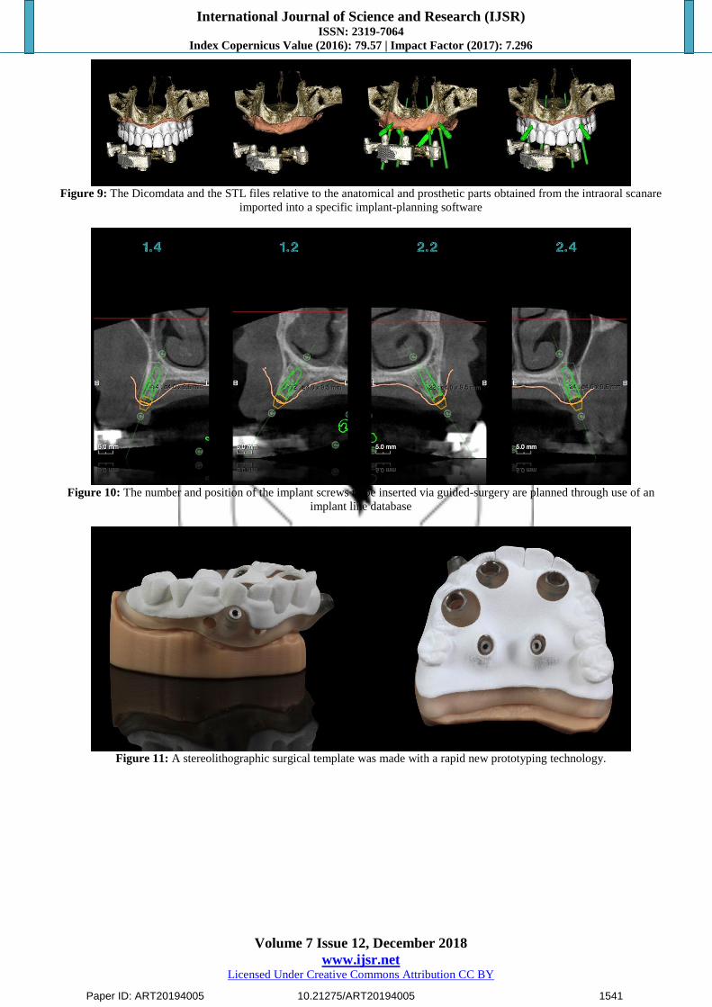
The Dicomdata resulting from the X-ray and the STL files
relative to the anatomical and prosthetic parts obtained from
the intraoral scan are imported in a specific implant planning
software (Realguide 5.0, 3 DIEMME, Italy)where, thanks to
adedicatedalgorhythm, are overlapped using a replicable and
controlled procedure (Fig. 9).Through use of the implant
line database used (Thommen Medical AG, Grenchen,
Switzerland) the number and position of the implant screws
to be inserted via guided-surgery are planned (Fig. 10).
After careful functional and esthetic evaluation and final
verification, the prosthetic-driven plan was approved, and a
stereolithographic surgical template was made using a newer
rapid prototyping technology (New Ancorvis, Bargellino,
Italy) (Fig. 11). Subsequently four prosthetic-driven
implants with a diameter of 4.0 mm and a length of 9.5 mm
(SPI ® CONTACT RC INICELL®, ThommenMedicalAG)
were placed, with a dedicated dedicated bur kit (Thommen
Medical Guided Surgery Kit), in the upper jaw, taking into
account the bone quality and quantity, soft-tissue thickness,
anatomical landmarks, and the type, volume and shape of
the final restoration.
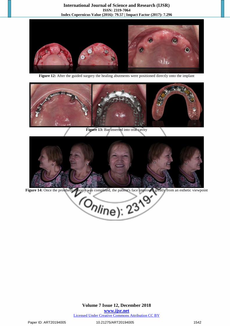
Healing abutments were positioned directly onto the implant
heads and the old denture was realigned with a temporary
soft material (Coe Soft (CS) GC America Inc.) (Fig. 12).
After four weeks,22
the final digital impression (Trios
3Shape) of the upper arch was made of the implant scan
bodies previously screwed into the fixture with a set torque.
In addition, the final volume of the clinicized prototype were
also reacquired so as to digitally superimpose the images
onto each other. Superimposing of various digital files has
proven to be a reliable procedure in digital workflows.23
Using a dedicated tool of the 3D software (Exocad®
software, Exocad GmbH), the project was executed to create
the upper retentive bar, using fixture scan transparencies to
assess the available spaces and the position of the teeth in
Paper ID: ART20194005 10.21275/ART20194005 1535
International Journal of Science and Research (IJSR) ISSN: 2319-7064
Index Copernicus Value (2016): 79.57 | Impact Factor (2017): 7.296
Volume 7 Issue 12, December 2018
www.ijsr.net Licensed Under Creative Commons Attribution CC BY
relation to their analogues, and to identify the type of
prosthetic structure and anchorage. The structure project was
sent to the milling center (New Ancorvis): indications were
given regarding the retentive system anchorage areas, the
use of dedicated threading, the type of metal, and the
execution technique (CAD/CAM technology).
After being checked in the dental technician's lab, the item
was sent to the dentist to clinically test the structure, in order
to verify its precision and passivity. The dentist took a pick-
up impression of the bar so that it was possible to make a
master model stone to finalize the prosthesis.
The process later continued with further polishing and
shining of the bar and the digital acquisition using a scanner.
The dental technician digitally designed the counter-bar,
always checking the available spaces, using final volume
superimposition and inserting retentive pins in the project
for the mechanical tightness of the teeth. It was made using
laser-melting technology that made it possible to obtain an
accurate item to which the acrylic resin adhered tightly
thanks to the presence of a retentive surface.24
It was then time for the complete application of the teeth
onto the counter-bar, using the clinicized prototype as a
fitting plane. The artificial teeth and the metal structure were
joined using a small amount of wax.
The prosthesis was finalized using a resin injection muffle
system (VertysystemR, a-gree srl) that transformed the wax
into resin with a codified protocol.25-26
The overdenture and
the finished and perfectly polished retention structure were
sent to the dental office.
The bar was then inserted into the oral cavity by the
clinician and tightened to the implant fixtures with a preset
torque (Fig 13). The dentist had to check that there were no
areas where the soft tissues were compressed and that there
was room for the use of dental devices: interdental brushes
and flosses.
Once the prosthetic therapy was completed, the patient's
face had improved greatly from an esthetic viewpoint. The
soft tissues of the face appeared firm and toned. A reduction
could be seen in the naso-labial folds and perilabial
wrinkles, both frontally and laterally. The vertical
dimension, which had been slightly increased, appeared
adequate and well tolerated esthetically. During phonation
and smiling dynamics the patient displayed natural looking
teeth that were perfectly integrated with her face (Fig.14).
3. Discussion
In prosthetic therapy, for both fixed and removable
prostheses, communication with the patient is a vital part of
the treatment. Effective digital previsualization is the ideal
way to explain esthetic changes to a patient and to receive
their approval. Until now, many digital previsualization
methods have been used in dentistry solely for this purpose.
In this article the Digital Smile approach was introduced in a
complex digital workflow. DSS not only allowed the patient
to see her future appearance, but it also enabled production
of a prototype (Patent Pending Workflow, Just Digital, Italy)
for the functional check of the digital project carried out.
The fact that the patient can see the possible future esthetic
results through digital rendering, including the possibility of
changes if desired, reduces overall clinical practice time.
Additionally, the construction of a prototype, based on the
virtual assembly, minimizes the number of errors in the
manufacture of the final productand becomes a fundamental
instrument for prosthetic-driven surgery.
Use of the prototype as a radiological stent during the
examination of the CBCT, and its transformation into a
surgery-driven guide make it possible to position implants
according to the digital study done with DSS and approved
by the patient.
Some phases of the described workflow require a learning
curve by the clinical operator and technician. For example,
the photos taken by the clinician for the DSS, must be taken
in the exact manner as previously described to facilitate the
superimposing of the photo of the patient's face, with the
scan of the model and the old denture. Another important
stage is that in which the teeth from the databank of the 3D
software are matched with the outlines obtained by digital
previsualization with DSS. In this case, if the matching is
not precise, the prototype will not correspond perfectly with
that approved by the patient.
4. Conclusions
The use of digital technologies is now vastly widespread in
the field of dentistry and, in particular, in prosthetic therapy.
In removable, traditional, and implant-support prosthetic
therapy, digital technology can play an essential role. The
clinical case described was almost entirely resolved with an
innovative digital workflow, both from a clinical and
technical viewpoint. In particular, guided-surgery has come
into the digital workflow through a simplified approach that
is closely dependent on the esthetic and functional aspects of
the patient.The human component is still fundamental and
not all stages can be carried out digitally. However, it is
expected that technical development will rapidly lead to
more and more digitalized therapies with an increase in the
end quality of the therapy and less conditioned by the skills
of the individual operator.
5. Acknowledgments
The authors thank Mr. Gianni Ortensi, CDT, Mr. Marco
Ortensi,CDT,who made the bar retained overdenture.
References
[1] Coachman, C., & Calamita, M. A. (2014). VIRTUAL
Esthetic Smile Design. Journal of Cosmetic Dentistry,
29(4).
[2] Ortensi,L., Stefani, R., Caviggioli, I., Molinelli, F.
(2011). La prima visita in odontoiatria protesica:
aspetti innovativi.Il Dentista Moderno.
[3] Zimmermann, M., &Mehl, A. (2015). Virtual smile
design systems: a current review. International journal
of computerized dentistry, 18(4), 303-317.
Paper ID: ART20194005 10.21275/ART20194005 1536
International Journal of Science and Research (IJSR) ISSN: 2319-7064
Index Copernicus Value (2016): 79.57 | Impact Factor (2017): 7.296
Volume 7 Issue 12, December 2018
www.ijsr.net Licensed Under Creative Commons Attribution CC BY
[4] Coachman, C. (2012). Calamita M. Digital Smile
Design: a tool for treatment planning and
communication in esthetic dentistry. QDT.
[5] Goodacre, C. J., Garbacea, A., Naylor, W. P., Daher,
T., Marchack, C. B., & Lowry, J. (2012). CAD/CAM
fabricated complete dentures: concepts and clinical
methods of obtaining required morphological data. The
Journal of prosthetic dentistry, 107(1), 34-46.
[6] Infante, L., Yilmaz, B., McGlumphy, E., & Finger, I.
(2014). Fabricating complete dentures with
CAD/CAM technology. The Journal of prosthetic
dentistry, 111(5), 351-355.
[7] Kattadiyil, M. T., Jekki, R., Goodacre, C. J., & Baba,
N. Z. (2015). Comparison of treatment outcomes in
digital and conventional complete removable dental
prosthesis fabrications in a predoctoral setting. The
Journal of prosthetic dentistry, 114(6), 818-825.
[8] Rossi M, Molinelli F, Caviggioli I.(2015). Latest
trends in prosthetics - Total maxillary rehabilitation
with a Toronto Bridge using digital technologies.
CAD/CAM. International magazine of
digitaldentistry;1:28-31.
[9] Ortensi L, Stefani R, Lavorgna L, Caviggioli I, Vitali
T. (2018). A Digital Workflow for an Implant Retained
over Denture: A New Approach. Biomed J Sci&Tech
Res 6(5).
[10] Vercruyssen, M., Laleman, I., Jacobs, R., &Quirynen,
M. (2015). Computer‐supported implant planning and
guided surgery: a narrative review. Clinical oral
implants research, 26, 69-76.
[11] Tallarico, M., Esposito, M., Xhanari, E., Caneva, M.,
&Meloni, S. M. (2018). Computer-guided vs freehand
placement of immediately loaded dental implants: 5-
year post-loading results of a randomised controlled
trial. European journal of oral implantology, 11(2).
[12] Ortensi, L., Martini, M., Montanari, M., &Galassini,
G. (2017). A Simplified Method to Identify Patient
Face Type for a Prosthodontic Treatment Plan. J Dent
Health Oral DisordTher, 8(4), 00291.
[13] McLaren E.A., Terry D. (2001). Photography in
dentistry. J Calif Dental Ass;29.10:735-742.
[14] Kim, J. E., Amelya, A., Shin, Y., & Shim, J. S. (2017).
Accuracy of intraoral digital impressions using an
artificial landmark. The Journal of prosthetic dentistry,
117(6), 755-761.
[15] Kim, J. E., Kim, N. H., & Shim, J. S. (2017).
Fabrication of a complete, removable dental prosthesis
from a digital intraoral impression for a patient with an
excessively tight reconstructed lip after oral cancer
treatment: A clinical report. The Journal of prosthetic
dentistry, 117(2), 205-208.
[16] Stefani R, Caviggioli I, Molinelli F, Ortensi L. (2012).
L’impiego delle tecnologie digitali nella diagnosi
protesica e nella realizzazione della protesi. Il
DentistaModerno.
[17] Jokstad, A. (2017). Computer‐assisted technologies
used in oral rehabilitation and the clinical
documentation of alleged advantages–a systematic
review. Journal of oral rehabilitation, 44(4), 261-290.
[18] Lombardi, R. E. (1977). Factors mediating against
excellence in dental esthetics. Journal of Prosthetic
Dentistry, 38(3), 243-248.
[19] Joda, T., Brägger, U., & Gallucci, G. (2015).
Systematic literature review of digital three-
dimensional superimposition techniques to create
virtual dental patients. International journal of oral &
maxillofacial implants, 30(2).
[20] Zhang, C., Liu, T., Liao, W., Yang, T., & Jiang, L.
(2017). Computer-aided design of dental inlay
restoration based on dual-factor constrained
deformation. Advances in Engineering Software, 114,
71-84.
[21] Rengier, F., Mehndiratta, A., Von Tengg-Kobligk, H.,
Zechmann, C. M., Unterhinninghofen, R., Kauczor, H.
U., &Giesel, F. L. (2010). 3D printing based on
imaging data: review of medical applications.
International journal of computer assisted radiology
and surgery, 5(4), 335-341.
[22] Burkhardt, M. A., Waser, J., Milleret, V., Gerber, I.,
Emmert, M. Y., Foolen, J., ... & Vogel, V. (2016).
Synergistic interactions of blood-borne immune cells,
fibroblasts and extracellular matrix drive repair in an in
vitro peri-implant wound healing model. Scientific
reports, 6, 21071.
[23] Coachman, C., Calamita, M. A., Coachman, F. G.,
Coachman, R. G., &Sesma, N. (2017). Facially
generated and cephalometric guided 3D digital design
for complete mouth implant rehabilitation: A clinical
report. The Journal of prosthetic dentistry, 117(5),
577-586.
[24] Koutsoukis, T., Zinelis, S., Eliades, G., Al‐Wazzan,
K., Rifaiy, M. A., & Al Jabbari, Y. S. (2015). Selective
Laser Melting Technique of Co‐Cr Dental Alloys: A
Review of Structure and Properties and Comparative
Analysis with Other Available Techniques. Journal of
Prosthodontics, 24(4), 303-312.
[25] Goodacre, B. J., Goodacre, C. J., Baba, N. Z.,
&Kattadiyil, M. T. (2018). Comparison of denture
tooth movement between CAD-CAM and
conventional fabrication techniques. The Journal of
prosthetic dentistry, 119(1), 108-115.
[26] Marco, M., Giuliano, B., & Luca, O. (2016). Oral
Rehabilitation with Implant-Supported Overdenture
and a New Protocol for Bar Passivation. Global
Journal of Oral Science, 2, 10-19.
Paper ID: ART20194005 10.21275/ART20194005 1537

International Journal of Science and Research (IJSR) ISSN: 2319-7064
Index Copernicus Value (2016): 79.57 | Impact Factor (2017): 7.296
Volume 7 Issue 12, December 2018
www.ijsr.net Licensed Under Creative Commons Attribution CC BY
Photo Captions
Figure 1: Initial state of the face: a reduction of the vertical dimension is observed with an increase in perilabial wrinkles.
Figure 2: A digital impression was made of the edentulous upper arch, old denturesand of the opposite arch.
Figure 3: The first facial photo was taken asking the patient to smile and show as many teeth as possible and the second facial
photograph was taken with cheek retractors
Paper ID: ART20194005 10.21275/ART20194005 1538

International Journal of Science and Research (IJSR) ISSN: 2319-7064
Index Copernicus Value (2016): 79.57 | Impact Factor (2017): 7.296
Volume 7 Issue 12, December 2018
www.ijsr.net Licensed Under Creative Commons Attribution CC BY
Figure 4: Patient’sdigital pre-rendering with the DSS software
Figure 5: The dental technician used the outline of the virtual smile obtained to place a tooth from the library or to create
customized teeth to convert the virtual 2D teeth arrangement into a 3D teeth arrangement
Figure 6: A prototype of the digital work was obtained with a dedicated resin
Paper ID: ART20194005 10.21275/ART20194005 1539

International Journal of Science and Research (IJSR) ISSN: 2319-7064
Index Copernicus Value (2016): 79.57 | Impact Factor (2017): 7.296
Volume 7 Issue 12, December 2018
www.ijsr.net Licensed Under Creative Commons Attribution CC BY
Figure 7: Face of patient with prototypes inserted into the mouth to check esthetics
Figure 8: The prototype was used as a radiological stent during the conduct of the CBCT
Paper ID: ART20194005 10.21275/ART20194005 1540
International Journal of Science and Research (IJSR) ISSN: 2319-7064
Index Copernicus Value (2016): 79.57 | Impact Factor (2017): 7.296
Volume 7 Issue 12, December 2018
www.ijsr.net Licensed Under Creative Commons Attribution CC BY
Figure 9: The Dicomdata and the STL files relative to the anatomical and prosthetic parts obtained from the intraoral scanare
imported into a specific implant-planning software
Figure 10: The number and position of the implant screws to be inserted via guided-surgery are planned through use of an
implant line database
Figure 11: A stereolithographic surgical template was made with a rapid new prototyping technology.
Paper ID: ART20194005 10.21275/ART20194005 1541
International Journal of Science and Research (IJSR) ISSN: 2319-7064
Index Copernicus Value (2016): 79.57 | Impact Factor (2017): 7.296
Volume 7 Issue 12, December 2018
www.ijsr.net Licensed Under Creative Commons Attribution CC BY
Figure 12: After the guided surgery the healing abutments were positioned directly onto the implant
Figure 13: Bar inserted into oral cavity
Figure 14: Once the prosthetic therapy was completed, the patient's face improved greatly from an esthetic viewpoint
Paper ID: ART20194005 10.21275/ART20194005 1542