Supramolecularly cross-linked amphiphilic block copolymer assembly … · 2019-11-29 · 1...
20
1 Supporting Information for Supramolecularly cross-linked amphiphilic block copolymer assembly by the dipolar interaction of a merocyanine dye Aritra Rajak, a Chandan Kumar Karan, a Patrick Theato, b,c and Anindita Das a * *Corresponding author; Email: [email protected] a School of Applied and Interdisciplinary Sciences, Indian Association for the Cultivation of Science, Jadavpur, Kolkata –700032, India b Institute for Chemical Technology and Polymer Chemistry Karlsruhe Institute of Technology (KIT), Engesser Str. 18, Building 11.23 D-76131 Karlsruhe c Soft Matter Synthesis Laboratory, Institute for Biological Interfaces III Karlsruhe Institute of Technology (KIT), Herrmann-von-Helmholtz-Platz 1 D-76344 Eggenstein-Leopoldshafen Page No. Materials and Methods 2 Synthesis and Characterization 2-6 Experimental Procedures 6-8 Additional Figures 9-14 X-ray Structure Determination 14-15 NMR and HRMS Spectra 16-20 References 20 Electronic Supplementary Material (ESI) for Polymer Chemistry. This journal is © The Royal Society of Chemistry 2019
Transcript of Supramolecularly cross-linked amphiphilic block copolymer assembly … · 2019-11-29 · 1...

1
Supporting Information for
Supramolecularly cross-linked amphiphilic block copolymer assembly
by the dipolar interaction of a merocyanine dye
Aritra Rajak,
a Chandan Kumar Karan,
a Patrick Theato,
b,cand Anindita Das
a*
*Corresponding author; Email: [email protected]
aSchool of Applied and Interdisciplinary Sciences,
Indian Association for the Cultivation of Science, Jadavpur, Kolkata –700032, India
bInstitute for Chemical Technology and Polymer Chemistry
Karlsruhe Institute of Technology (KIT), Engesser Str. 18, Building 11.23
D-76131 Karlsruhe
cSoft Matter Synthesis Laboratory, Institute for Biological Interfaces III
Karlsruhe Institute of Technology (KIT), Herrmann-von-Helmholtz-Platz 1
D-76344 Eggenstein-Leopoldshafen
Page No.
Materials and Methods 2
Synthesis and Characterization 2-6
Experimental Procedures 6-8
Additional Figures 9-14
X-ray Structure Determination 14-15
NMR and HRMS Spectra 16-20
References 20
Electronic Supplementary Material (ESI) for Polymer Chemistry.This journal is © The Royal Society of Chemistry 2019
2
Materials and Methods: All Chemicals were purchased from commercial suppliers and no
further purification was done unless otherwise mentioned. Acryloyl chloride was purchased
from Alfa Aesar. Dried solvents for polymerization were purchased from Sigma-Aldrich. 1
H,
13C and
19F NMR spectra were measured on a Bruker 500 MHz, 400 MHz and 300 MHz
NMR spectrometer using CDCl3 and DMSO-D6 as solvent. Chemical shifts (δ) are reported
in ppm unit with TMS as the internal standard. The coupling constant (J) is reported in hertz
(Hz). HRMS were done on XEVO G2-XS Q Tof and Micromass Q-Tof Micro machine.
Column chromatography was carried out on silica gel (100-200 mesh). Spectroscopic grade
solvents were used for UV-Vis studies, and UV-Vis spectra were recorded in a JASCO V-
750 spectrophotometer. Transmission Electron Microscopy (TEM) was performed in JEOL-
2010EX machine operating at an accelerating voltage of 200KV. Fluorescence spectra were
recorded in a FluoroMax-3 spectrophotometer, from Horiba Jobin Yvon. FTIR spectra were
recorded in a Perkin Elmer Spectrum 100 FT-IR Spectrometer. Olympus IX73 model was
used for fluorescence microscopy images. Number average molecular weight (Mn) and
dispersity (Ð) of the polymers were measured by size exclusion chromatography (SEC) at
30°C using a Waters machine equipped with a 515 HPLC pump, Waters 2414 RI detector,
and HSPgel HT 4.0/HSPgel HT 2.5 columns connected in a series. THF was used as an
eluent. For size exclusion chromatography in DMF solvent, Acquity Advanced Polymer
Chromatography system was used. Dynamic Light Scattering (DLS) measurements were
recorded in Malvern instrument.
Synthesis and Characterization
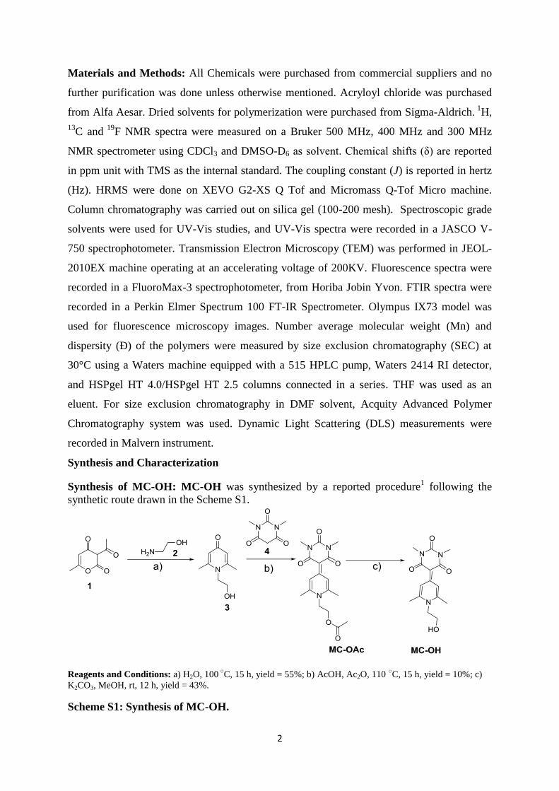
Synthesis of MC-OH: MC-OH was synthesized by a reported procedure1 following the
synthetic route drawn in the Scheme S1.
Reagents and Conditions: a) H2O, 100 ○
C, 15 h, yield = 55%; b) AcOH, Ac2O, 110 ○C, 15 h, yield = 10%; c)
K2CO3, MeOH, rt, 12 h, yield = 43%.
Scheme S1: Synthesis of MC-OH.

3
Synthesis of Compound 3: Dehydroacetic acid (1) (10.0 g, 59.47 mmol) and ethanolamine
(2) (5.38 mL, 89.2 mmol) were taken in a round bottom flask with 150 mL of water and the
mixture was refluxed for 15 h at 100 ○C. After 15 h, the mixture was cooled to room
temperature. Excess water was evaporated under reduced pressure and the brown crude
obtained was further refluxed with 35 mL ethanol. The solution was initially cooled to room
temperature and then kept in freezer overnight to obtain yellowish white crystals (5.5 g,
55%). M.P. = 116-118 ○C;
1H NMR (400 MHz, DMSO-d6, TMS): δ (ppm) = 5.92 (s, 2H),
5.07 (t, J = 5.3 Hz, 1H), 3.97-3.93 (m, 2H,), 3.60 (q, J = 5.7 Hz, 2H), 2.28 (s, 6H); 13
C NMR
(100 MHz, DMSO-d6): δ (ppm) = 20.21, 48.39, 59.81, 117.57, 149.53, 177.26. HRMS (ESI):
m /z calcd for C9H14NO2 [M + H]+:168.1019; found: 168.1018.
Synthesis of MC-OAc: Compound (3) (5.50 g, 32.9 mmol) and 1, 3-Dimethylbarbituric acid
(4) (6.16 g, 39.48 mmol) were taken together in a round bottom flask. To that acetic acid (10
mL) and acetic anhydride (70 mL) were added and the reaction mixture was heated at 112 ○C
under argon atmosphere. After 15 h, the reaction was stopped and the mixture was cooled to
room temperature and excess acetic acid was evaporated under rotary evaporator to obtain a
brown solid. The crude was purified by column chromatography using silica gel (100-200
mesh) as a stationary phase and 2-4% MeOH in CHCl3 as eluent to obtain the desired product
as light brown solid (1.1 gm, 10 %). M.P. = 251 ○C;
1H NMR (400 MHz, CDCl3, TMS): δ
(ppm) = 9.04 (s, 2H), 4.40-4.31 (m, 4H), 3.36 (s, 6H), 2.63 (s, 6H), 2.08 (s, 3H); 13
C-NMR
(100 MHz, CDCl3,): δ (ppm) = 25.57, 29.78, 47.42, 61.44, 90.05, 121.69, 148.02, 152.29,
156.19, 164.61, 170.51. HRMS (ESI): m/z calcd for C17H21N3O5Na [M+Na]+: 370.1379;
found: 370.1378.
Synthesis of MC-OH: MC-OAc (0.90 g, 2.6 mmol) and K2CO3 (0.72 g, 5.2 mmol) were
taken in a round bottom flask with 45 mL methanol and the mixture was stirred at room
temperature for 12 h. Excess methanol was removed under rotary evaporator and the crude
was dissolved in CH2Cl2 and filtered. The residue was washed multiple times with CH2Cl2.
The desired product was obtained as light yellow solid by removing the organic CH2Cl2 phase
(330 mg, 43 %). M.P. = 295 ○C;
1H NMR (400 MHz, DMSO-d6, TMS): δ (ppm) = 8.83 (s,
2H), 4.34 (t, J = 5.4 Hz, 2H), 3.79-3.73 (m, 2H), 3.16 (s, 6H), 2.63 (s, 6H); 13
C NMR (100
MHz, DMSO-d6): δ (ppm) = 21.88, 27.83, 52.19, 59.85, 87.51, 120.90, 150.35, 152.08,
154.58, 163.38. HRMS (ESI): m/z calculated for C15H20N3O4 [M+H]+: 306.1448; found:
306.1445.
4
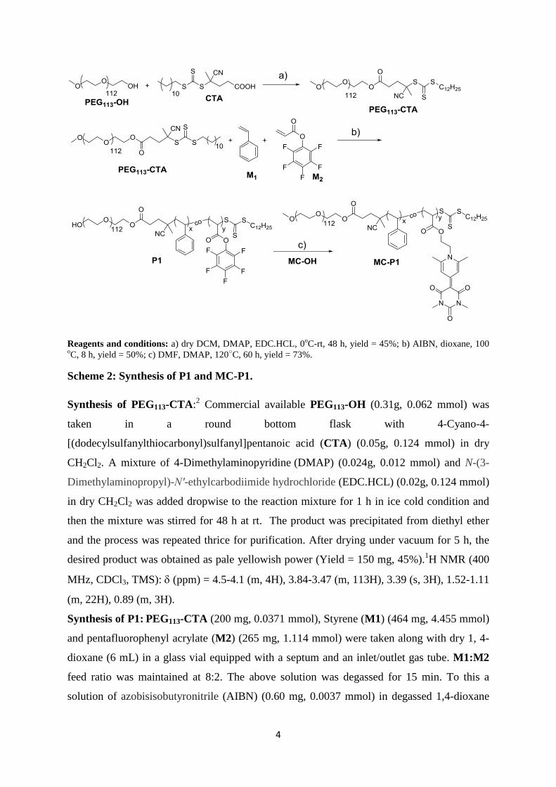
Reagents and conditions: a) dry DCM, DMAP, EDC.HCL, 0oC-rt, 48 h, yield = 45%; b) AIBN, dioxane, 100
oC, 8 h, yield = 50%; c) DMF, DMAP, 120
○C, 60 h, yield = 73%.
Scheme 2: Synthesis of P1 and MC-P1.
Synthesis of PEG113-CTA:2 Commercial available PEG113-OH (0.31g, 0.062 mmol) was
taken in a round bottom flask with 4-Cyano-4-
[(dodecylsulfanylthiocarbonyl)sulfanyl]pentanoic acid (CTA) (0.05g, 0.124 mmol) in dry
CH2Cl2. A mixture of 4-Dimethylaminopyridine (DMAP) (0.024g, 0.012 mmol) and N-(3-
Dimethylaminopropyl)-N′-ethylcarbodiimide hydrochloride (EDC.HCL) (0.02g, 0.124 mmol)
in dry CH2Cl2 was added dropwise to the reaction mixture for 1 h in ice cold condition and
then the mixture was stirred for 48 h at rt. The product was precipitated from diethyl ether
and the process was repeated thrice for purification. After drying under vacuum for 5 h, the
desired product was obtained as pale yellowish power (Yield = 150 mg, 45%).1H NMR (400
MHz, CDCl3, TMS): (ppm) = 4.5-4.1 (m, 4H), 3.84-3.47 (m, 113H), 3.39 (s, 3H), 1.52-1.11
(m, 22H), 0.89 (m, 3H).
Synthesis of P1: PEG113-CTA (200 mg, 0.0371 mmol), Styrene (M1) (464 mg, 4.455 mmol)
and pentafluorophenyl acrylate (M2) (265 mg, 1.114 mmol) were taken along with dry 1, 4-
dioxane (6 mL) in a glass vial equipped with a septum and an inlet/outlet gas tube. M1:M2
feed ratio was maintained at 8:2. The above solution was degassed for 15 min. To this a
solution of azobisisobutyronitrile (AIBN) (0.60 mg, 0.0037 mmol) in degassed 1,4-dioxane
5
was added and the reaction vial was immersed in a preheated oil bath at 100 °C for 8 h. The
reaction was quenched by exposing to air. The solution was diluted with CH2Cl2 and purified
by precipitating from hexane to afford a white solid. The process was repeated thrice to
obtain a pure polymer which was dried under vacuum at 40 °C for 48 h (Yield = 456 mg, 50
%). 1H NMR of P1 (400 MHz, CDCl3, TMS): δ (ppm) = 7.22-6.88 (bs, 3H), 6.85-6.29 (bs,
2H), 3.64 (s, 4H), 3.33 (s, 3H), 2.53-1.15 (multiple bs, 6H), 0.86 (m, 3H). FTIR shows
characteristic peak for PFP-ester carbonyl stretching at 1783 cm-1
. From the crude 1H NMR
and 19
F NMR, M1 and M2 conversion were calculated to be 32% and 62%, from which the
degree of polymerization (DP) for M1 and M2 were estimated to be 38 and 19, respectively.
Molar mass calculated from 1H NMR = 13,800 g/ mol; GPC (PS standard, THF): Mn = 3760
g/mol, Ð =1.18 for P1 and Mn =1470 g/mol, Ð =1.23 for PEG113-CTA. The values obtained
were under estimated; GPC (PMMA standard, DMF): Mn = 24800 g/mol, Ð =1.07 for P1
and Mn =19020 g/mol, Ð =1.23 for PEG113-CTA. The values obtained were over estimated.
Synthesis of MC-P1: The transesterification of P1 with MC-OH was performed following
literature procedure as follows:1
P1 (60 mg, 0.013 mmol of PFP-ester, 1.0 equiv) and DMAP (4.64 mg, 0.003 mM, 0.2 equiv)
were taken with 2.0 mL dry DMF and added with MC-OH (7.7 mg, 0.025 mM, 2.0 equiv).
The mixture was stirred at 120 °C for 60 h. The formation of the product was monitored by
FTIR measurements. The conversion was quantitative as no PFP-ester carbonyl peak at 1783
cm-1
was seen after the reaction. The mixture was dissolved in CH2Cl2 and purified by
precipitation from hexane. The precipitation method was repeated thrice to obtain solid
brown polymer MC-P1 (yield = 50 mg, 73%). 1H NMR (400 MHz, CDCl3, TMS): δ (ppm) =
δ 8.97 (s, 2H), 7.07 (bs, 3H), 6.58 (bs, 2H), 3.64 (s, 4H), 3.36 (s, 3H), 2.08-1.15 (multiple bs,
6H); By comparing the integrals of aromatic protons of MC and styrene with methylene
proton signals of PEG113 in MC-P1, the degree of polymerization (DP) of M1 and M2 were
estimated to be 45 and 18, respectively, which was comparable with the value obtained from
the crude 1H NMR data of P1 (M1:M2:: 38:19). Molar mass from
1H NMR of MC-P1:
15,500 g / mol. GPC (PS standard, THF): Mn = 1577 g/ mol, Ð =1.6 which was under
estimated.
Synthesis of P2: The PEG113-CTA (100 mg, 0.02 mmol) and Styrene (M1) (312 mg, 3.0
mmol) were taken along with dry 1, 4-dioxane (200 µL) in a glass vial equipped with a
septum and an inlet/outlet gas tube. The above solution was degassed for 15 min. To this a

6
solution of AIBN (0.82 mg, 0.005 mmol) in degassed 1,4-dioxane was added and the reaction
vial was immersed in a preheated oil bath at 72 °C for 22 h. The solution was diluted with
CH2Cl2 and purified by precipitating from cold diethyl ether to afford off white solid. The
process was repeated thrice to obtain a pure polymer which was dried under vacuum at 40 °C
for 4 h (Yield = 70 mg, 16 %). 1H NMR (500 MHz, CDCl3, TMS): δ (ppm) = 7.26-6.87 (bs,
3H), 6.72-6.33 (bs, 2H), 3.66 (s, 4H), 3.38 (s, 3H), 1.97-1.21 (multiple bs, 6H). Molar mass
calculated from 1H NMR = 10,080 g/ mol; GPC (PS standard, THF): Mn = 2155 g/mol, Ð
=1.15. This value was under estimated.
Experimental procedures:
Sample preparation in water: Measured amount (1.0 mg) of polymer MC-P1 was taken in
a glass vial and dissolved in 100 µL THF. To this 1.0 mL water was added dropwise and the
THF was evaporated by gently heating the solution. The aqueous solution was further heated
to 90 °C to break any pre-aggregated structure and then cooled to room temperature to obtain
a clear solution. The final MC-P1 concentration becomes 1.0 mg / mL in water. The solution
was allowed to equilibrate at room temperature for 1.0 h before any physical studies were
performed.
Sample preparation in toluene: Measured amount (1.0 mg) of polymer MC-P1 was taken
in a glass vial. To this 1.0 mL toluene was added and the solution was heated to 90 ○C to
break any pre-aggregated structure and cooled to room temperature to obtain a clear solution.
The final concentration becomes 1.0 mg/ mL in toluene. The solution was allowed to
equilibrate at room temperature for 1.0 h before any physical studies were done.
UV-Vis studies: Solvent-dependant UV-Vis absorption spectroscopic experiments were
performed using a quartz cuvette of 1.0 mm path length at 25 °C. For variable temperature
UV-Vis studies, two separate solutions of MC-P1 (1.0 mg/ mL) in water and toluene were
heated from 25°C to 90 °C at an interval of 10 °C. Before taking the measurements, each time
sample was allowed to stand for 5.0 min after the desired temperature was reached.
Photoluminescence studies: Samples were prepared in the same way as discussed earlier.
The emission intensity for MC-P1 solutions were recorded in a quartz cuvette of 10 mm path
length. The excitation wavelength (ex) was maintained at = 372 nm. Excitation and emission
band width = 3 nm each.

7
Dynamic light scattering studies: Experiments were carried out with a solution of MC-P1
(1.0 mg/ mL) in water and toluene separately. The solutions were prepared following the
procedure described earlier. Samples were equilibrated for 4.0 h before taking the reading.
For variable temperature studies, the solution was heated from 25 oC to higher temperatures
and the spectral measurements were carried out at different temperatures.
Transmission Electron Microscopy (TEM) studies: 0.5 mg/mL solution of MC-P1 in
water and toluene separately were drop casted on copper grids and slowly air dried for 48 h
prior to imaging.
Dye encapsulation studies: A stock solution of Nile red in THF (1× 10⁻⁴
M) was freshly
prepared. 10 µL of stock solution was taken in a vial. To this, 1.0 mL solution of MC-P1 (1
mg/ mL) in water was added dropwise (final dye concentration = 1 × 10⁻⁶
M) and trace
amount of THF was removed by slowly heating the solution. The solution was sonicated for
15 min and allowed to equilibrate for 2.0 h at room temperature before taking the
photoluminescence spectra.
Similar procedure was followed for encapsulating Rodamine B (stock = 1× 10⁻⁴
M in THF)
in MC-P1 aggregates in toluene. Final concentration of MC-P1 = 1.0 mg/ mL and Rodamine
B = 1 × 10⁻⁶
M in toluene.
Fluorescence microscopy (FM): Nile red and Rodamine B encapsulated solutions of MC-
P1 (0.5 mg/mL) in water and toluene, respectively were drop-casted on glass slides and air
dried overnight before FM images were captured. For fluorescence microscopy images of
MC-P1 aggregates in water and toluene, MC-P1 concentration was kept at 1.0 mg / mL. The
samples were drop-casted on glass slides and air dried overnight before FM images were
captured.
CMC Determination: For determination of CMC of MC-P1 in water, 10 µL stock solution
of Nile red in THF (1×10⁻⁴
M) was transferred to 10 different vials. To these, different
measured quantity of a stock solution of MC-P1 in THF (1.0 mg/ mL) was added and the
solvent was removed by heating to obtain a thin film. To this, water was added dropwise and
the final volume was adjusted to 1.0 mL. All the solutions were sonicated for 10 min and then
allowed to equilibrate for 2.0 h prior to measuring the spectra. All the resulting solutions had
same Nile red concentration (1×10⁻⁶
M) but varying concentrations of polymer (1.0 - 0.0
mg/mL). The emission spectra were recorded for each solution at an excitation wavelength
(ex) = 530 nm. Both excitation and emission band width were kept at 3.0 nm.

8
For determination of CMC of MC-P1 in toluene by UV-Vis spectroscopy, 1.0 mg/ mL
solution of MC-P1 in toluene was prepared. The solution was diluted stepwise (from 1.0 mg/
mL to 0.008 mg/mL) with measured quantity of toluene and in each step the UV-Vis spectra
of MC-P1 were recorded in a quartz cuvette of 1.0 mm path length.
Förster Resonance Energy Transfer (FRET) Studies: Stock solutions of the donor dye
DiO (3,3´-di-octadecyloxacarbocyanine perchlorate) and the acceptor dye DiI (1,10-
dioctadecyl-3,3,30,30-tetramethylindocarbocyanine perchlorate) were prepared in THF (1 ×
10-4
M). 10 µL each of DiO and DiI stock solutions were taken in two separate vials and THF
was removed. To these two vials, measured quantity (0.6 mg in 30 L THF) of MC-P1 was
added followed by dropwise addition of 600 µL water to make the final polymer
concentration 1.0 mg/ mL. Finally THF was removed by slowly heating the solution. Both
the solutions were sonicated for 30 min in a water bath at room temperature and allowed to
equilibrate for 2.0 h and filtered to remove any non-encapsulated dye. Final dye concentration
= 1.6 × 10⁻⁶
M.
Kinetic Study: 500 µL each of individual DiO and DiI loaded MC-P1 (1.0 mg/mL)
solutions were mixed in 1:1 ratio and emission spectra were recorded to monitor
FRET with time.
Dilution Experiment with THF: 5.0 µL of each DiO and DiI stock solutions (1 x10⁻⁴
M) in THF were taken in a single vial. To this, measured quantity (0.6 mg in 30 L
THF) of MC-P1 was added followed by dropwise addition of 600 µL water. Finally
the THF was removed by slowly heating the solution. The final concentration of MC-
P1 = 1.0 mg /mL in water and coencapsulated DiO and DiI concentration = 0.8 x10⁻⁶
M each. The solution was equilibrated for 30 min before taking the measurements. To
this solution, measured volume of THF was added stepwise and the emission spectra
were recorded to monitor FRET.
Dilution Experiment with water: Similar procedure was followed for the sample
preparation as done for dilution experiment with THF. Final MC-P1 concentration =
1.0 mg / mL and coencapsulated DiO and DiI concentration = 0.8 x 10⁻⁶
M.

9
Additional Figures:
Figure S1: UV-Vis absorption spectrum of MCOH in THF showing max = 378 nm
(Conc. = 0.5 mg / mL). Path length = 1.0 mm.
Figure S2: 19
F NMR spectrum of P1 and MC-P1 in CDCl3.
Figure S3: a) GPC chromatogram in THF and b) in DMF. No signal for MC-P1 was
observed in DMF GPC chromatogram. c) GPC chromatogram of P2 in THF.
10
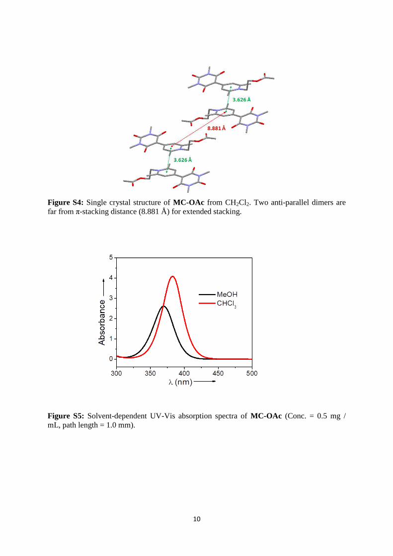
Figure S4: Single crystal structure of MC-OAc from CH2Cl2. Two anti-parallel dimers are
far from π-stacking distance (8.881 Å) for extended stacking.
Figure S5: Solvent-dependent UV-Vis absorption spectra of MC-OAc (Conc. = 0.5 mg /
mL, path length = 1.0 mm).

11
Figure S6: Structure of DiO and DiI dyes (Top); Emission intensity from an aqueous
solution of DiO+DiI coencapsulated in MC-P1 micelle and in absence of MC-P1 micelle
(Bottom). Dye Conc. = 0.8 x10⁻⁶
M, ex = 450 nm.
Figure S7: a) Concentration-dependent emission spectra of Nile red (Conc. = 1 × 10⁻⁶
M, ex
= 530 nm) loaded P2 in water; b) Nile red emission vs. P2 concentration plot; CMC = 0.29
mg/mL ≈ 2.9 x 10⁻⁵
M.

12
Figure S8: UV-Vis absorption spectrum of MC-P1 in water below CMC (Conc. = 0.013 mg
/ mL); max = 372 nm, Path length = 1.0 mm.
Figure S9: Variable-temperature DLS of a) P1 and b) P2 (Conc. = 1.0 mg /mL).
Figure S10: UV-Vis absorption spectrum of MC-P1 in water with 62% THF (Conc. = 1.0
mg / mL); max = 372 nm, Path length = 1.0 mm.

13
Figure S11: DLS data of MC-P1 in Toluene at 25 ○
C (Conc. = 1.0 mg / mL).
Figure S12: Concentration-dependent UV-Vis absorption spectra of MC-P1 in toluene; Path
length = 1.0 mm.
Figure S13: Fluorescence microscopy image from an aggregated solution of MC-P1 in
toluene (Conc. = 1.0 mg / mL).

14
Figure S14: a) TEM image from an aqueous solution of P1; b) Nile red loaded aqueous
solution of P1 under fluorescence microscope. Conc. of P1 = 1.0 mg / mL, Nile red = 1 ×
10−6
M.
Figure S15: a) Concentration-dependent emission spectra of Nile red (Conc. = 1 × 10⁻⁶
M,
ex = 530 nm) loaded P1 in water; b) Nile red emission vs. P1 concentration plot; CMC =
0.14 mg/mL ≈ 1.0 x 10⁻⁵
M.
X-ray Structure Determination:
The molecular structure of MC-OAc was determined by single crystal X-ray structure
determination. Diffraction-quality crystals were obtained by slow evaporation from a
concentrated solution in CH2Cl2. Single crystal for MC-OAc was coated with Parabar oil and
was mounted under a nitrogen cold stream. Data collection was performed at 100 K on a
Bruker D8VENTURE Microfocus diffractometer equipped with PHOTON II Detector and
Mo Kα radiation (λ = 0.71073 Å), controlled by the APEX3 (v2017.3-0) software package.
Space group was assigned by systematic absences (determined by XPREP) and analysis of
metric symmetry was further checked by PLATON.3,4
Structure was solved by direct method
and refined against all data in the reported 2θ ranges by full-matrix least squares on F2 using

15
the SHELXL program suite5 in the WinGX
6 interface. Non-hydrogen atoms were refined
anisotropically, and hydrogen atoms were fixed at calculated positions and refined using a
riding model. The details of crystal data collection and refinement of complexes are
summarized in Table S1.
Table S1: Crystallographic Data for MC-OAc
Compound MC-OAc
Empirical formula C18 H23 Cl2 N3 O5
FW 432.29
T, K 119(2)
Crystal system Monoclinic
Space group P21/c
a, Å 11.6895(10)
b, Å 23.580(2)
c, Å 7.2918(7)
α, deg 90.00°
β, deg 94.989°(3)
γ, deg 90.00°
V, Å3 2002.3(3)
Z, ρ, Mg m-3
4, 1.434
μ, mm-1
0.359
F(000) 904
Refln. collected 17289
Ind. Reflen 2907
Data/restn./param 2907 / 0 / 259
GOF on F2 1.046
Final R indices [I>2σ(I)] 0.0684, 0.1682

16
NMR & HRMS Spectra
1H NMR spectrum of Compound-3; (*) denotes residual solvent peak from DMSO-d6.
13C NMR spectrum of Compound-3; (*) denotes residual solvent peak from DMSO-d6.

17
HRMS Spectrum of Compound-3.
1H NMR spectrum of MC-OAc; (*) denotes residual solvent peak for CDCl3.

18
13C NMR spectrum of MC-OAC; (*) denotes residual solvent peak for CDCl3.
HRMS Spectrum of MC-OAc.

19
1H NMR spectrum of MC-OH; (*) denotes residual solvent peak for DMSO-d6.
13C NMR spectrum of MC-OH; (*) denotes residual solvent peak from DMSO-d6.

20
HRMS Spectrum of MC-OH.
References:
1. A. Das, S. Lin and P. Theato, ACS Macro Lett., 2017, 6, 50-55.
2. P. Pramanik and S. Ghosh, J. Polym. Sci., Part A: Polym. Chem., 2015, 53, 2444-
2451.
3. A. L. Spek, J. Appl. Crystallogr., 2003, 36, 7-13.
4. A. L. Spek, Acta Crystallogr. Sect. D., 2009, 65, 148-155.
5. G. M. Sheldrick, Acta Crystallogr. Sect. A, 2008, 64, 112-122.
6. L. J. Farrugia, J. Appl. Crystallogr., 1999, 32, 837-838.