Supporting Information€¦ · Supporting Information Yeh et al. 10.1073/pnas ... quantitative...
6
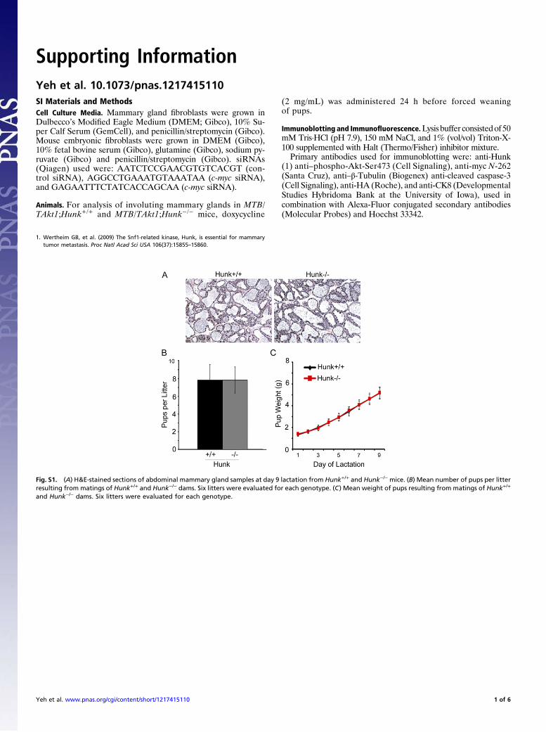
Supporting Information Yeh et al. 10.1073/pnas.1217415110 SI Materials and Methods Cell Culture Media. Mammary gland fibroblasts were grown in Dulbecco’s Modified Eagle Medium (DMEM; Gibco), 10% Su- per Calf Serum (GemCell), and penicillin/streptomycin (Gibco). Mouse embryonic fibroblasts were grown in DMEM (Gibco), 10% fetal bovine serum (Gibco), glutamine (Gibco), sodium py- ruvate (Gibco) and penicillin/streptomycin (Gibco). siRNAs (Qiagen) used were: AATCTCCGAACGTGTCACGT (con- trol siRNA), AGGCCTGAAATGTAAATAA (c-myc siRNA), and GAGAATTTCTATCACCAGCAA (c-myc siRNA). Animals. For analysis of involuting mammary glands in MTB/ TAkt1;Hunk +/ + and MTB/TAkt1;Hunk -/ - mice, doxycycline (2 mg/mL) was administered 24 h before forced weaning of pups. Immunoblotting and Immunofluorescence. Lysis buffer consisted of 50 mM Tris·HCl (pH 7.9), 150 mM NaCl, and 1% (vol/vol) Triton-X- 100 supplemented with Halt (Thermo/Fisher) inhibitor mixture. Primary antibodies used for immunoblotting were: anti-Hunk (1) anti–phospho-Akt-Ser473 (Cell Signaling), anti-myc N-262 (Santa Cruz), anti–β-Tubulin (Biogenex) anti-cleaved caspase-3 (Cell Signaling), anti-HA (Roche), and anti-CK8 (Developmental Studies Hybridoma Bank at the University of Iowa), used in combination with Alexa-Fluor conjugated secondary antibodies (Molecular Probes) and Hoechst 33342. 1. Wertheim GB, et al. (2009) The Snf1-related kinase, Hunk, is essential for mammary tumor metastasis. Proc Natl Acad Sci USA 106(37):15855–15860. Fig. S1. (A) H&E-stained sections of abdominal mammary gland samples at day 9 lactation from Hunk +/+ and Hunk -/- mice. (B) Mean number of pups per litter resulting from matings of Hunk +/+ and Hunk -/- dams. Six litters were evaluated for each genotype. (C ) Mean weight of pups resulting from matings of Hunk +/+ and Hunk -/- dams. Six litters were evaluated for each genotype. Yeh et al. www.pnas.org/cgi/content/short/1217415110 1 of 6
Transcript of Supporting Information€¦ · Supporting Information Yeh et al. 10.1073/pnas ... quantitative...
Supporting InformationYeh et al. 10.1073/pnas.1217415110SI Materials and MethodsCell Culture Media. Mammary gland fibroblasts were grown inDulbecco’s Modified Eagle Medium (DMEM; Gibco), 10% Su-per Calf Serum (GemCell), and penicillin/streptomycin (Gibco).Mouse embryonic fibroblasts were grown in DMEM (Gibco),10% fetal bovine serum (Gibco), glutamine (Gibco), sodium py-ruvate (Gibco) and penicillin/streptomycin (Gibco). siRNAs(Qiagen) used were: AATCTCCGAACGTGTCACGT (con-trol siRNA), AGGCCTGAAATGTAAATAA (c-myc siRNA),and GAGAATTTCTATCACCAGCAA (c-myc siRNA).
Animals. For analysis of involuting mammary glands in MTB/TAkt1;Hunk+/+ and MTB/TAkt1;Hunk−/− mice, doxycycline
(2 mg/mL) was administered 24 h before forced weaningof pups.
Immunoblotting and Immunofluorescence.Lysis buffer consistedof 50mM Tris·HCl (pH 7.9), 150 mM NaCl, and 1% (vol/vol) Triton-X-100 supplemented with Halt (Thermo/Fisher) inhibitor mixture.Primary antibodies used for immunoblotting were: anti-Hunk
(1) anti–phospho-Akt-Ser473 (Cell Signaling), anti-myc N-262(Santa Cruz), anti–β-Tubulin (Biogenex) anti-cleaved caspase-3(Cell Signaling), anti-HA (Roche), and anti-CK8 (DevelopmentalStudies Hybridoma Bank at the University of Iowa), used incombination with Alexa-Fluor conjugated secondary antibodies(Molecular Probes) and Hoechst 33342.
1. Wertheim GB, et al. (2009) The Snf1-related kinase, Hunk, is essential for mammarytumor metastasis. Proc Natl Acad Sci USA 106(37):15855–15860.
Fig. S1. (A) H&E-stained sections of abdominal mammary gland samples at day 9 lactation from Hunk+/+ and Hunk−/− mice. (B) Mean number of pups per litterresulting from matings of Hunk+/+ and Hunk−/− dams. Six litters were evaluated for each genotype. (C) Mean weight of pups resulting from matings of Hunk+/+
and Hunk−/− dams. Six litters were evaluated for each genotype.
Yeh et al. www.pnas.org/cgi/content/short/1217415110 1 of 6
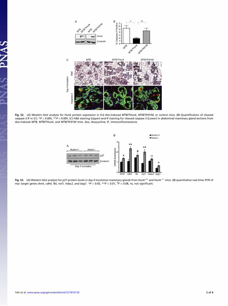
Fig. S2. (A) Western blot analysis for Hunk protein expression in 4-d dox-induced MTB/THunk, MTB/TK91M, or control mice. (B) Quantification of cleavedcaspase-3 IF in (C). *P < 0.005, **P = 0.005. (C) H&E staining (Upper) and IF staining for cleaved caspase-3 (Lower) in abdominal mammary gland sections fromdox-induced MTB, MTB/THunk, and MTB/TK91M mice. dox, doxycycline; IF, immunofluorescence.
Fig. S3. (A) Western blot analysis for p27 protein levels in day 4 involution mammary glands from Hunk+/+ and Hunk−/− mice. (B) quantitative real-time–PCR ofmyc target genes shmt, cdk4, fbl, nol1, hdac2, and bag1. *P < 0.05, **P < 0.01, #P = 0.08, ns, not significant.
Yeh et al. www.pnas.org/cgi/content/short/1217415110 2 of 6
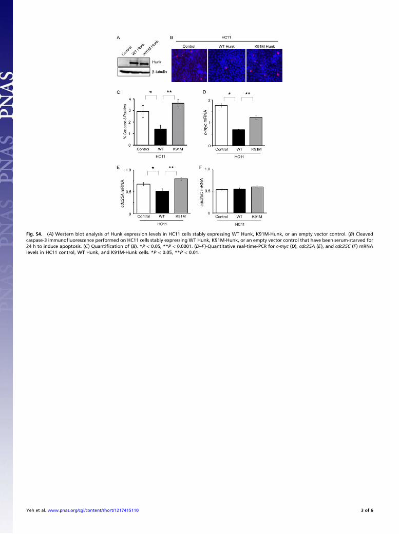
Fig. S4. (A) Western blot analysis of Hunk expression levels in HC11 cells stably expressing WT Hunk, K91M-Hunk, or an empty vector control. (B) Cleavedcaspase-3 immunofluorescence performed on HC11 cells stably expressing WT Hunk, K91M-Hunk, or an empty vector control that have been serum-starved for24 h to induce apoptosis. (C) Quantification of (B). *P < 0.05, **P < 0.0001. (D–F)-Quantitative real-time-PCR for c-myc (D), cdc25A (E), and cdc25C (F) mRNAlevels in HC11 control, WT Hunk, and K91M-Hunk cells. *P < 0.05, **P < 0.01.
Yeh et al. www.pnas.org/cgi/content/short/1217415110 3 of 6
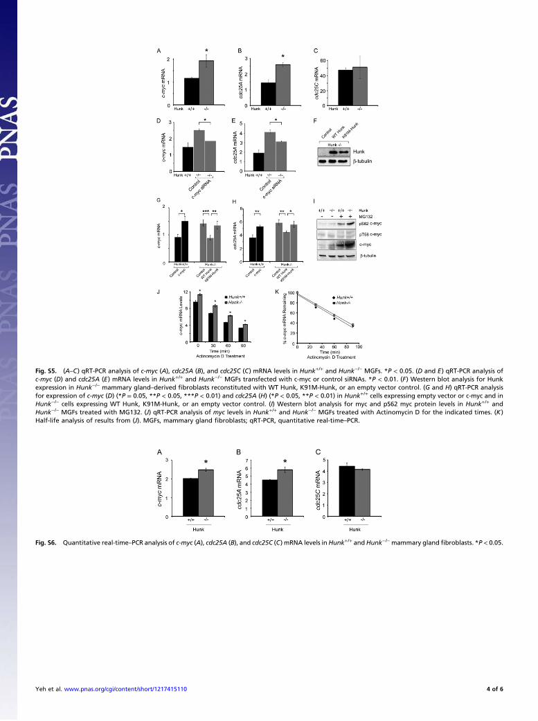
Fig. S5. (A–C) qRT-PCR analysis of c-myc (A), cdc25A (B), and cdc25C (C) mRNA levels in Hunk+/+ and Hunk−/− MGFs. *P < 0.05. (D and E) qRT-PCR analysis ofc-myc (D) and cdc25A (E) mRNA levels in Hunk+/+ and Hunk−/− MGFs transfected with c-myc or control siRNAs. *P < 0.01. (F) Western blot analysis for Hunkexpression in Hunk−/− mammary gland–derived fibroblasts reconstituted with WT Hunk, K91M-Hunk, or an empty vector control. (G and H) qRT-PCR analysisfor expression of c-myc (D) (*P = 0.05, **P < 0.05, ***P < 0.01) and cdc25A (H) (*P < 0.05, **P < 0.01) in Hunk+/+ cells expressing empty vector or c-myc and inHunk−/− cells expressing WT Hunk, K91M-Hunk, or an empty vector control. (I) Western blot analysis for myc and pS62 myc protein levels in Hunk+/+ andHunk−/− MGFs treated with MG132. (J) qRT-PCR analysis of myc levels in Hunk+/+ and Hunk−/− MGFs treated with Actinomycin D for the indicated times. (K)Half-life analysis of results from (J). MGFs, mammary gland fibroblasts; qRT-PCR, quantitative real-time–PCR.
Fig. S6. Quantitative real-time–PCR analysis of c-myc (A), cdc25A (B), and cdc25C (C) mRNA levels inHunk+/+ andHunk−/−mammary gland fibroblasts. *P < 0.05.
Yeh et al. www.pnas.org/cgi/content/short/1217415110 4 of 6
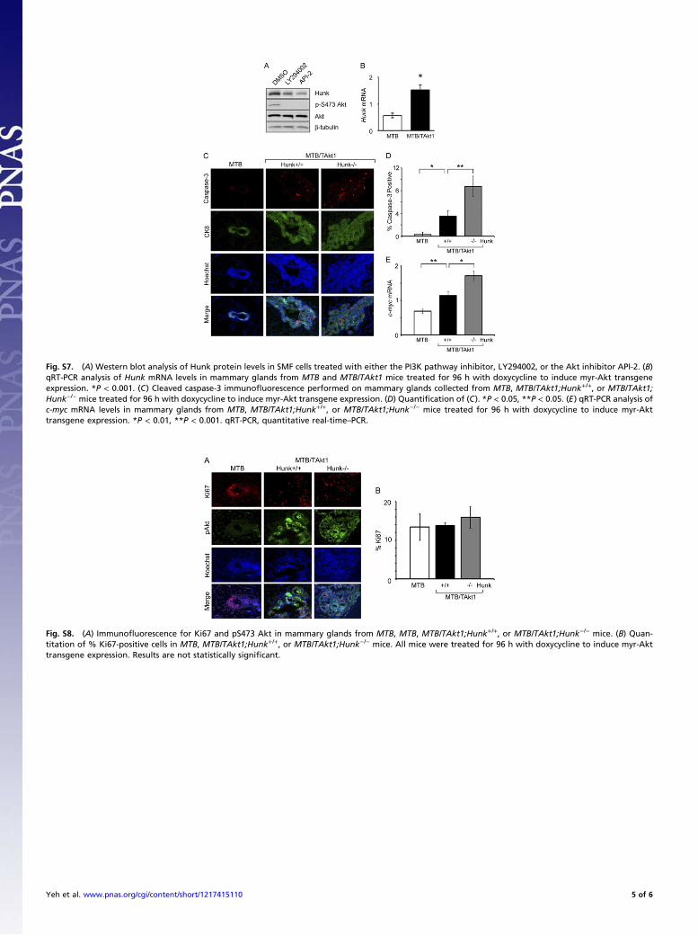
Fig. S7. (A) Western blot analysis of Hunk protein levels in SMF cells treated with either the PI3K pathway inhibitor, LY294002, or the Akt inhibitor API-2. (B)qRT-PCR analysis of Hunk mRNA levels in mammary glands from MTB and MTB/TAkt1 mice treated for 96 h with doxycycline to induce myr-Akt transgeneexpression. *P < 0.001. (C) Cleaved caspase-3 immunofluorescence performed on mammary glands collected from MTB, MTB/TAkt1;Hunk+/+, or MTB/TAkt1;Hunk−/− mice treated for 96 h with doxycycline to induce myr-Akt transgene expression. (D) Quantification of (C). *P < 0.05, **P < 0.05. (E) qRT-PCR analysis ofc-myc mRNA levels in mammary glands from MTB, MTB/TAkt1;Hunk+/+, or MTB/TAkt1;Hunk−/− mice treated for 96 h with doxycycline to induce myr-Akttransgene expression. *P < 0.01, **P < 0.001. qRT-PCR, quantitative real-time–PCR.
Fig. S8. (A) Immunofluorescence for Ki67 and pS473 Akt in mammary glands from MTB, MTB, MTB/TAkt1;Hunk+/+, or MTB/TAkt1;Hunk−/− mice. (B) Quan-titation of % Ki67-positive cells in MTB, MTB/TAkt1;Hunk+/+, or MTB/TAkt1;Hunk−/− mice. All mice were treated for 96 h with doxycycline to induce myr-Akttransgene expression. Results are not statistically significant.
Yeh et al. www.pnas.org/cgi/content/short/1217415110 5 of 6
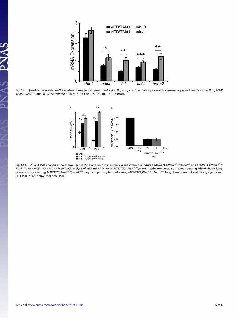
Fig. S9. Quantitative real-time–PCR analysis of myc target genes shmt, cdk4, fbl, nol1, and hdac2 in day 4 involution mammary gland samples fromMTB, MTB/TAkt1;Hunk+/+, and MTB/TAkt1;Hunk−/− mice. *P < 0.05, **P < 0.01, ***P < 0.001.
Fig. S10. (A) qRT-PCR analysis of myc target genes shmt and nol1 in mammary glands from 4-d induced MTB/TTC1;Ptenco/co;Hunk+/+ and MTB/TTC1;Ptenco/co;Hunk−/−. *P < 0.05, **P < 0.01. (B) qRT-PCR analysis of rtTA mRNA levels in MTB/TTC1;Ptenco/co;Hunk+/+ primary tumor, non–tumor-bearing Friend virus B lung,primary tumor-bearing MTB/TTC1;Ptenco/co;Hunk+/+ lung, and primary tumor-bearing MTB/TTC1;Ptenco/co;Hunk−/− lung. Results are not statistically significant.QRT-PCR, quantitative real-time–PCR.
Yeh et al. www.pnas.org/cgi/content/short/1217415110 6 of 6


![Untitled-1 [rupaonlinestore.com]rupaonlinestore.com/pdf-promotion/Frontline-Catalogue-JUNE.pdf · FRONTLINE KIDZ HUNK DRAWER SLIPS 'Dray,er Rupa & Company Limited Metro Tower, 1 ,](https://static.fdocuments.net/doc/165x107/5f18a4d69f95a7670e5af31b/untitled-1-frontline-kidz-hunk-drawer-slips-drayer-rupa-company-limited.jpg)