
Supporting Online Material for - · PDF fileOther Supporting Online Material for this...
46
www.sciencemag.org/cgi/content/full/science.1213506/DC1 Supporting Online Material for Evolutionarily Assembled cis-Regulatory Module at a Human Ciliopathy Locus Jeong Ho Lee, Jennifer L. Silhavy, Ji Eun Lee, Lihadh Al-Gazali, Sophie Thomas, Erica E. Davis, Stephanie L. Bielas, Kiley J. Hill, Miriam Iannicelli, Francesco Brancati, Stacey B. Gabriel, Carsten Russ, Clare V. Logan, Saghira Malik Sharif, Christopher P. Bennett, Masumi Abe, Friedhelm Hildebrandt, Bill H. Diplas, Tania Attié-Bitach, Nicholas Katsanis, Anna Rajab, Roshan Koul, Laszlo Sztriha, Elizabeth R. Waters, Susan Ferro-Novick, Geoffrey C. Woods, Colin A. Johnson, Enza Maria Valente, Maha S. Zaki, Joseph G. Gleeson* *To whom correspondence should be addressed. E-mail: [email protected] Published 26 January 2012 on Science Express DOI: 10.1126/science.1213506 This PDF file includes: Materials and Methods SOM Text Figs. S1 to S19 Tables S1 to S7 References Other Supporting Online Material for this manuscript includes the following: (available at www.sciencemag.org/cgi/content/full/science.1213506/DC1) Movies S1 to S11
Transcript of Supporting Online Material for - · PDF fileOther Supporting Online Material for this...

www.sciencemag.org/cgi/content/full/science.1213506/DC1
Supporting Online Material for
Evolutionarily Assembled cis-Regulatory Module at a Human Ciliopathy Locus
Jeong Ho Lee, Jennifer L. Silhavy, Ji Eun Lee, Lihadh Al-Gazali, Sophie Thomas, Erica E. Davis, Stephanie L. Bielas, Kiley J. Hill, Miriam Iannicelli, Francesco Brancati,
Stacey B. Gabriel, Carsten Russ, Clare V. Logan, Saghira Malik Sharif, Christopher P. Bennett, Masumi Abe, Friedhelm Hildebrandt, Bill H. Diplas, Tania Attié-Bitach,
Nicholas Katsanis, Anna Rajab, Roshan Koul, Laszlo Sztriha, Elizabeth R. Waters, Susan Ferro-Novick, Geoffrey C. Woods, Colin A. Johnson, Enza Maria Valente, Maha S. Zaki,
Joseph G. Gleeson*
*To whom correspondence should be addressed. E-mail: [email protected]
Published 26 January 2012 on Science Express
DOI: 10.1126/science.1213506
This PDF file includes:
Materials and Methods SOM Text Figs. S1 to S19 Tables S1 to S7 References
Other Supporting Online Material for this manuscript includes the following: (available at www.sciencemag.org/cgi/content/full/science.1213506/DC1)
Movies S1 to S11

2
Materials and Methods Research subjects We used standard methods to isolate genomic DNA from peripheral blood of the affected children and family members. Informed consents were obtained from all participating families and the studies were approved by the Casa Sollievo della Sofferenza, and the University of California, San Diego. Genetic mapping Genome wide linkage was performed using a 5K Illumina Linkage IVb mapping panel and analyzed with easyLinkage-Plus software. Multipoint LOD scores were calculated using Allegro version 1.2c where parameters were set to autosomal recessive with full penetrance and disease allele frequency of 0.001. Mutation screening Bioinformatics strategies involved design of primers to amplify 1643 of the 1667 potential JBTS2 target amplicons from 25 individuals (2 affected and both parents from each of 7 families) resulting in 100% sequence coverage at 1x and 80% sequence coverage at 2x using bidirectional Sanger-based sequencing. The resultant 82,150 sequence reads were annotated according to filters to exclude (1) any polymorphism heterozygous in an affected, (2) any polymorphism reported as homozygous in dbSNP, (3) any polymorphism homozygous in both parents and affected. From this data, we identified TMEM138 mutations in all remaining JBTS2 families. To test for additional TMEM138 mutations in a cohort of 460 individuals with JBTS, we applied high-resolution melting using a LightCycler 480 (Roche Applied Science), with the same TMEM138 primers and optimized PCR conditions. Mutations were validated by Sanger sequencing. All mutations segregated properly according to a single recessive disease mode. All mutations were excluded in 200 Central Asian (predominantly Pakistani and Egyptian) and 200 European (predominantly British) individuals, as well as a cohort of 96 ethnically diverse individuals by Sanger sequencing or high-resolution melting followed by Sanger sequencing. Two mutations were seen in more than one family, correlating with their country of origin, each observed on a shared haplotype. No TMEM216-mutated patients had mutations in TMEM138, or vice versa, suggesting independent mutational events. The phenotype associated with TMEM138 mutations is characterized by the presence of the MTI and extreme variability of other ciliopathy features that is characteristic of the JBTS2 locus (Table S1). Bioinformatics Genetic location is based on Human Genome Browser build hg18 of the human genome. Genetic locations for the other genomes were obtained from UCSC Genome Browser and Ensemble web site (Table S2-3) (1). The conserved genomic regions (>50% sequence similarity) were searched using mVISTA, ECR browser, and PhastCons of UCSC genome browser (2-4). Possible regulatory elements were searched using VISTA Browser and ENCODE Regulation database through UCSC Genome Browser (5, 6). To predict the protein structure of TMEM138, Phobius and TMHMM web-based tools were

3
used (7). The sequence alignment of TMEM138 and TMEM17/TMEM80/TMEM216 was performed with the Geneious program (http://www.geneious.com). Pairwise alignments of TMEM138 and TMEM216 were performed using EMBOSS-Align and BLAST. GeneCards microarray data was obtained from http://www.genecards.org. Phylogenetic analysis Phylogenetic analysis was conducted in order to examine the relationships of the TMEM proteins. The identified TMEM138, TMEM216, TMEM80, and TMEM17 proteins were aligned by clustal W using BioEdit Sequence Alignment Editor (8). The aligned sequences were manually trimmed on the N- and C-terminal ends to remove weak or ambiguous alignments. Phylogenetic analysis was performed using MEGA4 (9). We used default settings for the Neighbor-Joining method (JTT Matrix model). To estimate the reliability of the phylogenetic tree, bootstrap analysis was performed with 500 replicates of full heuristic searches. Cloning Full-length TMEM138 was cloned into the pcDNA3.1 vector and then shuttled into mCherry-, EGFP- and Flag-containing vectors (all tags on C-terminus of TMEM138). Mutations were introduced into TMEM138-pEGFP-N3 by QuikChange mutagenesis (Stratagene). The TMEM138 open reading frame was also cloned into pCS2+ vector to make RNA for injection into zebrafish embryos. TMEM216 related plasmids have been described previously (10). Zebrafish orthologs of human TMEM138 (XM_001333589) and TMEM216 (XM_678677) was cloned into the pcDNA3.1 and then shuttled into EGFP- and mCherry-containing vector respectively. Myc-TRAPPC9 and RFX4-Flag were gifts from Dr. Ferro-Novick (11) and Dr. Peterson (12), respectively. Cells, antibodies, and other reagents IMCD3, COS7, hTERT-RPE, HEK293T, and ZF4 (zebrafish embryonic fibroblasts) cells were grown in DMEM or DMEM-Ham’s F12 supplemented with 10% (vol/vol) fetal bovine serum (FBS) at 37 °C or 30 °C (for ZF4) in 5% CO2. Patient fibroblasts were maintained in MEM supplemented with 20% (vol/vol) FBS. Normal, non-diseased control fibroblasts were from ATCC. Mouse antisera specific to TMEM138 and TMEM216 were raised against the peptide sequence CYFYKRTAVRLGDPHFYQDS (131–150aa) and MLPRGLKMAPRGKRL (1-15aa) respectively by A&G Pharmaceutical Inc. Rabbit anti-TMEM216 antibody has been described (10). Primary antibodies used were mouse anti-EGFP antibody (Covance MMS-118R); mouse or rabbit anti-FLAG (Sigma-Aldrich); mouse anti-α-tubulin, (Sigma-Aldrich); mouse anti-glutamylated tubulin (GT335) (13); rabbit anti-ADP-ribosylation factor-like 13B (Arl13b) (14); rabbit anti-Cep290 (15); rabbit anti-γ tubulin (Sigma-Aldrich); rabbit anti-detyrosinated alpha tubulin (glu-tubulin) (Millipore); mouse anti-early endosome antigen1 (EEA1) (BD Bioscience); mouse anti-GM130 (Abcam); rabbit anti-LAMP2 (Abcam); MitoTracker (Molecular Probes). Secondary antibodies were Alexa-Fluor 488-, Alexa-Fluor 594- and Alexa-Fluor 568- conjugated goat anti-mouse IgG and goat anti-rabbit IgG (Molecular Probes), and horseradish peroxidase–conjugated goat anti-mouse and goat anti-rabbit (Dako). Nocodazole was purchased from Sigma-Aldrich.

4
Biochemical assays Constructs encoding wild-type or mutants of tagged (EGFP or flag) TMEM138 were transfected into 293T cells. Cells were lysed after 36-48 h and samples analyzed by protein blotting with α-EGFP (1:500), α-TMEM138 (1:1000), α-FLAG (1:2000). Whole-cell extracts were prepared from healthy control and patients fibroblasts in 100-mm tissue culture dishes and analyzed by protein blotting with α-TMEM216 (1:1000) antibody. Normalized loading levels or transfection efficiency were confirmed by blotting with α-tubulin (1:2,000) or the co-transfection with pTK β-gal (1:10 ratio) and subsequent β-gal assay respectively. Transfection and siRNAs For transfection with plasmids, cells at 70-80% confluency were transfected using Lipofectamine 2000 (Invitrogen). Cells were incubated for 24-36 h before lysis or immunostaining. For RNA-interference knockdown in IMCD3, hTRET-RPE, and COS7 cells, siRNAs (Table S7 and Fig. S19) were designed against different regions of the human and mouse TMEM138 and TMEM216, human TRAPPC9, p115, and COG1 human RFX1 to 5. siGENOME Non-Targeting siRNA pools (Dharmacon) were used as scrambled siRNA controls. Individual siRNAs (20 nM) or siRNA pools (total 60 nM) were transfected into each cells at 30-40% confluency using Lipofectamine 2000 RNAiMAX (Invitrogen). Further assays were carried out 36-48 h after transfection. For the complementation experiments, the cells were co-transfected with an empty or expression vector that expresses myc-TRAPPC9* and then immunostained 36-48 h later. The vector myc-TRAPPC9* contains three silent mutations (2558T to G, 2559C to T, 2561C to A) in the CDS region where siRNA1 against human TRAPPC9 targeted. In situ hybridization in human embryos Human embryos were collected from terminated pregnancies using the mefiprestone protocol in agreement with French bioethics laws (94-654 and 04-800). Embryos were fixed in 11% formaldehyde, 60% ethanol and 10% acetic acid, embedded in paraffin and sectioned at 5 μm. TMEM216 riboprobes used were those described previously (10). Primers for TMEM138 (TMEM138F_HIS: TCGAAGGTGACCTCTTGTCA, TMEM138R_HIS: TCTGTCCCAGGCTAAAGGAA) were selected on the 3 prime end of the cDNA for PCR amplification on human DNA to be used as template for generating the riboprobes, as described previously (16). Sections were hybridized with a Digoxygenin labeled probe at 70°C overnight, and digoxygenin was detected with an anti-DIG-Fab’ antibody (Roche) at 1:1000. Chromatin immunoprecipitation Chromatin immunoprecipitation (ChIP) assay was performed as described (17) with minor modifications. The cross-linked chromatins (first 2mM DSG and then 1% formaldehyde) in hTERT-RPE cells were sheared to ~300-500 bp fragments with a Bioruptor UCD-200TM (Diagenode). The crosslinked adducts extracts were pre-cleared with Precelaring Matrix F (Santa Cruz Biotech) and subsequently incubated with 1-2 ug rabbit IgG or RFX4 antibodies (18) overnight at 4 °C and followed by Sepharose A beads for 2 hr. After extensive washing of the beads, DNA-proteins complexes were eluted and crosslinking was reversed overnight at 65 °C. After DNA purification with QIAquick

5
spin column (Qiagen), real-time qPCR was performed. Primer sets: RE1 #1(F: CAGACAAGGTTGCAGAAAAGC, R:AGGCTCTTTGACTCCCCAAT), RE1 #2 (F: CCTAAAATTCTAGAGTACGGAGAACC, R:GGGCAATGCTTGGATTCTTA), RE2 #1(F:CCAGTATGCAACCCCATTTC, R:TAGTTGCGTGACTTGCCTTG), RE2 #2 (F:TCAAGCCTGTGCTTGTCTTG, R: AGCTCCAATGAAGCCTTGTC), Intergenic control (F:AAAGAGGCGGACAGATGAGA, R: TGCTTCAGTGTGCCTTTGAC) Immunofluorescence and confocal microscopy IMCD3, COS7, patient fibroblasts, or hTERT-RPE cells were seeded eight-well microscope chamber slides and fixed in cold methanol (10 min at 4°C) or 4% (vol/vol) paraformaldehyde (10 min at room temperature) depending on antigens. Primary antibodies and Alexa 488- or Alexa 594- or Alexa 647- conjugated secondary antibodies (Molecular Probes) were applied for 1•h at room temperature. Cells were viewed using a Deltavision deconvolution microscope (100x objective, 1025x1025 resolution with 4x digital magnification for high resolution images) (Applied Precision). For 3D reconstruction, confocal images with Z-stacks were obtained using a Nikon Eclipse TE2000-E system, controlled by EZ-C1 3.50 (Nikon) software. 3D images were processed using Volocity imaging software (PerkinElmer). Live cell imaging Cells were seeded at ~30% confluency on eight-well Lab-Tek chambered coverglass (Nunc) and transfected with plasmids or siRNAs for 24 h. Images were acquired for 3-5min with ~3s interval using an Olympus IX70 microscope and a cooled charge-coupled device system with temperature and CO2 control. For FRAP (Fluorescence recovery after Photobleaching), TMEM216-GFP expressing cells were exposed to the maximum strength of light source for 20min and photobleaching of GFP was confirmed before the acquisition of images for 30min with 30s interval. Acuquired images were deconvolved using Deltavision system (Applied Precision). Numbers of individual vesicles and tethered vesicles were measured using Volocity imaging software (PerkinElmer). Immunoelectron microscopy Immuo-EM method has been described previously (11). In brief, 293T cell were transfected with TMEM138-flag or –EGFP and TMEM216-flag or –EGPF for 24h and were fixed by adding 4% freshly prepared formaldehyde. Cells were stored until further processing in 1% formaldehyde at 4°C. Processing of cells for ultrathin cryosectioning and immunolabeling according to the protein A-gold method. The cell pellet was solidified on ice and cut into small blocks. For cryoprotection, blocks were infiltrated overnight with 2.3 M sucrose at 4°C and then mounted on aluminum pins and frozen in liquid nitrogen. To pick up ultrathin cryosections, a 1:1 mixture of 2.3 M sucrose and 1.8% methylcellulose was used. Anti-flag antibody and anti-EGFP antibody were diluted 1:50-1:100. Multiple grids dual-labeled for tagged TMEM138 and TMEM216 were examined and quantitated by electron microscopy. The number of gold particles
associated with vesicles and vesiculotubular structures was measured. Gene expression analyses with quantitative real-time PCR

6
For the relative quantification of gene expression, we used quantitative real-time PCR for the standard curve method. We collected total RNA from cells and representative tissues of zebrafish and mouse using RNeasy Mini kit (Qiagen). Total RNA (300ng) was reverse-transcribed using the Superscript III first-strand cDNA system (Invitrogen). PCR analysis of cDNA was performed primers (Table S7) specific for human or mouse and zebrafish TMEM138 and TMEM216, human RFX1 to 5, human TRAPPC9 , p115 and COG1 after optimization to eliminate primer-dimers and subsequent confirmation by analysis of amplimer dissociation curves after a quantitative PCR run. Each reaction was run in triplicate. Amplimer levels were quantified continuously with the SYBR green Master (Roche) using LightCycler480 instrument (Roche). β-actin (for mouse and human), 36B4 (for mouse), or Rpl13a (for zebrafish) RNA were amplified for normalization. Identification of ciliary-defect phenotypes in zebrafish To knock down tmem138 or tmem216 (10) in wild-type or Tg(cmlc2:egfp, a myocardium-specific transgenic reporter line) zebrafish, morpholinos (MO) (Table S7) or control oligonucleotide (Gene Tools) was microinjected (1-6ng) into one- to two-cell-stage embryos, obtained from natural spawning of wild-type (AB) zebrafish lines (for gross morphology and gastrulation) or Tg(cmlc2:egfp) zebrafish (for determining L/R axis of heart). The mRNA encoding full-length human TMEM138 or TMEM216 was co-injected. 3-5 days after fertilization, the morphological phenotypes of morphants were quantified under bright-field microscopy on the basis of ciliary defects (hydrocephalus, pericardial effusion and curved or kinked tail) or embryonic-lethal phenotypes. Morphants at the 9-10 somite stage were scored live according to previously described objective criteria. RNA in situ hybridization and morphometric analyses Embryos were fixed at the 9-10 somite stage in 4% paraformaldehyde, hybridized in situ with krox20, pax2, and myoD riboprobes according to standard procedures, and flat-mounted for morphometric analyses (n=8-10 embryos/injection batch). Images were captured at 8x magnification on a Nikon AZ100 stereoscope and analyzed with NIS Elements software

7
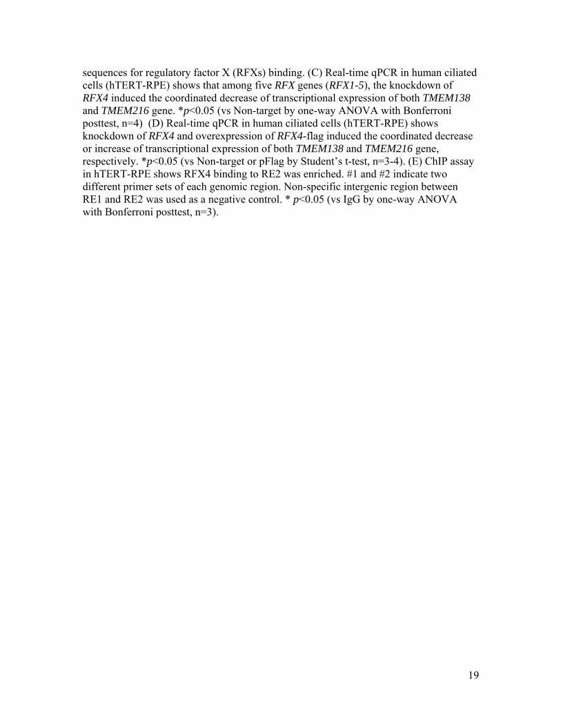
SOM text Coordinated expressions of adjacent TMEM138 and TMEM216 mediated by non-coding intergenic region We found two candidate REs, RE1 and RE2 with conserved DNaseI hypersensitive sites located in the dips surrounded by H3K4 monomethylation (H3K4me1) peaks (19), suggesting that DNA binding factors might regulate the transcription of these two genes (Fig. S7A-B). Regulatory factor X (RFX) paralogues binding to symmetrical X-box DNA elements are known to transcriptionally regulate genes necessary for ciliary formation (12, 20), and we found that the REs contained X-box consensus sequence motifs (Fig. S7B). Using knockdown, overexpression, and chromatin immunoprecipitation experiments, we further found that among the five known mammalian RFXs, RFX4 serves in transcriptional activation, associating with RE2 to mediate coordinated expressions of TMEM138 and TMEM216 (Fig. S7C-E). TMEM138 and TMEM216 vesicles differentially associate with cilia-targeted proteins Meckelin, a ciliary protein required for ciliogenesis was reported to interact with TMEM216 (10). Among other ciliary proteins, CEP290, controlling flagella protein content and crucial for ciliary formation, is known to localize at the transition zone and pericentriole matrix of cilia where TMEM138 is localized but where TMEM216 is adjacent (Fig. 3B and Fig. S9C) (15, 21), making it a good candidate for TMEM138 cargo. We indeed found that CEP290 specifically localized with TMEM138 vesicles (Fig. S13A), but not TMEM216. This data suggests that that TMEM138 and TMEM216 mark two distinct but linked vesicle pools, each associated with unique cilia-targeted proteins. TRAPPII mediates tethering of TMEM138 and TMEM216 vesicles crucial for ciliary assembly Among potential tethering proteins related to tethering of Golgi vesicles, TRAPPC9 (a core subunit of TRAPPII complex) is uniquely known to be mutated in a recessive human disease, and phenocopies some aspects of Joubert syndrome including cerebellar affection and mental retardation, making it a good candidate for linking the two TMEM proteins (22). We genetically tested each of these potential tethering proteins using siRNAs. We found that the siRNA disruption of TRAPPC9 de-tethered the two vesicular pools (Fig. 4A and Fig. S15A; Movie S7-10). As expected, this resulted in defects of ciliary assembly (Fig. S14C and Fig. S15B). Then, we tested whether the TRAPPII complex localizes where both TMEMs meet using aTRAPPC10 antibody, another core subunit of TRAPPII complex known to interact with TRAPPC9 (11). We found that TRAPPC10 overlaps where TMEM138 and TMEM216 signal is adjacent, as one might expect from a tethering protein, suggesting that TRAPPII or components thereof mediate tethering of TMEM138 and TMEM216 vesicles (Fig. S16). Our findings suggest that TRAPPII mediated tethering of TMEM138 and TMEM216 is crucial for ciliary assembly.
8
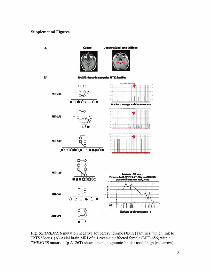
Supplemental Figures
Fig. S1 TMEM216 mutation negative Joubert syndrome (JBTS) families, which link to JBTS2 locus. (A) Axial brain MRI of a 1-year-old affected female (MIT-656) with a TMEM138 mutation (p.A126T) shows the pathognomic ‘molar tooth’ sign (red arrow)

9
indicating cerebellar malformation in JBTS. (B) Pedigrees of 6 JBTS families negative for TMEM216 mutations and their linkage plots used in this study are shown. Double bar indicates documented consanguinity. Male=square, Female=circle, Termination of pregnancy=triangle, Hashed line=deceased. LOD score plots present chromosome along x-axis and multipoint pLod along y-axis, as calculated by Allegro v1.2c. Note peak on chromosome 11 in all families.

10
Fig. S2 Genetic heterogeneity at JBTS2 locus and homozygous deleterious mutations of TMEM138. (A) Real-time qPCR of patient fibroblasts (MTI-656) and (B) Western blot analysis of whole lysates shows that TMEM216 mRNA and protein expressions are intact, implying the genetic heterogeneity at JBTS2 locus. Lysates from pcDNA3.1-TMEM216 expressing HEK293T cells were used as a positive control. (C) Missense and splicing mutation sites of TMEM138 found in JBTS2 linked families are evolutionary

11
conserved residues. (D) Shown is a protein blot of whole lysates of HEK293T cells transfected with a cDNA encoding wild-type TMEM138 or one of the missense mutations found in affected individual. Proteins levels were measured by densitometer. α-tubulin blot and β-gal assays (co-transfection with pTK β-gal at 1:10 ratio) were used for normalization of loading and transfection efficiency, respectively. Note that TMEM138 p.H96R resulted in the production of ~40% of wild-type protein level. *p<0.05 (vs WT by one-way ANOVA with Bonferroni posttest, n=4)

12
Fig. S3 Synteny of intergenic sequences between TMEM138 and TMEM216 using ECR Browser. The conservation profiles of human in comparison with chimpanzee, monkey, mouse, cow, dog, opposum, and anolis (submitted sequences based on coordinates of TMEM138 and TMEM216 homologs) are shown. Each line represents an ungapped alignment with the vertical height indicating the nucleotide identity. Pink bars of every layer show evolutionary conserved regions. Conserved alignment is red: intergenic region, green: transposons and simple repeats, yellow: UTRs, and blue: coding exons.

13
Fig. S4 TMEM138 and TMEM216 protein homologs. The schematic picture shows conserved protein regions (white regions: <50% sequence similarity to human) and percent protein homology to human (Table S4-6). DM: TMEM17 superfamily domain.
14
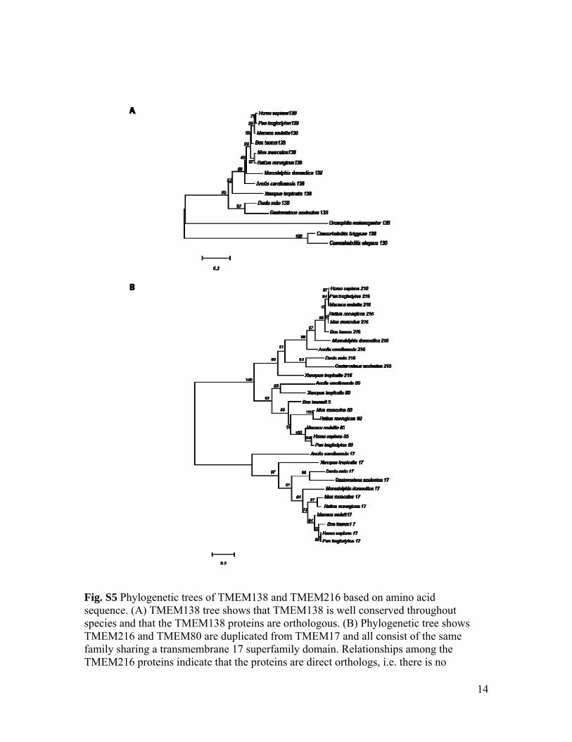
Fig. S5 Phylogenetic trees of TMEM138 and TMEM216 based on amino acid sequence. (A) TMEM138 tree shows that TMEM138 is well conserved throughout species and that the TMEM138 proteins are orthologous. (B) Phylogenetic tree shows TMEM216 and TMEM80 are duplicated from TMEM17 and all consist of the same family sharing a transmembrane 17 superfamily domain. Relationships among the TMEM216 proteins indicate that the proteins are direct orthologs, i.e. there is no

15
evidence of gene duplication creating paralogous proteins. Scale bar represents the average number of substitutions per amino acid residue. Confidence measurements from bootstrap analysis (500 iterations) are shown in nodes of trees. 216,TMEM216; 17,TMEM17; 80,TMEM80; 138,TMEM138.
16
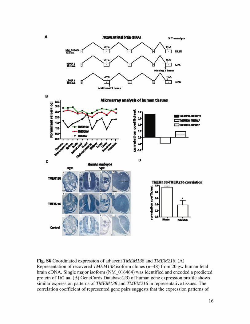
Fig. S6 Coordinated expression of adjacent TMEM138 and TMEM216. (A) Representation of recovered TMEM138 isoform clones (n=48) from 20 gw human fetal brain cDNA. Single major isoform (NM_016464) was identified and encoded a predicted protein of 162 aa. (B) GeneCards Database(23) of human gene expression profile shows similar expression patterns of TMEM138 and TMEM216 in representative tissues. The correlation coefficient of represented gene pairs suggests that the expression patterns of

17
TMEM138 and TMEM216 in human tissues are highly correlated, compared to TMEM67, another known JBTS causative gene (24). (C) Similar expression patterns of TMEM138 and TMEM216 based upon in situ hybridization in human embryonic tissues. TMEM138 antisense (a-d), TMEM216 antisense (e-h) and sense TMEM138 control probes (i-l). At 4gw, transverse sections show that TMEM138 as well as TMEM216 are ubiquitously expressed within embryonic tissues (nt: neural tube, h: heart primordium) (a, e, and i). At 5gw TMEM138 and TMEM216 expression became more intense particularly in neural tube (nt), dorsal root ganglia (drg), mesonephros (mn), gonadal ridge (go) and limb bud (b-d and f-h). (D) Real-time qPCR of TMEM138 and TMEM216 mRNA in tested tissues of mouse and zebrafish indicates that the average correlation coefficient in mouse (r=0.984) compare to those of zebrafish (r=0.386). *p<0.05 (by Student’s t-test, three independent experiments)

18
Fig. S7 Coordinated expressions of adjacent TMEM138 and TMEM216 mediated by non-coding intergenic region. (A) UCSC genome browser shows possible regulatory elements (RE1 and RE2) in the non-coding intergenic region, which are evolutionarily conserved and DNaseI hypersensitivity clusters. The shading is proportional to the maximum signal strength observed in any cell line. (B) RE1(~200bp) and RE2(~400bp) located in the dips surrounded by H3K4 monomethylation (H3K4me1) peaks contain X-box consensus
19
sequences for regulatory factor X (RFXs) binding. (C) Real-time qPCR in human ciliated cells (hTERT-RPE) shows that among five RFX genes (RFX1-5), the knockdown of RFX4 induced the coordinated decrease of transcriptional expression of both TMEM138 and TMEM216 gene. *p<0.05 (vs Non-target by one-way ANOVA with Bonferroni posttest, n=4) (D) Real-time qPCR in human ciliated cells (hTERT-RPE) shows knockdown of RFX4 and overexpression of RFX4-flag induced the coordinated decrease or increase of transcriptional expression of both TMEM138 and TMEM216 gene, respectively. *p<0.05 (vs Non-target or pFlag by Student’s t-test, n=3-4). (E) ChIP assay in hTERT-RPE shows RFX4 binding to RE2 was enriched. #1 and #2 indicate two different primer sets of each genomic region. Non-specific intergenic region between RE1 and RE2 was used as a negative control. * p<0.05 (vs IgG by one-way ANOVA with Bonferroni posttest, n=3).

20
Fig. S8 Knockdown of TMEM138 and TMEM216 in IMCD3 cells causes short cilia and defects in ciliogenesis (defined as having cilia <1 μm long). * p<0.05, **p<0.01 (vs Non-target by one-way ANOVA with Bonferroni posttest, n=60-70). Scale bars, 5um.

21
Fig. S9 Adjacent localization of TMEM138 and TMEM216 in IMCD3 and ciliated COS7 cells. (A) Western blot analysis of whole lysates from pcDNA3.1-TMEM138, pcDNA3.1-TMEM138-flag, and empty vector transfected HEK293T cells and (B) immunostaining of endogenous Tmem138 in Tmem138 knockdown IMCD3 cells (with shorter cilia) show specificity of generated mouse polyclonal TMEM138 antibody. The protein size of human TMEM138 is ~18kDa. (C) In IMCD3 cells, endogenous Tmem138 or Tmem216

22
were stained for cilia (Arl13b) or cilia plus centrosome (GT335), indicating the localization of Tmem138 in the ciliary axonem/basal body and Tmem216 in the basal body, respectively. Double labeling shows adjacent vesicular pattern of Tmem138 and Tmem216. A: ciliary axoneme, T: transition zone, and B: basal body. (D) hTMEM138-EGFP and hTMEM216-mCherry expressing COS7 cells show non-overlapping but closely adjacent vesicular patterns of surrounding the centrosome (γ-tubulin). (E) After 4h serum starvation, COS7 cells (>80-90% of confluence) were stained with ciliary markers (Arl13b and GT335), indicating that 40-50% cells display evidence of cilia. The side view (corresponding to the white box in XY plane view) of cilia in nuclear-labeled cells was shown. Boxes show insets at high power. Scale bars, (B), (C), and (E), 5um.

23
Fig. S10 Subcellular localizations of TMEM138 and TMEM216. Immunostaining in TMEM138-flag or TMEM216-flag expressing COS7 cells with various intracellular organelle markers including Golgi apparatus (GM130), microtubule (α-tubulin), mitochondria (Mitotracker), early endosome (EEA1), and late-endosome (Lamp2). TMEM proteins showed Golgi patterns (A) (also see Fig. S11) and aligned with microtubules (B). However, mitochondria, early-endosome, late-endosome marker did not overlap with TMEM138-flag and TMEM216-flag (C). Boxes show insets at high power. Scale bars 5um.
24
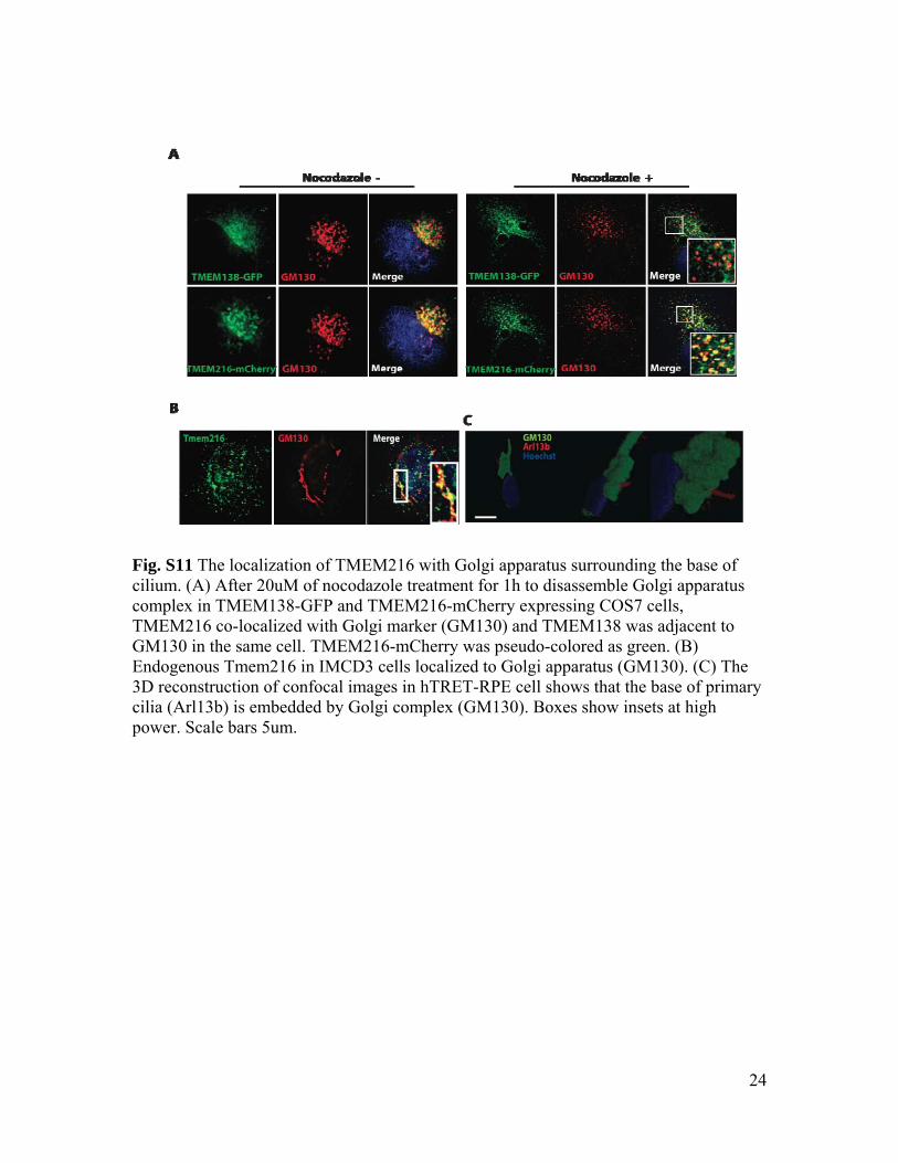
Fig. S11 The localization of TMEM216 with Golgi apparatus surrounding the base of cilium. (A) After 20uM of nocodazole treatment for 1h to disassemble Golgi apparatus complex in TMEM138-GFP and TMEM216-mCherry expressing COS7 cells, TMEM216 co-localized with Golgi marker (GM130) and TMEM138 was adjacent to GM130 in the same cell. TMEM216-mCherry was pseudo-colored as green. (B) Endogenous Tmem216 in IMCD3 cells localized to Golgi apparatus (GM130). (C) The 3D reconstruction of confocal images in hTRET-RPE cell shows that the base of primary cilia (Arl13b) is embedded by Golgi complex (GM130). Boxes show insets at high power. Scale bars 5um.

25
Fig. S12 TMEM138 and TMEM216 marked two distinct vesicular pools moving toward primary cilia. (A) Immunoelectron microscopy analysis in TMEM138-GFP and TMEM216-flag expressing HEK293T cells showing TMEM138 and TMEM216 are on adjacent but unique vesicles. Two 12nm gold particles (green arrows) tagging a TMEM138 containing vesicle are adjacent but not overlapping with six 6nm particles (red arrows) tagging TMEM216. Quantification from vesicles with two or more particles shows predominance of a single TMEM. **p<0.001(vs Single by Student’s t-test, n>270). (B) In COS7 cells producing primary cilia (Fig. S9E), live cell imaging and fluorescence recovery after photobleaching (FRAP) show that TMEM216-tagged vesicles move toward the centrosome (Centrin2, the central part of the basal body) and the accumulation of GFP signal around it, indicating that the net flux of vesicles is toward the centrosome (Movie S1 and S2). Arrows indicate existing or newly emerging vesicles that move toward the centrosomal marker. (C) Notably, TMEM138 and TMEM216 show

26
adjacent vesicles that moved together in a hand-in-hand fashion (Movie S3), consistent with adjacent vesicular movements in other ciliated cells (hTERT-RPE) (Movie. S4). Arrows indicate examples of vesicles moving in a hand-in-hand fashion. Boxes show insets at high power. Scale bars, 5um.

27
Fig. S13 Tmem138 and Tmem216 vesicles differentially associate with Cep290. (A) Visualized endogenous Cep290 together with cilia plus centrosomal marker (GT335), indicating the pericentrosomal location of Cep290 in IMCD3 cells. Cep290 has overlapping localization with Tmem138, whereas it is adjacent to Tmem216. (B) The knockdown of Tmem216, but not Tmem138 disrupts the trafficking of Cep290 to the base of cilia. Tmem138 is not necessary for Cep290 trafficking. Scale bar (A) and (B) 5um.

28
Fig. S14 Functional relatedness of TMEM138 and TMEM216. (A) Live cell imaging in TMEM138-GFP expressing COS7 shows that TMEM216 knockdown severely disrupts TMEM138 vesicular movements. Arrows indicate vesicles moving toward the centrosomal marker (Movie S5-6). (B) Immunostaining of Tmem138 in IMCD cells show that Tmem216 knockdown severely disrupts Tmem138 vesicular trafficking to the primary cilium. Arrows indicate intact or disrupted cilia. (C) In hTERT-RPE cells, knockdown of TRAPPC9 disrupted ciliogenesis induced by 24h serum starvation and largely rescued by myc-TRAPPC9* (see materials and methods). ** p<0.01, ***p<0.001 (by one-way ANOVA with Bonferroni posttest, n=90-110). Scale bars, (A) to (C), 5um.

29
Fig. S15 Effect of p115 and COG1 versus TRAPPC9 knockdown on TMEM138 and TMEM216 vesicular tethering and ciliogenesis. (A) Live cell imaging shows that knockdown of p115 and COG1 do not significantly affect vesicular tethering of TMEM138 and TMEM216 in transfected COS7 cells (Movie S9 and 10). (B) In hTERT-RPE cells, knockdown of p115 and COG1 do not significantly affect ciliogenesis induced by 24h serum starvation. Both effects were prominently disrupted following TRAPPC9 knockdown.

30
Fig. S16 TRAPPII localizes where TMEM138 and TMEM216 vesicles are adjacent. Endogenous TRAPPC10 (blue), a major subunit of TRAPPII, was stained in TMEM138-GFP (green) and TMEM216-mCherry (red) expressing COS7 cells. Images of each pair of colors presented. Merged image showed that white signal are strong at sites where green (TMEM138) and red (TMEM216) vesicles adjacent, indicating that TRAPPC10 overlays the adjacency of TMEM138 and TMEM216. Scale bar, 5um.

31
Fig. S17 Defects of L/R heart axis in tmem138 and tmem216 morphants. Injection of MOs in Tg(cmlc2:egfp) zebrafish embryos shows more severe L/R patterning defects of heart in tmem216 morphants than tmem138 (>50 embryos each condition).

32
Fig. S18 Analysis of tmem138 and tmem216 morphants. (A) Injection of translation-blocking antisense morpholinos (MO) to tmem138, tmem216, or double in wild-type (AB) zebrafish embryos caused ciliary phenotypes including hydrocephalus (only in tmem216 morphants), pericardial effusion, kinked tail, and lethality at 3 days post fertilization in a synergistic and dose-dependent manner. (B) tmem138 or tmem216 morphant phenotypes were mostly rescued by co-injection of RNA encoding human

33
TMEM138 or TMEM216. Trans-rescues showed that tmem138 morphants were largely rescued by TMEM216 RNA, whereas TMEM138 RNA did not effectively rescue tmem216 morphants (especially, hydrocephalus phenotypes). Trans-rescues by cognate human RNA showed some functional redundancy of TMEM216 in tmem138 morphants but not vice versa. “Partial rescue” means any remaining of weak ciliary defect phenotypes (i.e. mild curved tail). (>50 embryos each condition). (C) At early stage of zebrafish embryos (9-10 somites), MO-based suppression of tmem138 or tmem216 give rise to similar gastrulation defects quantified based on previously described objective criteria. Class I, shortened body axis, small anterior structures, broad somites; Class II, severely shortened anterior-posterior axis, small anterior structures, severely broadened somites with kinking of the notochord. Representative lateral (top panels) and dorsal (bottom panels) views of live embryos are shown. n=55-81 embryos/injection batch with masked scoring, repeated twice with similar results. (D). Whole embryo flat mounts hybridized in situ with a cocktail of krox20, pax2, and myoD riboprobes. Morphometrics demonstrate phenotypic defects in two different axes of development. w, width spanning the lateral ends of the fifth appreciable somites counted from the anterior end of the embryo; l, length of the notochord as indicated by adaxial cell labeling; w/l, ratio of measurements is indicated, n=8-10 embryos/injection batch. P-values denoted as * (P<0.01) or *** (P<0.0001).
34
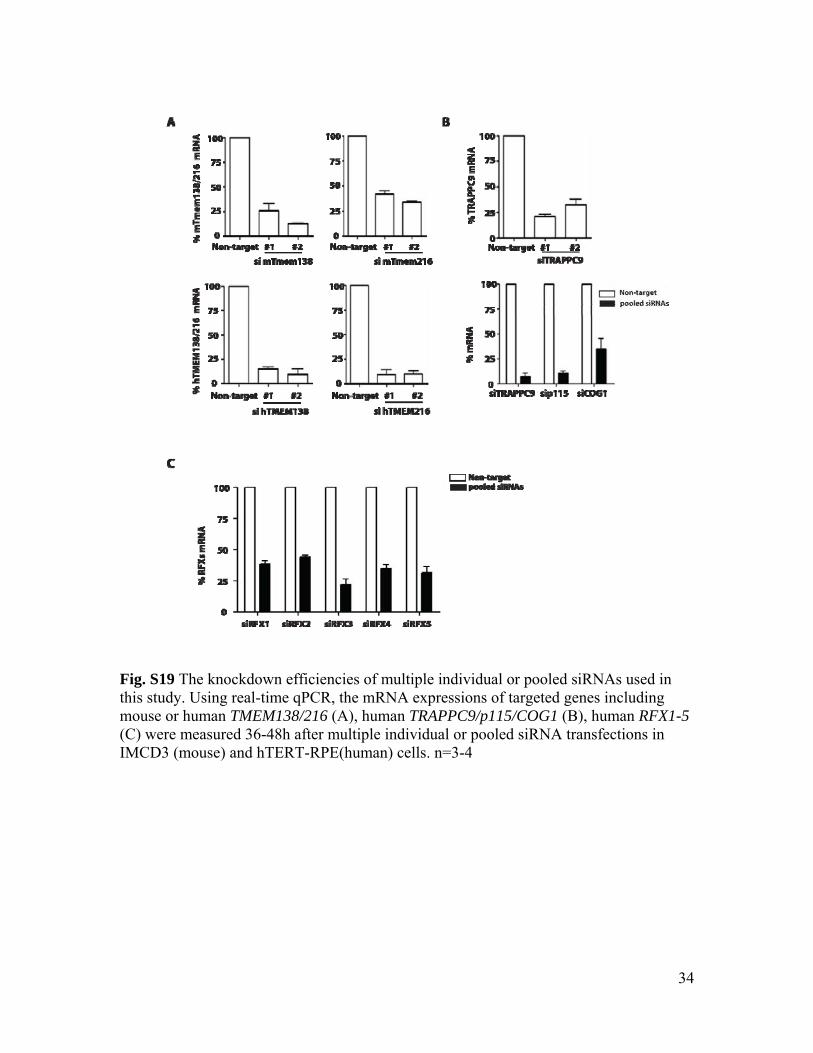
Fig. S19 The knockdown efficiencies of multiple individual or pooled siRNAs used in this study. Using real-time qPCR, the mRNA expressions of targeted genes including mouse or human TMEM138/216 (A), human TRAPPC9/p115/COG1 (B), human RFX1-5 (C) were measured 36-48h after multiple individual or pooled siRNA transfections in IMCD3 (mouse) and hTERT-RPE(human) cells. n=3-4

35
Table S1. Clinical and molecular data from TMEM138 mutated families

36
Table S2. Chromosomal locations of TMEM216 and TMEM138 homologs

37
Table S3. Accession numbers of TMEM17, TMEM216, TMEM80, and TMEM138 homologs

38
Table S4. Protein distance of TMEM138 and TMEM216 homologs

39
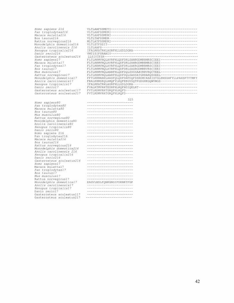
Table S5. The alignment of TMEM138 homologs 1 10 20 30 40 50 60 | | | | | | | Homo sapiens -MLQTSNYSLVLSLQFLLLSYDLFVNSFS-ELLQKTPVIQLVLFI---IQDIAVLFNIII Pan troglodytes (chimp) -MLQTSNYSLVLSLQFLLLSYDLFVNSFS-ELLQKTPVIQLVLFMCVHIQDIAVLFNIII Macaca mulatta (rhesus) -MLQTSNYSLVLSLQFLLLSYDLFVNSFS-ELLRKAPVIQLVLFI---IQDIAVLFNIII Bos taurus (cow) -MLQTSNYSLVLSLQFLLLSYDLFVNSFS-ELLRMAPVIQLVLFI---IQDIAILFNIII Mus musculus(mouse) -MLQTGNYSLVLSLQFLLLSYDLFVNSFS-ELLRMAPVIQLVLFI---IQDIAILFNIII Rattus norvegicus (rat) -MLQTSNYSLVLSLQFLLLCYDLFVNSFS-ELLRMAPVIQLVLFI---IQDIAILFNIII Monodelphis domestica(opossum) -MLQTSNYSLVLSLQLVLLLYDLLVNAFS-ELLRSAPVVQLVLFI---IQDIAILFNVII Anolis carolinensis -MLQTSNYSLVLSLQFLLLSFDLFVNSFS-ELLRMAPVIQLVLFI---IQDIAILFNIII Xenopus tropicalis -MLQPGNYSLVLSLQFLLLLFDLFVNSFS-ELLRDPPVNQLVLFI---LQDVGILFAAIV Danio rerio (zebrafish) -MLQTNNYSLVLLIQLALLTFDLFVNSFS-ELLRSAPVIQLVLFI---IQDIGILFNVII G.aculeatus1(stickleback) -MLQTGNYSLVLLIQMTLLTYDLFVNSFG-ELLRGAPVIQLVLFI---IQDIAILFNVII G.aculeatus2(stickleback) ---QTSNYSLVLLIQLSLLSFDLFVNSFS-ELLRHEPAVQLVLFI---IQDISILFNLII Caenorhabditis briggsae ---MTSKYPYVLTTQFLMLTIDIFFNALS-ILCYGDNMALLLIYI---LQDTLLIMSSLV C. elegans ---MTSKYPYVLTTQVFMLSIDILFNALG-VLCYGNNMTLLLIYI---LQDTLLIMSSLV Drosophila melanogaster MKLTLRRYSWVLLFQFALLGVDLFCNAFGPSLARNRLQTAIILFV---TQDALIIAEYLL 61 70 80 90 100 110 120 | | | | | | | Homo sapiens IFLMFFNTFVFQAGLVNLLFHKFKGTIILTAVYFALSISLHVWVMNLRWK--NSNSFIWT Pan troglodytes IFLMFFNTFVFQAGLVNLLFHKFKGTIILTAVYFALSISLHVWVMNLRWK—-NSNSFIWT Macaca mulatta IFLMFFNTFVFQAGLVNLLFHKFKGTIILTAVYFALSISLHVWVMNLRWK--NSNSFIWT Bos Taurus IFLMFFNTFVFQAGLVNLLFHKFKGTIILTAVYFALSISLHVWVMNLRWK--NSNCFVWT Mus musculus IFLMFFNTFVFQAGLVNLLFHKFKGTIILTSVYLALSISLHVWVMNVRWK--NSSSFSWT Rattus norvegicus IFLMFFNTFVFQAGLVNLLFHKFKGTIILTSVYLALSISLHVWVMNVRWK--DSSSFRWT Monodelphis domestica IFLMFFNTFVFQAGLINLLFRKFKGTIILTAFYLSLSIPLHVWVLNLRWT--DSKHYVWT Anolis carolinensis IFLMFFNTFVFQAGLVNLLFHKFKGTIVLSGTYLALSVSFHIWIMNLRWR--SSNYFVWT Xenopus tropicalis LFLMLFNTFVFQAGLVSLLCQRFQVTVILCAVYIALSISLHVWLMNLRWT—-GANRFVWS Danio rerio ILLMMFNTYVFQVGLVSLLLERFRAMLILSALYLTLSICFHCWVMNLRWM--ESNRFVWT Gasterosteus aculeatus1 ILLMMFNTYAFQVGLVSLLLERFRALLIFSAVYLTLSIGFHCWVLNLRWF--ESNRFVWT Gasterosteus aculeatus2 ILLMLFNTYVFQIGLVAVLLERFRAALMLSALYLTFSIILHSWLMNLRWL--NTNRFIWT C. briggsae LFVSFTATFVFQLGLIHIVLVQFLPTIIMSIFYTFVSIGYHYTSLSSTWED-QTVNIFLE C. elegans LFVSFTATFVFQLGLIHIVIFQFLPTVIISILYTFVSIGYHYASLSSTWED-RTVNIFMN D. melanogaster FTLALHSTCVYQVGASHIILRNCKLFMASITIYFLLSASQHFWIIYQYRQPPEEDGHHWP 121 130 140 150 160 170 180 | | | | | | | Homo sapiens DGLQMLFVFQRLAAVLYCYFYKRTAVRLGDPHFYQDSL-WLRKEFMQVRR---------- Pan troglodytes DGLQTLFVFQRLAAVLYCYFYKRTAVRLGDPRFYQDSL-WLRKEFMQVRR---------- Macaca mulatta DGLQTLFVFQRLAAVLYCYFYKRTAIRLGDPHFYQDSL-WLRKEFMQVRR---------- Bos Taurus DGLQTLFVFQRLAAVLYCYFYKRTAVRLGDPRFYQDSL-WLRMEFMQVRR---------- Mus musculus NGLQTLFVFQRLAAVLYCYFYKRTAVRLGDPRFYQDSL-WLRKEFMQVRR---------- Rattus norvegicus NGLQTLFVFQRLAAVLYCYFYKRTAVRLGDPRFYQDSL-WLRKEFMQVRR---------- Monodelphis domestica DGLQTLFVFQRLAAVLYCYFYKRTAIRLGDPRFYQDSL-WLRKEFTQVRS---------- Anolis carolinensis DGLQTLFVFQRLAAVLYYYFYKRTAVHLGDPRFYQDSL-WLRKEFAQVRS---------- Xenopus tropicalis DGLLALFVLQRFVAVLYFYYYKRTALSMGDSRFYHDSL-WLRKEFARVRG---------- Danio rerio DGLQVLFVFQRIAAVLYYYFYKRTTEYLGDPRLYEDSP-WLRDAFARARQ---------- Gasterosteus aculeatus1 DGLQALFVFQRTGAVMYYYLYKRTAEYMGDPRLYEDSL-WLREAFARGRQ---------- Gasterosteus aculeatus2 DGLQVLFVFQRVASVLYYYLYKRTSEYLGDPRLYEDSP-WLRELFARHERGSPLPKQK C. briggsae THLLIFFIFHKVISCIFYSFYKRTALQISDPKYNSDST-WLRELFIKHMNDKAAKLEARN C. elegans GPLLLFFIIHKIVSCVFYAYYKRTALQISDPKYNSDST-WLRELFIKHMNDKAAKIEART D. melanogaster LGLIALSVAQRIMSVFYYYSSKSTALTMADPRFKEEHLDWIADQLGDK------------ 185 Homo sapiens ----- Pan troglodytes ----- Macaca mulatta ----- Bos taurus ----- Mus musculus ----- Rattus norvegicus ----- Monodelphis domestica ----- Anolis carolinensis ----- Xenopus tropicalis ----- Danio rerio ----- Gasterosteus aculeatus1 ----- Gasterosteus aculeatus2 SPQNT C. briggsae AAAAT C. elegans AAENN D. melanogaster -----

40
Table S6. The alignment of TMEM80, TMEM216, and TMEM17 homologs 1 10 20 30 40 50 60 | | | | | | | Homo sapiens 80 ------------------------------------------------------------ Pan troglodytes80 ------------------------------------------------------------ Macaca mulatta80 ------------------------------------------------------------ Bos taurus80 ------------------------------------------------------------ Mus musculus80 ------------------------------------------------------------ Rattus norvegicus80 ------------------------------------------------------------ Monodelphis domestica80 ------------------------------------------------------------ Anolis carolinensis80 ----------------------------------------------------------MS Xenopus tropicalis80 ------------------------------------------------------------ Danio rerio80 ------------------------------------------------------------ Homo sapiens216 ------------------------------------------------------------ Pan troglodytes216 MCLECMVSTQDMEDGNQPSGMQKNSDWSCRRRRRGWSPLSLRSGEGTSERGPFAARPELV Macaca mulatta216 ------------------------------------------------------------ Bos taurus216 ------------------------------------------------------------ Rattus norvegicus216 ------------------------------------------------------------ Monodelphis domestica216 ------------------------------------------------------------ Anolis carolinensis 216 ------------------------------------------------------------ Xenopus tropicalis216 ------------------------------------------------------------ Danio rerio216 ------------------------------------------------------------ Gasterosteus aculeatus216 ------------------------------------------------------------ Homo sapiens17 ------------------------------------------------------------ Macaca mulatta17 ------------------------------------------------------------ Pan troglodytes17 ------------------------------------------------------------ Bos taurus17 ------------------------------------------------------------ Mus musculus17 ------------------------------------------------------------ Rattus norvegicus17 ------------------------------------------------------------ Monodelphis domestica17 ------------------------------------------------------------ Anolis carolinensis17 ------------------------------------------------------------ Xenopus tropicalis17 ------------------------------------------------------------ Danio rerio17 ------------------------------------------------------------ Gasterosteus aculeatus1-17 ------------------------------------------------------------ Gasterosteus aculeatus2-17 ------------------------------------------------------------ 61 70 80 90 100 110 120 | | | | | | | Homo sapiens80 ---------------------------MAEGARARGPRGCRDRDGPAGGAGRGSSTVLSS Pan troglodytes80 -------------------MAEGARARGPRGCRDRDGPAGGAGKMAAPRRGRGSSTVLSS Macaca mulatta80 ------------------------------------------------------------ Bos taurus80 --------------------------------------------MAAPRRGKASSTVLSS Mus musculus80 ------------------------------------------------------------ Rattus norvegicus80 -----------------MAAVRPGRGSFKVVSCCCCFPRPDKHSPTGLHHIKLQTDTLSS Monodelphis domestica80 ------------------------------------------------------------ Anolis carolinensis80 PEWFRGGAESDEAARSVPREQQRTEKTPRRLRKDCRETGWGGAGIEREGPKWRLSSELSS Xenopus tropicalis80 ---------------------------------------------MALVGRGKSSVVLSS Danio rerio80 ------------------------------------------------MAAHGRQPVLSS Homo sapiens216 -------------------------------------------MLPRGLKMAPRGKRLSS Pan troglodytes216 QLRARPAPGDHRVPRAGRGAGNPGRFVPVSGSAALLREPLWQRMLPRGLKMAPRGKRLSS Macaca mulatta216 --------------------------------------------------MAPRGKRLSS Bos taurus216 --------------------------------------------------MAPRGKRLSS Rattus norvegicus216 --------------------------------------------------MAPRDKRLSS Monodelphis domestica216 ------------------------------------------------------------ Anolis carolinensis 216 ------------------------------------------------------------ Xenopus tropicalis216 --------------MAQAAGVRRQLDSLTRNIFLRDVGRTVPEKSGAP--LTGDSEVAPS Danio rerio216 ------------------------------------------------MAAHGRQPVLSS Gasterosteus aculeatus216 ------------------------------------------------------------ Homo sapiens17 --------------MELPDPVRQRLGNFSRAVFS-DSNRTGPESNEGP-----ENEMVSS Macaca mulatta17 ---------------------------------------------MEQLLDIAENEMVSS Pan troglodytes17 --------------MELPDPVRQRLGNFSRAVFS-DSNRTGPESNEGP-----ENEMVSS Bos taurus17 --------------MELPDPVRQRLGNFSRTVFS-DSNRTGPEYNEGP-----DNEMVSS Mus musculus17 --------------MELPDPVRQRLSNLSLTVFG-DSSRTGPESSDAA-----DNEMVSS Rattus norvegicus17 --------------MELPDPVRQRLSNLSLTMFG-DSSRTGPESSEAA-----DNEMVSS Monodelphis domestica17 --------------MELPDPVRERLGNFSRSLFT-DTNRTGPENNSNL-----NNEIVSS Anolis carolinensis17 --------------MALQTPLPPNLRRGLTTFSGSLFVNNKTGDAGAAQVYHPGHAVLSS Xenopus tropicalis17 --------------MAQAAGVRRQLDSLTRNIFLRDVGRTVPEKSGAP--LTGDSEVAPS Danio rerio17 --------------MDLPEPIRRRLGDFSRTVFV-DQSRTQPSFEEHANFLDQNKDVVSS Gasterosteus aculeatus117 --------------MDLPDGIRKRLEDFSRNVLF-DQSGTRTVARENDTFLPHDKRLLSS Gasterosteus aculeatus217 --------------MDLPDAIRKRLEDFSRNVLF-DQSGTRTVARENDTFLPHDKRLLSS 121 130 140 150 160 170 180 | | | | | | | Homo sapiens80 VPLQMLFYLSGTYYALYFLATLLMITYKSQVFSYPHRYLVLDLALLFLMGILEAVRLYLG

41
Pan troglodytes80 VPLQMLFYLSGTYYALYFLATLLMITYKSQVFSYPHRYLVLDLALLFLMGMLEAVRLYLG Macaca mulatta80 ----MLFYLSGTYYALYFLATLLMITYKGQVFSCPHHYLVLDLALLFLMGILEALRLYLG Bos taurus80 LPLQMLLCLSGTYYALYFLATLLLLVYKSQVFTYPHSCLVLDLTLLFLMGILEAIRLYFG Mus musculus80 ----MLFHLSGLYSALYFLATLLMIVYKSQVFSYPCNCLALDLVLLLLMGILKVAQLYLG Rattus norvegicus80 VPLQMLFHLSGLYYALYFLATLLMIVYKSQVFSYPSNCLVLDLVLLLLMGIFEVAQLYLG Monodelphis domestica80 --------------------------------------------MLFLYLGIEITRIFFG Anolis carolinensis80 VPLQILFYLNGVYYIFYFFAALLMIIYKSQVFTYPDNFLTLDLILLFIMALLETIRLYFG Xenopus tropicalis80 VPLQMLLYLNIIYYVFYFLATLLMIIYKSQVFSYPGSNLALDLCLLFLMGILEPVRLYLG Danio rerio80 TPLQILFHLNGWYFAAFFVAEILMFIYKGVILPYPQDNLILDVVLLLLFSGLETLRLFYG Homo sapiens 216 TPLEILFFLNGWYNATYFLLELFIFLYKGVLLPYPTANLVLDVVMLLLYLGIEVIRLFFG Pan troglodytes216 TPLEILFFLNGWYNATYFLLELFIFLYKGVLLPYPTANLVLDVVMLLLYLGIEVIRLFFG Macaca mulatta216 TPLEILFFLNAWYNATYFLLELFIFLYKGVLLPYPTANLVLDVVMLLLYLGIEVIRLFFG Bos taurus216 TPLEILFFLNGWYYATYFLLELFIFLYKGLLLPYPTANLVLDVVMLFLYLGVEVIRLFFG Rattus norvegicus216 TPLEILFFLNGWYYATYFLLELLIFLYKGLLLPYPTANLVLDVVMLLLYLGIEVIRLFFG Monodelphis domestica216 --------------------------------------------MLFLYLGIEITRIFFG Anolis carolinensis 216 --------------------------------------------MLFLYLGIEVTRIFFG Xenopus tropicalis216 VSLQIFLYFNAFYFPFWWVCYVIMLQLKYVLL--PDYYKFILVVLLILMSVIEVIRLYLG Danio rerio216 TPLQILFHLNGWYFAAFFVAEILMFIYKGVILPYPQDNLILDVVLLLLFSGLETLRLFYG Gasterosteus aculeatus216 ----VLFYLNSWYFAAFYLAEVLMFIYKGNSLPYPSDNLVLDVVLLVLFLGLEMLRIFYG Homo sapiens17 LALQMSLYFNTYYFPLWWVSSIMMLHMKYSIL--PDYYKFIVITVIILITLIEAIRLYLG Macaca mulatta17 LALQMSLYFNTYYFPLWWVSSIMMLHMKYSIL--PDYYKFIVITVIILITLIEAIRLYLG Pan troglodytes17 LALQMSLYFNTYYFPLWWVSSIMMLHMKYSIL--PDYYKFIVITVIILITLIEAIRLYLG Bos taurus17 LALQMSLYFNTYFFPLWWVSSIMMLQMKYSIL--PDYYKFIVVTIIILITLIEAIRLYLG Mus musculus17 LALQMSLYFNSYFFPLWWVSCIVMLHLKYSIL--PDYYKFIVVTVVILITLIEAIRLYLG Rattus norvegicus17 LPLQMSLYFNSYFFPLWWVSCIVMLHLKYSVL--PDYYKFIVITVVILITLIEAIRLYLG Monodelphis domestica17 LALQMSLYFNIYFFPFWWVSSIIMLLLKYPVL--PDYYKFILVTVVILITLIEAIRLYLG Anolis carolinensis17 LPLQMMLYFNAFYFPFWCLSEGIMLQLKFSLL--PAYNQFLLLSAYLTITLAEVLRLYLG Xenopus tropicalis17 VSLQIFLYFNAFYFPFWWVCYVIMLQLKYVLL--PDYYKFILVVLLILMSVIEVIRLYLG Danio rerio17 LPLQMSLYFNMWFFPFWWISEVVMLDLKYSAL--ADYYKFILITILIVMTLIEAIRLYLG Gasterosteus aculeatus1-17 LHLQMSLYFNMWFFPLWWISEAVMLHLKYPAL--PDYYKFILVTVLILMTLIEAIRLFLG Gasterosteus aculeatus2-17 LHLQMSLYFNMWFFPLWWISEAVMLHLKYPAL--PDYYKFILVTVLILMTLIEAIRLFLG 181 190 200 210 220 230 240 | | | | | | | Homo sapiens80 TRGNLTEAERPLAASLALTAGTALLSAHFLLWQALV--LWADWALSATLLALHGLEAVLQ Pan troglodytes80 TRGNLTEAERPLAASLALTAGTALLSAHFLLWQTLV--LWADWALSATLLALHSLEAILQ Macaca mulatta80 TRGNLTEAERPLAASLALTAGTALLSAYFLLWQTLV--LWADWALSATLLALHGLEAVLQ Bos taurus80 TTGNLMEAEVPLAASLVLTVGSALLSAYFLLWQTLV--LRADSALGAPLLALHGLEAVLQ Mus musculus80 TKGNLMEAEVPLAASLAFTAVGGLLSVHFLLWQTLV--LWMDSVLSTVLLVLHGLEAGLQ Rattus norvegicus80 TQGNLTEAEVPLAASLAFTAVSGLLSVHFLLWQTLV--LWMDSVLSTVLLVLHGLEAGLQ Monodelphis domestica80 TKGNLCQRMMPLGISLALTFPSAMMASYYLLLQTYV--LRLEAVMGTILLLFCALEVILE Anolis carolinensis80 TKGNLTEDEAPLGFSLVITAGCVILSVYFLALQTYV--LKADVIINVVLLVIYGLEGILQ Xenopus tropicalis80 TQGNLAEEEIPLGSSLLVTVGNIFLAIYFLVWQTYI--LRADLIINIILLVIYGLEVILE Danio rerio80 WKGNLCQRSLALFVSVAILVPCAVLSVYYLLLQTFV--LRLEFVLNAVLLCFYGFELVLG Homo sapiens216 TKGNLCQRKMPLSISVALTFPSAMMASYYLLLQTYV--LRLEAIMNGILLFFCGSELLLE Pan troglodytes216 TKGNLCQRKMPLSISVALTFPSAMMASYYLLLQTYV--LRLEAIMNGILLFFCGSELLLE Macaca mulatta216 TKGNLCQRKMPLSISVALTFPSAMMASYYLLLQTYV--LRLEAIMNGILLFFCGSELLLE Bos taurus216 TKGNLCQRKMPLGISVALTFPSTMMASYYLLLQTYV--LRLEAIMNSILLFFCGSELLLE Rattus norvegicus216 TKGNLCQRKMPLGISVALTFPSAMMASYYLLLQTYV--LRLEAIMNSILLFFCGSELLLE Monodelphis domestica216 TKGNLCQRMMPLGISLALTFPSAMMASYYLLLQTYV--LRLEAVMGTILLLFCALEVILE Anolis carolinensis 216 SKGNLCQRKVPLAISLALTFPAAVMAAYYLLLQTYA--LRLEAILNAILLLFYAVELLLG Xenopus tropicalis216 YSGNLQEKVPELAGFCLLSILLQLPLLLFLLCDPGLEPLPLERAVHGILTAFLLIQIPIS Danio rerio216 WKGNLCQRSLALFVSVAILVPCAVLSVYYLLLQTFV--LRLEFVLNAVLLCFYGFELVLG Gasterosteus aculeatus216 WKGNLCESSLASCAALLVLLPCAALAVYFLLLQTYV--LRLEFILGAVLLCFYGRELLLG Homo sapiens17 YVGNLQEKVPELAGFWLLSLLLQLPLILFLLFNEGLTNLPLEKAIHIIFTLFLAFQVVAA Macaca mulatta17 YMGNLQEKVPELAGFWLLSLLLQLPLILFLLFNEGLTNLPLEKAIHIIFTLFLAFQVVAA Pan troglodytes17 YVGNLQEKVPELAGFWLLSLLLQLPLILFLLFNEGLTNLPLEKAIHIIFTLFLAFQVVAA Bos taurus17 YMGNLQEKVPELAGFWLLSLLLQLPLILFLLFNEGLTNLPLEKAVHIIFTIFLTFQVVSA Mus musculus17 CMGNLQEKVPELAGFWLLSLLLQLPLILFLLLNDGLRNLPLEKAIHIIFTIFLTFQVISA Rattus norvegicus17 CMGNLQEKVPELAGFWLLSLLLQLPLLLFLLLNEGLKNLPLEKAIHSIFTVFLTFQVISA Monodelphis domestica17 YMGNLQEKVPELAGFWLLSFLLQLPLILFLLFNEGLKSQPLERAVHIIFTIFLTLQVVLA Anolis carolinensis17 YIGNLQEKVPELAGFLLLSLMIEVPTVLFILADMYTLRLPLETAVNLLLLLFLVAEIAAA Xenopus tropicalis17 YSGNLQEKVPELAGFCLLSILLQLPLLLFLLCDPGLEPLPLERAVHGILTAFLLIQIPIS Danio rerio17 NAGNLQEKVPELAGFWLLTFLLQFPLILFQLFNEAVLVQPLERGVHIILALFIFAEVLFG Gasterosteus aculeatus117 YSGNLQEQVPELAGFWLLSILLQFPLILFQLFNEAILIQPLERGVHIVLAIFILAQALSG Gasterosteus aculeatus217 YSGNLQEQVPELAGFWLLSILLQFPLILFQLFNEAILIQPLERGVHIVLAIFILAQALSG 241 250 260 270 280 290 300 | | | | | | | Homo sapiens80 VVAIAAFTR--------------------------------------------------- Pan troglodytes80 VVAIAAFTR--------------------------------------------------- Macaca mulatta80 VVAIAAFTR--------------------------------------------------- Bos taurus80 VVAIAAFVS--------------------------------------------------- Mus musculus80 VVVIADFIR--------------------------------------------------- Rattus norvegicus80 VVVIADFIR--------------------------------------------------- Monodelphis domestica80 VLTLSTFSSIT------------------------------------------------- Anolis carolinensis80 IIAIAAFVS--------------------------------------------------- Xenopus tropicalis80 VLTVAAFFRY-------------------------------------------------- Danio rerio80 VMTISIFSRANIY-----------------------------------------------
42
Homo sapiens 216 VLTLAAFSSMDTI----------------------------------------------- Pan troglodytes216 VLTLAAFSSMDRI----------------------------------------------- Macaca mulatta216 VLTLAAFSSMDRI----------------------------------------------- Bos taurus216 VLTLTAFSSMDR------------------------------------------------ Rattus norvegicus216 MLTLATFSSMDRI----------------------------------------------- Monodelphis domestica216 VLTLSTFSSIT------------------------------------------------- Anolis carolinensis 216 ILTLAAFS---------------------------------------------------- Xenopus tropicalis216 IFALRKATRHLAGRFHLLGDLDGRA----------------------------------- Danio rerio216 VMTISIFSRANIY----------------------------------------------- Gasterosteus aculeatus216 LLSIYTFSR--------------------------------------------------- Homo sapiens17 FLTLRKMVNQLAVRFHLQDFDRLSANRGDMRRMRSCIEEI-------------------- Macaca mulatta17 FLTLRKMVNQLAVRFHLQDFDRLSANRGDMRRMRSCIEEI-------------------- Pan troglodytes17 FLTLRKMVNQLAVRFHLQDFDRLSAKRGDMRRMRSCIEEI-------------------- Bos taurus17 FLTLRKMVNQLATRFHLQDFDRLSASRGDMRRVRSCIEEI-------------------- Mus musculus17 FLTLKKMVNQLAARFHLQDFDQLSSSSAAVRRVRQCTEEL-------------------- Rattus norvegicus17 FLTLKKMVNQLAARFHLQDFDQLSASSATGRRARQSSEEL-------------------- Monodelphis domestica17 FFTLKKMANHLAASFQLQDFDRFDQFSSRDMIRAKISTGLENSGHFYLLPAHSPTYTMPT Anolis carolinensis17 FRALKHMSKQLAMQFYLKQFEEGVGQTPGDGHKGQWVWGS-------------------- Xenopus tropicalis17 IFALRKATRHLAGRFHLLGDLDGRA----------------------------------- Danio rerio17 FVALRTMVRHTESRFHLRQFHGIQELRT-------------------------------- Gasterosteus aculeatus117 FVTLRDMVRHTGRQFHLRQFD--------------------------------------- Gasterosteus aculeatus217 FVTLRDMVRHTGRQFHLRQFD--------------------------------------- 325 Homo sapiens80 ------------------------- Pan troglodytes80 ------------------------- Macaca mulatta80 ------------------------- Bos taurus80 ------------------------- Mus musculus80 ------------------------- Rattus norvegicus80 ------------------------- Monodelphis domestica80 ------------------------- Anolis carolinensis80 ------------------------- Xenopus tropicalis80 ------------------------- Danio rerio80 ------------------------- Homo sapiens 216 ------------------------- Pan troglodytes216 ------------------------- Macaca mulatta216 ------------------------- Bos taurus216 ------------------------- Rattus norvegicus216 ------------------------- Monodelphis domestica216 ------------------------- Anolis carolinensis 216 ------------------------- Xenopus tropicalis216 ------------------------- Danio rerio216 ------------------------- Gasterosteus aculeatus216 ------------------------- Homo sapiens17 ------------------------- Macaca mulatta17 ------------------------- Pan troglodytes17 ------------------------- Bos taurus17 ------------------------- Mus musculus17 ------------------------- Rattus norvegicus17 ------------------------- Monodelphis domestica17 EAGVLNSLFQNKGNGIFDKNKTFGP Anolis carolinensis17 ------------------------- Xenopus tropicalis17 ------------------------- Danio rerio17 ------------------------- Gasterosteus aculeatus117 ------------------------- Gasterosteus aculeatus217 -------------------------

43
Table S7. siRNAs, primers, and morpholino antisense oligonucleotides used in this study siRNAs target sequences Mouse TMEM138 siRNA#1 5’-GGGUCAUGAACGUGCGAUG Mouse TMEM138 siRNA#2 5’-AGGUUAGCCUGUAGAGAGA Mouse TMEM216 siRNA#1 5’-GAACCCAUUUAGUAGUUUA Mouse TMEM216 siRNA#2 5’-GUGAAAAGUGGUAGGUCGA Human TMEM138 siRNA#1 5’-CAAAGGAUGCGAAGAGUGA Human TMEM138 siRNA#2 5’-CAUAUGGACAGAUGGACUU Human TMEM216 siRNA#1 5’-GGCAAGCAACUCAGCAUAA Human TMEM216 siRNA#2 5’-CCACAUGCUCCCUGUACGA Human TRAPPC9 siRNA#1 5’-CGAAAGUUCUCACCACUAA Human TRAPPC9 siRNA#2 5’-CCUAUUACAGCAAGUAUAA Human RFX1 siRNA pool (5’-CAACACAGGCGUAUACUGA,
UCACAGAGCUCGACCUCCA, CCGCACAGAUCAACCAGAU, CCACGUGGCUCAAGAGGUG)
Human RFX2 siRNA pool (5’-UGACACAGGCCAUCCGUAA, GAACAGCCUACACCUACAA, GCACAGCACUCCGGAACAG, GAGAGACGCCGAUCGCUGU)
Human RFX3 siRNA pool (5’-GAACAACAACGUAUCCUUA, GCGAUACUGUCUAUACCAA, GUAUAUGGCUAUGAGACAA, CGAAGAUACACGUCGCUUA)
Human RFX4 siRNA ON-TARGETplus SMARTpool (L-0135577-00-0005, Dharmacon) Human RFX5 siRNA ON-TARGETplus SMARTpool (L-011103-00-0005, Dharmacon) Human TRAPPC9 siRNA pool (5’-GCAGACGACUGGAACGAUU,
CGAAAGUUCUCACCACUAA, CCUAUUACAGCAAGUAUAA, GAAAGUCAGCAACUAAUCA)
Human p115 siRNA pool (5’-CCAGGGAAGUUAUACGUAA, AGAAAUUGGUAGAUUGCGA, GCUCAUAUAUUCAACGUAU, GUACAAGGAGAGACCGAGA)
Human COG1 siRNA pool (5’-CCAGGAAAUUAAUCGGGUU, UGAAGGGUCUCGCGGGAAU, AACCAGAAACAUCGAAACA, CUGUAUGGUUCAAGUAGUA)
Primers for quantitative real-time PCR Mouse TMEM138 F 5’- CATGAACGTGCGATGGAAAA Mouse TMEM138 R 5’- TGTTTGCAGGCCGTTTGTC

44
Mouse TMEM216 F 5’- CGTGCTCCGCCTAGAAGCTA Mouse TMEM216 R 5’- AGCAGCTCTGAGCCACAAAAG Human TMEM138 F 5’- CATCCTGACAGCTGTGTACTTTGC Human TMEM138 R 5’- CGTAAGTTCATGACCCAGACATG Human TMEM216 F 5’- TGCTGCTGCAGACCTACGTACT Human TMEM216 R 5’- CAGAAGAAGAGCAAGATGCCATT Human TRAPPC9 F 5’- ACGGTTACCATACCACGGTCTTC Human TRAPPC9 R 5’- TTTATTCCCGGCAGGTTATCC Human RFX1 F 5’- CCCGAGCGGAGTTGGTAAC Human RFX1 R 5’- TGGAACAGATAAGGCCATGATG Human RFX2 F 5’- TCACCGGCTCCACCAAAT Human RFX2 R 5’- CCCCCGCTTGAGTCAAAGTA Human RFX3 F 5’- GCCTCCCCAGCGACAAT Human RFX3 R 5’- TGGACAGACCGTCAGACTTTTG Human RFX4 F 5’- CCCATGCAGAGCCAGTATCC Human RFX4 R 5’- TGGAGTGGCCCAGAGATAGC Human RFX5 F 5’- GGCAGGCTCCACCAGCTAA Human RFX5 R 5’- TTGGCATCACTTGCTGTATCCT Human and mouse b-actin F 5’- GACCCAGATCATGTTTGAGACC Human and mouse b-actin R 5’- GGCCATCTCTTGCTCGAAGTC Mouse 36B4 F 5’- GCCAGCTCAGAACACTGGTCTA Mouse 36B4 R 5’- ATGCCCAAAGCCTGGAAGA Zebrafish tmem138 F 5’- CGGAGACCCCAGACTTTACG Zebrafish tmem138 R 5’- GCGCTCTTGCGAAAGCAT Zebrafish tmem216 F 5’- TGGCTTCGAGCTGGTTCTG Zebrafish tmem216 R 5’- AGATGTTGGCCCTGGAAAAA Zebrafish Rpl13a F 5’- GTCTGAAACCCACACGCAAA Zebrafish Rpl13a R 5’- GCCAACTTCATGGGCCAAT Translational blocking morpholino antisense oligonucleotide for zebrafish tmem138 5’-GTAGTTATTTGTCTGGAGCATTTCT
45
Movie S1. Representative time-lapse sequence of TMEM216-EGFP and dsRed-Centrin2 expressing COS7 cells. TMEM216 tagged vesicles (green) move toward and around the centrosome (red). Images were captured every 3s for 400s . Sacle bar 5um. Selective images from this movie are presented in Fig. S12B. Movie S2. Representative time-lapse sequence of the fluorescence recovery after photobleaching (FRAP) of EGFP in TMEM216-EGFP and dsRed-Centrin2 expressing COS7 cells. Newly emerging TMEM216 tagged vesicles (green) move toward and around the centrosome (red). The accumulation of GFP signal around the centrosome is shown. Images were captured every 30s for 30min. Sacle bar 5um. Selective images from this movie are presented in Fig. S12B. Movie S3. Representative time-lapse sequence of TMEM138-EGFP and TMEM216-mCherry expressing COS7 cells. TMEM138 (green) and TMEM216 (red) tagged vesicles move together in a hand-in-hand fashion. Images were captured every 3s for 120s. Scale bar 5um. Selective images form this movie are presented in Fig. 3C. Movie S4. Representative time-lapse sequence of TMEM138-EGFP and TMEM216-mCherry expressing hTERT-RPE cells. TMEM138 (green) and TMEM216 (red) tagged vesicles move together in a hand-in-hand fashion. Images were captured every 3s for 140s. Scale bar 5um. Movie S5. Representative time-lapse sequence of TMEM138-EGFP and dsRed-Centrin2 expressing COS7 cells after non-target siRNA transfection. TMEM138 tagged vesicles move toward and around the centrosome. Images were captured every 3s for 120s (9 frames per second). Scale bar 5um. Selective images from this movie are presented in Fig. 4A. Movie S6. Representative time-lapse sequence of TMEM138-EGFP and dsRed-Centrin2 expressing COS7 cells after siTMEM216 RNA transfection. TMEM216 knockdown disrupts TMEM138 vesicular movement to the centrosome. Images were captured every 3s for 120s (9 frames per second). Scale bar 5um. Selective images from this movie are presented in Fig. 4A. Movie S7. Representative time-lapse sequence of TMEM138-EGFP and TMEM216-mCherry expressing COS7 cells after non-target siRNA transfection. Non-target siRNAs do not change ‘hand-in-hand fashion’ movements of TMEM138 and TMEM216 labeled vesicles. Images were captured every 3s for 120s. Scale bar 5um. Selective images form this movie are presented in Fig. 4B. Movie S8. Representative time-lapse sequence of TMEM138-EGFP and TMEM216-mCherry expressing COS7 cells after siTRAPPC9 RNA transfection. TRAPPC9 knockdown detaches tethered TMEM138 and TMEM216 vesicles and inhibited TMEM138 vesicular trafficking. Images were captured every 3s for 120s. Scale bar 5um. Selective images form this movie are presented in Fig. 4B.

46
Movie S9. Representative time-lapse sequence of TMEM138-EGFP and TMEM216-mCherry expressing COS7 cells after sip115 RNA transfection. Images were captured every 3s for 120s. Scale bar 5um. Movie S10. Representative time-lapse sequence of TMEM138-EGFP and TMEM216-mCherry COS7 cells after siCOG1 RNA transfection. Images were captured every 3s for 120s. Scale bar 5um Movie S11. Representative time-lapse sequence of tmem138-EGFP and tmem216-mCherry expressing ZF4 (zebrafish embryonic fibroblasts) cells. Images were captured every 3s for 120s. Scale bar 5um. References 1. E. Birney et al., Genome Res 14, 925 (2004). 2. I. Ovcharenko, M. A. Nobrega, G. G. Loots, L. Stubbs, Nucleic Acids Res 32,
W280 (2004). 3. K. A. Frazer, L. Pachter, A. Poliakov, E. M. Rubin, I. Dubchak, Nucleic Acids Res
32, W273 (2004). 4. A. Siepel et al., Genome Res 15, 1034 (2005). 5. E. Birney et al., Nature 447, 799 (2007). 6. A. Visel, S. Minovitsky, I. Dubchak, L. A. Pennacchio, Nucleic Acids Res 35,
D88 (2007). 7. L. Kall, A. Krogh, E. L. Sonnhammer, Nucleic Acids Res 35, W429 (2007). 8. T. A. Hall, Nucleic Acids Symposium Series 41, 95 (1999). 9. K. Tamura, J. Dudley, M. Nei, S. Kumar, Mol Biol Evol 24, 1596 (2007). 10. E. M. Valente et al., Nat Genet 42, 619 (2010). 11. A. Yamasaki et al., Mol Biol Cell 20, 4205 (2009). 12. A. M. Ashique et al., Sci Signal 2, ra70 (2009). 13. K. Rogowski et al., Cell 143, 564 (2010). 14. T. Caspary, C. E. Larkins, K. V. Anderson, Dev Cell 12, 767 (2007). 15. J. Kim, S. R. Krishnaswami, J. G. Gleeson, Hum Mol Genet 17, 3796 (2008). 16. S. S. Trueba et al., J Clin Endocrinol Metab 90, 455 (2005). 17. Y. W. Cho et al., Cell Metab 10, 27 (2009). 18. R. Araki et al., J Biol Chem 279, 10237 (2004). 19. N. D. Heintzman et al., Nat Genet 39, 311 (2007). 20. B. P. Piasecki, J. Burghoorn, P. Swoboda, Proc Natl Acad Sci U S A 107, 12969
(2010). 21. B. Craige et al., J Cell Biol 190, 927 (2010). 22. G. H. Mochida et al., Am J Hum Genet 85, 897 (2009). 23. M. Safran et al., Database (Oxford) 2010, baq020. 24. L. Baala et al., Am J Hum Genet 80, 186 (2007).