Supporting Information - PNAS · Supporting Information Schindler and and Schekman...
3
Supporting Information Schindler and and Schekman 10.1073/pnas.0910342106 SI Methods In Vitro Budding Assay. CHO-ATF6 cells were permeabilized with 40 g/mL digitonin for 5 min, washed, and incubated in B88 buffer (20 mM Hepes, pH 7.4/150 mM KOAc/5 mM MgOAc/250 mM sorbitol) with 4 mg/mL rat liver cytosol, 1 mM ATP, 50 M GDP-mannose, 40 mM creatine phosphate, and 200 g/mL creatine phosophokinase. Rat liver cytosol was harvested as described in ref. 1, except DTT was excluded. For COPII-only assays, cells were treated with 1 M KoAc in B88 for 15 min on ice before the reaction, GTP was raised to 300 M, and the amount of donor membranes raised from 40 to 100 g. Prebudding Assay. Reactions were performed in 150 L of buffer E [50 mM Hepes, pH 7.2/70 mM KOAc/5 mM EGTA/2.5 mM Mg(OAc) 2 /250 mM sorbitol] with 500 M GTP, 10 g purified Sec23/24, and 5 g GST-Sar1. Permeabilized cells were incu- bated for 20 min at 30 °C, washed in buffer E, and lysed in 300 L lysis buffer (0.5% digitonin/50 mM Hepes, pH 7.2/150 mM KCl/1 mM MgCl 2 /0.5 mg BSA) for 1 h at 4 °C with sample rotation. Lysates were cleared with unconjugated Sepharose CL-6B beads (Sigma) for 30 min, and bound for 1 h to gluta- thione Sepharose 4B beads (Amersham). Beads were washed 3 with lysis buffer. Sample buffer was added, beads were heated in boiling water for 5 min, and aliquots evaluated by SDS/PAGE. Fluorescence Microscopy. Cells were grown on coverslips in a 24-well plate to 50–60% confluency and toxins added for indicated times. After treatment, cells were fixed with 4% paraformaldehyde and permeabilized with 100% methanol. Antibodies were added in 0.3% BSA for 1 h. Secondary -FITC and -TRITC antibodies were from Jackson Immunoresearch. Cells were imaged with a Zeiss AxioObserver Z1 fluorescent microscope, and images captured with Metamorph software (Molecular Devices). Merges of images were performed with Photoshop (Adobe). Protein Purification. Human GST-Sar1 WT, H79G, and T39N were purified from Escherichia coli as described in ref. 1. FLAG-Sec23/His-Sec24 and HA-Sec13/His-Sec31 complexes were purified using Ni-NTA affinity chromatography as de- scribed in ref 1. For prebudding assays, purified Sec23/24 was exchanged with buffer E using NAP columns (GE Healthcare). 1. Kim J, Hamamoto S, Ravazzola M, Orci L, Schekman R (2005) Uncoupled packaging of amyloid precursor protein and presenilin 1 into coat protein complex II vesicles. J Biol Chem 280:7758 –7768. Schindler and Schekman www.pnas.org/cgi/content/short/0910342106 1 of 4
Transcript of Supporting Information - PNAS · Supporting Information Schindler and and Schekman...
Supporting InformationSchindler and and Schekman 10.1073/pnas.0910342106SI MethodsIn Vitro Budding Assay. CHO-ATF6 cells were permeabilized with40 �g/mL digitonin for 5 min, washed, and incubated in B88buffer (20 mM Hepes, pH 7.4/150 mM KOAc/5 mM MgOAc/250mM sorbitol) with 4 mg/mL rat liver cytosol, 1 mM ATP, 50 �MGDP-mannose, 40 mM creatine phosphate, and 200 �g/mLcreatine phosophokinase. Rat liver cytosol was harvested asdescribed in ref. 1, except DTT was excluded. For COPII-onlyassays, cells were treated with 1 M KoAc in B88 for 15 min onice before the reaction, GTP was raised to 300 �M, and theamount of donor membranes raised from 40 to 100 �g.
Prebudding Assay. Reactions were performed in 150 �L of bufferE [50 mM Hepes, pH 7.2/70 mM KOAc/5 mM EGTA/2.5 mMMg(OAc)2/250 mM sorbitol] with 500 �M GTP, 10 �g purifiedSec23/24, and 5 �g GST-Sar1. Permeabilized cells were incu-bated for 20 min at 30 °C, washed in buffer E, and lysed in 300�L lysis buffer (0.5% digitonin/50 mM Hepes, pH 7.2/150 mMKCl/1 mM MgCl2/0.5 mg BSA) for 1 h at 4 °C with samplerotation. Lysates were cleared with unconjugated SepharoseCL-6B beads (Sigma) for 30 min, and bound for 1 h to gluta-
thione Sepharose 4B beads (Amersham). Beads were washed 3�with lysis buffer. Sample buffer was added, beads were heated inboiling water for 5 min, and aliquots evaluated by SDS/PAGE.
Fluorescence Microscopy. Cells were grown on coverslips in a24-well plate to 50–60% confluency and toxins added forindicated times. After treatment, cells were fixed with 4%paraformaldehyde and permeabilized with 100% methanol.Antibodies were added in 0.3% BSA for 1 h. Secondary �-FITCand �-TRITC antibodies were from Jackson Immunoresearch.Cells were imaged with a Zeiss AxioObserver Z1 fluorescentmicroscope, and images captured with Metamorph software(Molecular Devices). Merges of images were performed withPhotoshop (Adobe).
Protein Purification. Human GST-Sar1 WT, H79G, and T39Nwere purified from Escherichia coli as described in ref. 1.FLAG-Sec23/His-Sec24 and HA-Sec13/His-Sec31 complexeswere purified using Ni-NTA affinity chromatography as de-scribed in ref 1. For prebudding assays, purified Sec23/24 wasexchanged with buffer E using NAP columns (GE Healthcare).
1. Kim J, Hamamoto S, Ravazzola M, Orci L, Schekman R (2005) Uncoupled packaging ofamyloid precursor protein and presenilin 1 into coat protein complex II vesicles. J BiolChem 280:7758–7768.
Schindler and Schekman www.pnas.org/cgi/content/short/0910342106 1 of �4
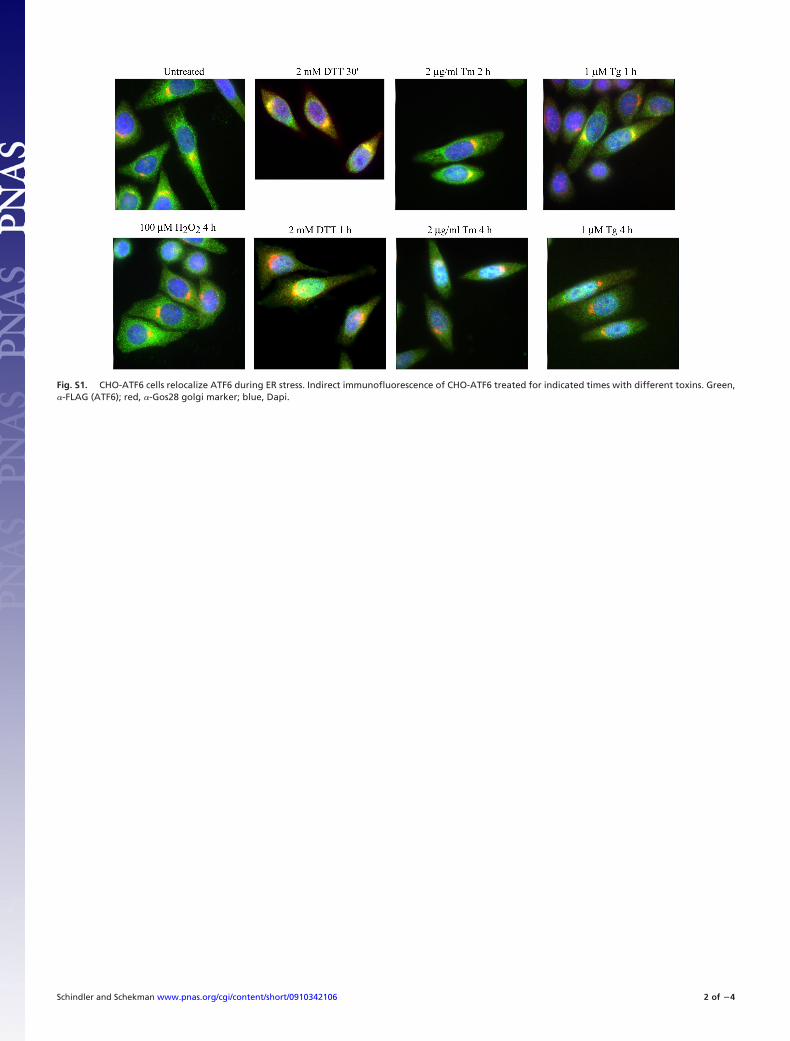
Fig. S1. CHO-ATF6 cells relocalize ATF6 during ER stress. Indirect immunofluorescence of CHO-ATF6 treated for indicated times with different toxins. Green,�-FLAG (ATF6); red, �-Gos28 golgi marker; blue, Dapi.
Schindler and Schekman www.pnas.org/cgi/content/short/0910342106 2 of �4
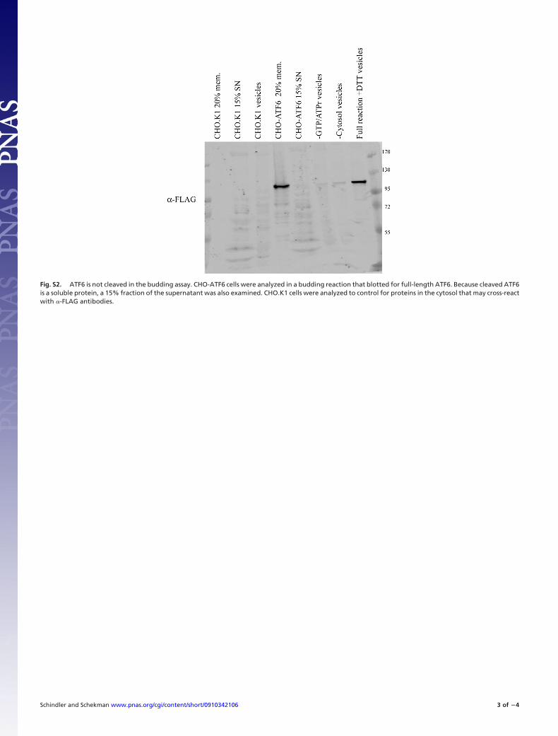
Fig. S2. ATF6 is not cleaved in the budding assay. CHO-ATF6 cells were analyzed in a budding reaction that blotted for full-length ATF6. Because cleaved ATF6is a soluble protein, a 15% fraction of the supernatant was also examined. CHO.K1 cells were analyzed to control for proteins in the cytosol that may cross-reactwith �-FLAG antibodies.
Schindler and Schekman www.pnas.org/cgi/content/short/0910342106 3 of �4