Supporting Information - pnas.org · and subjected to quantitative RT-PCR (qRT-PCR) analysis. TPB,...
5
Supporting Information Zhang et al. 10.1073/pnas.1214156110 SI Materials and Methods Cell Culture and Reagents. HEK293T, SaoS2, and HeLa cells were cultured in DMEM (GIBCO) supplemented with 10% (vol/vol) FBS. HCT116 WT, HCT116 p53 -/- , and U2OS cells were cul- tured in McCoy’s 5A supplemented with 10% FBS. The fol- lowing reagents were used: MG132 (Calbiochem), nocodazole (Sigma), imidazole (Sigma), GSH-Sepharose (GE Health- care), Ni-Sepharose (GE Healthcare), doxorubicinorubicin (Sigma), and etoposide (Sigma). Plasmids and Antibodies. Tripartite motif 39 (TRIM39), p21, and Cdt2 were cloned into expression vectors, and deletion mutants were generated by a PCR-based approach. All constructs were sequenced before use. For Western blot analysis, the following antibodies were used: anti-p21 (BD Pharmingen/Santa Cruz Biotechnology), anti-Cul4 (Santa Cruz Biotechnology), anti-Myc (Cell signaling technology; Santa Cruz Biotechnology), anti– DNA damage-binding protein 1 (DDB1; Bethyl), anti-Cdt2 (Bethyl), anti-Skp2 (Santa Cruz Biotechnology), anti-FLAG (Sigma), anti-HA (Roche), anti-phosphohistone H3 (Millipore), anti-puma (Sigma), anti-p27 (BD Pharmingen), anti-p53 (Santa Cruz Biotechnology), anti-Mdm2 (BD Pharmingen), anti– β-actin (Sigma), anti-proliferating cell nuclear antigen (Abcam), anti-His (Sigma), and anti-GFP (Sigma). Real-Time PCR. Total RNA was extracted with TRIzol (Invitrogen). RNA (4 μg) was reverse-transcribed in a 20-μL reaction using the PrimeScript RT-PCR Kit (TAKARA). After RNase H treat- ment at 37 °C for 20 min and after inactivation by incubating samples at 95 °C for 2 min, the RT reaction was diluted. cDNA was used for RT-PCR or real-time PCR assay. Primer sequences were as follows: TRIM39α:5′-GGCTTCGAGATGCTTAA GGATGT-3′,5′- CCCGTGGACAGATCGTTGA-3′ TaqMan probe: 5′FAM-CTCTCCTTGGATTAGTAAAA-3′ MGB; TRIM39β: 5′-AGGGTCACATCCGCAATTAG-3′, 5′-GTC AAAAGTACCCTGGAAAAATGTG-3′ TaqMan probe: 5′FAM-CCCTGG AAAAATGTGAAAA-3′ MGB In Vivo Ubiquitylation Assay. HCT116 WT or HCT116 p53 -/-- cells expressing HA-ubiquitin were infected with indicated lentivi- ruses. Cells were treated with 20 μM MG132 for 6 h before being harvested by modified radioimmunoprecipitation assay buffer (50 mM Tris·HCl at pH 8.0, 0.1% SDS, 1% sodium deoxycholine acid, 1% Triton X-100, 0.15 M NaCl, 5 mM N-ethylmaleimide, 1 mM Na 4 VO 3 , 10 mM NaF, and 1 mM EDTA). Then ubiq- uitylation assay was performed as previously described (1). Flow Cytometry. Cells were harvested and fixed in 70% ethanol. For cell cycle and apoptosis assay, the fixed cells were washed once with PBS and then incubated with PBS containing 10 μg/mL propidium iodide and 20 μg/mL RNaseA for 30 min at 37 °C. Cells were analyzed by flow cytometry using an Epics XL flow cytometer (Beckman Coulter). Immunofluorescence. HCT116 p53 +/+ cells grown in 10-cm 2 dishes were transfected with TRIM39-Myc (2 μg). Twenty-four hours later, cells were split into new 10-cm 2 dishes with coverslips. At an additional 24 h later, the culture medium was removed and coverslips were carefully washed three times with PBS. Cells were then fixed with 4% paraformaldehyde for 5 min at room temperature and subsequently washed twice with PBS and twice with washing buffer. Cells were then permeabilized for 5 min with 0.5% Triton X-100, blocked in PBS plus 1% BSA, and subsequently incubated with appropriate primary/secondary an- tibodies and DAPI. Confocal fluorescence images were obtained with a confocal microscope (LSM 780 NLO; Carl Zeiss). Immunohistochemistry. Formalin-fixed, paraffin-embedded 5-μm tissue sections were deparaffinized in xylenes and rehydrated through a graded series of alcohols. After antigen retrieval was performed, all sections were blocked at room temperature in avidin/biotin blocking buffer (Vector Laboratories) and then in 3% BSA for 30 min. Staining with anti-p21 antibody was con- ducted at room temperature for 60 min. Sections were rinsed twice in PBS, and protein staining was performed using a dia- minobenzidine substrate kit. Samples were counterstained with hematoxylin. Immunohistochemistry images were obtained using an upright microscope (Nikon). In Vitro Binding Assay. Bacterially expressed GST or GST-Cdt2 was bound to glutathione-Sepharose beads and incubated with puri- fied His-p21 and/or His-TRIM39α, His-TRIM39β, or His–TRIM39- N in TN buffer (50 mM Tris·HCl at pH 8.0, 250 mM NaCl, 10 mM β-glycerol phosphate, 10 mM NaF, 1 mM Na 4 VO 3 , and 0.5% Triton X-100) for 1 h at 4 °C. The beads were washed with TN buffer and subjected to SDS/PAGE. 1. Kim Y, Starostina NG, Kipreos ET (2008) The CRL4Cdt2 ubiquitin ligase targets the degradation of p21Cip1 to control replication licensing. Genes Dev 22(18):2507–2519. Zhang et al. www.pnas.org/cgi/content/short/1214156110 1 of 5
Transcript of Supporting Information - pnas.org · and subjected to quantitative RT-PCR (qRT-PCR) analysis. TPB,...
Supporting InformationZhang et al. 10.1073/pnas.1214156110SI Materials and MethodsCell Culture and Reagents. HEK293T, SaoS2, and HeLa cells werecultured in DMEM (GIBCO) supplemented with 10% (vol/vol)FBS. HCT116 WT, HCT116 p53−/−, and U2OS cells were cul-tured in McCoy’s 5A supplemented with 10% FBS. The fol-lowing reagents were used: MG132 (Calbiochem), nocodazole(Sigma), imidazole (Sigma), GSH-Sepharose (GE Health-care), Ni-Sepharose (GE Healthcare), doxorubicinorubicin(Sigma), and etoposide (Sigma).
Plasmids and Antibodies. Tripartite motif 39 (TRIM39), p21, andCdt2 were cloned into expression vectors, and deletion mutantswere generated by a PCR-based approach. All constructs weresequenced before use. For Western blot analysis, the followingantibodies were used: anti-p21 (BD Pharmingen/Santa CruzBiotechnology), anti-Cul4 (Santa Cruz Biotechnology), anti-Myc(Cell signaling technology; Santa Cruz Biotechnology), anti–DNA damage-binding protein 1 (DDB1; Bethyl), anti-Cdt2(Bethyl), anti-Skp2 (Santa Cruz Biotechnology), anti-FLAG(Sigma), anti-HA (Roche), anti-phosphohistone H3 (Millipore),anti-puma (Sigma), anti-p27 (BD Pharmingen), anti-p53 (SantaCruz Biotechnology), anti-Mdm2 (BD Pharmingen), anti–β-actin (Sigma), anti-proliferating cell nuclear antigen (Abcam),anti-His (Sigma), and anti-GFP (Sigma).
Real-Time PCR.Total RNAwas extracted with TRIzol (Invitrogen).RNA (4 μg) was reverse-transcribed in a 20-μL reaction using thePrimeScript RT-PCR Kit (TAKARA). After RNase H treat-ment at 37 °C for 20 min and after inactivation by incubatingsamples at 95 °C for 2 min, the RT reaction was diluted. cDNAwas used for RT-PCR or real-time PCR assay. Primer sequenceswere as follows:
TRIM39α: 5′-GGCTTCGAGATGCTTAA GGATGT-3′, 5′-CCCGTGGACAGATCGTTGA-3′TaqMan probe: 5′FAM-CTCTCCTTGGATTAGTAAAA-3′MGB; TRIM39β: 5′-AGGGTCACATCCGCAATTAG-3′,5′-GTC AAAAGTACCCTGGAAAAATGTG-3′TaqMan probe: 5′FAM-CCCTGG AAAAATGTGAAAA-3′MGB
In Vivo Ubiquitylation Assay.HCT116WT or HCT116 p53−/−− cellsexpressing HA-ubiquitin were infected with indicated lentivi-ruses. Cells were treated with 20 μMMG132 for 6 h before being
harvested by modified radioimmunoprecipitation assay buffer(50 mM Tris·HCl at pH 8.0, 0.1% SDS, 1% sodium deoxycholineacid, 1% Triton X-100, 0.15 M NaCl, 5 mM N-ethylmaleimide,1 mM Na4VO3, 10 mM NaF, and 1 mM EDTA). Then ubiq-uitylation assay was performed as previously described (1).
Flow Cytometry. Cells were harvested and fixed in 70% ethanol.For cell cycle and apoptosis assay, the fixed cells were washedonce with PBS and then incubated with PBS containing 10 μg/mLpropidium iodide and 20 μg/mL RNaseA for 30 min at 37 °C.Cells were analyzed by flow cytometry using an Epics XL flowcytometer (Beckman Coulter).
Immunofluorescence.HCT116 p53+/+ cells grown in 10-cm2 disheswere transfected with TRIM39-Myc (2 μg). Twenty-four hourslater, cells were split into new 10-cm2 dishes with coverslips. Atan additional 24 h later, the culture medium was removed andcoverslips were carefully washed three times with PBS. Cellswere then fixed with 4% paraformaldehyde for 5 min at roomtemperature and subsequently washed twice with PBS and twicewith washing buffer. Cells were then permeabilized for 5 minwith 0.5% Triton X-100, blocked in PBS plus 1% BSA, andsubsequently incubated with appropriate primary/secondary an-tibodies and DAPI. Confocal fluorescence images were obtainedwith a confocal microscope (LSM 780 NLO; Carl Zeiss).
Immunohistochemistry. Formalin-fixed, paraffin-embedded 5-μmtissue sections were deparaffinized in xylenes and rehydratedthrough a graded series of alcohols. After antigen retrieval wasperformed, all sections were blocked at room temperature inavidin/biotin blocking buffer (Vector Laboratories) and then in3% BSA for 30 min. Staining with anti-p21 antibody was con-ducted at room temperature for 60 min. Sections were rinsedtwice in PBS, and protein staining was performed using a dia-minobenzidine substrate kit. Samples were counterstained withhematoxylin. Immunohistochemistry images were obtained usingan upright microscope (Nikon).
In Vitro Binding Assay.Bacterially expressed GST or GST-Cdt2 wasbound to glutathione-Sepharose beads and incubated with puri-fiedHis-p21 and/or His-TRIM39α, His-TRIM39β, or His–TRIM39-N in TN buffer (50 mM Tris·HCl at pH 8.0, 250 mM NaCl, 10 mMβ-glycerol phosphate, 10 mM NaF, 1 mM Na4VO3, and 0.5%Triton X-100) for 1 h at 4 °C. The beads were washed with TNbuffer and subjected to SDS/PAGE.
1. Kim Y, Starostina NG, Kipreos ET (2008) The CRL4Cdt2 ubiquitin ligase targets thedegradation of p21Cip1 to control replication licensing. Genes Dev 22(18):2507–2519.
Zhang et al. www.pnas.org/cgi/content/short/1214156110 1 of 5
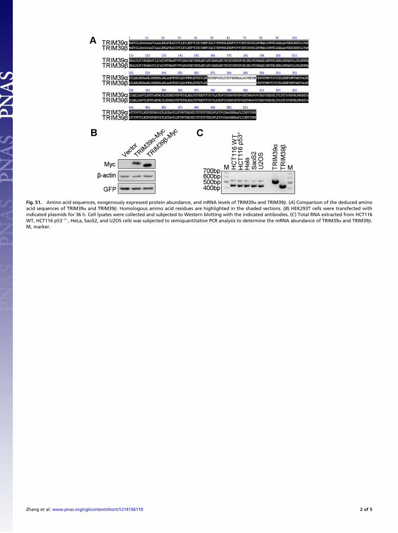
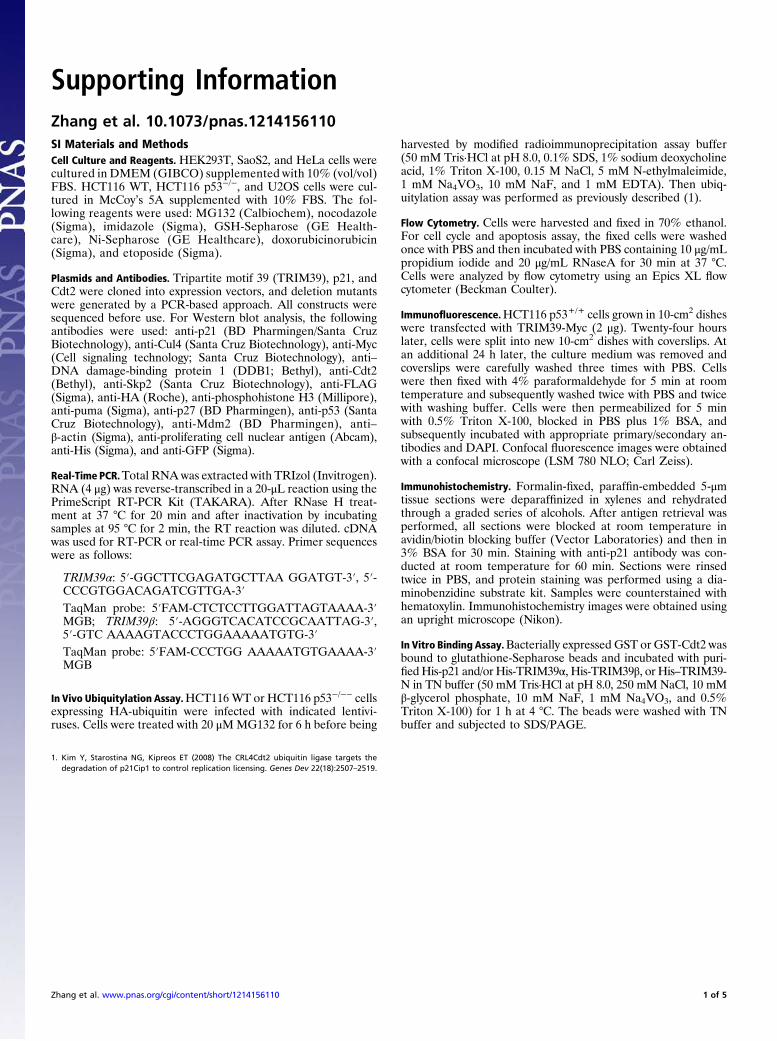
Fig. S1. Amino acid sequences, exogenously expressed protein abundance, and mRNA levels of TRIM39α and TRIM39β. (A) Comparison of the deduced aminoacid sequences of TRIM39α and TRIM39β. Homologous amino acid residues are highlighted in the shaded sections. (B) HEK293T cells were transfected withindicated plasmids for 36 h. Cell lysates were collected and subjected to Western blotting with the indicated antibodies. (C) Total RNA extracted from HCT116WT, HCT116 p53−/−, HeLa, SaoS2, and U2OS cells was subjected to semiquantitative PCR analysis to determine the mRNA abundance of TRIM39α and TRIM39β.M, marker.
Zhang et al. www.pnas.org/cgi/content/short/1214156110 2 of 5
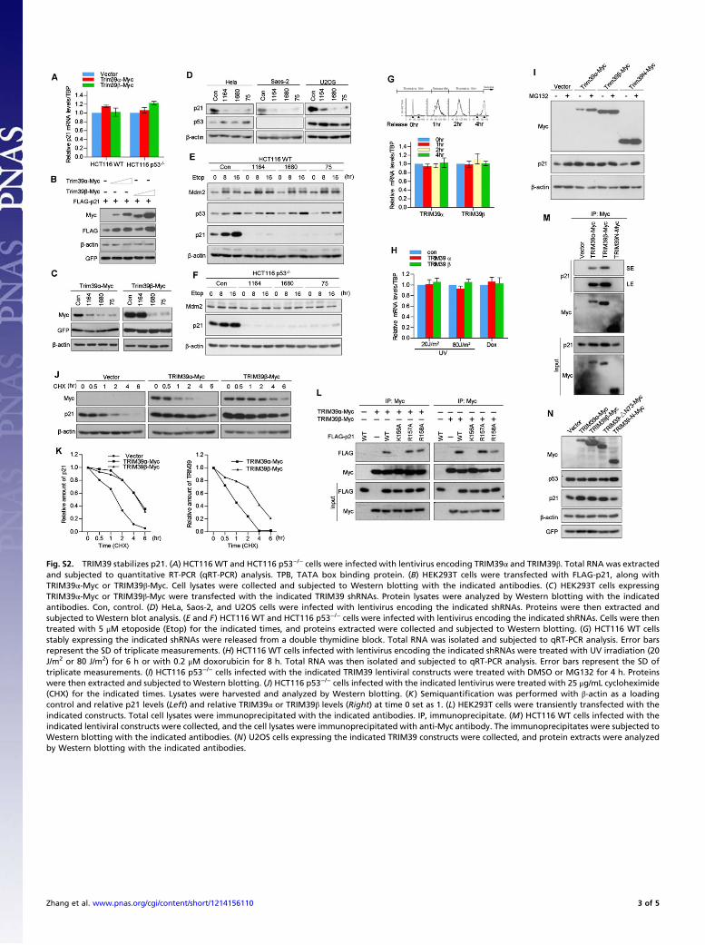
Fig. S2. TRIM39 stabilizes p21. (A) HCT116 WT and HCT116 p53−/− cells were infected with lentivirus encoding TRIM39α and TRIM39β. Total RNA was extractedand subjected to quantitative RT-PCR (qRT-PCR) analysis. TPB, TATA box binding protein. (B) HEK293T cells were transfected with FLAG-p21, along withTRIM39α-Myc or TRIM39β-Myc. Cell lysates were collected and subjected to Western blotting with the indicated antibodies. (C) HEK293T cells expressingTRIM39α-Myc or TRIM39β-Myc were transfected with the indicated TRIM39 shRNAs. Protein lysates were analyzed by Western blotting with the indicatedantibodies. Con, control. (D) HeLa, Saos-2, and U2OS cells were infected with lentivirus encoding the indicated shRNAs. Proteins were then extracted andsubjected to Western blot analysis. (E and F) HCT116 WT and HCT116 p53−/− cells were infected with lentivirus encoding the indicated shRNAs. Cells were thentreated with 5 μM etoposide (Etop) for the indicated times, and proteins extracted were collected and subjected to Western blotting. (G) HCT116 WT cellsstably expressing the indicated shRNAs were released from a double thymidine block. Total RNA was isolated and subjected to qRT-PCR analysis. Error barsrepresent the SD of triplicate measurements. (H) HCT116 WT cells infected with lentivirus encoding the indicated shRNAs were treated with UV irradiation (20J/m2 or 80 J/m2) for 6 h or with 0.2 μM doxorubicin for 8 h. Total RNA was then isolated and subjected to qRT-PCR analysis. Error bars represent the SD oftriplicate measurements. (I) HCT116 p53−/− cells infected with the indicated TRIM39 lentiviral constructs were treated with DMSO or MG132 for 4 h. Proteinswere then extracted and subjected to Western blotting. (J) HCT116 p53−/− cells infected with the indicated lentivirus were treated with 25 μg/mL cycloheximide(CHX) for the indicated times. Lysates were harvested and analyzed by Western blotting. (K) Semiquantification was performed with β-actin as a loadingcontrol and relative p21 levels (Left) and relative TRIM39α or TRIM39β levels (Right) at time 0 set as 1. (L) HEK293T cells were transiently transfected with theindicated constructs. Total cell lysates were immunoprecipitated with the indicated antibodies. IP, immunoprecipitate. (M) HCT116 WT cells infected with theindicated lentiviral constructs were collected, and the cell lysates were immunoprecipitated with anti-Myc antibody. The immunoprecipitates were subjected toWestern blotting with the indicated antibodies. (N) U2OS cells expressing the indicated TRIM39 constructs were collected, and protein extracts were analyzedby Western blotting with the indicated antibodies.
Zhang et al. www.pnas.org/cgi/content/short/1214156110 3 of 5
Fig. S3. TRIM39α and TRIM39β are both required to regulate p21 stability. (A–D) HCT116 p53−/− cells stably expressing shRNA-resistant forms of TRIM39α or/and TRIM39β were infected with indicated lentiviral shRNA constructs. Cells were then harvested, and lysates were subjected to immunoblotting with theindicated antibodies. Con, control. (E and F) TRIM39α interacts with TRIM39β in vivo. HEK293T cells transfected with the indicated constructs were lysed,followed by immunoprecipitation with anti-Myc antibody. The immunoprecipitates (IP) were then blotted with the indicated antibodies. (G) TRIM39α interactswith TRIM39β in vitro. Purified His-TRIM39β was incubated with GST or GST-TRIM39α coupled to GSH-Sepharose. Proteins retained on Sepharose were thenblotted with the indicated antibodies. (H) TRIM39β stabilizes TRIM39α. HCT116 p53−/− cells infected with the indicated lentivirus were lysed, and lysates wereblotted with the indicated antibodies. (I) HCT116 p53−/− cells infected with the indicated lentivirus were treated with 25 μg/mL cycloheximide (CHX) for theindicated times. Lysates were harvested and analyzed by Western blotting. (Right) Semiquantification was performed with β-actin as a loading control andrelative p21 levels at time 0 set as 1.
Zhang et al. www.pnas.org/cgi/content/short/1214156110 4 of 5
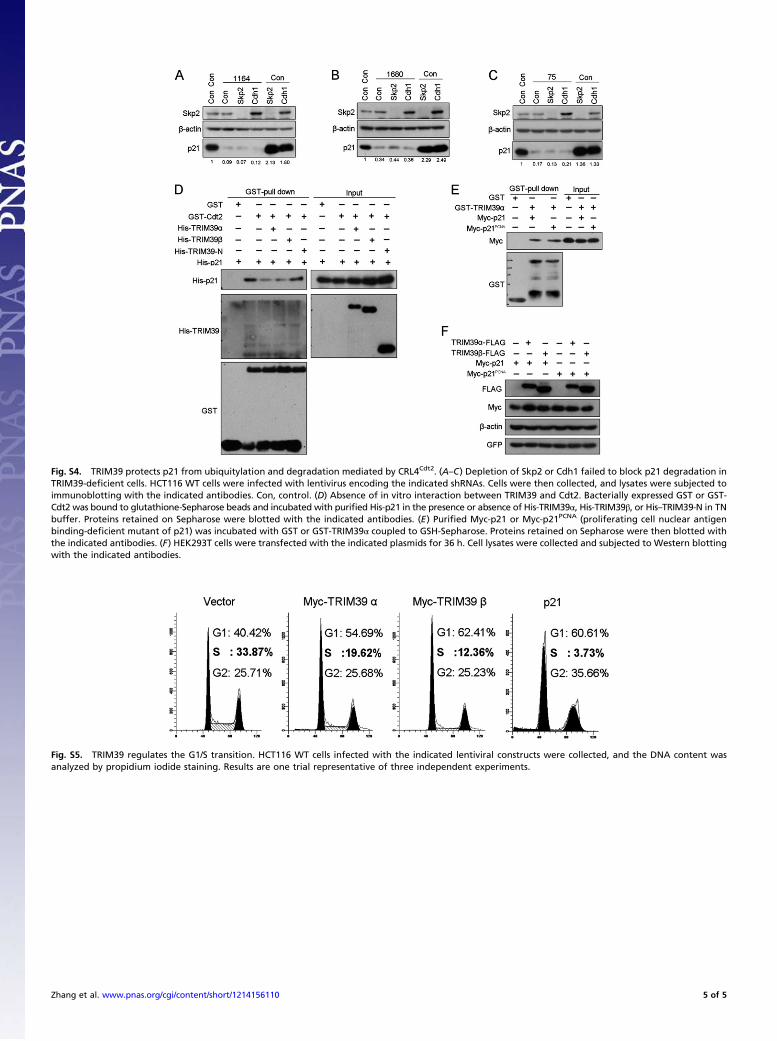
Fig. S4. TRIM39 protects p21 from ubiquitylation and degradation mediated by CRL4Cdt2. (A–C) Depletion of Skp2 or Cdh1 failed to block p21 degradation inTRIM39-deficient cells. HCT116 WT cells were infected with lentivirus encoding the indicated shRNAs. Cells were then collected, and lysates were subjected toimmunoblotting with the indicated antibodies. Con, control. (D) Absence of in vitro interaction between TRIM39 and Cdt2. Bacterially expressed GST or GST-Cdt2 was bound to glutathione-Sepharose beads and incubated with purified His-p21 in the presence or absence of His-TRIM39α, His-TRIM39β, or His–TRIM39-N in TNbuffer. Proteins retained on Sepharose were blotted with the indicated antibodies. (E) Purified Myc-p21 or Myc-p21PCNA (proliferating cell nuclear antigenbinding-deficient mutant of p21) was incubated with GST or GST-TRIM39α coupled to GSH-Sepharose. Proteins retained on Sepharose were then blotted withthe indicated antibodies. (F) HEK293T cells were transfected with the indicated plasmids for 36 h. Cell lysates were collected and subjected to Western blottingwith the indicated antibodies.
Fig. S5. TRIM39 regulates the G1/S transition. HCT116 WT cells infected with the indicated lentiviral constructs were collected, and the DNA content wasanalyzed by propidium iodide staining. Results are one trial representative of three independent experiments.
Zhang et al. www.pnas.org/cgi/content/short/1214156110 5 of 5


![Research Paper HUWE1 controls the development of non-small ... · ACTAGCACAG-AAT3´ [22] and TP53: 5´CGGCGCA CAGAGGAAGAGAAT3´ [23]. The DNA vectors were co-transfected into HEK293T](https://static.fdocuments.net/doc/165x107/5f4af818e356352b74783c76/research-paper-huwe1-controls-the-development-of-non-small-actagcacag-aat3.jpg)