Support- supports body and gives it shape Protection- for delicate organs, heart, lungs, brain, any...
59
-
Upload
alan-hensley -
Category
Documents
-
view
216 -
download
0
Transcript of Support- supports body and gives it shape Protection- for delicate organs, heart, lungs, brain, any...
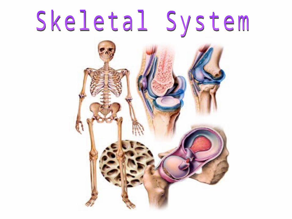

• Support- supports body and gives it shape
• Protection- for delicate organs, heart, lungs, brain, any internal organs
• Movement- bones act as levers for muscles
• Mineral storage- calcium
• Blood cell formation

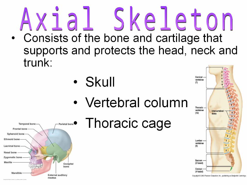
• There are 206 bones in the adult body and they fall into 2 Categories:
• 1. Axial Skeleton
• 2. Appendicular Skeleton


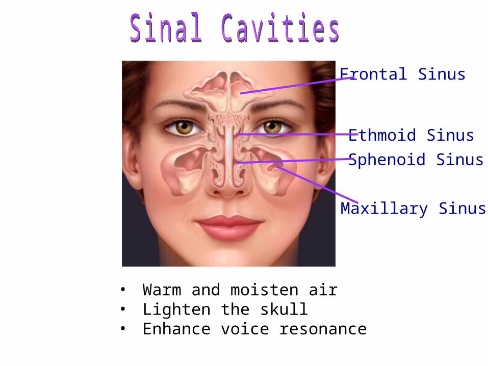
• Warm and moisten air• Lighten the skull• Enhance voice resonance
Frontal Sinus
Ethmoid Sinus
Sphenoid Sinus
Maxillary Sinus
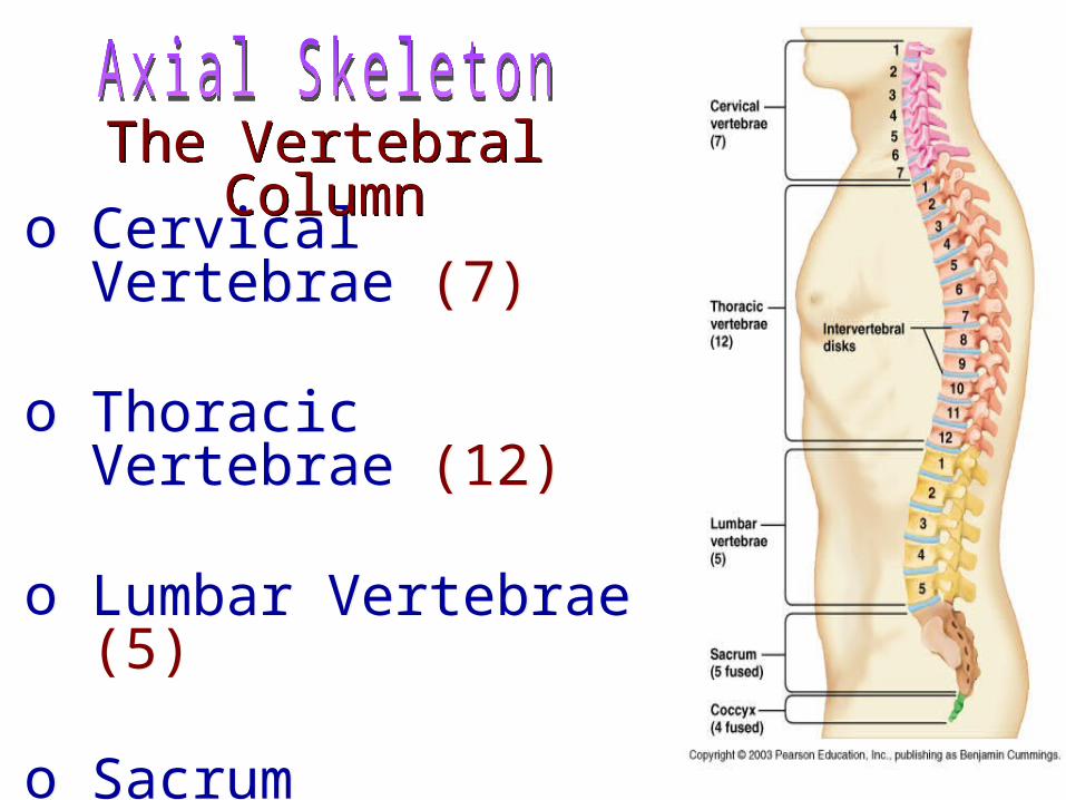
o Cervical Vertebrae (7)
o Thoracic Vertebrae (12)
o Lumbar Vertebrae (5)
o Sacrumo Coccyx
o Cervical Vertebrae (7)
o Thoracic Vertebrae (12)
o Lumbar Vertebrae (5)
o Sacrumo Coccyx
The Vertebral ColumnThe Vertebral Column

Sternum
Ribs (12 total)
Sternum
Ribs (12 total)
The Thoracic CageThe Thoracic Cage

Sacrum & CoccyxSacrum & Coccyx

• Includes:
• 1. Pectoral Girdle
• 2. Upper Limbs
• 3. Pelvic Girdle
• 4. Lower Limbs

Bones of the Pectoral GirdleBones of the Pectoral Girdle

Humerus
Ulna
Radius
8 Carpals
14 Phalanges
5 Metacarpals
Bones of the Upper LimbsBones of the Upper Limbs
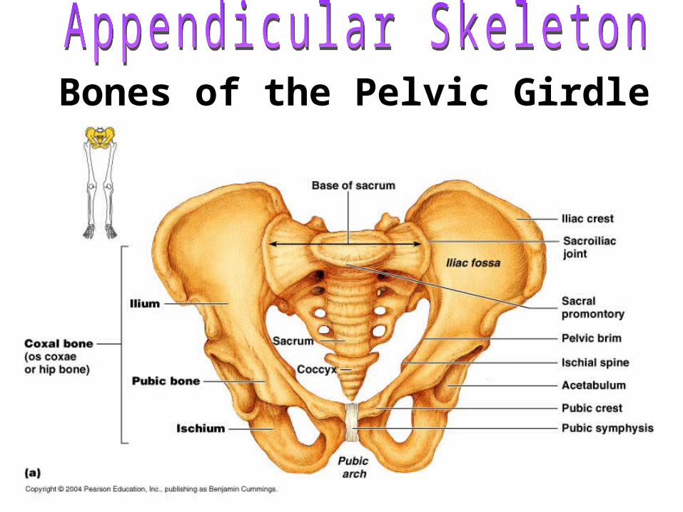
Bones of the Pelvic Girdle
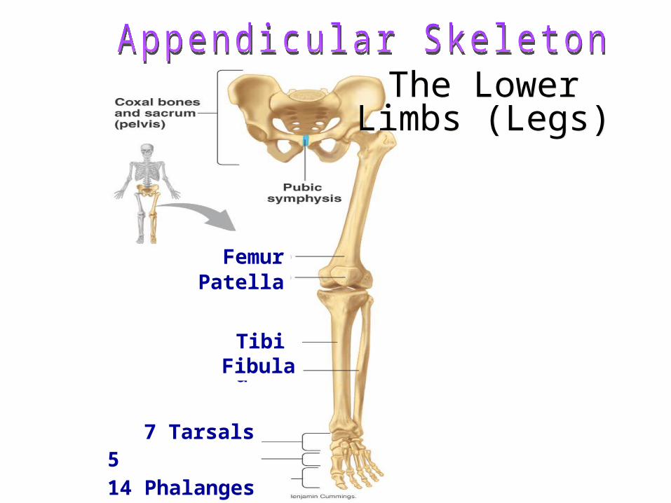
Patella
The Lower Limbs (Legs)
The Lower Limbs (Legs)
Femur
TibiaFibula
5 Metatarsals14 Phalanges
7 Tarsals

WARM UP
• How many bones does the adult skeleton have?
• What is the appendicular skeleton and the axial skeleton?

• Long Bones- longer than they are wide. Ex: metacarples, phalangies, humerus, ulna, radius…
• Short Bones- circular/square shape. Ex: carpals, tarsals
• Flat Bones- rib, scapula, skull, sternum
• Irregular Bones- vertebrae, facial bones
• Other: patella
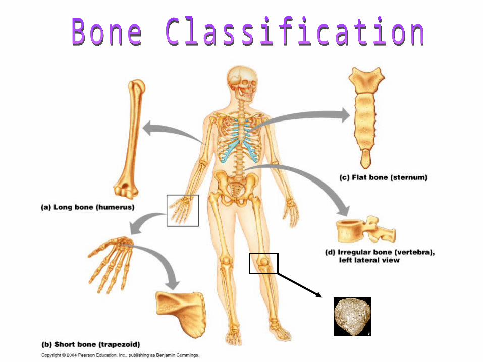

What type of bones do you think the following are?
1. Ribs2. Phalanges3. Pelvic bone4. Skull bones5. Ulna/ Radius6. Nasal bone7. Mandible

• Bones are organs which means they are composed of many types of tissues:1. Fibrocartilage connective tissue.2. Hayline Cartilage.3. Blood tissue.4. Lymphatic tissue.5. Adipose tissue.6. Nervous tissue.
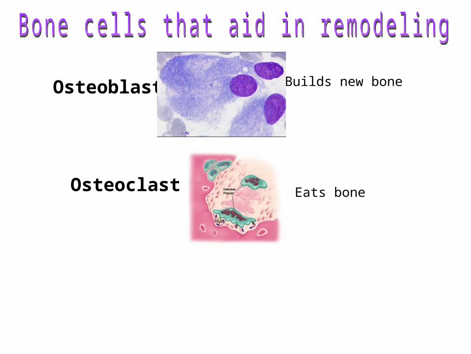
Osteoblast
Osteoclast Eats bone
Builds new bone

1.1. Epiphysis:Epiphysis: The end of each long bone2.2. Diaphysis: Diaphysis: The shaft of the bone3.3. Hyaline Cartilage: Hyaline Cartilage: on the outer surface of the
epiphysis to assist growth4.4. Periosteum: Periosteum: Completely encloses or wraps
around the bone (very tough)5.5. Compact bone: Compact bone: Very thick with no gaps around
the diaphysis6.6. Spongy bone: Spongy bone: numerous branching plates and
spaces in the epiphysis. 7.7. MarrowMarrow: Red or yellow in the center of bones.

Distal
epiphysis
Proximal
epiphysis
diaphysis
yellow marrow
epiphyseal line
periosteum
compact bone
spongy bone
Endosteum
hyaline cartilage
Sharpey’s fibers
STOP!! 1. Label the human skeleton
worksheet2. You have 10 minutes to quiz
each other on the skeleton. 3. With a partner you will then
construct out of toothpicks and other materials the skeleton.
1. Label the human skeleton worksheet
2. You have 10 minutes to quiz each other on the skeleton.
3. With a partner you will then construct out of toothpicks and other materials the skeleton.
WARM UP
• T/F the largest bone is the pelvic bone.• T/F the 'femur', in the thigh, makes up almost
one quarter of the body's total height.• T/F bone rots quicker than cartilage after
death. • What does the term ossification mean?• What is the purpose of the growth plate?

Answers
• True – it’s made up of 6 bones total• True – it’s the longest bone in the body• False, cartilage rots faster, This is why the
skulls of skeletons have no nose or ears.

1. Most bones develop from masses of hyaline cartilage shaped like future bones.
1. Most bones develop from masses of hyaline cartilage shaped like future bones.
This occurs while still in the uterus as a fetus- usually the first two months.
This occurs while still in the uterus as a fetus- usually the first two months.

2. The cartilage begins to break down and disappear first in the diaphysis. At the same time the periosteum begins to form, the cartilage calcifies and blood vessels begin.
2. The cartilage begins to break down and disappear first in the diaphysis. At the same time the periosteum begins to form, the cartilage calcifies and blood vessels begin.
cartilage
calcified cartilage
periosteum

3. Compact bone begins to form. The epiphysis cartilage continues to grow and calcify.
3. Compact bone begins to form. The epiphysis cartilage continues to grow and calcify.
Compact bonejust before birthjust before birth
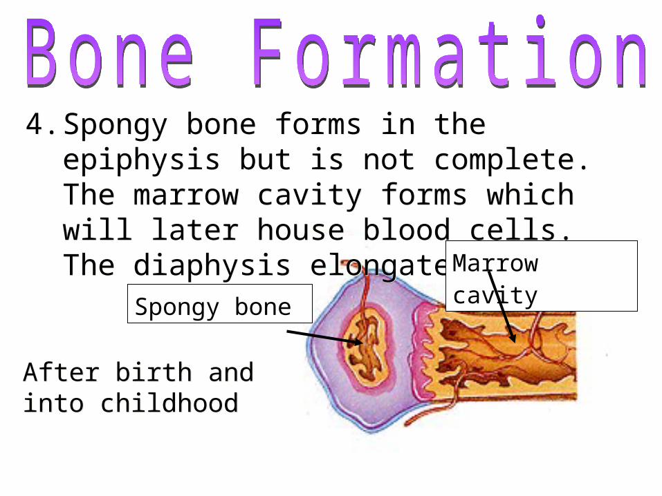
4. Spongy bone forms in the epiphysis but is not complete. The marrow cavity forms which will later house blood cells. The diaphysis elongates.
4. Spongy bone forms in the epiphysis but is not complete. The marrow cavity forms which will later house blood cells. The diaphysis elongates.
Spongy bone
After birth and into childhoodAfter birth and into childhood
Marrow cavity
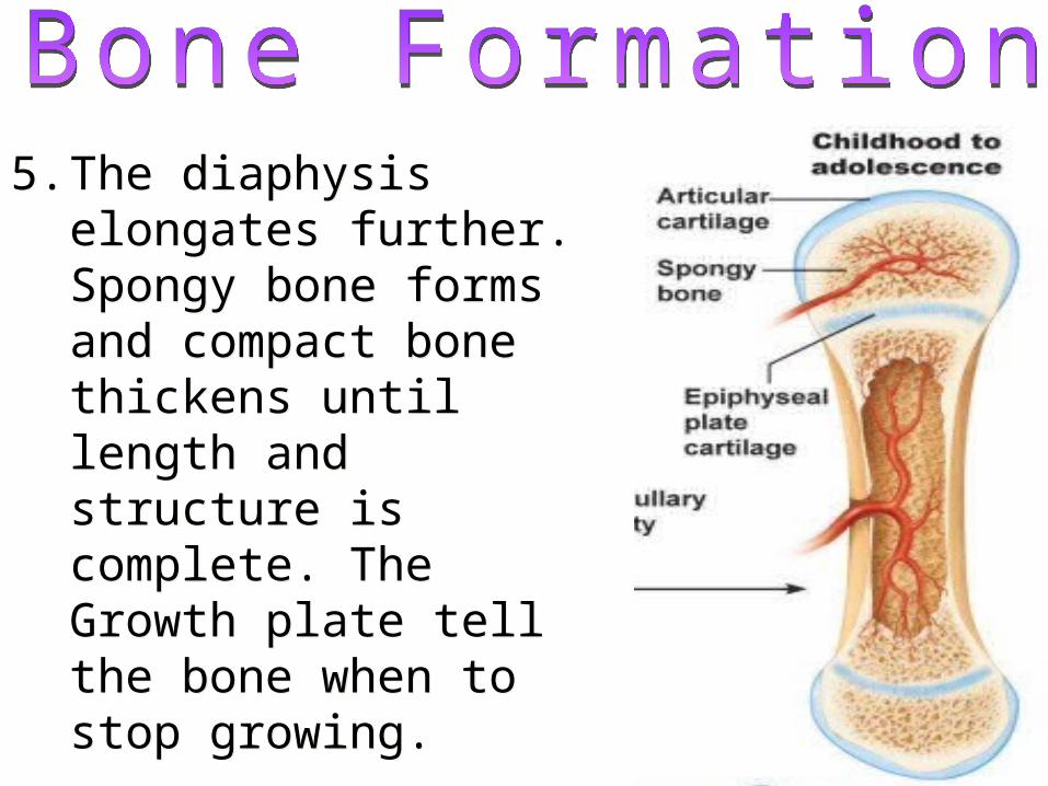
5. The diaphysis elongates further. Spongy bone forms and compact bone thickens until length and structure is complete. The Growth plate tell the bone when to stop growing.
5. The diaphysis elongates further. Spongy bone forms and compact bone thickens until length and structure is complete. The Growth plate tell the bone when to stop growing.

6. Adult bone: everything is fully grown! Cartilage is only left at the ends of the epiphysis. Marrow is now fully formed also
6. Adult bone: everything is fully grown! Cartilage is only left at the ends of the epiphysis. Marrow is now fully formed also
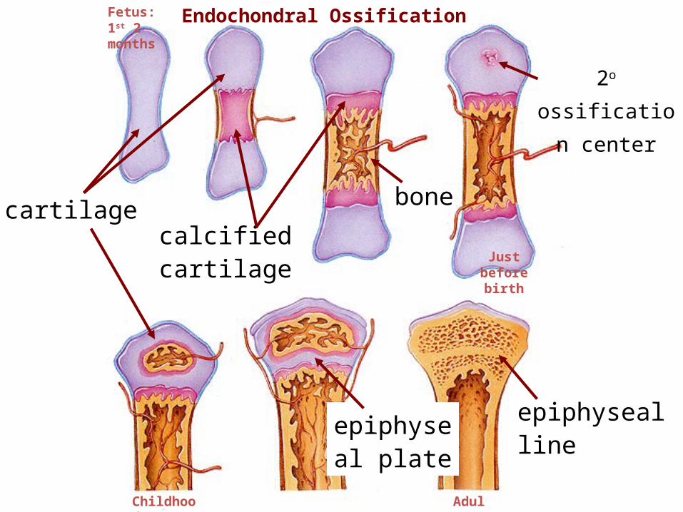
cartilagecalcified cartilage
bone
epiphyseal plate
epiphyseal line
Endochondral Ossification
2o ossification
center
Fetus: 1st 2 months
AdultChildhood
Just before birth
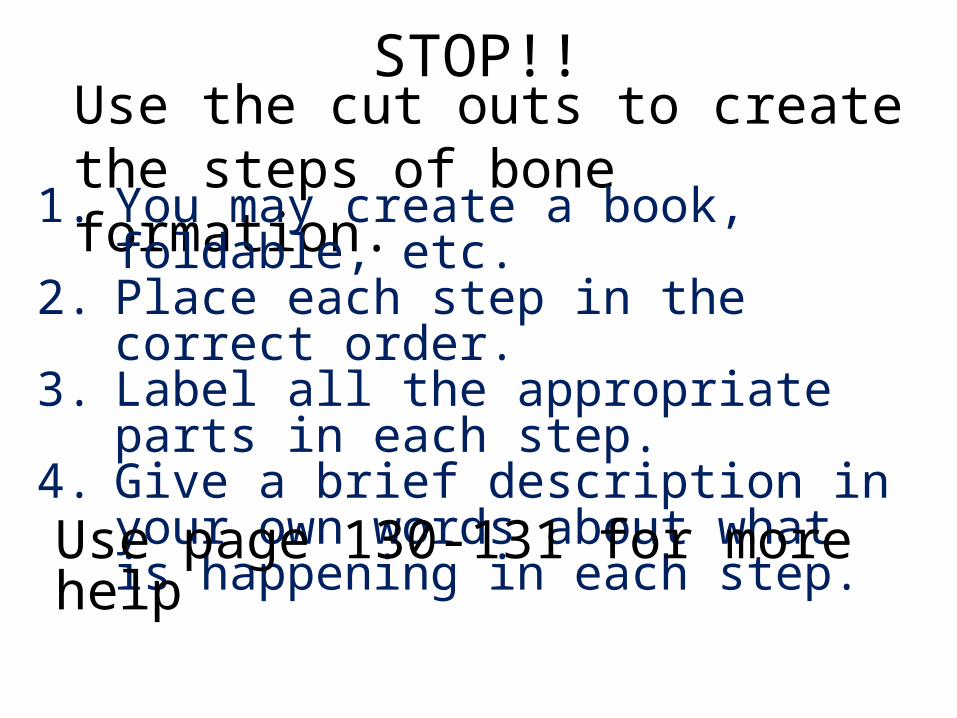
Use the cut outs to create the steps of bone formation.
STOP!!
1. You may create a book, foldable, etc. 2. Place each step in the correct order. 3. Label all the appropriate parts in each
step. 4. Give a brief description in your own
words about what is happening in each step. Use page 130-131 for more help

Did you finish your work?
Yes?• Put the skeleton back
where you found it.
• Place the bone formation booklet in the block 1 bin.
• Have a great night!
No? • Finish the skeleton and
place it back where you found it.
• Complete your bone formation book for HW. Remember it is 25points product.


Types of bone breaks
1.Simple- skin is not pierced (most breaks)2.Compound- skin is pierced3.Complete- bone is broken in half4.Partial- broken lengthwise but not two parts5.Comminuted- broken into several pieces6.Spiral- twisted
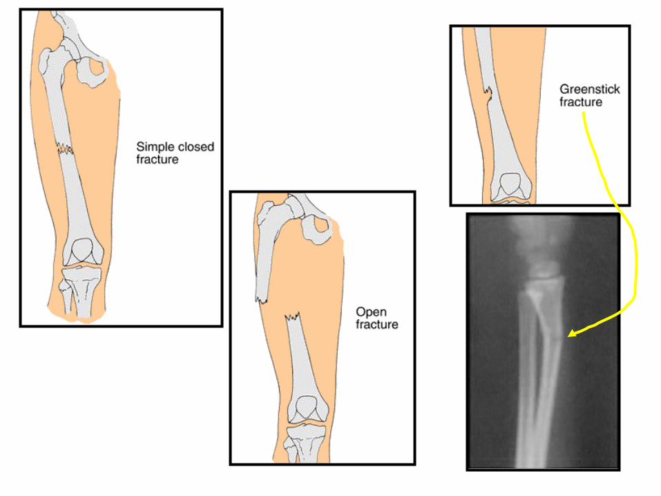

What kind of fracture is this?It’s kind of tough to tell, but this is a _ _ _ _ _ _ fracture.

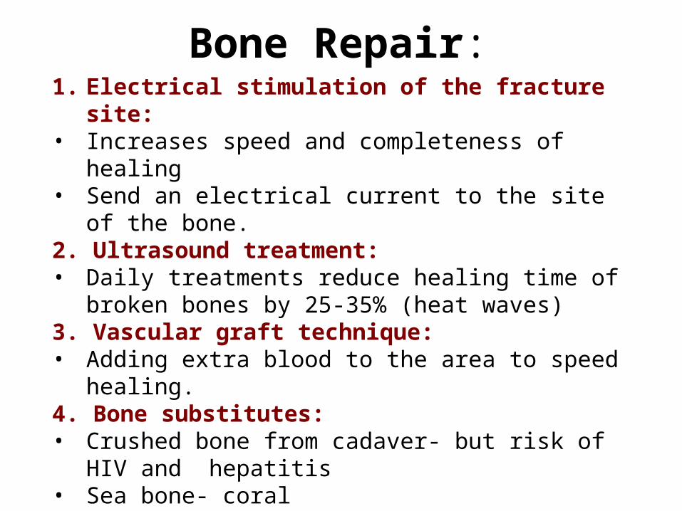
Bone Repair:1. Electrical stimulation of the fracture site:• Increases speed and completeness of healing• Send an electrical current to the site of the bone. 2. Ultrasound treatment:• Daily treatments reduce healing time of broken bones
by 25-35% (heat waves)3. Vascular graft technique:• Adding extra blood to the area to speed healing.4. Bone substitutes:• Crushed bone from cadaver- but risk of HIV and
hepatitis• Sea bone- coral• Artificial bone- ceramic


STOP!!
Discuss these questions with a partner:
1.What type of bones break the easiest? Why? 2. What type of bones do not break easily, why? 3.Why are blood vessels important in bones and in repairing them?

Bone strength lab

UEQ: How does our skeleton move?
LEQ: what are the three kinds of skeletal joints and how do
they allow us to move?
VOCAB: joints, fibrous, cartilaginous, synovial, saddle, ball and socket, hinge, pivot, gliding
WARM UP: complete the handout
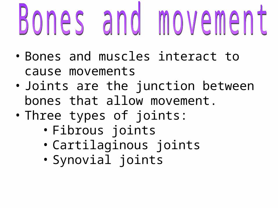
• Bones and muscles interact to cause movements
• Joints are the junction between bones that allow movement.
• Three types of joints: • Fibrous joints• Cartilaginous joints• Synovial joints

Immovable Joints (formed by a thin layer of dense connective tissue)
suturesuture
pubis symphisispubis symphisis
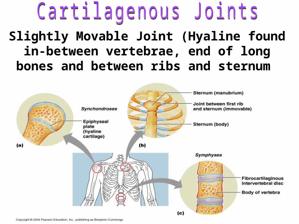
Slightly Movable Joint (Hyaline found in-between vertebrae, end of long bones and
between ribs and sternum
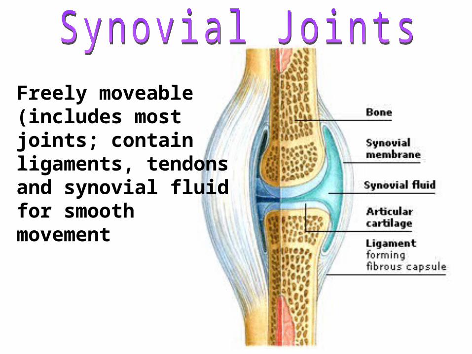
Freely moveable (includes most joints; contain ligaments, tendons and synovial fluid for smooth movement


Types of freely movable joints1.1.SaddleSaddle: carpal and metacarpal bones of thumb
2.2.Ball and socketBall and socket: shoulder and hip joints
3.3.PivotPivot- rotation only: proximal end of radius and ulna
4.4.HingeHinge- up and own movement in one plane: knee and elbow
5.5.Gliding-Gliding- sliding and twisting: wrist and ankle

Gliding
Pivot
Saddle
Hinge
Condyloid
Ball and socket

Range of Motion activity

Diseases of the Skeletal System
Diseases of the Skeletal System:
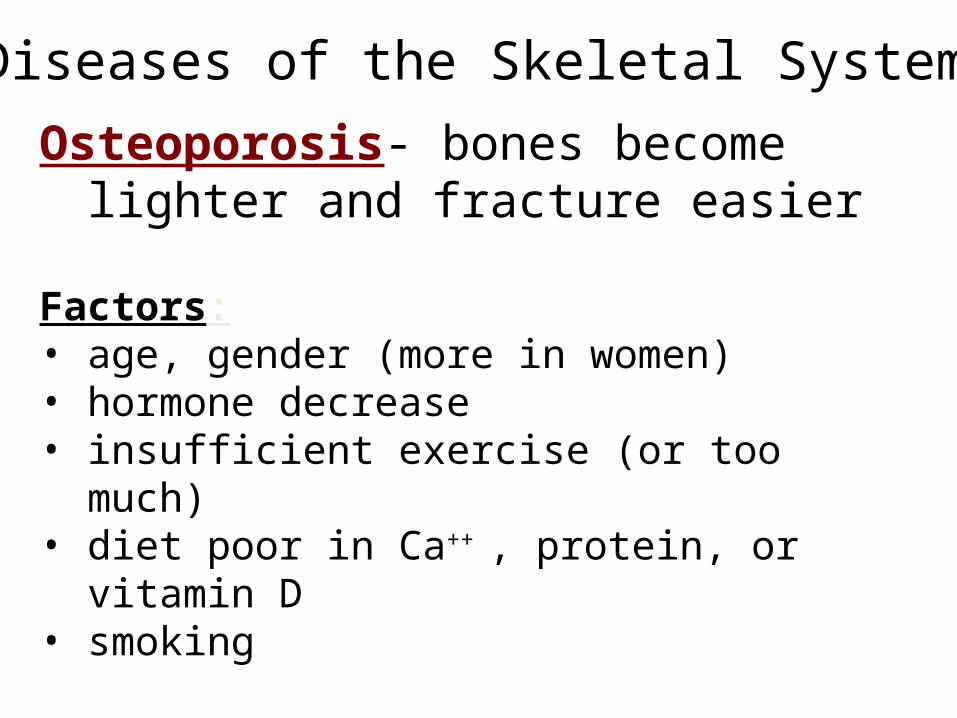
Osteoporosis- bones become lighter and fracture easier
Factors: • age, gender (more in women)• hormone decrease• insufficient exercise (or too much)• diet poor in Ca++ , protein, or vitamin D• smoking

Osteoporosis
2929 4040 8484 9292

Rickets- vitamin D deficiency
Osteomalacia- soft bones, inadequate minerals in bones, lack of vitamin D
Pagets Disease- breaks down old bone faster than it rebuilds new bone. Common in old age.
Rheumatoid arthritis- autoimmune reaction effecting the joints surrounding bones and movement.
Diseases of the Skeletal System:

Case study and research
Finish children story
Book work and practice labeling and worksheets