Supplementaryminformation extreme biomi etic conditions · Supplementaryminformation Synthesis of...
5
This journal is © The Royal Society of Chemistry 2013 J. Name ., 2013, 00 , 1-3 | 1 Supplementary information Synthesis of nanostructured chitin-hematite materials under extreme biomimetic conditions Marcin Wysokowski, a Mykhailo Motylenko, b Juliane Walter ,c Grzegorz Lota, d Jarosław Wojciechowski, d Hartmut Stöcker, c Roberta Galli, e Allison L. Stelling, f Cameliu Himcinschi, g Elke Niederschlag, h Enrico Langer, i Vasilii V. Bazhenov, c Tomasz Szatkowski, a Jakub Zdarta, a Iaroslav Pertenko, c Zoran Kljajić, j Tilmann Leisegang, k Serguei L. Molodtsov, c,l Dirk C. Meyer, c Teofil Jesionowski, a and Hermann Ehrlich* c a Institute of Chemical Technology and Engineering, Poznan University of Technology, 60965 Poznan, Poland b Institute of Materials Science, TU Bergakademie Freiberg, 09599 Freiberg, Germany c Institute of Experimental Physics, TU Bergakademie Freiberg, Leipziger 23, 09599 Freiberg, Germany; Tel: 0049393731392867; E-mail: [email protected] d Institute of Chemistry and Technical Electrochemistry, Faculty of Chemical Technology, Poznan University of Technology, 60965 Poznan, Poland e Faculty of Medicine Carl Gustav Carus, Department of Anesthesiology and Intensive Care Medicine, Clinical Sensoring and Monitoring, Technische Universität Dresden, 01069 Dresden, Germany f Department of Mechanical Engineering and Materials Science, Duke University, 27708 Durham, NC, USA g Institute of Theoretical Physics, TU Bergakademie Freiberg, Leipziger 23, 09599 Freiberg, Germany h Insitut für NE-Metallurgie und Reinstoffe, TU Bergakademie Freiberg, Leipziger Str. 34, 09599 Freiberg, Germany i Institut für Oberflächenphysik und Mikrostrukturphysik, TU Dresden 01062 Dresden j Institute of Marine Biology, University of Montenegro, 85330 Kotor, Montenegro k Fraunhofer-Technologiezentrum Halbleitermaterialien THM, Am St.-Niclas-Schacht 13, 09599 Freiberg, Germany l European X-Ray Free-Electron Laser Facility (XFEL) GmbH, 22761 Hamburg Materials and methods Scanning electron microscopy (SEM) The surface morphology and microstructure of the Fe 2 O 3 nanoparticles were examined on the basis of the SEM images recorded on Ultra 55 (Carl Zeiss AG, Germany). Before testing, the samples were coated with carbon over a period of 5 s for 1 min using an Edwards S150B sputter coater. Raman spectroscopy The Raman measurements were performed using an LabRam HR 800 spectrometer (from Horiba Jobin Yvon) equipped with a 600 grooves/mm grating and a Peltier cooled CCD detector. The spectra were measured in the backscattering configuration with the laser light focused on the samples and collected through a 100x objective of an Olympus microscope. For excitation the 785 nm line of a diode laser was used. The laser power was chosen in order to avoid the damage to the sample surface during the measurements. The power was set at 1.2 mW for the measurement Electronic Supplementary Material (ESI) for RSC Advances. This journal is © The Royal Society of Chemistry 2014
Transcript of Supplementaryminformation extreme biomi etic conditions · Supplementaryminformation Synthesis of...
This journal is © The Royal Society of Chemistry 2013 J. Name., 2013, 00, 1-3 | 1
Supplementary informationSynthesis of nanostructured chitin-hematite materials under extreme biomimetic conditions Marcin Wysokowski,a Mykhailo Motylenko,b Juliane Walter,c Grzegorz Lota,d Jarosław Wojciechowski,d Hartmut Stöcker,c Roberta Galli,e Allison L. Stelling,f Cameliu Himcinschi,g Elke Niederschlag,h Enrico Langer,i Vasilii V. Bazhenov,c Tomasz Szatkowski,a Jakub Zdarta,a Iaroslav Pertenko,c Zoran Kljajić,j Tilmann Leisegang,k Serguei L. Molodtsov,c,l Dirk C. Meyer,c Teofil Jesionowski,a and Hermann Ehrlich*c
a Institute of Chemical Technology and Engineering, Poznan University of Technology, 60965 Poznan, Poland b Institute of Materials Science, TU Bergakademie Freiberg, 09599 Freiberg, Germanyc Institute of Experimental Physics, TU Bergakademie Freiberg, Leipziger 23, 09599 Freiberg, Germany; Tel: 0049393731392867; E-mail: [email protected] Institute of Chemistry and Technical Electrochemistry, Faculty of Chemical Technology, Poznan University of Technology, 60965 Poznan, Polande Faculty of Medicine Carl Gustav Carus, Department of Anesthesiology and Intensive Care Medicine, Clinical Sensoring and Monitoring, Technische Universität Dresden, 01069 Dresden, Germanyf Department of Mechanical Engineering and Materials Science, Duke University, 27708 Durham, NC, USAg Institute of Theoretical Physics, TU Bergakademie Freiberg, Leipziger 23, 09599 Freiberg, Germanyh Insitut für NE-Metallurgie und Reinstoffe, TU Bergakademie Freiberg, Leipziger Str. 34, 09599 Freiberg, Germanyi Institut für Oberflächenphysik und Mikrostrukturphysik, TU Dresden 01062 Dresdenj Institute of Marine Biology, University of Montenegro, 85330 Kotor, Montenegrok Fraunhofer-Technologiezentrum Halbleitermaterialien THM, Am St.-Niclas-Schacht 13, 09599 Freiberg, Germanyl European X-Ray Free-Electron Laser Facility (XFEL) GmbH, 22761 Hamburg
Materials and methodsScanning electron microscopy (SEM)
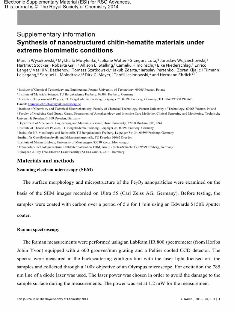
The surface morphology and microstructure of the Fe2O3 nanoparticles were examined on the
basis of the SEM images recorded on Ultra 55 (Carl Zeiss AG, Germany). Before testing, the
samples were coated with carbon over a period of 5 s for 1 min using an Edwards S150B sputter
coater.
Raman spectroscopy
The Raman measurements were performed using an LabRam HR 800 spectrometer (from Horiba
Jobin Yvon) equipped with a 600 grooves/mm grating and a Peltier cooled CCD detector. The
spectra were measured in the backscattering configuration with the laser light focused on the
samples and collected through a 100x objective of an Olympus microscope. For excitation the 785
nm line of a diode laser was used. The laser power was chosen in order to avoid the damage to the
sample surface during the measurements. The power was set at 1.2 mW for the measurement
Electronic Supplementary Material (ESI) for RSC Advances.This journal is © The Royal Society of Chemistry 2014
ARTICLE Journal Name
2 | J. Name., 2012, 00, 1-3 This journal is © The Royal Society of Chemistry 2012
of the hematite structures, and 10 mW for the measurements on the chitin/hematite or
sponge/hematite systems.
Thermogravimetric analysis
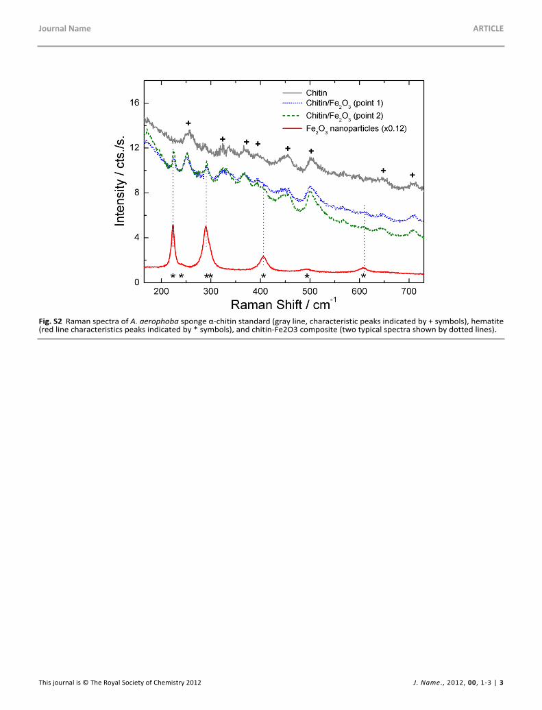
The thermogravimetric analyzer (TG, model Jupiter STA 449F3, Netzsch) was used to investigate
the thermal decomposition behavior of the samples. Measurements were carried out under flowing
nitrogen (10 cm3/min) at a heating rate of 10°C/min over a temperature range of 25–1000°C, with
an initial sample weight of approximately 5.5 mg.
Results
Fig. S1 SEM image of Fe2O3 nanoparticles prepared without the chitinous template
Journal Name ARTICLE
This journal is © The Royal Society of Chemistry 2012 J. Name., 2012, 00, 1-3 | 3
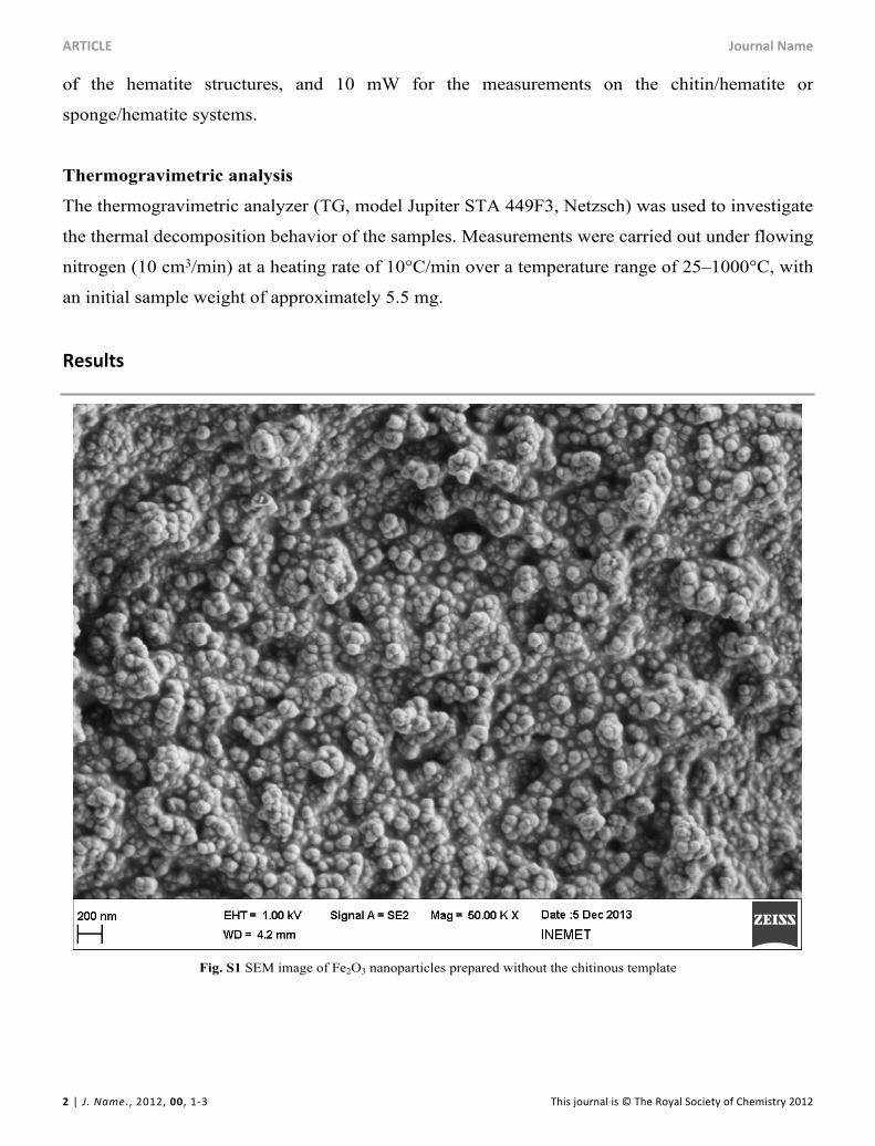
Fig. S2 Raman spectra of A. aerophoba sponge α-chitin standard (gray line, characteristic peaks indicated by + symbols), hematite (red line characteristics peaks indicated by * symbols), and chitin-Fe2O3 composite (two typical spectra shown by dotted lines).
ARTICLE Journal Name
4 | J. Name., 2012, 00, 1-3 This journal is © The Royal Society of Chemistry 2012
Fig. S3 TG/ DTA curves of α-chitin (green line) and α-chitin-Fe2O3 composite (red line)
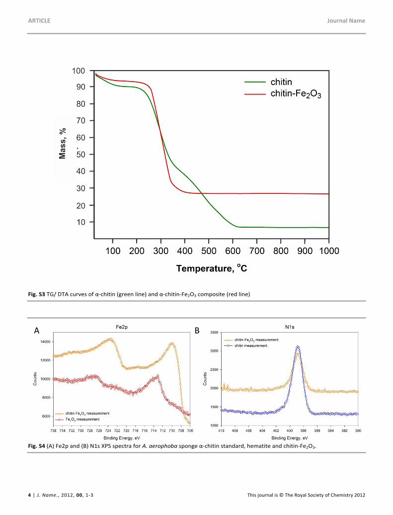
Fig. S4 (A) Fe2p and (B) N1s XPS spectra for A. aerophoba sponge α-chitin standard, hematite and chitin-Fe2O3.
Journal Name ARTICLE
This journal is © The Royal Society of Chemistry 2012 J. Name., 2012, 00, 1-3 | 5
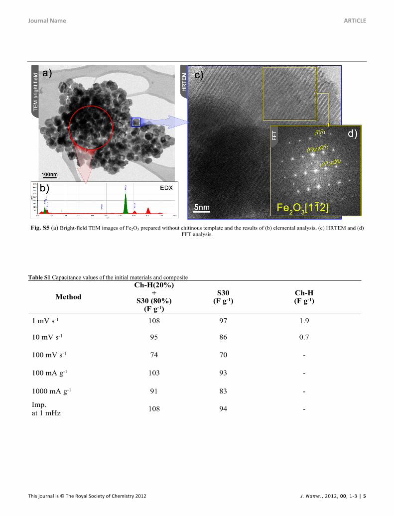
Fig. S5 (a) Bright-field TEM images of Fe2O3 prepared without chitinous template and the results of (b) elemental analysis, (c) HRTEM and (d) FFT analysis.
Table S1 Capacitance values of the initial materials and composite
MethodCh-H(20%)
+ S30 (80%)
(F g-1)
S30(F g-1)
Ch-H(F g-1)
1 mV s-1 108 97 1.9
10 mV s-1 95 86 0.7
100 mV s-1 74 70 -
100 mA g-1 103 93 -
1000 mA g-1 91 83 -
Imp. at 1 mHz 108 94 -