Supplemental Information Direct Signaling between ... · 1 Cancer Cell, 20 Supplemental Information...
24
1 Cancer Cell, 20 Supplemental Information Direct Signaling between Platelets and Cancer Cells Induces an Epithelial-Mesenchymal-Like Transition and Promotes Metastasis Myriam Labelle, Shahinoor Begum, and Richard O. Hynes Inventory of Supplemental Information Figure S1, related to Figure 1 Table S1, related to Figure 2. Provided as an Excel File. Table S2, related to Figure 2 Figure S2, related to Figure 3 Table S3, related to Figure 3 Figure S3, related to Figure 4 Table S4, related to Figure 4 Figure S4, related to Figure 6 Supplemental Experimental Procedures
Transcript of Supplemental Information Direct Signaling between ... · 1 Cancer Cell, 20 Supplemental Information...

1
Cancer Cell, 20 Supplemental Information
Direct Signaling between Platelets and Cancer
Cells Induces an Epithelial-Mesenchymal-Like
Transition and Promotes Metastasis
Myriam Labelle, Shahinoor Begum, and Richard O. Hynes Inventory of Supplemental Information Figure S1, related to Figure 1 Table S1, related to Figure 2. Provided as an Excel File. Table S2, related to Figure 2 Figure S2, related to Figure 3 Table S3, related to Figure 3 Figure S3, related to Figure 4 Table S4, related to Figure 4 Figure S4, related to Figure 6 Supplemental Experimental Procedures

2
SUPPLEMENTAL INFORMATION

3
Figure S1, related to Figure 1. Pretreatment of Tumor Cells with Platelets Induces an
EMT-Like Phenotype in Mouse and Human Cell Lines.
(A) Immunofluorescence staining for platelets (CD41;red) in cell suspension (prepared as for
tail-vein injection) of MC38GFP or Ep5 cells stably expressing GFP. Two representative cells
for each condition are shown. Note that very few platelets remain attached to tumor cells treated
with platelets or the platelet pellet fraction from WT mice, or with platelets from Pf4-cre+;
TGF1fl/fl mice. Scale bar=10µm.
(B) Immunofluorescence stainings for E-cadherin and N-cadherin (red) in MC38GFP cells or
Ep5 cells stably expressing GFP treated with buffer or platelets for 40h. Scale bar=50µm.
(C) Phase-contrast micrographs of MCF10A or HMLER cells treated with buffer or platelets for
24h. Scale bar=50µm.
(D) Relative fold change in mRNA expression in human breast epithelial MCF10A or HMLER
human cells treated with buffer or platelets for 40h (n=3). Values are normalized to GAPDH
expression. Bars represent the mean SEM. **p<0.01, ***p<0.001 were determined by
Student’s t-test.
(E) Zymography for MMP-9 in the conditioned medium of MCF10A or HMLER human cells
treated as in (D).
4
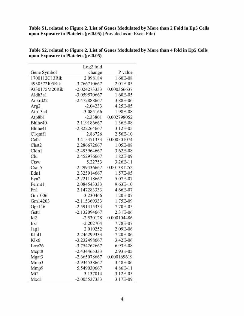
Table S1, related to Figure 2. List of Genes Modulated by More than 2 Fold in Ep5 Cells upon Exposure to Platelets (p<0.05) (Provided as an Excel File) Table S2, related to Figure 2. List of Genes Modulated by More than 4 fold in Ep5 Cells upon Exposure to Platelets (p<0.05)
Gene Symbol Log2 fold
change P value1700112C13Rik 2.098184 1.60E-084930572J05Rik -3.766710667 2.01E-059330175M20Rik -2.024273333 0.000366637Aldh3a1 -3.059570667 1.60E-05Ankrd22 -2.472888667 3.88E-06Arg2 -2.04233 4.25E-05Atp13a4 -3.085166 1.98E-08Atp8b1 -2.33801 0.002798052Bhlhe40 2.119186667 1.36E-08Bhlhe41 -2.822264667 3.12E-05C1qtnf1 2.86726 2.56E-10Ccl2 3.415371333 0.000501074Chst2 2.286672667 1.05E-08Cldn1 -2.493964667 3.62E-08Clu 2.452976667 1.82E-09Ctsw 5.22753 3.26E-11Cxcl5 -2.299436667 0.001381252Edn1 2.325914667 1.57E-05Eya2 -2.221118667 5.07E-07Fermt1 2.084543333 9.63E-10Fn1 2.147283333 4.66E-07Gm1006 -3.230466 1.20E-07Gm14203 -2.115369333 1.75E-09Gpr146 -2.591415333 7.70E-05Gstt1 -2.132094667 2.31E-06Id2 -2.530128 0.000104486Irs1 -2.202704 7.78E-07Jag1 2.010252 2.09E-06Klhl1 2.246299333 7.20E-06Klk6 -3.232498667 3.42E-06Lrrc26 -3.754262667 6.93E-08Mcpt8 -2.434465333 2.93E-05Mgat3 -2.665078667 0.000169619Mmp3 -2.934538667 3.48E-06Mmp9 5.549030667 4.86E-11Mt2 3.137014 3.12E-05Mxd1 -2.005537333 3.17E-09

5
Ncam1 3.060405333 2.89E-09Npnt -2.537079333 7.44E-06Nt5e 2.16121 2.99E-05Padi1 -2.58373 4.09E-07Pdia5 2.178992 4.35E-05Ppl -2.346288 3.27E-05Rhpn2 -2.334857333 3.05E-07Rom1 -2.792601333 1.39E-06Serpinb2 -2.026295333 0.003003047Serpine1 4.16097 4.47E-09Sgk2 -3.672803333 1.48E-05Slc25a35 -2.232655333 0.001304895Slc26a9 -2.669384667 9.74E-06Slc44a3 -3.123658 7.31E-05Styk1 -2.362784 4.20E-06Tc2n -2.454634 0.000866249Tmem71 -2.043573333 0.000126913Tns4 -2.508330667 7.11E-09Vegfc 3.681836 1.39E-07Vim 2.430557333 0.0143674Wisp1 4.779488 9.59E-11

6
Figure S2, related to Figure 3. Stumpy Deletion Does Not Affect Metastasis
(A) Numbers of metastatic foci at the surface of lungs (2 largest lobes) 14 days after tail-vein
injection of MC38GFP cells in wild-type (WT), Pf4-cre+; TGF1fl/fl, Pf4-cre+; TGF1fl/+ mice,
Pf4-cre+; TGF1fl/- mice or TGF1fl/- mice. Each bar represents the mean SEM of n=4-9.
*p<0.05, **p<0.01 vs WT were determined by one-way ANOVA followed by Tuckey’s post
test.
(B) Micrographs of lungs 14 days after tail-vein injection of MC38GFP cells in wild-type (WT),
Pf4-cre+; TGF1fl/fl, Pf4-cre+; TGF1fl/+ mice, Pf4-cre+; TGF1fl/- mice, or TGF1fl/- mice.
7
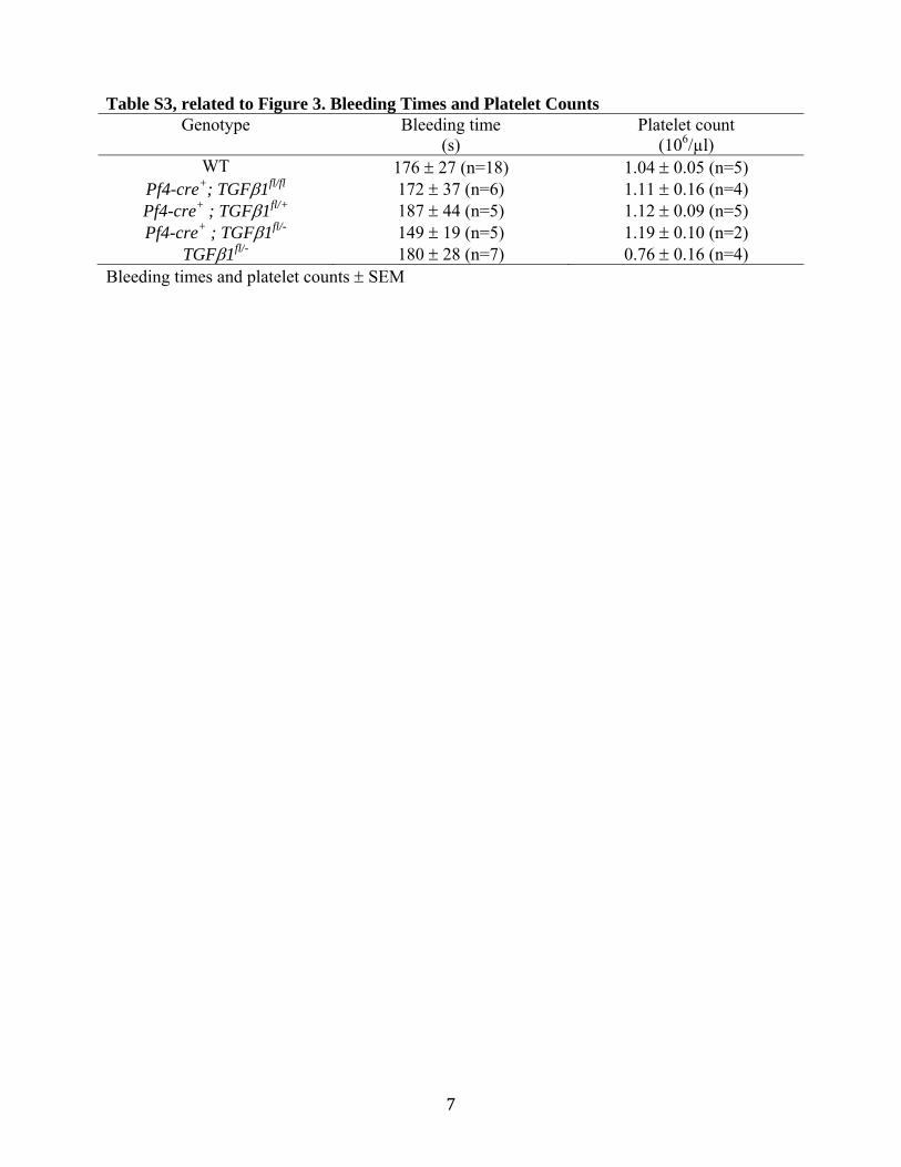
Table S3, related to Figure 3. Bleeding Times and Platelet Counts Genotype Bleeding time
(s) Platelet count
(106/µl) WT 176 27 (n=18) 1.04 0.05 (n=5)
Pf4-cre+; TGF1fl/fl 172 37 (n=6) 1.11 0.16 (n=4) Pf4-cre+ ; TGF1fl/+ 187 44 (n=5) 1.12 0.09 (n=5) Pf4-cre+ ; TGF1fl/- 149 19 (n=5) 1.19 0.10 (n=2)
TGF1fl/- 180 28 (n=7) 0.76 0.16 (n=4) Bleeding times and platelet counts SEM
8

9
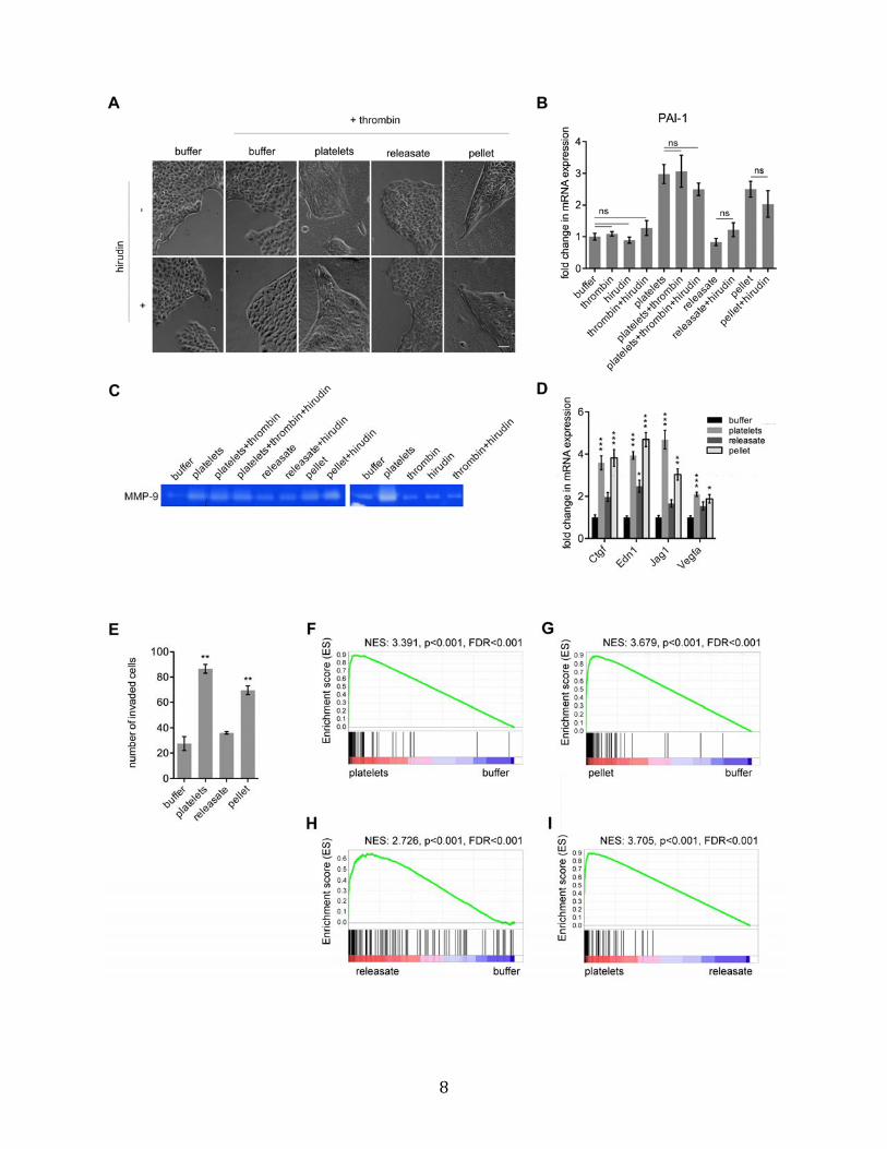
Figure S3, related to Figure 4. Platelet-Derived TGF1 and Platelet-Bound Factors
Cooperate to Promote Metastasis
(A) Phase-contrast micrographs of Ep5 cells treated with buffer, platelets, releasate from
activated platelets (releasate), or the pellet fraction from activated platelets (pellet) +/- thrombin
and hirudin for 24h. The releasate and pellet fractions were generated by treating platelets with
thrombin (0.5U/ml) and separated by centrifugation. For some conditions, thrombin was blocked
with hirudin (5U/ml) prior dilution in culture medium and co-incubation with the tumor cells.
Scale bar=50µm.
(B) Relative fold change in PAI-1 mRNA expression in Ep5 cells treated as in (A) for 40h (n=3).
Values are normalized to Gapdh expression. ns (p>0.05) was determined by one-way ANOVA
followed by Tuckey’s post test.
(C) Zymography for MMP-9 in the conditioned medium of Ep5 cells treated as in (B).
(D) Relative fold change in mRNA expression in Ep5 cells treated with buffer, platelets,
releasate from activated platelets (releasate), or the pellet fraction from activated platelets (pellet)
(n=3). Values are normalized to Gapdh expression. Bars represent the mean SEM, and
*p<0.05, **p<0.01, ***p<0.001 vs buffer were determined by one-way ANOVA followed by
Tuckey’s post test.
(E) Ep5 cells were added at the top of transwells coated with Matrigel and treated with buffer,
platelets, releasate from activated platelets (releasate), or the pellet fraction from activated
platelets (pellet). The total numbers of cells that invaded to the bottom of the transwell were
counted after 48h. Each bar represents the mean SEM of n=2. **p<0.01 vs buffer were
determined by one-way ANOVA followed by Tuckey’s post test.
(F-I) Enrichment plots for the platelet-induced gene signature (genes upregulated by more than 2
fold; Table S1) in an independent set of microarray data generated with Ep5 cells treated with

10
buffer, platelets, releasate from activated platelets (releasate), or the pellet fraction from
activated platelets (pellet) (n=3). Enrichment in platelet-, platelet pellet- or releasate-treated cells
versus untreated cells (buffer) are shown in F, G and H. Enrichment in the platelet-treated cells
in comparison to the releasate-treated cells is presented in I. Each vertical black line represents a
platelet-induced gene. The left-to-right position of each line indicates the relative position of the
gene within the rank ordering of the 13,243 genes represented in the dataset from the gene most
upregulated upon platelet treatment (position 1 on the left) to the most down-regulated (position
13,243 on the right). The genes near the middle are unaffected by the platelet treatment. The
platelet-induced gene signature is clearly enriched in the platelet-treated Ep5 cells (E; p<0.001,
FDR<0.001), as evidenced by the cluster of vertical black lines at the very left of the distribution
and the positive enrichment score marked by the green line, validating the platelet-induced gene
signature in this data set. Similarly, the gene signature is also highly enriched in the pellet-treated
cells (F; p<0.001, FDR<0.001). Interestingly, while the platelet-induced gene signature is overall
also enriched in releasate-treated cells (G; p<0.001, FDR<0.001), there is a subset of genes
which are less affected by this treatment and are redistributed towards the right of the plot,
suggesting that treatment with the releasate only induces partial gene expression changes in
comparison to treatment with platelets in Ep5 cells. The overall lower magnitude of gene
expression changes observed in the releasate-treated cells in comparison with platelet-treated
cells is further illustrated by the enrichment of the platelet-induced gene signature in platelet-
treated cells directly compared to releasate-treated cells (H; p<0.001, FDR<0.001). The NES
(normalized enrichment score), p-value and FDR (false discovery rate) are indicated at the top of
each plot.
11
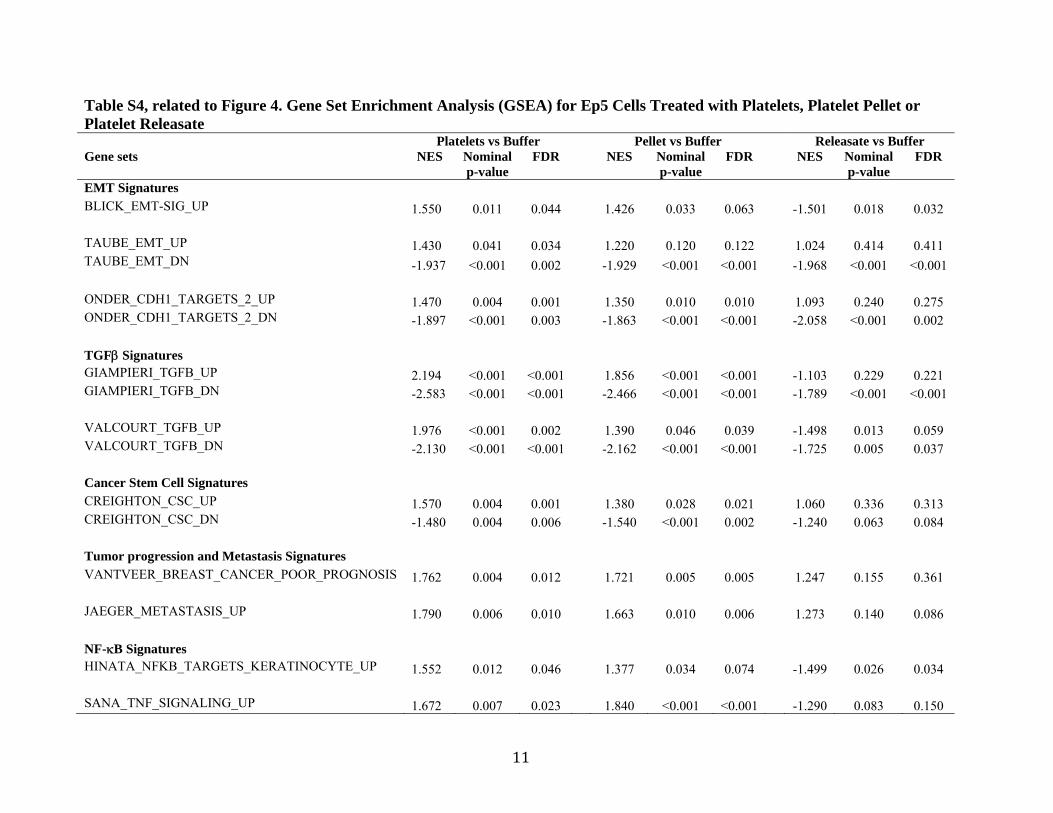
Table S4, related to Figure 4. Gene Set Enrichment Analysis (GSEA) for Ep5 Cells Treated with Platelets, Platelet Pellet or Platelet Releasate Platelets vs Buffer Pellet vs Buffer Releasate vs Buffer Gene sets NES Nominal
p-value FDR NES Nominal
p-value FDR NES Nominal
p-value FDR
EMT Signatures BLICK_EMT-SIG_UP 1.550 0.011 0.044 1.426 0.033 0.063 -1.501 0.018 0.032 TAUBE_EMT_UP 1.430 0.041 0.034 1.220 0.120 0.122 1.024 0.414 0.411 TAUBE_EMT_DN -1.937 <0.001 0.002 -1.929 <0.001 <0.001 -1.968 <0.001 <0.001 ONDER_CDH1_TARGETS_2_UP 1.470 0.004 0.001 1.350 0.010 0.010 1.093 0.240 0.275 ONDER_CDH1_TARGETS_2_DN -1.897 <0.001 0.003 -1.863 <0.001 <0.001 -2.058 <0.001 0.002 TGF Signatures GIAMPIERI_TGFB_UP 2.194 <0.001 <0.001 1.856 <0.001 <0.001 -1.103 0.229 0.221 GIAMPIERI_TGFB_DN -2.583 <0.001 <0.001 -2.466 <0.001 <0.001 -1.789 <0.001 <0.001 VALCOURT_TGFB_UP 1.976 <0.001 0.002 1.390 0.046 0.039 -1.498 0.013 0.059 VALCOURT_TGFB_DN -2.130 <0.001 <0.001 -2.162 <0.001 <0.001 -1.725 0.005 0.037 Cancer Stem Cell Signatures CREIGHTON_CSC_UP 1.570 0.004 0.001 1.380 0.028 0.021 1.060 0.336 0.313 CREIGHTON_CSC_DN -1.480 0.004 0.006 -1.540 <0.001 0.002 -1.240 0.063 0.084 Tumor progression and Metastasis Signatures VANTVEER_BREAST_CANCER_POOR_PROGNOSIS 1.762 0.004 0.012 1.721 0.005 0.005 1.247 0.155 0.361 JAEGER_METASTASIS_UP 1.790 0.006 0.010 1.663 0.010 0.006 1.273 0.140 0.086 NF-B Signatures HINATA_NFKB_TARGETS_KERATINOCYTE_UP 1.552 0.012 0.046 1.377 0.034 0.074 -1.499 0.026 0.034 SANA_TNF_SIGNALING_UP 1.672 0.007 0.023 1.840 <0.001 <0.001 -1.290 0.083 0.150

12
Enrichment of gene sets from the literature. Positive normalized enrichment score (NES) indicates enrichment in either platelet-, pellet-
or releasate-treated Ep5 cells; negative NES indicates enrichment in untreated Ep5 cells (buffer). FDR (false discovery rate).
NF-B signatures are enriched in platelet- or pellet-treated Ep5 cells, but not in releasate-treated Ep5 cells (note the negative NES for
releasate-treated cells), suggesting a dependence on platelet-bound factors for activation of this pathway. Similarly, while genes
upregulated during EMT, upon TGF treatment, in cancer stem cells or during tumor progression and metastasis are significantly
enriched in platelet- or pellet-treated cells (p<0.05 and/or FDR<0.25), this enrichment is not observed in releasate-treated cells (p>0.05
and/or FDR>0.25). However, genes downregulated upon TGF treatment and during EMT are significantly depleted upon all three
treatments, suggesting that partial TGF and EMT responses are maintained in releasate-treated cells.

13

14
Figure S4, related to Figure 6. The NF-B Signaling Pathway Is Activated by Platelets in a
Contact-Dependent Manner and Cooperates with TGF Signaling to Induce an EMT-Like
Transition
(A) Ep5 cells were transfected with firefly luciferase reporters of NF-B or JNK activity and
constitutively active control Renilla luciferase reporters. 24h after transfection, the cells were
treated with buffer, platelets, releasate from activated platelets (releasate), or the pellet fraction
from activated platelets (pellet) for 20h, and the relative luciferase activity (RLU) was measured
(n=3).
(B) MCP-1 concentration in the conditioned medium from MC38GFP or Ep5 cells incubated
with buffer, platelets, releasate from activated platelets (releasate), or the pellet fraction from
activated platelets (pellet) for 40h (n=3).
(C) Detection of phospho-Smad2 and total Smad2 protein levels by immunoblotting in Ep5 cells
stably expressing an IB super-repressor (Ep5-IkBSR) or a control vector (Ep5-vector) and
treated with buffer or platelets for 40h. -tubulin is used as loading control.
(D) Zymography for MMP-9 in the conditioned medium from Ep5 cells treated with buffer,
platelets, JSH-23, or platelets + JSH-23 for 40h.
(E) Detection of vimentin protein levels by immunoblotting in Ep5 cells treated with buffer,
platelets, JSH-23 5 µM or platelets + JSH-23 5 µM. -tubulin is used as loading control.
(F) Relative luciferase activity in MLEC cells stably expressing a luciferase reporter under the
control of the PAI-1 promoter construct and treated with buffer, platelets, JSH-23, platelets +
JSH-23, or platelets + TGF1 (1 ng/ml) + JSH-23 for 20h (n=2).

15
(G) Relative luciferase activity in Ep5 cells stably expressing a luciferase reporter under the
control of the SBE promoter and treated with buffer, platelets, JSH-23 or platelets + JSH-23 for
20h (n=2).
For panels A, B, F and G, bars represent the mean SEM, and *p<0.05, **p<0.01, ***p<0.001
vs buffer were determined by one-way ANOVA followed by Tuckey’s post test.

16
SUPPLEMENTAL EXPERIMENTAL PROCEDURES
Tissue Culture
Ep5 (EpRas) cells (obtained from R. Weinberg), MC38GFP cells (obtained from A. Varki), and
MLEC cells stably expressing a PAI-1 promoter-luciferase reporter construct (Abe et al., 1994)
were cultivated in DMEM 10% FCS, 1% Penicillin/Streptomycin and 2 mM L-Glutamine.
HMLER cells (H-Ras transformed human mammary epithelial cell line; obtained from R.
Weinberg) were cultivated as described previously (Elenbaas et al., 2001). MCF10A cells (Soule
et al., 1990) were maintained in DMEM/F12 (50:50) supplemented with 5% horse serum, 10mM
HEPES, 10µg/ml insulin, 20ng/ml epidermal growth factor, 0.5µg/ml hydrocortisone, 100ng/ml
cholera toxin, 1% Penicillin/Streptomycin.
Generation of Cell Lines Stably Expressing ZsGreen and IkBSR Reporters
Retroviral vectors coding for ZsGreen or IBα super-repressor and GFP (IkBSR) were
transduced in Ep5 cells. Packaging of the vectors was obtained by cotransfection of 293FT cells
(ATCC) with 1ng transfer vector, 1µg MLV gag-pol, and 1µg VSVg expression vectors using
Fugene6 (Roche) as described previously (Stern et al., 2008). 48h after transfection, 293FT cell-
conditioned medium was collected, filtered through a 0.45µm filter, and applied to Ep5 cells
with 4µg/ml polybrene (Sigma). Ep5 cells were then selected on the basis of GFP or ZsGreen
expression by FACS sorting.
Genotyping
DNA from mouse tail biopsies was amplified by PCR using the primers listed in the table below.

17
Genotyping Primers For detection of the Tgfb1
flox and WT alleles forw 5’CCCAGGCTAGCCTTGAACTTCT3’ (Li et al.,
2007) rev 5’AGGGGTGGAGATGTAGTTTGG3’ For simultaneous detection
of the Tgfb1 null (egfp knockin) and WT alleles
forw 5’CGCATCCCACCTTTGCCGAG3’ (Li et al.,
2007) rev1 5’GGCGTCAGCACTAGAAGCCA3’ rev2 5’GCCGTAGGTCAG GGTGGTCA3’
For detection of the Pf4-cre transgene
forw 5’CCCATACAGCACACCTTTTG3’ (Tiedt et al., 2007) rev 5’TGCACAGTCAGCAGGTT3’
In Vivo Metastasis Assays
For lung metastasis assays, cells treated with platelets for 40h were washed in PBS, and either
trypsinized (Ep5) or lifted with 2mM EDTA in PBS (MC38GFP). Cells were then rinsed and
centrifuged twice to remove platelets, and resuspended in HBSS at a constant number of cells for
all mice in a given experiment (250,000 to 1,000,000 cells/injection). 100µl of cell suspension
were then injected via the tail vein of syngeneic mice. After 14 days, animals were sacrificed and
the numbers of metastatic foci at the surface of lungs were counted under a fluorescence
stereomicroscope (Nikon SMZ1500). For 3h and 48h time points, pictures were taken
(magnification 3x) and the number of cells/picture was automatically counted with Cell Profiler
(Lamprecht et al., 2007). For metastasis experiments with the Pf4-cre+; TGF1fl/fl mice,
untreated MC38GFP cells cultivated in DMEM 10%FCS were lifted and washed as described
above and 100µl of a 1x10E7/ml cell suspension were injected via the tail vein.
RT-qPCR Analysis
RNA was isolated from total cell lysates using RNeasy Mini kit (Qiagen), and reverse-
transcribed with TaqMan Reverse Transcription Reagents and random hexameric primers
(Applied Biosystems). Human specific PCR primers were designed using Primer3 and BLAST.
RT-qPCR was performed with the iQ SYBR Green Supermix (Biorad). Primers used are listed in

18
the tables below. Data were normalized to GAPDH expression. Relative mRNA levels were
calculated using the comparative CT method.
RT-qPCR Primers used with mouse cells Gapdh forw 5’CAGTATGACTCCACTCACGGC3’ Gapdh rev 5’GAGGGGCCATCCACAGTCTTC3’ Snail forw 5’GGAAGCCCAACTATAGCGAGC3’ Snail rev 5’CAGTTGAAGATCTTCCGCGAC3’
Fibronectin forw 5’CGTAAATTGCCCCATTGAGTG3’ Fibronectin rev 5’GAGGGTCTGCTAACATCACTG3’
Serpine1 (PAI-1) forw 5’CCCGCCTCCTCATCCTGCCT3’ Serpine1 (PAI-1) rev 5’GCCACTGTGCCGCTCTCGTT3’
Claudin-1 forw 5’GCGTTTCGCAAAGCACCGGG3’ Claudin-1 rev 5’GGCTCGGGTTGCCTGCAAAGT3’ Vimentin forw 5’AATGCTTCTCTGGCACGTCT3’ Vimentin rev 5’GCTCCTGGATCTCTTCATCG3’
Slug forw 5’CATCCTTGGGGCGTGTAAGTC3’ Slug Rev 5’GCCCAGAGAACGTAGAATAGGTC3’
Twist forw 5’GGACAAGCTGAGCAAGATTCA3’ Twist rev 5’CGGAGAAGGCGTAGCTGAG3’ Zeb1 forw 5’CGCCATGAGAAGAACGAGGAC3’ Zeb1 rev 5’TGTATGCAAAGGTGTAACTGCAC3’
Zeb2 forw 5’CAGGCTCGGAGACAGATGAAG3’ Zeb2 rev 5’CTTGCAGAATCTCGCCACTG3’ Ctgf forw 5’CTCCACCCGAGTTACCAATG3’ Ctgf rev 5’TGGCGATTTTAGGTGTCC3’
Edn1 forw 5’TTTCCCGTGATCTTCTCTCTGC3’ Edn1 rev 5’CTGAGTTCGGCTCCCAAGAC3’ Jag1 forw 5’TTCAGTTTCGCCTGGCCGAG3’ Jag1 rev 5’TCAGTGTCTGCCATTGCCGG3’
Vegfa forw 5’CTTGTTCAGAGCGGAGAAAGC3’ Vegfa rev 5’ACATCTGCAAGTACGTTCGTT3’
RT-qPCR Primers used with human cells
GAPDH forw 5’GGTCTCCTCTGACTTCAACA3’ GAPDH rev 5’GTGAGGGTCTCTCTCTTCCT3’ Snail forw 5’TCGGAAGCCTAACTACAGCGA3’ Snail rev 5’AGATGAGCATTGGCAGCGAG3’
Fibronectin forw 5’CCATCGCAAACCGCTGCCAT3’ Fibronectin rev 5’AACACTTCTCAGCTATGGGCTT3’ Serpine1 forw 5’ACCGCAACGTGGTTTTCTCA3’

19
Serpine1 rev 5’TTGAATCCCATAGCTGCTTGAAT3’ N-cadherin forw 5’ATCCTACTGGACGGTTCG3’ N-cadherin rev 5’TTGGCTAATGGCACTTGA3’ Vimentin forw 5’GAACGCCAGATGCGTGAAATG3’ Vimentin rev 5’CCAGAGGGAGTGAATCCAGATTA3’
Slug forw 5’AAGCATTTCAACGCCTCCAAA3’ Slug Rev 5’GGATCTCTGGTTGTGGTATGACA3’
Twist forw 5’CCGGAGACCTAGATGTCATTG3’ Twist rev 5’CCACGCCCTGTTTCTTTG3’ Zeb1 forw 5’GATGATGAATGCGAGTCAGATGC3’ Zeb1 rev 5’ACAGCAGTGTCTTGTTGTTGT3’
Zeb2 forw 5’GGAGACGAGTCCAGCTAGTGT3’ Zeb2 rev 5’CCACTCCACCCTCCCTTATTTC3’
Zymography
Conditioned media from cells treated for 48h were collected and centrifuged to remove cellular
debris and platelets. Volumes of conditioned media normalized to the number of cells were then
mixed with 2X Laemmli sample buffer (BioRad) and loaded onto a 7.5%
acrylamide/bisacrylamide separating gel containing 0.2% (w/v) gelatin. After electrophoresis, the
gel was incubated in 2.5% Triton X-100, rinsed in distilled water and incubated at 37°C in buffer
containing 50mM Tris pH 7.6, 20mM NaCl, 5mM CaCl2. Finally, the gel was stained in 0.1%
Coomassie blue R-250, 30% methanol, 10% acetic acid, and destained in the same solution
without the Coomassie blue dye.
Immunoblotting
Immunoblot analysis was performed as described previously (Labelle et al., 2008). The primary
antibodies were anti--tubulin (Sigma), anti E-cadherin (BD Biosciences), anti-Smad2/3, anti-
phosphoSmad2 (Cell Signaling Technology) and anti-vimentin (Sigma). The secondary
antibodies were horseradish peroxidase-conjugated anti-rabbit or anti-mouse IgG (Jackson

20
Immunoresearch).
Invasion Assay
Invasion assays were performed in 24-well BD BiocoatTM MatrigelTM Invasion Chambers (8 µm
pore size; BD Biosciences). 50,000 cells were plated in transwell inserts and either left untreated,
treated with SB431542 (10 µM), anti-TGF1 blocking antibody (6µg/ml) or the different platelet
fractions. Both upper and lower chambers contained DMEM. After 48h, cells remaining in the
upper part of the transwell were removed with a cotton swab. Migrated cells were then stained
with Crystal Violet 0.5% and the total number of cells was counted with a Zeiss Axiovert 200
microscope.
Immunofluorescence Staining
For visualization of blood vessels and quantification of extravasation, lungs were fixed by
tracheal perfusion with PBS, 4% formaldehyde, 0.3% Triton X-100 for 15 min and removed en
bloc. Lung lobes were then cut in 4 pieces, washed in PBS, 0.3% triton X-100 and blocked in
PBS 10% normal goat serum followed by the primary antibody (anti-PECAM-1, BD
Biosciences). After washing, samples were incubated with Alexa 594-conjugated goat anti-rat
IgG (Molecular probes) and DAPI. Images of lobe pieces from 3 or more mice per group were
taken at 60X with Z-sections every 1µm on an Olympus FV10i inverted confocal microscope.
3D rendering and XYZ views were generated with Volocity (Perkin Elmer). XYZ views were
then examined and cells clearly within blood vessels (directly surrounded by PECAM-1 staining;
red) were scored as intravascular, while cells outside blood vessels were scored as extravascular.
For platelet immunostaining in lungs, 20 µm-thick sections were fixed in acetone and stained

21
with anti-GP1b (Emfret). The secondary antibody was Alexa 594-conjugated goat anti-rat IgG
(Molecular Probes). Images were taken at 60X with Z-sections every 1µm on an Olympus FV10i
inverted confocal microscope.
For platelet immunostaining in cell suspensions, cells were prepared as for in vivo metastasis
assays. The suspensions were then stained with anti-CD41 (BD Biosciences), washed once with
PBS and incubated with Alexa 594-conjugated goat anti-rat IgG (Molecular probes). After a final
wash in PBS, cells were resuspended in PBS and 5µl of cell suspensions were loaded on a
microscope slide.
For immunostaining of tumor cells in tissue culture, cells were rinsed with PBS, fixed with 4%
formaldehyde and stained with anti-E-cadherin or anti-N-cadherin (BD Biosciences). The
secondary antibody was Alexa 594-conjugated goat anti-rat IgG (Molecular probes). Images
were taken with a Zeiss LSM510 microscope.
Tail Bleeding Assay
For tail bleeding assays, mice were anesthetized with 2.5% isoflurane in oxygen. The tail was cut
at 5mm and bled onto a Whatman filter paper. The filter paper was dabbed to the wound every
30 seconds without disrupting the forming clot. The experiment was continued until bleeding
stopped completely.
Microarray Analysis
For data presented in Fig. S3 and Table S4, total RNA was isolated from Ep5 cells treated with
buffer, platelets, platelet pellet or platelet releastate (n=3). Samples where then processed with
22
the Nugen Applause® WT-Amp Plus ST System and hybridized on Affymetrix Mouse Gene 1.0
ST arrays, according to manufacturer's instructions (Affymetrix).
Data are deposited in Gene Expression Omnibus (GEO) under accession number GSE27456.
Gene Set Enrichment Analysis (GSEA)
GSEA was performed using GSEA v2.07 (www.broadinstitute.org/gsea; Mootha et al., 2003;
Subramanian et al., 2005). The signal-to-noise metric and permutation of gene sets were used to
rank the genes and calculate nominal p-values and FDR. Probe sets were collapsed to unique
gene symbols and used to interrogate the gene sets from the literature listed in the table below,
some of which were provided by the Molecular Signatures Database (MSigDB;
www.broadinstitute.org/gsea/msigdb).
Gene set Source BLICK_EMT-SIG (Blick et al.) TAUBE_EMT (Taube et al.) ONDER_CDH1_TARGETS_2 MSigDB, (Onder et al., 2008) GIAMPIERI_TGFB (Giampieri et al., 2009) VALCOURT_TGFB (Valcourt et al., 2005) HINATA_NFKB_TARGETS_KERATINOCYTE MSigDB, (Hinata et al., 2003) SANA_TNF_SIGNALING MSigDB, (Sana et al., 2005) CREIGHTON_CSC (Creighton et al., 2009) VANTVEER_BREAST_CANCER_POOR_PROGNOSIS MSigDB, (van 't Veer et al., 2002) JAEGER_METASTASIS MSigDB, (Jaeger et al., 2007)
23
SUPPLEMENTAL REFERENCES
Blick, T., Hugo, H., Widodo, E., Waltham, M., Pinto, C., Mani, S.A., Weinberg, R.A., Neve, R.M., Lenburg, M.E., and Thompson, E.W. Epithelial mesenchymal transition traits in human breast cancer cell lines parallel the CD44(hi/)CD24 (lo/-) stem cell phenotype in human breast cancer. J Mammary Gland Biol Neoplasia 15, 235-252. Creighton, C.J., Li, X., Landis, M., Dixon, J.M., Neumeister, V.M., Sjolund, A., Rimm, D.L., Wong, H., Rodriguez, A., Herschkowitz, J.I., et al. (2009). Residual breast cancers after conventional therapy display mesenchymal as well as tumor-initiating features. Proc Natl Acad Sci U S A 106, 13820-13825. Elenbaas, B., Spirio, L., Koerner, F., Fleming, M.D., Zimonjic, D.B., Donaher, J.L., Popescu, N.C., Hahn, W.C., and Weinberg, R.A. (2001). Human breast cancer cells generated by oncogenic transformation of primary mammary epithelial cells. Genes Dev 15, 50-65. Hinata, K., Gervin, A.M., Jennifer Zhang, Y., and Khavari, P.A. (2003). Divergent gene regulation and growth effects by NF-kappa B in epithelial and mesenchymal cells of human skin. Oncogene 22, 1955-1964. Jaeger, J., Koczan, D., Thiesen, H.J., Ibrahim, S.M., Gross, G., Spang, R., and Kunz, M. (2007). Gene expression signatures for tumor progression, tumor subtype, and tumor thickness in laser-microdissected melanoma tissues. Clin Cancer Res 13, 806-815. Lamprecht, M.R., Sabatini, D.M., and Carpenter, A.E. (2007). CellProfiler: free, versatile software for automated biological image analysis. Biotechniques 42, 71-75. Mootha, V.K., Lindgren, C.M., Eriksson, K.F., Subramanian, A., Sihag, S., Lehar, J., Puigserver, P., Carlsson, E., Ridderstrale, M., Laurila, E., et al. (2003). PGC-1alpha-responsive genes involved in oxidative phosphorylation are coordinately downregulated in human diabetes. Nat Genet 34, 267-273. Onder, T.T., Gupta, P.B., Mani, S.A., Yang, J., Lander, E.S., and Weinberg, R.A. (2008). Loss of E-cadherin promotes metastasis via multiple downstream transcriptional pathways. Cancer Res 68, 3645-3654. Sana, T.R., Janatpour, M.J., Sathe, M., McEvoy, L.M., and McClanahan, T.K. (2005). Microarray analysis of primary endothelial cells challenged with different inflammatory and immune cytokines. Cytokine 29, 256-269. Soule, H.D., Maloney, T.M., Wolman, S.R., Peterson, W.D., Jr., Brenz, R., McGrath, C.M., Russo, J., Pauley, R.J., Jones, R.F., and Brooks, S.C. (1990). Isolation and characterization of a spontaneously immortalized human breast epithelial cell line, MCF-10. Cancer Res 50, 6075-6086. Subramanian, A., Tamayo, P., Mootha, V.K., Mukherjee, S., Ebert, B.L., Gillette, M.A., Paulovich, A., Pomeroy, S.L., Golub, T.R., Lander, E.S., et al. (2005). Gene set enrichment

24
analysis: a knowledge-based approach for interpreting genome-wide expression profiles. Proc Natl Acad Sci U S A 102, 15545-15550. Taube, J.H., Herschkowitz, J.I., Komurov, K., Zhou, A.Y., Gupta, S., Yang, J., Hartwell, K., Onder, T.T., Gupta, P.B., Evans, K.W., et al. Core epithelial-to-mesenchymal transition interactome gene-expression signature is associated with claudin-low and metaplastic breast cancer subtypes. Proc Natl Acad Sci U S A 107, 15449-15454. Valcourt, U., Kowanetz, M., Niimi, H., Heldin, C.H., and Moustakas, A. (2005). TGF-beta and the Smad signaling pathway support transcriptomic reprogramming during epithelial-mesenchymal cell transition. Mol Biol Cell 16, 1987-2002. van 't Veer, L.J., Dai, H., van de Vijver, M.J., He, Y.D., Hart, A.A., Mao, M., Peterse, H.L., van der Kooy, K., Marton, M.J., Witteveen, A.T., et al. (2002). Gene expression profiling predicts clinical outcome of breast cancer. Nature 415, 530-536.