Supplement to THE JOURNAL OF Allergy Clinical Immunology...rhinitis, occupational rhinitis,...
38
Sinusitis develops in approximately 31 million Amer- icans each year. An average of 4 days are lost from work each year because of acute sinusitis. Acute sinusitis typi- cally follows a viral upper respiratory infection or an allergic reaction. Swelling of the nasal mucous mem- branes may obstruct drainage from the sinus outflow tracts, causing mucus to collect in the paranasal sinuses. This pool of retained secretions may become infected, leading to congestion and inflammation of the sinus mucosa and the classic symptoms associated with sinus- itis. Treatment is aimed at killing the overgrown bacteria and cleansing the sinuses. Four documents comprise this practice parameter on sinusitis: (1) an executive summary that reviews, in nar- rative format, the key clinical issues considered in the parameter documents; (2) a management algorithm with narrative annotations designed to assist clinical decision making; (3) a document listing only numbered summary statements that is intended to promote rapid review and identification of material comprehensively discussed in the final document; and (4) the complete guidelines doc- ument, which is organized so that the numbered, key summary statements precede relevant supporting text and citations of evidence-based publications. This format provides a ready reference for any physician who evalu- ates and treats a patient with suspected sinusitis. In par- ticular, the algorithm and its accompanying annotations are designed to present a global and useful approach to both diagnosis and management. Clinical decision points are clearly shown, and each of these proceeds step-wise to logical implementation strategies. If further justifica- tion is required at any step in the algorithm, the eviden- tiary-based guidelines text can and should be consulted. In addition, guidance about appropriate referral of refrac- tory cases, either because of treatment failure or for fur- ther investigation of possible associated conditions, is provided. The greatest majority of patients with sinusitis seek care from their primary care physician; in fact, there are more than 18 million office visits to primary care physi- cians for this diagnosis each year. Various subspecialists (allergists and otolaryngologists) also see cases of sinus- itis, especially in those patients who are more difficult to treat. It is incumbent on all physicians treating sinusitis to be knowledgeable concerning the latest information on pathophysiology, diagnosis, and management, especially in light of the rapidity with which infective organisms are able to change their character. This practice parameter includes anatomic, allergic, immunologic, and physiologic considerations, as well as clinical diagnosis, differential diagnosis, diagnostic test- ing, and treatment. Predisposing factors, such as allergy, upper respiratory infections, anatomic abnormalities, immotile cilia syndrome, cystic fibrosis, immune defi- ciencies, and environmental factors (eg, smoking and pollution), will be addressed. Medical and surgical ther- apies will be discussed. An initial draft of parameters was prepared by a work group of experts in the field who carefully reviewed the current medical literature. This material then underwent extensive peer review, revision, and annotation by exter- nal reviewers and by the Joint Task Force on Practice Parameters for Allergy and Immunology, a national panel of allergist-immunologists appointed by its cosponsoring organizations: the American Academy of Allergy, Asth- ma and Immunology; the American College of Allergy, Asthma and Immunology; and the Joint Council of Aller- gy, Asthma and Immunology. The parameters were reviewed and approved by the cosponsoring organiza- tions and thereby represent an evidence-based, broadly accepted consensus opinion. The Joint Task Force is grateful for the cosponsoring organizations’ financial support and encouragement. The Joint Task Force would especially like to thank the many individuals who have donated substantial time and effort in producing this document that is intended to improve the quality of care of many millions of sinusitis patients. Preface S107 Reprint requests: Joint Council of Allergy, Asthma and Immunology, 50 N. Brockway St., #3-3, Palatine, IL 60067. J Allergy Clin Immunol 1998;102:S107-44. Copyright © 1998 by Mosby, Inc. 0091-6749/98 $5.00 + 0 1/0/94390 VOLUME 102 NUMBER 6, PART 2 T HE J OURNAL OF AllergyAND Clinical Immunology Supplement to
Transcript of Supplement to THE JOURNAL OF Allergy Clinical Immunology...rhinitis, occupational rhinitis,...
Sinusitis develops in approximately 31 million Amer-icans each year. An average of 4 days are lost from workeach year because of acute sinusitis. Acute sinusitis typi-cally follows a viral upper respiratory infection or anallergic reaction. Swelling of the nasal mucous mem-branes may obstruct drainage from the sinus outflowtracts, causing mucus to collect in the paranasal sinuses.This pool of retained secretions may become infected,leading to congestion and inflammation of the sinusmucosa and the classic symptoms associated with sinus-itis. Treatment is aimed at killing the overgrown bacteriaand cleansing the sinuses.
Four documents comprise this practice parameter onsinusitis: (1) an executive summary that reviews, in nar-rative format, the key clinical issues considered in theparameter documents; (2) a management algorithm withnarrative annotations designed to assist clinical decisionmaking; (3) a document listing only numbered summarystatements that is intended to promote rapid review andidentification of material comprehensively discussed inthe final document; and (4) the complete guidelines doc-ument, which is organized so that the numbered, keysummary statements precede relevant supporting text andcitations of evidence-based publications. This formatprovides a ready reference for any physician who evalu-ates and treats a patient with suspected sinusitis. In par-ticular, the algorithm and its accompanying annotationsare designed to present a global and useful approach toboth diagnosis and management. Clinical decision pointsare clearly shown, and each of these proceeds step-wiseto logical implementation strategies. If further justifica-tion is required at any step in the algorithm, the eviden-tiary-based guidelines text can and should be consulted.In addition, guidance about appropriate referral of refrac-
tory cases, either because of treatment failure or for fur-ther investigation of possible associated conditions, isprovided.
The greatest majority of patients with sinusitis seekcare from their primary care physician; in fact, there aremore than 18 million office visits to primary care physi-cians for this diagnosis each year. Various subspecialists(allergists and otolaryngologists) also see cases of sinus-itis, especially in those patients who are more difficult totreat. It is incumbent on all physicians treating sinusitisto be knowledgeable concerning the latest information onpathophysiology, diagnosis, and management, especiallyin light of the rapidity with which infective organisms areable to change their character.
This practice parameter includes anatomic, allergic,immunologic, and physiologic considerations, as well asclinical diagnosis, differential diagnosis, diagnostic test-ing, and treatment. Predisposing factors, such as allergy,upper respiratory infections, anatomic abnormalities,immotile cilia syndrome, cystic fibrosis, immune defi-ciencies, and environmental factors (eg, smoking andpollution), will be addressed. Medical and surgical ther-apies will be discussed.
An initial draft of parameters was prepared by a workgroup of experts in the field who carefully reviewed thecurrent medical literature. This material then underwentextensive peer review, revision, and annotation by exter-nal reviewers and by the Joint Task Force on PracticeParameters for Allergy and Immunology, a national panelof allergist-immunologists appointed by its cosponsoringorganizations: the American Academy of Allergy, Asth-ma and Immunology; the American College of Allergy,Asthma and Immunology; and the Joint Council of Aller-gy, Asthma and Immunology. The parameters werereviewed and approved by the cosponsoring organiza-tions and thereby represent an evidence-based, broadlyaccepted consensus opinion.
The Joint Task Force is grateful for the cosponsoringorganizations’ financial support and encouragement. TheJoint Task Force would especially like to thank the manyindividuals who have donated substantial time and effortin producing this document that is intended to improvethe quality of care of many millions of sinusitis patients.
Preface
S107
Reprint requests: Joint Council of Allergy, Asthma and Immunology, 50 N.Brockway St., #3-3, Palatine, IL 60067.
J Allergy Clin Immunol 1998;102:S107-44.Copyright © 1998 by Mosby, Inc.0091-6749/98 $5.00 + 0 1/0/94390
VOLUME 102 NUMBER 6, PART 2
THE JOURNAL OF
AllergyANDClinicalImmunology
Supplement to
S108
Sinusitis, defined as inflammation of one or moreparanasal sinuses, has been characterized as “acute”when lasting 3 to 8 weeks and “chronic” when lastinglonger. Viral upper respiratory infections frequently pre-cede subsequent bacterial invasion of the sinuses byStreptococcus pneumoniae, Haemophilus influenzae, andMoraxella catarrhalis. These bacteria may also be foundin patients with chronic sinusitis, as well as organismssuch as Pseudomonas aeruginosa, group A Streptococ-cus, Staphylococcus aureus, and certain anaerobes. Vari-ous fungi can also be found, especially in immunocom-promised individuals.
Predisposing factors for sinusitis include allergicrhinitis, occupational rhinitis, vasomotor rhinitis, nasalpolyps, rhinitis medicamentosa, and immunodeficiency.In addition, sinusitis is found more commonly in patientswith cystic fibrosis, Wegener’s granulomatosis, HIVinfection, Kartagener’s syndrome, immotile cilia syn-drome, and tumors. Certain anatomic variations can alsopredispose to sinusitis.
Prominent symptoms of sinusitis include nasal con-gestion, purulent rhinorrhea, postnasal drip, facial ordental pain, headache, hyposmia, and cough. Typicalsigns include tenderness over the sinus cavities, mucosaledema, purulent nasal secretions, increased posteriorpharyngeal secretions, and periorbital edema.
Although transillumination and endoscopy may behelpful in the diagnosis of sinusitis in selected individu-als, standard radiographs, or better yet computerizedtomography, confirm the diagnosis. Magnetic resonanceimaging is preferred when fungal sinusitis and varioustumors are suspected.
Laboratory evaluation of chronic or recurrent sinusitismay include the following: nasal cytology, sweat chloridetests, ciliary function studies, and tests for immunodefi-ciency. Nasal cytology is useful in the clinical evaluationof underlying allergic rhinitis, nonallergic rhinitis witheosinophilia syndrome, nasal polyposis, and aspirin-sen-sitive patients. Quantitative sweat chloride tests for diag-nosis of cystic fibrosis should be considered in childrenwith nasal polyps and/or colonization of the nose andsinuses with Pseudomonas sp. Tests for immunodeficien-cy (eg, quantitative immunoglobulins, functional anti-body tests, and serum IgE) complement components maybe useful if either congenital or acquired immunodefi-ciency is suspected in cases of recurrent sinusitis.
Paranasal sinus biopsy specimens may be required todetermine whether a lesion is neoplastic, confirm thepresence of suspected fungal disease, or assess the possi-bility of granulomatous disease. Nasal mucosa from theposterior portion of the inferior turbinates is the preferredbiopsy site for primary ciliary dysfunction.
Medications used as primary or secondary therapy forsinusitis include the following. (1) Antibiotics specificfor the spectrum of sinusitis organisms. Although a 14-day course is usually adequate for patients with acute dis-ease, chronic sinusitis should be treated for 8 weeks orlonger. The choice of antibiotic is based on the predictedeffectiveness in specific locations. However, other fac-tors, such as increased β−lactamase resistance, cost, andpotential side effects have to be considered. (2) Antihist-amines. Although they are not indicated for use in acutebacterial sinusitis, they may have a secondary role inameliorating chronic sinusitis in patients with concomi-tant allergic rhinitis. (3) α-Adrenergic decongestants.These act to reduce turbinate swelling and improve ostialpatency by their vasoconstrictive effects. (4) Nasal glu-cocorticoids. These have been shown to be efficaciousadjuncts to antibiotic therapy. (5) Adjunctive therapiessuch as saline irrigations, mucolytics, and expectorants.These may provide symptomatic benefit in selectedcases. (6) Intravenous gamma globulin. The use of thistreatment modality for sinusitis is indicated only in indi-viduals with proven functional impairment of humoralimmunity.
Although children with cystic fibrosis and concomi-tant sinusitis generally respond to prolonged treatmentwith conventional oral antibiotics, older children andadults who are colonized with Pseudomonas aeruginosafrequently require oral quinolones or intravenoustobramycin or ceftazidime to control an acute exacerba-tion.
There is an association between sinusitis and asthma.The incidence of sinusitis in asthmatic subjects rangesfrom 40% to 75%. Effective medical or surgical man-agement of sinusitis results in both objective and sub-jective improvement of asthma. In patients for whommedical therapy has failed, functional endoscopicsurgery in particular and occasionally other forms of tra-ditional sinus surgery, may result in significant improve-ment of not only the underlying sinus disease but also ofasthma.
Consultation with a specialist should be sought when(1) there is a need to clarify the allergic or immunologicbasis for sinusitis; (2) sinusitis is refractory to the usualantibiotic treatment; (3) sinusitis is recurrent; (4) sinus-itis is associated with unusual opportunistic infections;and/or (5) sinusitis significantly affects performance andquality of life. Consultation is also appropriate whenconcomitant conditions are present that complicateassessment or treatment, including chronic otitis media,bronchial asthma, nasal polyps, recurrent pneumonia,immunodeficiencies, allergic fungal disease, granulo-mas, and multiple antibiotic sensitivities.
I. Executive summary of sinusitis practiceparameters
Sheldon L. Spector, MD, and I. Leonard Bernstein, MD
S109
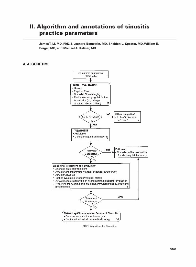
FIG 1 Algorithm for Sinusitus
II. Algorithm and annotations of sinusitispractice parameters
James T. Li, MD, PhD, I. Leonard Bernstein, MD, Sheldon L. Spector, MD, William E.
Berger, MD, and Michael A. Kaliner, MD
A. ALGORITHM
S110 Sinusitus Practice Parameters J ALLERGY CLIN IMMUNOLDECEMBER 1998
B. ANNOTATIONS
1. Symptoms suggestive of acute sinusitis
• Acute sinusitis typically presents as a persistent upperrespiratory infection.
• In adults, prominent symptoms include nasal conges-tion, purulent rhinorrhea, postnasal drainage, facial ordental pain, headache, and cough frequently with amore severe nocturnal component.
• Any patient with orbital pain, swelling of forehead,and/or diplopia should be urgently scheduled for eval-uation.
• Children with acute sinusitis may also exhibitincreased irritability and vomiting occurring in associ-ation with gagging on mucus and/or prolonged cough.
• In all age groups, less frequent symptoms associatedwith acute sinusitis include fever, nausea, malaise,fatigue, halitosis, hyposmia, and sore throat.
2. Office visit
• Review medical history for diagnosis of sinusitis andunderlying risk factors.
• General examination includes an evaluation forsigns of upper airway and sinus inflammation asso-ciated with nasal mucosa edema, purulent secre-tions, and increased localized blood flow. Typicalclinical signs include tenderness overlying the sinus-es, dark circles beneath the eyes, and/or periorbitaledema. Pharyngeal erythema, lymphoid hyperplasia,and posterior pharyngeal purulent material is alsofrequently observed.
• Nasal examination in patients with acute sinusitis mayreveal mucosal erythema and purulent secretions.Nasal polyps may contribute to nasal congestion andcan be a source of recurrent sinusitis by obstructingthe sinus ostia. In adults, nasal polyps may be associ-ated with nonsteroidal antiinflammatory drug sensitiv-ity and asthma. Nasal polyps are relatively uncommonin children, and their presence should prompt evalua-tion for possible cystic fibrosis. Ear examination inpatients with suspected acute sinusitis frequently willreveal middle ear abnormalities and associatedeustachian tube dysfunction.
• Acute or chronic sinusitis may initiate or worsen asth-ma and bronchial hyperresponsiveness. Accordingly,chest auscultation and other objective measurements ofairflow obstruction, such as spirometry, should be con-sidered in any patient with possible sinusitis and cough.
• Patients with obvious acute sinusitis should be carefullyreviewed for any possible evidence of complicating fac-tors, including the presence of external facialswelling/erythema over an involved sinus, visualchanges, abnormal extraocular movements, proptosis,periorbital inflammation/edema/erythema/cellulitis, anysuggestion of intracranial involvement, or central ner-vous system manifested as abnormal neurologic signs.
• In general, radiographs are not necessary in making thediagnosis of acute sinusitis. Occasionally imagingstudies may be useful to support the diagnosis or pro-
vide evidence of the degree of mucosal involvement,thereby guiding more aggressive therapy. Radiograph-ic signs compatible with sinusitis include greater than 6mm mucosal thickening in the maxillary sinuses,greater than 33% loss of air space volume within themaxillary sinuses, or opacification/air fluid levels inany of the paranasal sinuses. Waters view radiographsare a useful screen in adults and children over 1 year ofage because nearly all patients will have a maxillarycomponent to their sinusitis. A limited coronal sinuscomputed tomographic scan should be consistent if iso-lated ethmoid sinusitis is suspected. Axial sinus com-puted tomography is indicated in suspected orbitalinvolvement, and sinus magnetic resonance imagingcan provide useful information with related soft tissueinvolvement.
• Nasal cultures are not reliable for establishing thediagnosis or for determining a specific causativemicroorganism. Maxillary antrum aspiration for cul-ture is definitive but is indicated only when precisemicrobial identification is essential.
3. Acute sinusitis
• Acute sinusitis is defined as symptoms and signs for 3to 4 weeks, although others have modified the defini-tion to 8 weeks. The diagnosis of acute sinusitis isbased primarily on the clinical history, the physicalexamination, and possibly other ancillary evaluations,including nasal cytology or radiographic imaging. Inmost instances, the diagnosis is made presumptively,and treatment is initiated. Clinical resolution usuallyoccurs within 3 to 4 weeks.
4. Other diagnoses
Differential diagnoses include:• allergic and nonallergic rhinitis• upper respiratory infection• nasal septum deviation• nasal polyps• nasopharyngeal tumor, granulomata
5.Treatment
Antibiotics
• Amoxicillin or trimethoprim-sulfamethoxazole (TMP/ SMX)are generally effective, inexpensive, and well tolerated. Forpatients allergic to both amoxicillin and TMP/SMX, alterna-tives would include quinolones, cephalosporins, ormacrolides. These alternatives should also be considered in sit-uations where microorganisms resistant to amoxicillin orTMP/SMX are prevalent.
• Acute sinusitis generally responds to treatment for 10to 14 days. Some physicians continue treatment for 7days after the patient is well. It is important to instructthe patient to complete the course of antibiotics.
• One approach is to start the patient on amoxicillin orTMP/SMX for 5 to 7 days and determine whether thesigns and symptoms are improving (clearing of secre-tions and generally improved well-being). If the
J ALLERGY CLIN IMMUNOLVOLUME 102, NUMBER 6, PART 2
Sinusitus Practice Parameters S111
patient is improving, continue this treatment until thepatient is well for 7 days (generally a 10- to 14-daycourse). If after 5 to 7 days the patient has not shownimprovement, switch to cefuroxime axetil, amoxi-cillin/clavulanate, or clarithromycin (or other appro-priate, broad spectrum, potent antibiotics) until thepatient is well for 7 days.
Corticosteroids
• The use of nasal corticosteroids is rational particularlyin patients with underlying rhinitis or associatedbronchial hyperresponsiveness.
• Although efficacy has not yet been proven, the short-term use of oral corticosteroids as an adjunct in treat-ing patients with acute sinusitis is reasonable when thepatient has had significant anatomic obstruction, inva-sive nasal polyposis, or has demonstrated markedmucosal edema radiographically.
Saline/mucolytics
• Saline nasal sprays or lavage may be a useful adjunctby liquefying secretions and decreasing the risk ofcrusting near the sinus ostia.
• There is no conclusive evidence that mucolytics,such as guaifenesin, are useful adjuncts in treatingacute sinusitis.
α-Adrenergic decongestants
• Topical decongestants (eg, oxymetazoline andphenylephrine) and oral decongestants (eg, pseu-doephedrine) reduce mucosal blood flow, decrease tis-sue edema and nasal resistance, and may enhancedrainage of secretions from the sinus ostia.
• The use of topical decongestants beyond 3 to 5 daysmay induce rhinitis medicamentosa with associatedincreased congestion and refractoriness to subsequentdecongestant therapy.
Education
• The following comfort measures may be helpful: ade-quate rest, adequate hydration, analgesics as needed,warm facial packs, steamy showers, and sleeping withthe head of bed elevated.
• Prevention measures may include appropriate treatment ofallergies and viral upper respiratory tract infections and avoid-ance of adverse environmental factors, such as cigarettesmoke, pollution, and barotrauma.
• Patients should be instructed to phone if symptomsworsen (eg, especially with headache or high fever) orif symptoms have not resolved within 5 to 7 days oftreatment (see annotation #10).
6. Treatment successful?
Complete response
• Patient is improved symptomatically to near normal.
Partial response
• Patient is symptomatically improved but not back tonormal at the end of the first course of antibiotics.
Poor response
• Patient has little or no symptomatic improvement afterthe first course of antibiotic therapy.
7. Follow-up
• No further evaluation for resolved, uncomplicatedsinusitis.
• Consider further evaluation of underlying risk factors,such as allergic and nonallergic rhinitis and structuralabnormalities.
8. Additional treatment and evaluation
• For partial response, continue antibiotic treatment foranother 10 to 14 days or consider antibiotic choiceslisted under “poor responses.”
• For poor response to treatment with amoxicillin orTMP/SMX, an antibiotic should be prescribed thatcovers resistant bacteria. Appropriate choices includeamoxicillin/potassium clavulanate, cefuroxime, cefpo-doxime, cefprozil, cefixime, ceftibuten, loracarbef,azithromycin, and clarithromycin. In adults, expandedspectrum quinolones, such as ciprofloxacin, lev-ofloxacin, grepafloxacin, or trovafloxacin, may alsobe a consideration.
• Persistent sinusitis, defined as failure after 21 to 28days of initial antibiotic treatment may be caused bypathogens not adequately covered by prior antibi-otics, the presence of nasal polyps, or noncompli-ance. The use of broader spectrum single agentssuch as amoxicillin/potassium clavulanate, cefurox-ime, or cefpodoxime should be considered with orwithout the addition of anaerobic coverage with clin-damycin or metronidazole.
• Reinforce the comfort and prevention measures out-lined in annotation #5.
• Consider sinus computed tomographic scan if notalready done.
• Underlying risk factors should be evaluated in a moredetailed manner.
• Consider consultation with allergist/immunologist fortreatment of underlying allergic factors and evaluationof opportunistic infection, immunodeficiency, andstructural abnormality.
Recurrent sinusitis
• Repeated episodes of acute sinusitis typically 3 ormore times per year.
• Patients with chronic or recurrent sinusitis should beevaluated for underlying rhinitis, immunodeficiency,and anatomic abnormalities.
Rhinitis
• Patients with suspected allergic rhinitis in conjunctionwith sinusitis should be evaluated by an allergist/immu-nologist competent in the evaluation of IgE sensitiza-tion to inhalant allergens.
• Emphasis of therapy for allergic rhinitis includes environ-mental control, antihistamines and nasal corticosteroids,and, in selected patients, allergen immunotherapy.
S112 Sinusitus Practice Parameters J ALLERGY CLIN IMMUNOLDECEMBER 1998
• Other rhinitic conditions (vasomotor, NARES,rhinitis medicamentosa) may also lead to sinusi-tis, and the consultant must be capable of differ-entiating these conditions and initiating appro-priate course of therapy.
Immunodeficiency
• Referral to an allergist/immunologist is particularlyindicated in patients with chronic or recurrent sinusitisassociated with otitis media, bronchitis, bronchiectasis,or pneumonia and in patients who have undergoneprior surgical procedures and continue to experiencesinusitis. This evaluation may include measurement ofquantitative serum IgG, IgA, and IgM and assessmentof specific antibody responses to protein and polysac-charide antigens, such as tetanus toxoid or Pneumovax.
9. Treatment successful?
• See annotation #6.
10. Follow-up
• See annotation #7.
11. Chronic sinusitis
Chronic sinusitis
• Signs and symptoms compatible with sinusitis persist-ing 3 to 8 weeks or longer.
Chronic sinusitis with ostiomeatal obstruction
• If the patient has a significant nasal septal defect that
compresses the middle turbinate into the ostiomeatalcomplex or obstruction of the sinus outflow tractscaused by middle turbinate deformity or the presence ofaccessory structures that block sinus drainage, considerconsultation with a surgeon. The presence of obstruct-ing nasal polyps, after an appropriate course of treat-ment that may include a trial of oral corticosteroids, isalso an indication for referral. Finally, a patient withrecurrent or chronic symptoms despite aggressive med-ical management of 4 to 6 months may also benefit fromwidening of the ostiomeatal outflow tract.
• Evaluation should include coronal sinus computedtomography with extra cuts through the ostiomeatalcomplex to clarify the extent of disease and specificlocation or locations. Significant anatomic abnormali-ties might prompt evaluation by an otolaryngologist atthe same time that underlying rhinitis and immunode-ficiency is explored.
• Evaluation may also include nasal/sinus biopsy in sus-pected cases of neoplasia, fungal disease, granulomatousdisease, or abnormal ciliary structure and/or function.
• In patients with “borderline” anatomic abnormalities,every effort should be made to maximize treatment forunderlying rhinitis or immunodeficiency before pro-ceeding with surgical intervention.
• Contemporary surgical therapy involves functionalendoscopic sinus surgery.
• Most patients benefit from continued individualizedmedical therapy, including allergy management,after surgery.
S113
INDICATIONS FOR REFERRAL; INTERAC-
TIONS BETWEEN CONSULTANTS AND
REFERRING PHYSICIAN
• Referral should be considered when serious complica-tions (eg, otitis media, asthma, bronchiectasis, fungalsinusitis, multiple antibiotic allergies, etc.) occurand/or when quality of life is compromised.
• Referral is indicated if an underlying or immunologicbasis is suspected.
• The consultant should delineate imunologic/allergicfactors, assist in a treatment plan including avoidancemeasures, pharmacotherapy, education, and immuno-therapy.
• An otolaryngologic surgeon should be consulted whenanatomical factors impair optimal medical manage-ment, biopsy and/or cultures are required, or severecomplications ensue.
• The otolaryngologic surgeon should inform the con-sulting physician about the needs, risks, or possiblealternatives of surgical interventions.
DEFINITIONS, ANATOMIC CONSIDERATIONS,
SINUS PHYSIOLOGY, MICROBIOLOGY
A. Definitions
• Sinusitis is defined as inflammation of 1 or more ofthe paranasal sinuses. The most common cause ofsinusitis is infection. Classification of sinusitis isfrequently based on duration of symptoms and/orimaging characteristics.
• There is no universally accepted classification ofsinusitis, but commonly used terminology is as fol-lows:A. Acute sinusitis. Symptoms for 3 to 4 weeks (with
some clinicians modifying this definition to 8weeks) consisting of some or all of the following:persistent symptoms of an upper respiratory infec-tion, purulent rhinorrhea, postnasal drainage, anos-mia, nasal congestion, facial pain, headache, fever,cough and purulent discharge.
B. Chronic sinusitis. Symptoms for 3 to 8 weeks orlonger of varying severity consisting of thesame symptoms as seen in acute sinusitis. Inchronic sinusitis there should be abnormal find-ings on computed tomography or magnetic res-onance imaging. Some patients with chronicsinusitis may present with vague or insidioussymptoms.
C. Recurrent sinusitis. Three or more episodes ofacute sinusitis per year. Patients with recurrentsinusitis may be infected by different organisms atdifferent times.
B. Anatomic considerations
• The sinuses develop at different ages during childhood.• The optic nerve, cavernous sinus, carotid artery, and
sella turcica are adjacent to the sphenoid sinus.Tumors and infection in the sphenoid sinuses canprogress to involve these structures.
• The ethmoid bulla cells can occasionally enlarge intothe anterior attachment of the middle turbinate causingvariable degrees of pneumatization of the turbinate(concha bullosa). The enlarged turbinate can obstructventilation of the middle meatus and may causesinusitis. Frontal recess cells can be variable in num-ber and size. When present, they can impinge in thenasofrontal duct, leading to frontal sinusitis.
• Septal deviation can predispose to sinusitis if the devi-ation narrows the middle meatus. With long-standingdeviation, atrophy of the middle turbinate and narrow-ing of the ostiomeatal complex is commonly seen.
C. Sinus physiology
• The sinuses are air-filled cavities with classical pseu-dostratified ciliated columnar epithelium interspersedwith goblet cells. The cilia sweep mucus towards theostial opening.
• Obstruction of sinus ostia may lead to mucousimpaction and decreased oxygenation in the sinuscavities.
• During obstruction of the ostia, the pressure in thesinus cavity can decrease, which in turn causes thesymptom of pain, particularly in the frontal region.
D. Microbiology
Bacterial
• In acute sinus disease viral upper respiratory infec-tions frequently precede bacterial superinfection byStreptococcus pneumoniae, Haemophilus influenzae,and Moraxella catarrhalis.
• Both M catarrhalis and H influenzae may be β-lacta-mase producing and thereby amoxicillin resistant.
• Streptococcus pneumoniae are becoming more peni-cillin resistant. It is not uncommon for 40% of sinusisolates to be penicillin resistant.
• In addition to the organisms mentioned above, themost common organisms in chronic sinusitis, arePseudomonas aeruginosa, Group A streptococcus andStaphylococcus aureus, as well as anaerobes such asBacteroides spp, Fusobacteria and Propionibacteriumacnes.
• In contrast to community-acquired sinusitis, the usualpathogens in nosocomial sinusitis are gram-negativeenterics (such as P aeruginosa, Klebsiella pneumoniae,
III. Summary statements of sinusituspractice parameters
S114 Sinusitus Practice Parameters J ALLERGY CLIN IMMUNOLDECEMBER 1998
Enterobacter spp, Proteus mirabilis, Serratia marce-scens) and grampositive cocci (occasionally streptococ-ci and staphylococci).
Fungal
• Aspergillus fumigatus is said to be the most com-mon cause of fungal sinusitis in immunocompetentindividuals.
• Allergic fungal sinusitis may be caused by Aspergillusspp, Myriodontium keratinophilum, Bipolaris spp,Dreschlera spp, Curvularia spp, and Alternaria spp.
CLINICAL DIAGNOSIS
A. Clinical history
• Acute sinusitis is typically first seen as an upper res-piratory infection that has persisted beyond 5 to 7days.
• The diagnosis of sinusitis is based on a combination ofclinical history with physical examination, nasal cytol-ogy, and/or imaging studies.
• Factors that may predispose to sinusitis include aller-gic or occupational rhinitis, vasomotor rhinitis, nasalpolyps, rhinitis medicamentosa, and immunodeficien-cy. For many patients, the clinical history shouldaddress these factors.
• The differential diagnosis of sinusitis includes cysticfibrosis, Wegener’s granulomatosis, HIV infection,Kartagener’s syndrome, immotile cilia syndrome,and tumors.
B. Clinical examination
• The occurrence of a common cold or persistent aller-gic or nonallergic rhinitis are common precursors ofacute and chronic sinusitis.
• Prominent symptoms include nasal congestion, puru-lent rhinorrhea, postnasal drainage, facial or dentalpain, headache, hyposmia, and cough.
• Typical clinical signs include tenderness overlying thesinuses, mucosal erythema, nasal purulent secretions,increased posterior pharyngeal secretions, and perior-bital edema.
• If performed properly in adults, transillumination maybe a useful diagnostic technique when combined withabnormal signs and symptoms.
• More detailed examination for underlying risk factorsmay be required if sinusitis becomes chronic.
• Endoscopy is a quick and safe way to visualize theposterior nasal structures and may aid in the diagno-sis.
IMAGING STUDIES IN THE EVALUATION OF
SINUSITUS
• Imaging studies may be required when the symptomsare vague, physical findings are equivocal, or there ispoor response to the initial management.
• Standard radiographs may be used for detection of
acute sinus disease, but they are insensitive, especiallyin ethmoid disease.
• Computed tomography is the preferred imaging tech-nique for preoperative evaluation of the nose andparanasal sinuses secondary to obstruction of theostiomeatal complex.
• Although magnetic resonance imaging has limitationsin the definition of the bony anatomy, it is particularlysensitive for evaluation of the frontal, maxillary, andsphenoid sinuses for fungal sinusitis and tumors andthe differential diagnosis between inflammatory dis-eases and malignant tumors.
• Ultrasonography has limited utility but may be applic-able in pregnant women and for determining theamount of retained secretions.
LABORATORY TESTS
• Laboratory evaluation of chronic or recurrent sinusitismay include nasal cytology, a sweat chloride test, cil-iary function studies, and tests for immunodeficiency.
• Nasal cytology is useful in the clinical evaluation ofunderlying allergic rhinitis, the nonallergic rhinitiswith eosinophilia syndrome, nasal polyposis, andaspirin sensitivity.
• Quantitative sweat chloride tests for diagnosis of cys-tic fibrosis should be considered in children with nasalpolyps and/or colonization of the nose and sinuseswith Pseudomonas spp.
• Tests for immunodeficiency (eg, quantitativeimmunoglobulins, antibody tests, serum IgE, andcomplement components) may be useful if either con-genital or acquired immunodeficiency is suspected incases of recurrent sinusitis.
RHINOLARYNGOSCOPY
• Fiberoptic rhinoscopy permits more detailed examina-tion of the anterior and posterior nasal and pharyngealstructures.
• Fiberoptic rhinoscopy may be considered for somepatients with chronic or recurrent sinusitis to assessstructural abnormalities, fungal disease, or granuloma-tous lesions.
BIOPSY OF THE NOSE AND PARANASAL
SINUSES
• Paranasal sinus biopsies may be required to determinewhether a lesion is neoplastic or to confirm the pres-ence of suspected fungal disease and the possibility ofgranulomatous disease.
• Nasal mucosa harvested from the posterior portion ofthe inferior turbinate is the preferred biopsy site forprimary cilia dysfunction.
CONGENITAL OR ACQUIRED IMMUNODEFI-
CIENCY
• The majority of patients with congenital immune defi-ciency and associated sinusitis will have defects in
J ALLERGY CLIN IMMUNOLVOLUME 102, NUMBER 6, PART 2
Sinusitus Practice Parameters S115
humoral immunity. However, sinusitis is common inacquired immune deficiency (AIDS), in which bothhumoral and cellular impairments are present. Appro-priate laboratory studies of both humoral and cellularimmunity include quantitative immunoglobulin assays;specific antibody responses to tetanus toxoid, pneumo-coccal, or Hemophilus influenzae vaccines; mea-surement of complement components; and key cel-lular responses.
• The most common congenital immunodeficiency syn-dromes in this group are common-variable immuno-deficiency, IgA deficiency, Wiskott-Aldrich syn-drome, ataxia telangiectasia, X-linked immunodefi-ciency with normal, or elevated IgM and X-linkedagammaglobulinemia.
ASTHMA
• The association between sinusitis and asthma has longbeen appreciated and is generally stated to range from40% to 75%.
• Although a number of theories have been proposed toexplain this relationship, no direct causal factor hasyet been found.
• Studies in both adults and children have clearlyshown that medical and surgical management ofsinusitis results in objective and subjective improve-ment of asthma.
ALLERGIC RHINITIS
• Allergic rhinitis commonly precedes the developmentof recurrent or chronic sinusitis because the associatednasal obstruction and inflammation interrupts normalmucociliary clearance and leads to retention ofmucopurulent secretions within the sinus cavities.
• Patients with suspected allergic rhinitis in conjunctionwith sinusitis may benefit from evaluation by an aller-gist/immunologist who, in most instances, will per-form prick/puncture tests to clarify the role of aller-gies.
• Treatment of allergic rhinitis should include environ-mental control; medications such as systemic and top-ical antihistamines, decongestants, nasal cromolyn,anticholinergics, and glucocorticosteroids; and, inappropriate patients, allergen immunotherapy.
• Other forms of rhinitis (eg, vasomotor rhinitis andNARES) also commonly precede the development ofrecurrent or chronic sinusitis.
CYSTIC FIBROSIS
• Chronic sinusitis is an important source of morbidityin nearly all patients with cystic fibrosis, creatingnasal obstruction, post nasal drainage, headache, andpotential exacerbation of pulmonary obstruction.
• Pathogens in patients with cystic fibrosis and sinusitisinclude Pseudomonas aeruginosa, Escherichia coli,Staphylococcus aureus, and Aspergillus fumigatus, in
addition to the more common polysaccharide-encap-sulated organisms.
• Although children with cystic fibrosis and sinusitisgenerally respond to prolonged treatment with con-ventional oral antibiotics, older children and adultswho are colonized with P aeruginosa frequentlyrequire oral quinolones, intravenous tobramycin, orceftazadime to control acute exacerbations.
ANTIBIOTICS
• Antibiotics are the primary therapy for bacterial sinusitis.• The most common bacteria observed are polysaccha-
ride encapsulated organisms of which 30% to 40%produce β-lactamase.
• Appropriate duration of antibiotic therapy for acutesinusitis is not well defined, although a 14-day course isprobably adequate for most patients with acute disease.
• Chronic sinusitis should be treated until the patient iswell for 7 days before stopping therapy.
• Choice of antibiotic should be based on predictedeffectiveness, cost and side effects.
ANTIHISTAMINES
• There are no data presently to recommend the use ofH1 antihistamines in acute bacterial sinusitis.
• There may be a role for antihistamines in chronicsinusitis, especially in patients with allergic rhinitis.
α-ADRENERGIC DECONGESTANTS
• Both topical and oral decongestants are often used inthe therapy of acute or chronic sinusitis because it isthought that they may widen ostial patency and reduceturbinate swelling.
• Prospective studies are lacking and are needed toassess the value of α-adrenergic agents in the treat-ment of sinusitis.
GLUCOCORTICOSTEROIDS
• The use of systemic steroid therapy for sinus diseasehas not been studied systematically in a well-con-trolled or blinded manner.
• A few recent studies suggest that the addition ofintranasal steroids as an adjunct to antibiotic therapy isbeneficial in the treatment of sinusitis.
• The relative safety of directed therapy with inhaledsteroids makes their use a likely mode of treatmentfor sinusitis, at least for the treatment of any under-lying rhinitis.
ADJUNCTIVE THERAPIES INCLUDING SALINE,
MUCOLYTICS, AND EXPECTORANTS
• There are inadequate data to recommend use of wet-ting agents as individual therapy for sinusitis.
S116 Sinusitus Practice Parameters J ALLERGY CLIN IMMUNOLDECEMBER 1998
• Clinical practice, as well as folklore, supports the useof wetting agents for symptomatic treatment as part ofa pharmacologic regimen.
• There are several scientific studies that imply, but donot directly confirm, a role for these agents in sinusitis.
• The safety profile of each agent should be carefullyconsidered for individual patients.
• Use of all these agents as prophylaxis for exacerba-tions of chronic sinusitis is empiric and not supportedby clinical data.
INTRAVENOUS IMMUNE GLOBULIN
• Immunodeficiency is one of the underlying factors forthe development of chronic and recurrent sinusitis.
• Intravenous immunoglobulin is indicated for use inpatients with impaired humoral immunity.
SURGICAL CONSIDERATIONS
• Antral puncture and irrigation is an office procedurethat has a place in the management of acute ethmo-maxillary sinusitis refractory to medical therapy or inan immunosuppressed patient in whom early identifi-cation of pathogenic organisms is paramount.
• The term functional endoscopic sinus surgery is basedon the clinical and experimental findings that ostialobstruction is the final common pathway in the devel-opment of sinusitis.
S117
A. INDICATIONS FOR REFERRAL TO A CON-
SULTANT AND CONSULTATION WITH A
SPECIALIST
i. Indications for referral
a. When the condition or its treatment is interferingwith a patient’s performance or causing significantloss of school or work on a chronic or recurrentbasis or when the patient’s quality of life is signifi-cantly affected.
b. When there are complications of sinusitis, such as oti-tis, asthma, bronchiectasis nasal polyps, or bronchitis
c. When there is consideration for an allergic orimmunologic basis for the sinusitis.
d. When the condition becomes chronic, persists forseveral months, or recurs 2 to 3 times per yeardespite treatment by the primary care physician.
e. When there is the need for complex pharmacology,such as a patient with multiple antibiotic allergies,allergic fungal sinusitis, or resistant pathogens.
ii. What the consultant should provide to the
referring physician
a. Clarification of allergic, immunologic, or nonaller-gic etiologic basis for the patient’s condition
b. Assessment of nasal and sinus outflow track anato-my and any contribution these anatomic factorshave in the causation of the sinus problems.
c. Identification of specific allergens or other triggersfor the patient’s condition and education in ways toavoid exposure to these triggers.
d. Assistance in developing an effective treatmentplan, including patient education, allergy avoidance,pharmacotherapy, antiinfectious therapy, andimmunotherapy if appropriate
e. Provision of specialized services, such as prepara-tion of extracts and provision of immunotherapy.
iii. Indications for referral to an otolaryngo-
logic surgeon
a. When anatomical defects exist that obstruct thesinus outflow track, including the ostiomeatal com-plex and adenoidal tissues and are thought to becontributing to recurrent or chronic sinusitis.
b. When nasal polyps obstruct the sinus drainage andpersist despite appropriate medical treatment.
c. When there is recurrent or persistent sinusitis, despiteadequate trials of medical management. Adequate
medical management minimally involves multiplecourses of antibiotics chosen to cover the spectrum ofpathogens anticipated to be causing the disease.
d. For biopsy of the nasal mucosa to rule out granulo-matous disease, neoplasms, ciliary dyskinesia, orfungal infections.
e. When maxillary antral puncture for culture or reliefof pain is required.
f. For sinusitis complicated by brain abscess, menin-gitis, cavernous sinus thrombosis, or Pott’s tumor(infectious erosion of the ethmoid or frontal sinus).
iv. What the otolaryngologic surgeon should
provide the referring physician
a. Assessment of the need for surgical revision of abnor-mal anatomy and the likelihood that such revision willreduce the recurrent or chronic sinus disease.
b. Determination of whether an adequate trial of med-ical, allergic, or immunologic treatment has beenprovided before recommending surgery.
c. Recommendation of specific procedure or proce-dures for the specific patient and discussion of themerits of alternative approaches.
d. Conservative prediction of the discomfort, inconve-nience. and dangers from the procedures proposed.
B. DEFINITIONS, ANATOMIC CONSIDERA-
TIONS, SINUS PHYSIOLOGY, MICROBIOL-
OGY
Bi. Definitions
SUMMARY STATEMENTS
1. Sinusitis is defined as inflammation of 1 or more ofthe paranasal sinuses. The most common cause ofsinusitis is infection. Classification of sinusitis is fre-quently based on duration of symptoms and/or thespecific sinus involved.
2. There is no universally accepted classification of sinusi-tis, but commonly used terminology is as follows:a. Acute sinusitis. Symptoms for 3 to 4 weeks (with
some clinicians modifying this definition to 8weeks) consisting of some or all of the following:persistent symptoms of an upper respiratoryinfection, purulent rhinorrhea, postnasaldrainage, anosmia, nasal congestion, facial pain,headache, fever, and cough.
b. Chronic sinusitis. Symptoms for 3 to 8 weeks orlonger of varying severity consisting of the same
IV. Complete guidelines and references
Sheldon L. Spector, MD, I. Leonard Bernstein, MD, James T. Li, MD, PhD, William E.
Berger, MD, Michael A. Kaliner, MD, Diane E. Schuller, MD, Joann Blessing-Moore,
MD, Mark S. Dykewicz, MD, Stanley Fineman, MD, Rufus E. Lee, MD, and Richard
A. Nicklas, MD
S118 Sinusitus Practice Parameters J ALLERGY CLIN IMMUNOLDECEMBER 1998
symptoms as seen in acute sinusitis. In chronicsinusitis, there should be abnormal findings oncomputed tomography or magnetic resonanceimaging. Some patients with chronic sinusitis maybe first seen with vague or insidious symptoms.
c. Recurrent sinusitis. Three or more episodes ofacute sinusitis per year. Patients with recurrentsinusitis may be infected by different organismsat different times.
Sinusitis is defined as inflammation of 1 or more ofthe paranasal sinuses. The most common cause of sinusi-tis is infection. Classification of sinusitis is frequentlybased on duration of symptoms, the specific sinusinvolved, and/or imaging characteristics.1-7 There is nouniversally accepted classification of sinusitis, but com-monly used terminology is as follows:
1. Acute sinusitis. Symptoms of 3 to 4 weeks (someclinicians modify this definition to 8 weeks) con-sisting of some or all of the following: persistentsymptoms of an upper respiratory infection, puru-lent rhinorrhea, postnasal drainage, anosmia, nasalcongestion, facial pain, headache, fever, and cough.
2. Chronic sinusitis. symptoms for more than 4weeks of varying severity, consisting of the samesymptoms as seen in acute sinusitis (others havedefined chronic sinusitis as symptoms for up to 8weeks or longer and have used the term subacutesinusitis to define symptoms lasting 3 weeks). Inchronic sinusitis, there should be abnormal find-ings on computed tomography or magnetic reso-nance imaging. Some patients with chronic sinusi-tis may be first seen with vague or insidious symp-toms.
3. Recurrent sinusitis. Three or more episodes ofacute sinusitis per year. Patients with recurrentsinusitis may be infected by different organisms atdifferent times.
REFERENCES
1. Druce HM. Diagnosis and management of chronic sinusitis and its compli-cations. Immunol Allergy Clin North Am 1987;7:117-32.
2. Avant RF, Kennedy DW. Need for a national education program on appro-priate care of patients with sinusitis. Otolaryngol Head Neck Surg1990;103:855.
3. Stafford CT. The clinician’s view of sinusitis. Otolaryngol Head Neck Surg1990;103:870-5.
4. Newman L, Platts-Mills TAE, Phillips CD, Hazen KC, Gross CW. Chronicsinusitis: relation of computed tomographic findings to allergy, asthma, andeosinophilia. JAMA 1994;271:363-7.
5. Wald ER, Byers C, Guerra N, Casselbrant M, Beste D. Subacute sinusitis inchildren. J Pediatr 1989;115:28-32.
6. Bluestone CB. The diagnosis and management of sinusitis in children: pro-ceedings of a closed conference. Pediatr Infect Dis J 1985;6(suppl):S49-81.
7. Friedman WH, Katsantonis GP, Sivore M, Kay S. Computed tomographystaging of the paranasal sinuses in chronic hyperplastic rhinosinusitis.Laryngoscope 1990;100:1161-5.
Bii. Anatomic considerations
SUMMARY STATEMENTS
1. The sinuses develop at different ages during child-hood.
2. The optic nerve, cavernous sinus, carotid artery, andsella turcica are adjacent to the sphenoid sinus.Tumors and infection in the sphenoid sinuses canprogress to involve these structures.
3. The ethmoid bulla cells can occasionally enlargeinto the anterior attachment of the middle turbinatecausing variable degrees of pneumatization of theturbinate (concha bullosa). The enlarged turbinatecan obstruct ventilation of the middle meatus andmay cause sinusitis. Frontal recess cells can bevariable in number and size. When present, theycan impinge in the nasofrontal duct leading tofrontal sinusitis.
4. Septal deviation can predispose to sinusitis if thedeviation narrows the middle meatus. With longstanding deviation, atrophy of the middle turbinateand narrowing of the ostiomeatal complex is com-monly seen.
DEVELOPMENT
The maxillary sinus is the first to begin significantpneumatization between birth and 12 months. It begins toenlarge laterally along the floor of the orbit at about age3 years. The floor of the maxillary sinus reaches the levelof the nose by 12 years of age. Adult size is achieved inmidadolescence. Rudimentary ethmoid sinuses are pre-sent at birth but do not begin to enlarge until the child isbetween 3 and 7 years of age. Adult size of the ethmoidsinuses is reached at 12 to 14 years of age. The sphenoidsinuses, which originate from the nasal cupola, do notreach significant size until 4 to 5 years of age. Sphenoiddevelopment, which is complete by midadolescence, canbe highly variable in the degree of pneumatization of thegreater and lesser wings of the sphenoid and the ptery-goid processes. The frontal sinuses are the last to devel-op. Typically not present at birth, the frontal sinusesbegin as outgrowths of nasal mucosa into the frontalrecess from the middle meatus. They do not usually reachsignificant size until midadolescence. Like the sphenoidsinuses, the frontal sinuses can be quite variable in theirdegree of pneumatization. Difference in developmentbetween the right and left frontal sinuses are often foundwithin individuals.
ANATOMY
The ethmoid sinuses consist of a complex “honey-comb” of cells varying between 4 and 17 in number. Theaverage individual has 9 cells. The ethmoid sinuses arecommonly divided into 2 groups: the anterior cells andthe posterior cells. Some authors further separate the eth-moid bulla from the anterior cells, calling them the mid-dle ethmoid cells.
A common occurrence is the complete absence of afrontal sinus. The degree of pneumatization of the frontalsinus is variable.
The paired sphenoid sinuses are separated by anintrasinus septum. The optic nerve courses over the later-al superior surface of the sphenoid sinus. The carotidartery within the cavernous sinus is just lateral, and the
J ALLERGY CLIN IMMUNOLVOLUME 102, NUMBER 6, PART 2
Sinusitus Practice Parameters S119
maxillary nerve (part of the fifth cranial nerve) is infero-lateral anteriorly. The pituitary, located in the sella turci-ca, can be approached through the posterior superior wallof the sphenoid sinus. Tumors and infection in the sphe-noid sinuses can progress to involve these structures.
It is important to realize that ethmoid cells or sinusesare not completely contained in the ethmoid bone. Forexample, the ethmoid sinus in the course of its develop-ment may invade the middle turbinate forming conchabullosa. The ethmoid bulla cells can occasionally enlargeinto the anterior attachment of the middle turbinate, caus-ing variable degrees of pneumatization of the turbinate(concha bullosa). The enlarged turbinate obstructs venti-lation of the middle meatus and commonly causes later-alization of the uncinate process and narrowing of theethmoid infundibulum. Similarly, a cell invading themedial floor of the orbit is known as a Haller cell. Thosecells outside the ethmoid bone proper are called extra-mural. These cells are located in the medial inferior orbitand usually form the medial wall of the ethmoidinfundibulum. Because of this relationship, they canmarkedly narrow the infundibulum, causing the obstruc-tion of the maxillary and anterior ethmoid sinuses. ThusHaller cells are frequently present in association withsinus disease.
Frontal recess cells can be variable in number and size.When present, they can impinge in the nasofrontal duct,leading to frontal sinusitis.
Septal deviation can predispose to sinusitis if the devi-ation narrows the middle meatus. With long-standingdeviation, atrophy of the middle turbinate and narrowingof the ostiomeatal complex is commonly seen.
Biii. Sinus physiology
SUMMARY STATEMENTS
1. The sinuses are air-filled cavities with classical pseu-dostratified ciliated columnar epithelium inter-spersed with goblet cells. The cilia sweep mucustowards the ostial opening.
2. Obstruction of sinus ostia may lead to mucous impactionand decrease oxygenation in the sinus cavities.
3. During obstruction of the ostia, the pressure in thesinus cavity can decrease, which in turn causes thesymptom of pain, particularly in the frontal region.
The sinus cavities are air filled with classical pseudo-stratified ciliated columnar epithelium interspersed withgoblet cells. The cilia sweep mucus towards the ostialopening. Blood flow in the maxillary sinus is roughlyestimated to be 100 mL/100 g tissue per minute, which issimilar to that found in the nose but higher than thatfound in the brain. Obstruction of the ostia can lead tomucous impaction and decrease oxygenation in the sinuscavities. This in turn may lead to further complications(discussed in further sections). There is very little gasexchange in the sinuses except during ostial obstruction,when the oxygen concentrations can fall to close to 0%with purulent secretions but not with nonpurulent secre-
tions. The growth of bacteria is facilitated in this anaero-bic environment.
During obstruction of the ostia, the pressure in thesinus cavity can decrease, which in turn causes the symp-tom of pain, particularly in the frontal region.1 This pres-sure decrease may range from 20 to 30 mm H2O, with thelowest pressure being –66 mm H2O. Transudation maystart when the pressure is lower than 20 to 30 mm H2Obelow 0. This decrease in pressure is preceded by a tran-sient pressure increase caused by the rise in CO2, where-as the decrease in pressure is principally caused by O2absorption.2 However, in acute purulent sinusitis, thepressure may sometimes be as high as a 100 mm H2O.3
Purulent secretions have a low oxygen content and thepain may be due to a combination of inflammation orig-inating from the mucosa and pressure from the secretionson the inside walls of the sinus.
During deep sea diving, the change in sinus pressuremay be very high, causing transudation, bleeding, andedema, especially when pressures exceed 350 to 500 mmH2O. During flying, there is usually less change in pressurethan diving. When there is obstruction of the ostia, changesin sinus pressure similar to that of diving may occur.
REFERENCES
1. Aust R, Stierner P, Drettner B. Basic experimental study of ostial paten-cy and local metabolic environment of the maxillary sinus. Acta Oto-Laryngologica Suppl 1994;515:7-10.
2. Aust R, Drettner B. Oxygen tension in the human maxillary sinus during normal and pathological conditions. Acta Otolaryngol1974;78:264-9.
3. Aust R, Falck B, Svanholm H. Studies of gas exchange and pressure inthe maxillary sinus in normal and infected humans. Rhinology1979;17:245-51.
Biv. Microbiology
SUMMARY STATEMENTS
Bacterial• In acute sinus disease viral upper respiratory infec-
tions frequently precede bacterial superinfection byStreptococcus pneumoniae, Haemophilus influenzae,and Moraxella catarrhalis. Both M catarrhalis and Hinfluenzae may produce β-lactamase and thereby beamoxicillin resistant.
• Streptococcus pneumoniae are becoming more peni-cillin resistant. It is not uncommon for 40% of sinusisolates to be penicillin resistant.
• In addition to the organisms mentioned above, themost common organisms in chronic sinusitis, arePseudomonas aeruginosa, Group A streptococcus,and Staphylococcus aureus, as well as anaerobes, suchas Bacteroides spp, Fusobacteria, and P acnes.
• In contrast to community-acquired sinusitis, the usualpathogens in nosocomial sinusitis are gram-negativeenterics (such as Pseudomonas aeruginosa, Klebsiellapneumoniae, Enterobacter spp, Proteus mirabilis, Ser-ratia marcescens) and gram-positive cocci (occasion-ally streptococci and staphylococci).
S120 Sinusitus Practice Parameters J ALLERGY CLIN IMMUNOLDECEMBER 1998
Fungal• Aspergillus fumigatus is said to be the most common cause
of fungal sinusitis in immunocompetent individuals. • Allergic fungal sinusitis may be caused by Aspergillus,
Myriodontium Keratinophilum, Bipolaris spp, Dreschleraspp, Curvularia spp, and Alternaria spp.
1. Bacterial The microbiology of paranasal sinusinfections can be anticipated according to the age of thepatient, clinical presentation, and immunocompetency ofthe host.1-13 In acute sinus disease viral upper respirato-ry infections frequently precede bacterial superinfectionby Streptococcus pneumoniae, Haemophilus influenzaeand Moraxella catarrhalis.1-13 Both Morexellacatarrhalis and Haemophilus influenzae may produce β-lactamase and thereby be amoxicillin resistant.
The most common organisms in chronic sinusitis,in addition to the organisms mentioned above, arePseudomonas aeruginosa, Group A streptococcus, andStaphylococcus aureus, as well as anaerobes such asBacteroides spp, Fusobacteria, and P acnes.14,15
In contrast to community-acquired sinusitis, the usualpathogens in nosocomial sinusitis are gram-negativeenterics (such as Pseudomonas aeruginosa, Klebsiellapneumoniae, Enterobacter spp, Proteus mirabilis, andSerratia marcescens) and gram-positive cocci (occasion-ally streptococci and staphylococci).16-20
2. Fungal Fungal sinusitis is being increasingly recog-nized as a cause of chronic sinusitis in immunocompetenthosts.21-24
Aspergillus fumigatus is said to be the most commoncause of fungal sinusitis in immunocompetent individu-als.23 The organism can colonize the sinuses, externalauditory canal, or the tracheobronchial tree. It is nottransmitted between patients; sources of infection areendogenous. The disease may take 1 of 3 forms: nonin-vasive, invasive, and disseminated. The noninvasivetype is first seen as chronic rhinitis and nasal obstruc-tion and may be allergic or nonallergic. If undiagnosed,it may go on to cause invasion. This presentation is sim-ilar to an intracranial mass. Fulminant disseminateddisease occurs when the organism becomes locallyaggressive in immunosuppressed hosts and invades thebloodstream, seeding lungs, liver, spleen, and bone andcentral nervous systems.
Other fungal species reported to cause disease in nor-mal hosts include Aspergillus flavus, Aspergillus niger,25
Sporothrix schenkii,26 Schizophyllum commune,27
Emericella nidulans,28 Pseudoallescheria boydii,29-33
Paecilomyces spp,34 Candida spp,35 Mucor spp,36 Basi-dobolus haptosporus,37 Stemphyllium mucorsporidi-um,38 Penicillium melinii,38 and Bipolaris spp.39 Sinusi-tis has also been caused by dematiaceous fungi otherthan Bipolaris, including Drechslera hawaiiensis,40
Dreschslera spicifera,41 Alternaria spp,42,43 Exserohilumspp, and Curvularia lunata.44 These fungi are commonsaprophytes, and infection is acquired by inhalation offungal spores. Dematiaceous fungi are similar to
Aspergillus spp, which are characterized by histological-ly septate hyphal organisms. Some reports of Aspergillussinusitis without culture confirmation may actually becases caused by other dematiaceous fungi. Allergic fun-gal sinusitis may be caused by Aspergillus spp, Myri-odontium Keratinophilum, or Bipolaris, Dreschlera,Curvularia, and Alternaria spp.45-49
Patients particularly prone to fungal infections of theparanasal sinuses include diabetics, patients withleukemia and solid malignancies who are febrile andneutropenic (most of whom will have received broad-spectrum antimicrobial therapy), patients receiving high-dose steroid therapy (eg, patients with connective tissuedisease or transplant recipients), and patients with severeimpairment of cell-mediated immunity (transplant recip-ients or persons with congenital T-cell immunodeficien-cies).50,51
The most common cause of fungal sinusitis in immuno-suppressed patients are Aspergillus spp.16 Much lesscommonly, acute or chronic sinusitis may be caused byCandida spp or Mucor spp; the latter agent most frequent-ly affects diabetic patients. In addition, Pseudoallescheriaboydii, Alternaria spp, Exserohilum spp, and Bipolaris spphave been observed to cause sinusitis in immunosup-pressed patients.24,25,50
REFERENCES
1. Evans RD Jr, Sydnor JB, Moore WEC, et al. Sinusitis of the maxillaryantrum. N Engl J Med 1975;293:735-9.
2. Wald ER, Milmoe GJ, Bowen AD, Ledesma-Medina J, Salmon N, Blue-stone CD. Acute maxillary sinusitis in children. N Engl J Med1981;304:749-54.
3. Wald ER, Reilly JS, Casselbrant M, et al. Treatment of acute maxillarysinusitis in childhood: a comparative study of amoxicillin and cefaclor. JPediatr 1984;104:297-302.
4. Rodriguez RS, De La Torre C, Sanchez C, et al. Bacteriology and treat-ment of acute maxillary sinusitis in children: a comparative study of ery-thromycin-sulfisoxazole and amoxicillin. Abstracts of the InterscienceConference of Antimicrobial Agents and Chemotherapy (328); 1988; LosAngeles, California.
5. Wald ER, Byers C, Guerra N, Casselbrant M, Beste D. Subacute sinusitisin children. J Pediatr 1989;115:28-32.
6. Brook I. Bacteriologic features of chronic sinusitis in children. JAMA1981;246:967-9.
7. Muntz HR, Lusk RP. Bacteriology of the ethmoid bullae in children withchronic sinusitis. Arch Otolaryngol Head Neck Surg 1991;117:179-81.
8. Orobello PW, Park RI, Belcher LJ, et al. Microbiology of chronic sinusi-tis in children. Arch Otolaryngol Head Neck Surg 1991;117:980-3.
9. Tinkleman DG, Silk HJ. Clinical and bacteriologic features of chronicsinusitis in children. Am J Dis Child 1989;143:938-41.
10. Shapiro ED, Milmoe GJ, Wald ER, Rodnan JB, Bowen AD. Bacteriologyof the maxillary sinuses in patients with cystic fibrosis. J Infect Dis1982;146:589-93.
11. Friedman R, Ackerman W, Wald E, Casselbrant M, Friday G, Fireman P.Asthma and bacterial sinusitis in children. J Allergy Clin Immunol1984;74:185-9.
12. Goldenhersh MJ, Rachelefsky GS, Dudley J, et al. The bacteriology ofchronic maxillary sinusitis in children with respiratory allergy. J AllergyClin Immunol 1990;85:1030-9.
13. Gwaltney JM Jr, Scheld WM, Sande MA, Sydnor A. The microbial etiol-ogy and antimicrobial therapy of adults with acute community acquiredsinusitis: a fifteen year experience at the University of Virginia and reviewof other selected studies. J Allergy Clin Immunol 1992;90:457-61.
14. Frederick J, Braude AI. Anaerobic infection of the paranasal sinuses. NEngl J Med 1974;290:135-7.
J ALLERGY CLIN IMMUNOLVOLUME 102, NUMBER 6, PART 2
Sinusitus Practice Parameters S121
15. Karma P, Jokipii L, Sipila P, Luotonen J, Jokipii AMM. Bacteria inchronic maxillary sinusitis. Arch Otolaryngol 1979;105:386-90.
16. Morgan MA, Wilson WR, Neil HB III, Roberts GD. Fungal sinusitis inhealthy and immunocompromised individuals. Am J Clin Pathol1984;82:597-601.
17. Jahrsdoerfer RA, Ejercito VS, Johns MME, et al. Aspergillosis of thenose and paranasal sinuses. Am J Otolaryngol 1979;1:6-14.
18. Washburn RG, Kennedy DW, Gegley MG, Henderson DK, Bennett JE.Chronic fungal sinusitis in apparently normal hosts. Medicine1988;67:231-47.
19. Agger WA, Caplan RH, Maki DG. Ocular sporotrichosis mimickingmucormycosis in a diabetic. Ann Ophthalmol 1978;10:767-71.
20. Kern ME, Uecker FA. Maxillary sinus infection caused by the Homoba-sidiomycetous fungus Schizophylumm commune. J Clin Microbiol1986;23:1001-5.
21. Mitchell RG, Chaplin AJ, MacKenzie DWR. Emericella nidulans in amaxillary sinus fungal mass. J Med Vet Mycol 1987;25:339-41.
22. Bloom SM, Warner RRP, Weitzman I. Maxillary sinusitis: isolation ofScedosporium (Monosporium) apiospermum, anamorph of Petriellidium(Allescheria) boydii. Mt Sinai J Med 1982;49:492-4.
23. Bryan CS, DiSalvo AF, Kaufman L, et al. Petriellidium boydii infectionof the sphenoid sinus. Am J Clin Pathol 1980;74:846-51.
24. Hecht R, Montgomerie JZ. Maxillary sinus infection with Allescheriaboydii (Petriellidium boydii). Johns Hopkins Med J 1978;142:107-9.
25. Travis LB, Roberts GD, Wilson WR. Clinical significance ofPseudoallescheria boydii: a review of 10 years’ experience. Mayo ClinProc 1985;60:531-7.
26. Winn RE, Ramsey PD, McDonald JC, Dunlop KJ. Maxillary sinusitisfrom Pseudoallescheria boydii. Efficacy of surgical therapy. Arch Oto-laryngol 1983;109:123-5.
27. Rockhill RC, Klein MD. Paecilomyces lilacinus as the cause of chronicmaxillary sinusitis. J Clin Microbiol 1980;11:737-9.
28. Iwamoto H, Katsura M, Fujimaki T. Mycosis of the maxillary sinuses.Laryngoscope 1972;92:903-9.
29. Henderson LT, Robbins T, Weitzner S, et al. Benign Mucor coloniza-tion (fungus ball) associated with chronic sinusitis. South Med J1988;81:846-50.
30. Dworzack DL, Pollack AS, Hodges GR, et al. Zygomycosis of the max-illary sinus and palate caused by Basidiobolus haptosporus. Arch InternMed 1978;138:1274-6.
31. Bassiouny A, Maher A, Bucci TJ, et al. Noninvasive antromycosis (diag-nosis and treatment). J Laryngol Otol 1982;96:215-28.
32. Adam RD, Paquin ML, Petersen EA, et al. Phaeohyphomycosis causedby the fungal genera Bipolaris and Exserohilum. Medicine1986;65:203-17.
33. Young CN, Swart JG, Ackermann D, Davidge-Pitts K. Nasal obstructionand bone erosion caused by Dreschslera hawaiiensis. J Laryngol Otol1978;92:137-43.
34. Sobol SM, Love RG, Stutman HR, Pysher TJ. Phaeohyphomycosis of themaxilloethmoid sinus caused by Drechslera spicifera: a new fungalpathogen. Laryngoscope 1984;95:620-7.
35. Azar P, Acquavella JV, Smith RS. Keratomycosis due to an Alternariaspecies. Am J Ophthalmol 1975;79:881-2.
36. Shugar MA, Montgomery WW, Hyslop NE Jr. Alternaria sinusitis. AnnOtol 1981;90:251-4.
37. Zieske LA, Kopke RD, Hamill R. Dematiaceous fungal sinusitis. Oto-laryngol Head Neck Surg 1991;105:567-77.
38. Katzenstein A, Sale SR, Greenberger PA. Pathologic findings in allergicAspergillus sinusitis. Am J Surg Pathol 1983;7:439-43.
39. Maran AGD, Dwong K, Mine LJR, et al. Frontal sinusitis caused byMyriodontium keratinophilum. Br Med J 1985;290:207.
40. Gourley DS, Whisman BA, Jorgenson NL, et al. Allergic Bipolarissinusitis: clinical and immunopathologic characteristics. J Allergy ClinImmunol 1990;85:583-91.
41. Friedman GC, Hartwick RW, Ro JY, et al. Allergic fungal sinusitis.Report of three cases associated with dematiaceous fungi. Am J ClinPathol 1991;96:368-72.
42. Bartynski JM, McCaffrey TV, Frigas E. Allergic fungal sinusitis sec-ondary to dematiaceous fungi-Curvularia lunata and Alternaria. Oto-laryngol Head Neck Surg 1990;103:32-9.
43. Berlinger NT. Sinusitis in immunodeficient and immunosuppressedpatients. Laryngoscope 1985;95:29-33.
44. Eron LJ, Huckins C, Park CH, et al. Mycobacterium chelonei infects themaxillary sinus: a rare case. Virginia Med 1981;108:335-8.
45. Kavanaugh KT, Parham DM, Hughes WT, Chanin LR. Fungal sinusitis inimmunocompromised children with neoplasms. Ann Otol Rhinol Laryn-gol 1991;100:331-6.
46. McGill TJ, Simpson G, Healy GB. Fulminant aspergillosis of the nose andparanasal sinuses: a new clinical entity. Laryngoscope 1980;90:748-54.
47. Schubert MM, Peterson DE, Meyers JD, et al. Head and neck aspergillo-sis in patients undergoing bone marrow transplantation. Cancer1986;57:1092-6.
48. Douer D, Goldschmied-Reouven A, Segev S, Ben-Basset I. Humanexserohilum and bipolaris infections: report of Exserohilum nasal infec-tion in a neutropenic patient with acute leukemia and review of the liter-ature. J Med Vet Mycol 1987;25:235-41.
49. Davis JJ, Heymen MR. Cryptosporidiosis and sinusitis in an immunode-ficient adolescent. J Infect Dis 1988;158:649.
50. Gonzalez M, Gould E, Dickinson G, et al. Acquired immunodeficiencysyndrome associated with Acanthamoeba infection and other opportunis-tic organisms. Arch Pathol Lab Med 1986;110:749-51.
51. Gherman CR, Ward RR, Bassis ML. Pneumocystis carinii otitis mediaand mastoiditis as the initial manifestation of the acquired immunodefi-ciency syndrome. Am J Med 1988;85:250-2.
C. CLINICAL DIAGNOSIS
Ci. Clinical history
SUMMARY STATEMENTS
• Acute sinusitis is typically first seen as an upper respi-ratory infection that has persisted beyond 5 to 7 days.
• The diagnosis of sinusitis is based on a combination ofclinical history with physical examination, nasal cytol-ogy, and/or imaging studies.
• Factors that may predispose to sinusitis include aller-gic or occupational rhinitis, vasomotor rhinitis, nasalpolyps, rhinitis medicamentosa, and immunodeficien-cy. For many patients, the clinical history shouldaddress these factors.
• The differential diagnosis of sinusitis includes cysticfibrosis, Wegener’s granulomatosis, HIV infection,Kartagener’s syndrome, immotile cilia syndrome, andtumors.
The diagnosis of sinusitis is based on a combination ofclinical history with physical examination; laboratorystudies, including nasal cytology; and/or imaging studies(also see sections on Rhinolaryngoscopy and Biopsy).Acute sinusitis is typically first seen as an upper respira-tory infection that has persisted beyond 5 to 7 days.Prominent symptoms in adults include nasal congestion,purulent rhinorrhea, postnasal drainage, facial or dentalpain, headache, and cough (frequently with a moresevere nocturnal component).1,2 Children, in addition tothe symptoms seen in adults, frequently exhibit increasedirritability, vomiting that occurs in association with gag-ging on mucus, and/or prolonged cough.3,4 Less pre-dictable signs/symptoms include fever, nausea, malaise,fatigue, halitosis, or sore throat.
Symptoms of chronic sinusitis are similar to thoseobserved with acute sinusitis but may be less obvious.5
Patients with rhinitis may not realize that they havechronic sinusitis, but rather complain that their usualmedications are no longer efficacious.
S122 Sinusitus Practice Parameters J ALLERGY CLIN IMMUNOLDECEMBER 1998
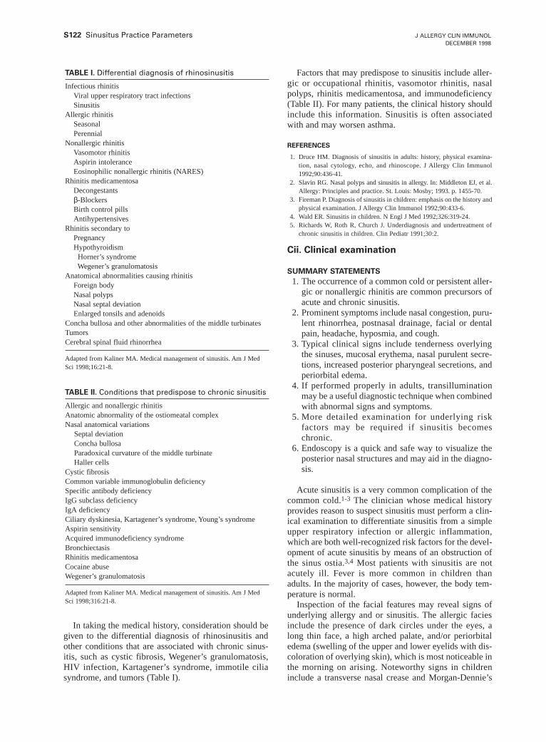
In taking the medical history, consideration should begiven to the differential diagnosis of rhinosinusitis andother conditions that are associated with chronic sinus-itis, such as cystic fibrosis, Wegener’s granulomatosis,HIV infection, Kartagener’s syndrome, immotile ciliasyndrome, and tumors (Table I).
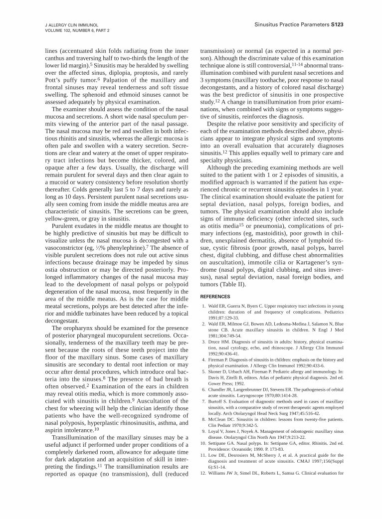
Factors that may predispose to sinusitis include aller-gic or occupational rhinitis, vasomotor rhinitis, nasalpolyps, rhinitis medicamentosa, and immunodeficiency(Table II). For many patients, the clinical history shouldinclude this information. Sinusitis is often associatedwith and may worsen asthma.
REFERENCES
1. Druce HM. Diagnosis of sinusitis in adults: history, physical examina-tion, nasal cytology, echo, and rhinoscope. J Allergy Clin Immunol1992;90:436-41.
2. Slavin RG. Nasal polyps and sinusitis in allergy. In: Middleton EJ, et al.Allergy: Principles and practice. St. Louis: Mosby; 1993. p. 1455-70.
3. Fireman P. Diagnosis of sinusitis in children: emphasis on the history andphysical examination. J Allergy Clin Immunol 1992;90:433-6.
4. Wald ER. Sinusitis in children. N Engl J Med 1992;326:319-24.5. Richards W, Roth R, Church J. Underdiagnosis and undertreatment of
chronic sinusitis in children. Clin Pediatr 1991;30:2.
Cii. Clinical examination
SUMMARY STATEMENTS
1. The occurrence of a common cold or persistent aller-gic or nonallergic rhinitis are common precursors ofacute and chronic sinusitis.
2. Prominent symptoms include nasal congestion, puru-lent rhinorrhea, postnasal drainage, facial or dentalpain, headache, hyposmia, and cough.
3. Typical clinical signs include tenderness overlyingthe sinuses, mucosal erythema, nasal purulent secre-tions, increased posterior pharyngeal secretions, andperiorbital edema.
4. If performed properly in adults, transilluminationmay be a useful diagnostic technique when combinedwith abnormal signs and symptoms.
5. More detailed examination for underlying riskfactors may be required if sinusitis becomeschronic.
6. Endoscopy is a quick and safe way to visualize theposterior nasal structures and may aid in the diagno-sis.
Acute sinusitis is a very common complication of thecommon cold.1-3 The clinician whose medical historyprovides reason to suspect sinusitis must perform a clin-ical examination to differentiate sinusitis from a simpleupper respiratory infection or allergic inflammation,which are both well-recognized risk factors for the devel-opment of acute sinusitis by means of an obstruction ofthe sinus ostia.3,4 Most patients with sinusitis are notacutely ill. Fever is more common in children thanadults. In the majority of cases, however, the body tem-perature is normal.
Inspection of the facial features may reveal signs ofunderlying allergy and or sinusitis. The allergic faciesinclude the presence of dark circles under the eyes, along thin face, a high arched palate, and/or periorbitaledema (swelling of the upper and lower eyelids with dis-coloration of overlying skin), which is most noticeable inthe morning on arising. Noteworthy signs in childreninclude a transverse nasal crease and Morgan-Dennie’s
TABLE I. Differential diagnosis of rhinosinusitis
Infectious rhinitisViral upper respiratory tract infectionsSinusitis
Allergic rhinitisSeasonalPerennial
Nonallergic rhinitisVasomotor rhinitisAspirin intoleranceEosinophilic nonallergic rhinitis (NARES)
Rhinitis medicamentosaDecongestantsβ-BlockersBirth control pillsAntihypertensives
Rhinitis secondary toPregnancyHypothyroidism
Horner’s syndromeWegener’s granulomatosis
Anatomical abnormalities causing rhinitisForeign bodyNasal polypsNasal septal deviationEnlarged tonsils and adenoids
Concha bullosa and other abnormalities of the middle turbinatesTumorsCerebral spinal fluid rhinorrhea
Adapted from Kaliner MA. Medical management of sinusitis. Am J MedSci 1998;16:21-8.
TABLE II. Conditions that predispose to chronic sinusitis
Allergic and nonallergic rhinitisAnatomic abnormality of the ostiomeatal complexNasal anatomical variations
Septal deviationConcha bullosaParadoxical curvature of the middle turbinateHaller cells
Cystic fibrosisCommon variable immunoglobulin deficiencySpecific antibody deficiencyIgG subclass deficiencyIgA deficiencyCiliary dyskinesia, Kartagener’s syndrome, Young’s syndromeAspirin sensitivityAcquired immunodeficiency syndromeBronchiectasisRhinitis medicamentosaCocaine abuseWegener’s granulomatosis
Adapted from Kaliner MA. Medical management of sinusitis. Am J MedSci 1998;316:21-8.
J ALLERGY CLIN IMMUNOLVOLUME 102, NUMBER 6, PART 2
Sinusitus Practice Parameters S123
lines (accentuated skin folds radiating from the innercanthus and traversing half to two-thirds the length of thelower lid margin).5 Sinusitis may be heralded by swellingover the affected sinus, diplopia, proptosis, and rarelyPott’s puffy tumor.6 Palpation of the maxillary andfrontal sinuses may reveal tenderness and soft tissueswelling. The sphenoid and ethmoid sinuses cannot beassessed adequately by physical examination.
The examiner should assess the condition of the nasalmucosa and secretions. A short wide nasal speculum per-mits viewing of the anterior part of the nasal passage.The nasal mucosa may be red and swollen in both infec-tious rhinitis and sinusitis, whereas the allergic mucosa isoften pale and swollen with a watery secretion. Secre-tions are clear and watery at the onset of upper respirato-ry tract infections but become thicker, colored, andopaque after a few days. Usually, the discharge willremain purulent for several days and then clear again toa mucoid or watery consistency before resolution shortlythereafter. Colds generally last 5 to 7 days and rarely aslong as 10 days. Persistent purulent nasal secretions usu-ally seen coming from inside the middle meatus area arecharacteristic of sinusitis. The secretions can be green,yellow-green, or gray in sinusitis.
Purulent exudates in the middle meatus are thought tobe highly predictive of sinusitis but may be difficult tovisualize unless the nasal mucosa is decongested with avasoconstrictor (eg, 1⁄2% phenylephrine).7 The absence ofvisible purulent secretions does not rule out active sinusinfections because drainage may be impeded by sinusostia obstruction or may be directed posteriorly. Pro-longed inflammatory changes of the nasal mucosa maylead to the development of nasal polyps or polypoiddegeneration of the nasal mucosa, most frequently in thearea of the middle meatus. As is the case for middlemeatal secretions, polyps are best detected after the infe-rior and middle turbinates have been reduced by a topicaldecongestant.
The oropharynx should be examined for the presenceof posterior pharyngeal mucopurulent secretions. Occa-sionally, tenderness of the maxillary teeth may be pre-sent because the roots of these teeth project into thefloor of the maxillary sinus. Some cases of maxillarysinusitis are secondary to dental root infection or mayoccur after dental procedures, which introduce oral bac-teria into the sinuses.8 The presence of bad breath isoften observed.2 Examination of the ears in childrenmay reveal otitis media, which is more commonly asso-ciated with sinusitis in children.9 Auscultation of thechest for wheezing will help the clinician identify thosepatients who have the well-recognized syndrome ofnasal polyposis, hyperplastic rhinosinusitis, asthma, andaspirin intolerance.10
Transillumination of the maxillary sinuses may be auseful adjunct if performed under proper conditions of acompletely darkened room, allowance for adequate timefor dark adaptation and an acquisition of skill in inter-preting the findings.11 The transillumination results arereported as opaque (no transmission), dull (reduced
transmission) or normal (as expected in a normal per-son). Although the discriminate value of this examinationtechnique alone is still controversial,11-14 abnormal trans-illumination combined with purulent nasal secretions and3 symptoms (maxillary toothache, poor response to nasaldecongestants, and a history of colored nasal discharge)was the best predictor of sinusitis in one prospectivestudy.12 A change in transillumination from prior exami-nations, when combined with signs or symptoms sugges-tive of sinusitis, reinforces the diagnosis.
Despite the relative poor sensitivity and specificity ofeach of the examination methods described above, physi-cians appear to integrate physical signs and symptomsinto an overall evaluation that accurately diagnosessinusitis.12 This applies equally well to primary care andspecialty physicians.
Although the preceding examining methods are wellsuited to the patient with 1 or 2 episodes of sinusitis, amodified approach is warranted if the patient has expe-rienced chronic or recurrent sinusitis episodes in 1 year.The clinical examination should evaluate the patient forseptal deviation, nasal polyps, foreign bodies, andtumors. The physical examination should also includesigns of immune deficiency (other infected sites, suchas otitis media15 or pneumonia), complications of pri-mary infections (eg, mastoiditis), poor growth in chil-dren, unexplained dermatitis, absence of lymphoid tis-sue, cystic fibrosis (poor growth, nasal polyps, barrelchest, digital clubbing, and diffuse chest abnormalitieson auscultation), immotile cilia or Kartagener’s syn-drome (nasal polyps, digital clubbing, and situs inver-sus), nasal septal deviation, nasal foreign bodies, andtumors (Table II).
REFERENCES
1. Wald ER, Guerra N, Byers C. Upper respiratory tract infections in youngchildren: duration of and frequency of complications. Pediatrics1991;87:129-33.
2. Wald ER, Milmoe GJ, Bowen AD, Ledesma-Medina J, Salamon N, Bluestone CB. Acute maxillary sinusitis in children. N Engl J Med1981;304:749-54.
3. Druce HM. Diagnosis of sinusitis in adults: history, physical examina-tion, nasal cytology, echo, and rhinoscope. J Allergy Clin Immunol1992;90:436-41.
4. Fireman P. Diagnosis of sinusitis in children: emphasis on the history andphysical examination. J Allergy Clin Immunol 1992;90:433-6.
5. Skoner D, Urbach AH, Fireman P. Pediatric allergy and immunology. In:Davis H, Zitelli B, editors. Atlas of pediatric physical diagnosis. 2nd ed.Gower Press; 1992.
6. Chandler JR, Langenbrunner DJ, Stevens ER. The pathogenesis of orbitalacute sinusitis. Laryngoscope 1970;80:1414-28.
7. Burtoff S. Evaluation of diagnostic methods used in cases of maxillarysinusitis, with a comparative study of recent therapeutic agents employedlocally. Arch Otolaryngol Head Neck Surg 1947;45:516-42.
8. McClean DC. Sinusitis in children: lessons from twenty-five patients.Clin Pediatr 1970;9:342-5.
9. Loyal V, Jones J, Noyek A. Management of odontogenic maxillary sinusdisease. Otolaryngol Clin North Am 1947;9:213-22.
10. Settipane GA. Nasal polyps. In: Settipane GA, editor. Rhinitis. 2nd ed.Providence: Oceanside; 1990. P. 173-83.
11. Low DE, Desrosiers M, McSherry J, et al. A practical guide for thediagnosis and treatment of acute sinusitis. CMAJ 1997;156(Suppl6):S1-14.
12. Williams JW Jr, Simel DL, Roberts L, Samsa G. Clinical evaluation for

S124 Sinusitus Practice Parameters J ALLERGY CLIN IMMUNOLDECEMBER 1998
sinusitis: making the diagnosis by history and physical examination. AnnIntern Med 1992;117:705-10.
13. Williams JW Jr, Simel DL. Does this patient have sinusitis? JAMA1993;270:1242-6.
14. Spector SL, Lotan A, English G, Philpot I. Comparison between transil-lumination and x-ray in diagnosing paranasal sinus disease. J AllergyClin Immunol 1981;67:22-6.
15. Haddad J, Brager R, Bluestone CD. Infections of the ears, nose and throatin children with primary immunodeficiencies. Arch Otolaryngol HeadNeck Surg 1992;118:138-41.
D. IMAGING STUDIES IN THE EVALUATION
OF SINUSITIS
SUMMARY STATEMENTS
1. Imaging studies may be required when the symptomsare vague, physical findings are equivocal, or there ispoor response to the initial management.
2. Standard radiographs may be used for detection ofacute sinus disease, but they are insensitive, especial-ly in ethmoid disease.
3. Computed tomography is the preferred imaging tech-nique for preoperative evaluation of the nose andparanasal sinuses secondary to obstruction of theostiomeatal complex.
4. Although magnetic resonance imaging has limita-tions in the definition of the bony anatomy, it is par-ticularly sensitive for evaluation of the frontal, max-illary, and sphenoid sinuses for fungal sinusitis andtumors and the differential diagnosis between inflam-matory diseases and malignant tumors.
5. Ultrasonography has limited utility but may beapplicable in pregnant women and for determiningthe amount of retained secretions.
Although sinusitis is a common medical problem,definitive diagnosis by history and physical examinationalone is often difficult. The greatest clinical challenge isto document the presence and extent of disease within theparanasal sinuses and to distinguish between sinusitisand rhinitis. Percussion, transillumination, and anteriorrhinoscopy often are of limited diagnostic value.1
Although sinus ultrasonography has occasionally beenused, roentgenographic examination has been the prima-ry diagnostic procedure used to confirm a clinicalimpression of sinusitis or document disease in the majorsinus cavities.2,3 However, computed tomography (CT)and magnetic resonance imaging (MRI) are becomingaccepted as the standard imaging techniques for defini-tive diagnostic evaluation of patients with both acute andchronic sinusitis. The clinical utility, relative safety, easeof performance, availability, and cost of these imagingtechniques vary considerably, and all have inherent limi-tations.
The indications for obtaining imaging confirmationare in most cases self-evident. Most often, these tech-niques are required when the symptoms are vague, phys-ical findings are equivocal, or there is poor response tothe initial courses of management. In a situation inwhich multiple antibiotic hypersensitivity exists and the
risk of reaction to a new antibiotic is greater than nor-mal, it is very important to document that sinusitis existsbefore treatment. Persistent refractory conjunctivitis,especially in children, may suggest the possibility ofunderlying chronic sinus disease, which needs to be con-firmed by imaging. Acute, severe eye pain or occipitalpain may occur as presenting symptoms, and imagingunder these conditions is essential to rule out frontaland/or sphenoid sinusitis. A patient with severe, chronicunderlying asthma may be suspected of having chronicsinus disease, which further aggravates the lowerinflammatory disease. Finally, in cases of chronic andunremitting sinus disease, unusual diseases, such asallergic sinus mycosis and Wegener’s granulomatosis,may be suggested by the newer, more sophisticatedimaging techniques.
Imaging techniques
UltrasonographyA-mode ultrasound is a safe, quick, noninvasive tech-
nique that has been proposed as a diagnostic screeningtool for sinus pathology.4,5
The rationale for use of A-mode ultrasound in theevaluation of sinus disease is based on the principle thatultrasound waves are reflected at the boundary of 2media with differing acoustic characteristics. A spike orecho is observed when the sound is reflected. Soundwaves are readily transmitted in fluid but are reflected byair. If the sinus contains fluid, an echo is expected fromthe bony back wall of the sinus, but if the sinus is nor-mally aerated, the sound will be reflected rather thantransmitted, and only the bony echo from the anteriorwall will be observed.4,5
In general, the usefulness of this method comparedwith routine radiography is very limited because ofinsensitivity.6-8
It was hoped that ultrasound would provide reliablediagnostic results that could facilitate early diagnosis inthe clinic or office setting at reduced cost and radiationexposure to the patient.8-12 It may have some utility as adiagnostic screen in pregnant women to avoid ionizingradiation.8 Additionally, because of the benign nature andease of performance of the examination, it was hopedthat ultrasound would have potential as a screeningmodality for easier detection of sinus disease. However,ultrasonography can be used to evaluate only the maxil-lary and frontal sinuses. In 1 comparative study betweenradiography and ultrasonography, it was concluded thatultrasonography better measured retained secretions(excluding very small amounts), whereas radiographymore accurately detected mucosal swelling and thicken-ing.13,14
In another study comparing ultrasound with radiogra-phy in 30 children and adults, the false-positive rate was,respectively, 39% to 45% and the false-negative rate was42% to 56%. The authors concluded that ultrasound isnot sufficiently accurate to be used as a substitute forradiography in diagnosing sinusitis.6
J ALLERGY CLIN IMMUNOLVOLUME 102, NUMBER 6, PART 2
Sinusitus Practice Parameters S125
Radiologic evaluation
The primary goals of radiologic imaging of theparanasal sinuses are to provide an accurate image of theregional anatomy and to establish the presence and extentof sinus disease. This information may assist in planningand monitoring medical therapy and provide an anatom-ic guide to facilitate surgical treatment.15
Radiography (X-ray)Standard radiographs are still the most frequently used
radiologic imaging modality for evaluating sinus disease.The Caldwell (anterior-posterior) and Waters views bestdemonstrate the frontal and maxillary sinuses. The later-al view is the best choice for visualization of the sphe-noid sinus and adenoidal tissue in children. Substitutinga single Waters view for a 4-view sinus series is anacceptable screening strategy for diagnosing maxillarysinusitis.16 The fine bony anatomy of the ethmoid sinus-es is not adequately displayed on any of the viewsbecause of the problem of structural superimposition.
Interpretation of standard radiographs may be contro-versial. Opacification, moderate-to-severe mucosal thick-ening, polyps, or air fluid levels in patients with persistentsymptoms are generally considered indicative of sinusitis.Some authors claim that any opacification is abnormaland that the only normal sinus is a clear sinus, but othersstress that sinus opacification is commonly observed inpatients with acute upper respiratory infections. Addition-ally, overlay of anatomic structures may mimic mucosalthickening or air-fluid levels, and a hypoplastic sinus maybe misinterpreted as pathologic opacification. Otherabnormalities may be related to residual postoperativechanges.
Although many consider standard sinus radiographsuseful in the diagnosis and monitoring of acute sinusitis,they are of limited value in the evaluation of chronicunremittant sinusitis. Standard radiographs are inade-quate for determination of the need for, or guidance of,endoscopic sinus surgery in both children and adults.17
Lateral views are of limited value in children less than 3years of age and tend to overestimate sphenoidal cloud-ing in children 3 to 5 years of age.17
Computed tomographyCT is currently the modality of choice in the preopera-
tive evaluation of the nose and paranasal sinuses and is thegold standard for precise delineation of inflammatorysinus disease secondary to obstruction of the ostiomeatalcomplex.18 High resolution CT can demonstrate diseasethat is not shown on routine x-ray films.17,19,20 It can alsodelineate pathologic variations and demonstrate anatom-ic structures inaccessible by physical examination orendoscopy. Because of its axial plane of view and superbcontrast resolution, CT is the method of choice for exam-ining the complex anatomy of the sinuses. If intravenouscontrast enhancement is used, CT can reliably distin-guish mucosal thickening from fluid in the maxillaryantrum. The coronal plane most closely correlates withthe surgical approach and best shows both the
ostiomeatal channels and the relationship between brain,fovea ethmoidalis, and ethmoid sinuses. Claustrophobiaand limitation of neck movements, particularly inpatients over the age of 60, are the most common factorsleading to suboptimal scans.21
In routine CT studies, the radiation dose to the patientis not negligible.8,18 Although low-dose CT increasesimage graininess, it does not induce many errors of inter-pretation, except that the thickness and integrity of theethmoid septa are sometimes more difficult to evaluate incases of extensive sinus disease.18 Thus low-dose CTmay be useful in the evaluation of inflammatory diseaseof the sinuses, complemented by 1 or 2 sections obtainedwith standard dose settings, focused on questionablyabnormal bone septa.18
Standard radiographs are frequently ordered in prefer-ence to CT because of reduced cost.22 However, an aver-age 4-slice, limited-cut, coronal-plane CT of the sinusestakes 20 minutes to perform at a cost only slightly greaterthan that of a standard set of radiographs.22 It provideshigh resolution bone and soft tissue imaging of the keysurgical areas.
Many otolaryngologists consider high-resolution CTto be a mandatory part of the chronic rhinosinusitis pre-operative assessment. The planning and safety of func-tional endoscopic sinus surgery is greatly improved byCT imaging.22,23 CT can provide excellent definition ofthe paranasal sinuses, particularly the ethmoids, which isa prerequisite for endoscopic surgery.23
CT may be especially helpful in the diagnosis of fun-gal sinusitis because it delineates unilateral lesions of 1or more sinuses, nodular mucoperiosteal thickening,focal areas of bone destruction, and very dense intrasinusconcretions.15,24 In most cases a combination of thesefindings suggests fungal disease.24
MRIMRI provides better imaging of soft tissue than CT,
but it is less suited to imaging the bony anatomy of thisregion. Because bone and air yield similar signal intensi-ties on MRI, precise definition of the ostiomeatal air pas-sages and their bony perimeters is more difficult by thistechnique. Furthermore, in the patient with extensiveinflammatory disease, the signal intensity of the patho-logic process is indistinguishable from the appearance ofthe normal mucosa in the edematous phase of the nasalcycle. These factors limit the MRI evaluation of underly-ing anatomy in a patient with chronic inflammatory dis-ease, especially because the mucosa in the ethmoid sinus“cycles” with the mucosa in the nasal cavity. AlthoughMRI has significant limitations in the definition of anato-my, it is extremely sensitive in evaluation of paranasalsinus mucosal disease in the frontal, maxillary, and sphe-noid sinuses because the mucosa in these sinuses doesnot undergo the cyclic edema.25
The etiology of certain disease processes may be bet-ter differentiated by MRI. MRI often aids in the differen-tial diagnosis between inflammatory diseases and malig-nant tumors.26,27 MRI is useful for cases complicated by
S126 Sinusitus Practice Parameters J ALLERGY CLIN IMMUNOLDECEMBER 1998
orbital or intracranial extension. Bacterial and viralinflammation have a high signal intensity on T2-weight-ed images, whereas neoplastic processes, primarily squa-mous cell carcinoma, demonstrate an intermediate brightsignal on T2-weighted images. Fungal concretions havea very low signal intensity on T2-weighted images simi-lar to that of air.15 MRI or CT is significantly more sen-sitive than plain radiography (P < .001) in defining theextent of the disease, particularly with posterior sinusinvolvement.28 Hypointense signals may be present onMRI sequences despite CT evidence of the presence ofhigh-attenuation material filling the sinus.
CT compared with MRIBoth CT and MRI are generally considered to be supe-
rior to x-ray tests in the diagnosis of inflammatory con-ditions of the nose and paranasal sinuses. When aninflammatory condition is known to be present, MRI ismore accurate than CT, but the latter (thanks to its opti-mal delineation of both bony walls and soft tissues) issuperior to MRI in the differential diagnosis of a genericdisease of the nose and paranasal sinuses (ie, when theinflammatory nature is questionable). CT is especiallyuseful in emphasizing the ostiomeatal channels and ispreferred for studying the ethmoid and sphenoid sinus-es.21,26,29,30 On balance, CT emerges as the diagnosticgold standard of sinus disease because of its diagnosticaccuracy, easy access, and lower cost.
REFERENCES
1. Godley FA. Chronic sinusitis: an update. Am Fam Physician1992;45:2190-9.
2. Mafee MF. Imagine methods for sinusitis. JAMA 1993;269:2608.3. Fireman P. Diagnosis of sinusitis in children: emphasis on the histo-
ry and physical examination. J Allergy Clin Immunol1992;90(suppl):S433-6.
4. Kuhn JP. Imaging of the paranasal sinuses. J Allergy Clin Immunol1986;77:6-8.
5. Jannert M, Andreasson L, Honer N-G, et al. Ultrasonic examinationof the paranasal sinuses. Acta Otolryngol 1982;389(suppl):1-51.
6. Shapiro GG, Furukawa CT, Pierson WE, et al. Blinded comparison ofmaxillary sinus radiography and ultrasound for diagnosis of sinusitis.J Allergy Clin Immunol 1986;77:59-64.
7. Rohr AS, Spector SL, Siegel SC, et al. Correlation between A-modeultrasound and radiography in the diagnosis of maxillary sinusitis. JAllergy Clin Immunol 1986;78:58-61.
8. Stafford CT. The clinician’s view of sinusitis. Otolaryngol HeadNeck Surg 1990;103(suppl):870-5.
9. Druce HM. Emerging techniques in the diagnosis of sinusitis. AnnAllergy 1991;66:132-6.
10. Herr RD. Acute sinusitis: diagnosis and treatment update. Am FamPhysician 1991;44:2056-62.
11. Anderson MH, Stafford CT. Comparison of imaging techniques inthe diagnosis of sinusitis [abstract]. Ann Allergy 1991;66:73.
12. Revonta M. Ultrasound in the diagnosis of maxillary and frontalsinusitis. Acta Otolaryngol 1980;370(suppl):1-54.
13. Suonpaa J, Revonta M. Diagnosis of frontal sinusitis: one-dimen-sional ultrasonography versus radiography. J Laryangol Otol1989;103:765-7.
14. Reilly JS, Hotaling AJ, Chiponis D, Wald ER. Use of ultrasound indetection of sinus disease in children. Int J Pediatr Otorhinolaryngol1989;17:225-30.
15. Zinreich SJ. Radiologic diagnosis of the nasal cavity and paranasalsinuses. In: Druce HM, editor. Sinusitis: pathophysiology and treat-ment. New York: Marcel Dekker, Inc; 1993.
16. Williams JW Jr, Roberts L Jr, Distell B, Simel DL. Diagnosingsinusitis by x-ray: Is a single Waters view adequate? J Gen InternMed 1992;7:481-5.
17. McAllister WH, Lusk R, Muntz HR. Comparison of plain radi-ographs and coronal CT scans in infants and children with recurrentsinusitis. Am J Radiol 1989;153:1259-64.
18. Duvoisin B, Landry M, Chapuis L, Krayenbuhl M, Schnyder P. Low-dose CT and inflammatory disease of the paranasal sinuses. Neuro-radiology 1991;33:403-6.
19. Rieden K, Lellig U, Schnepper U. Radiologic diagnosis of inflam-matory and tumorous disorders of the paranasal sinuses. Laryngorhi-nootologie 1989;68:543-6.
20. Lee HS, Majima Y, Sakakura Y, Inagaki M, Sugiyama Y, Nakamoto S.Conventional x-ray versus CT in diagnosis of chronic sinusitis in chil-dren [in Japanese]. Nippon Jibinkoka Gakkai Kaiho 1991;94:1250-6.
21. Babbel RW, Harnsberger HR. A contemporary look at the imagingissues of sinusitis: sinonasal anatomy, physiology, and computedtomography techniques. Semin Ultrasound CT MR 1991;12:526-40.
22. White PS, Robinson JM, Stewart IA, Doyle T. Computerized tomog-raphy mini-series: an alternative to standard paranasal sinus radi-ographs. Aust NZ J Surg 1990;60:25-9.
23. East CA, Annis JA. Preoperative CT scanning for endoscopic sinussurgery: a rational approach. Clin Otolaryngol 1992;17:60-6.
24. Harnsberger HR, Babbel RW, Davis WL. The major obstructiveinflammatory patterns of the sinonasal region seen on screening sinuscomputed tomography. Semin Ultrasound CT MR 1991;12:541-60.
25. Zinreich SJ. Paranasal sinus imagine. Otolaryngol Head Neck Surg1990;103(suppl):870-5.
26. Oddone M, Toma P, Scotto Di Santillo L, Tarantino V. Chronicsinusitis in children. Current role of diagnostic imaging. MinervaPediatr 1992;44:17-25.
27. Zinreich SJ. Imaging of chronic sinusitis in adults: X-ray, computedtomography, and magnetic resonance imaging. J Allergy ClinImmunol 1992;90(suppl):445-51.
28. Conner BL, Phillips K, Roach ES, et al. Nuclear magnetic resonance(NMR) imaging of paranasal sinuses: frequency of abnormalities. JAllergy Clin Immunol 1986;77:139.
29. Friedman WH, Katsantonis GP, Sivore M, Kay S. Computed tomog-raphy staging of the paranasal sinuses in chronic hyperplastic rhi-nosinusitis. Laryngoscope 1990;100:1161-5.
30. Calhoun K. Diagnosis and management of sinusitis in the allergicpatient. Otolaryngol Head Neck Surg 1992;107(suppl):850-4.
E. LABORATORY TESTS
SUMMARY STATEMENTS
1. Laboratory evaluation of chronic or recurrent sinus-itis may include any of the following: nasal cytology,sweat chloride test, ciliary function studies, and testsfor immunodeficiency.
2. Nasal cytology is useful in the clinical evaluation ofunderlying allergic rhinitis, the nonallergic rhinitiswith eosinophilia syndrome, nasal polyposis, andaspirin sensitivity.
3. Quantitative sweat chloride tests for diagnosis of cys-tic fibrosis should be considered in children withnasal polyps and/or colonization of the nose andsinuses with Pseudomonas spp.
4. Tests for immunodeficiency (eg, quantitativeimmunoglobulins, antibody tests, serum IgE, andcomplement components) may be useful if eithercongenital or acquired immunodeficiency is suspect-ed in cases of recurrent sinusitis.
Other diagnostic methods may be indicated for inves-tigating the patients with chronic or recurrent sinusitis,
J ALLERGY CLIN IMMUNOLVOLUME 102, NUMBER 6, PART 2
Sinusitus Practice Parameters S127
especially if bronchitis or bronchiectasis is present. Thissection will review the laboratory evaluation of patientswith chronic sinusitis for which no obvious causal linkhas been found (eg, allergic or infectious rhinitis). Labo-ratory studies that are of major importance include thefollowing: nasal cytology, the sweat chloride test, ciliarydysfunction studies, and other miscellaneous tests thataid in the confirmation of several diseases either causingor associated with chronic sinusitis.
Nasal cytology
Nasal cytology is a tool used in the evaluation of rhini-tis. It is a useful adjunct in determining whether chronicrhinitis conditions are associated with sinusitis. However,many centers do not include cytology as a diagnostic stepin evaluating possible sinusitis, and the clinician mustdecide if this procedure is significantly helpful to warrantits performance. Several methods of collecting nasalsecretions are described. The patient can blow secretionsinto wax paper or Saran wrap, which is the most simpleand effective way of collecting a sample. The sample canalso be collected by nasal lavage with a cytology brush,cotton swab, or a Rhinoprobe (Synbiotics Corporation,San Diego, Calif). By collecting secretions from theblowing technique, less cellular material may be collect-ed.1 This technique is useful for detecting nasaleosinophils in allergic rhinitis, especially during seasonalexacerbations when the patient is symptomatic. Becausenasal lavage is a dilution process, the sample needs to beconcentrated; it is minimally invasive but reflects ananatomically wider sample of nasal material.2
Once the sample is collected, it must be stained. Aftercytofixation or heat fixation, Hansel stain, a commonlyused stain to identify eosinophils, is often used. Otherstains, such as the Papanicolaou, hematoxylin-eosin,acidified toluidine blue, or May-Grunwald-Giemsastains, can be used to identify other elements, includingmast cells, basophils, neutrophils, nonciliated epithelialcells, squamous epithelial cells, pollen grains, anddebris. Nasal cytology may be affected by topicalsteroids.
Several distinct syndromes that may be associatedwith sinusitis have been defined by nasal cytology. Theseinclude allergic rhinitis, vasomotor rhinitis, nonallergicrhinitis with eosinophils, bacterial sinusitis, and metapla-sia secondary to environmental exposures.3 Both subjec-tive and semiquantitative methods exist for assessingnasal cytology, leading to limitations in the interpretationand quantitation of results.4-6 Studies defining the sensi-tivity and specificity of nasal cytology in these diseasesare limited. Nasal smear eosinophils may be present inallergic rhinitis, the nonallergic rhinitis with eosinophilssyndrome, nasal polyposis, and aspirin sensitivity.7,8
Comparative studies of nasal cytology with global andradiographic assessments of sinusitis are limited andconflicting. In these studies the specificity of nasal neu-trophils in sinusitis ranged from 40% to 90% and sensi-tivity varied between 67% and 80%.9,10 These disparateresults are further complicated by the fact that acute viral
infections can lead to neutrophilic nasal secretions andtransient abnormal imaging studies.10 Furthermore, it ispossible for patients to have concurrent allergy and bac-terial sinusitis (ie, a mixture of eosinophils and neu-trophils). Therefore the clinical usefulness of nasal cytol-ogy in evaluating sinusitis is limited.
Sweat test
The Gibson-Cooke sweat test (quantitative pilo-carpine iontophoresis) is the gold standard for identify-ing patients with cystic fibrosis.11 Chloride valuesgreater than 60 mEq/L in children are considered diag-nostic, provided at least 75 mg of sweat is collected.Adults can have values 10 to 15 mEq/L higher thanthose of children.12 Although sodium is measured insome laboratories, it is more variable than chloride,which should always be measured for a definitive diag-nosis.13 Indications for a sweat test in patients withsinusitis include the observation of nasal polyps and/orchronic colonization of the nose and sinuses withPseudomonas spp.14-19
Ciliary tests
Congenital abnormalities of ciliary structures areoccasionally considered as possible explanations forrecurrent sinusitis. These rare disorders are autosomalrecessive and occur in 1 in 20,000 live births but may befound in as many as 5% of patients with chronic respira-tory infections arising in the first weeks of life.20,21 Mostpatients with these disorders have a triad of recurrent oti-tis, recurrent sinusitis, and frequent pneumonia associat-ed with bronchiectasis.22-25 In half the cases situs inver-sus is present (Kartagener’s syndrome) and provides thebest clue to the presence of a ciliary disorder. It isbelieved that the mechanism of increased susceptibilityto bacterial infection is loss of normal mucociliary trans-port, although some defects are also associated withdefective neutrophil chemotaxis.23
Functional tests of cilia include visual and videoscop-ic measurements of tissue and mucociliary transport,such as the rate of saccharin or disc movement tests.26,27
Visual assessments of ciliary function have been done bycollecting ciliated cells on a cytology brush and suspend-ing the material on a glass slide.28,29 The ciliary motionis then observed by simple light or phase microscopy,with a ciliary beat frequency less than 10 beats/secondconsidered below the normal range.21
The saccharin test is used as a screening test.30 It is per-formed by placing a small volume of saccharin sodium atthe bottom of the inferior nasal meatus. After 3 minutes,the subjects swallow every 30 seconds until a sweet tasteis detected.26 Values greater than 15 to 30 minutes areconsidered abnormal.31 Definitive confirmation of ciliaryabnormalities requires electron microscopy.32-34
Among the criteria for ultrastructural analysis are thetype of defect (dynein defects being the most specific),the percent of cilia having the defect, the distribution ofthe defect (bronchial versus nasal), and the number ofcilia present.32-35
S128 Sinusitus Practice Parameters J ALLERGY CLIN IMMUNOLDECEMBER 1998
Miscellaneous tests in the differential diagnosis of chron-
ic sinusitis
Patients with immunodeficient states, both congenitaland acquired, are often first seen with severe, recurrentsinusitis. In such situations it is appropriate to evaluatequantitative immunoglobulin isotypes, including IgE(see section on congenital or acquired immunodeficien-cy). In rare instances total complement and complementcomponents may be obtained. The diagnosis of acquiredimmunodeficiency is established by HIV antibody and/orantigen tests.
In suspected cases of Wegener’s granulomatosis, anantineutrophil cytoplasmic antibody test may be a diag-nostic adjunct to tissue biopsy.36 Similarly, elevatedangiotensin-converting enzyme and soluble IL-2 receptorlevels may aid in the diagnosis of active sarcoid lesionsin nasal or sinus locations.37
The suspicion of sinus allergic mycoses may beenhanced by high serum IgE levels and the presence ofprecipitating IgG and/or specific IgE antibodies to avariety of saprophytic fungi, which have been culturedin this disease.
REFERENCES
1. Lara Becerra A, Gonzalez Diaz SN, Gonzalez Morales JE, Canseco Gonza-lez C. Determination of the eosinophil count in nasal mucus. Comparison oftwo techniques. Rev Alerg Mex 1990;37:123-6.
2. Kohler C, Stringini R,Moneret DA, Grignon G. Study of cells harvested innasal secretions after lavage. Improvement of the cytologic technique andapplication to ORL and bronchial pathology. Bull Assoc Anat 1992;76:43-6.
3. Calderon-Garciduenas L, Roy-Ocotla G. Nasal cytology in southwest met-ropolitan Mexico City inhabitants: a pilot intervention study. Environ HealthPerspect 1993;101:138-44.
4. Orgel HA, Meltzer EO, Kemp JP, Ostrom NK, Welch MJ. Comparison ofintranasal cromolyn sodium, 4%, and oral terfenadine for allergic rhinitis:symptoms, nasal cytology, nasal ciliary clearance, and rhinomanometry.Ann Allergy 1991;66:237-44.
5. Rivasi F, Bergamini G. Nasal cytology in allergic processes and other syn-dromes caused by hyperreactivity. Diagn Cytopathol 1988;4:99-105.
6. Lans DM, Alfano N, Rocklin R. Nasal eosinophilia in allergic and nonal-lergic rhinitis: usefulness of the nasal smear in the diagnosis of allergicrhinitis. Allergy Proc 1989;10:275-80.
7. Davidson AE, Miller SD, Settipane RJ, Ricci AR, Klein DE, Settipane GA.Delayed nasal mucociliary clearance in patients with nonallergic rhinitis andnasal eosinophilia. Allergy Proc 1992;13:81-4.
8. Cohen GA, MacPherson GA, Golembesky HE, Jalowayski AA, O’ConnorRD. Normal nasal cytology in infancy. Ann Allergy 1985;54:112-4.
9. Wilson NW, Jalowayski AA, Hamburger RN. A comparison of nasal cytologywith sinus x-rays for the diagnosis of sinusitis. Am J Rhinol 1988;2:55-9.
10. Gill FF, Neiburger JB. The role of nasal cytology in the diagnosis of chron-ic sinusitis. Am J Rhinol 1989;3:13-5.
11. Gibson LE, Cooke RE. Test for concentration of electrolytes in sweat in cys-tic fibrosis of the pancreas utilizing pilocarpine by iontophoresis. Pediatrics1959;23:45-9.
12. Hall SK, Stableforth DE, Green A. Sweat sodium and chloride concentra-tions—essential criteria for the diagnosis of cystic fibrosis in adults. AnnClin Biochem 1990;27:318-20.
13. Gleeson M, Henry RL. Sweat sodium or chloride? Clin Chem 1991;37:112.14. Ramsey B, Richardson MA. Impact of sinusitis in cystic fibrosis. J Allergy
Clin Immunol 1992;90:547-52.15. Hammond KB, Turcios NL, Gibson LE. Clinical evaluation of the macrod-
uct sweat collection system and conductivity analyzer in the diagnosis ofcystic fibrosis. J Pediatr 1994;255:60.
16. Yeung WH, Palmer J, Schidlow D, Bye MR, Huang NN. Evaluation of apaper-patch test for sweat chloride determination. Clin Pediatrics1984;23:603-7.
17. Warwick WJ, Hansen LG, Werness ME. Quantification of chloride in sweatwith the cystic fibrosis indicator system. Clin Chem 1990;36:96-8.
18. Simmonds E, Alfaham M, Prosser R, Penney MD. Fractional measurementsof sweat osmolality in patients with cystic fibrosis. Arch Dis Child1989;64:1717-20.
19. Rosenstein B, Laugbaum T. Sweat testing in CF: not to be taken lightly. JRespir Dis 1982;3:71-6.
20. Afzelius BA. Disorders of ciliary motility. Hosp Pract 1986;21:73-80.21. Buchdahl RM, Reiser J, Ingram D, Rutman A, Cole PJ, Warner JO. Ciliary
abnormalities in respiratory disease. Arch Dis Child 1988;63:238-43.22. Turner JAP, Corkey CWB, Lee JYC, Levison H, Sturgess J. Clinical expres-
sions of immotile cilia syndrome. Pediatrics 1981;67:805-10.23. Afzelius BA, Ewetz L, Palmblad J, Uden AM, Venizelos N. Structure and
function of neutrophil leukocytes from patients with the immotile-cilia syn-drome. Acta Med Scand 1980;208:145-54.
24. Carson JL, Collier AM, Hu SCS. Acquired ciliary defects in nasal epitheli-um of children with acute viral upper respiratory infections. N Engl J Med1985;312:463-8.
25. Phillips JL. Lack of cilia and squamous metaplasia in upper respiratory tractbiopsies from children. S Afr Med J 1989;76:355-7.
26. Duchateau GS, Graamans K, Zuidema J, Mekus FW. Correlation betweennasal ciliary beat frequency and mucus transport rate in volunteers. Laryn-goscope 1985;95:854-9.
27. Proctor DF. The nose, upper airway physiology and the atmospheric envi-ronment. Amsterdam: Elsevier Biomedical Press; 1982.
28. Boat TF, Wood RE, Tandler B, Stern RC, Orenstein DM, Doershuk CF. Ascreening test for the immotile cilia syndrome. Cleveland (OH): Departmentof Pediatrics and School of Dentistry, Case Western Reserve University.
29. Robson AM, Smallman LA, Drake-Lee AB. Factors affecting ciliary func-tion in vitro: a preliminary study. Clin Otolaryngol 1992;17:125-9.
30. Stanley P, MacWilliam L, Greenstone M, Mackay I, Cole P. Efficacy of asaccharin test for screening to detect abnormal mucociliary clearance. Br JDis Chest 1984;78:62-5.
31. Greenstone M, Cole PJ. Ciliary function in health and disease. Br J DisChest 1985;79:9-26.
32. Verra F, Fleury-Feith J, Boucherat M, Pinchon MC, Bignon J, Escudier E.Do nasal ciliary changes reflect bronchial changes? Am Rev Respir Dis1993;147:908-13.
33. Rautiinen M, Kiukaanniemi H, Nuutinen J, Collan Y. Ultrastructuralchanges in human nasal cilia caused by the common cold and recovery ofciliated epithelium. Ann Otol Rhinol Laryngol 1992;101:982-7.
34. Robson AM, Smallman LA, Gregory J, Drake-Lee AB. Ciliary ultrastruc-ture in nasal brushings. Cytopathology 1993;4:149-59.
35. Stockinger L, Sellner W, Ellinger A, Hofler H. Pathophysiology of the cili-ated epithelium of the respiratory mucosa in humans. Disorders of ciliogen-esis. Exp Lung Res 1989;15:925-41.
36. Rao JK, Weinberger EM, Oddone EZ, et al. The role of anti-neutrophil cyto-plasmic antibody (c-ANCA) testing in the diagnosis of Wegener’s granulo-matosis. Ann Intern Med 1995;123:925-32.
37. Keicho N, Kitamura K, Takaku F,Yotsumoto H. Serum concentration of sol-uble interleukin-2 receptor as a sensitive parameter of disease activity in sar-coidosis. Chest 1990;98:1125-9.
F. RHINOLARYNGOSCOPY
SUMMARY STATEMENTS
1. Fiberoptic rhinoscopy permits more detailed exami-nation of the anterior and posterior nasal and pharyn-geal structures.
2. Fiberoptic rhinoscopy may be considered for somepatients with chronic or recurrent sinusitis to assessstructural abnormalities, fungal disease, or granulo-matous lesions.
In some situations in which sinus disease is suspected,and the routine anterior nasal speculum examination hashad limited diagnostic value, endoscopy of the upper air-way should be considered to permit more detailed exam-
J ALLERGY CLIN IMMUNOLVOLUME 102, NUMBER 6, PART 2
Sinusitus Practice Parameters S129
ination of the anterior and posterior nasopharyngealrecessed structures, such as sinus ostia, sphenoethmoidalrecess, and eustachian tube ostia. Direct vision of theepiglottis, glottis, and vocal cords is an additional advan-tage of this technique.1,2
Instruments
Two types of endoscopes are available. The opticallyexcellent Hopkins rigid nasal endoscope permits exten-sive examination of the nasal cavities and portions of thenasopharynx, as well as high-quality photography of thestructures examined.3 Fiberoptic rhinoscopy is a conve-nient, cost-effective, and safe alternative. The fiberopticrhinoscope permits quick and thorough examination ofmost areas of the upper airway without discomfort to thepatient. Abnormalities of the septum, turbinates, mucosa,nasopharynx, adenoids, eustachian tube orifice, tonsils,posterior tongue, epiglottis, glottis, and vocal cords caneasily be seen.4 The origin and course of nasal polyps canbe identified, and the effect of treatment can be followed.
Indications for endoscopic evaluation for sinus disease
Fiberoptic rhinoscopy may be considered for somepatients with acute, chronic, or recurrent sinusitis. Thistechnique may reveal the presence or extent of nasalpolyps, septal deviation, and/or mucopurulent secretions.
REFERENCES
1. Messerklinger W. Endoscopy of the nose. Baltimore-Munich: Urban &Schwartzenber; 1978.
2. Lancer JM, Jones AS. Flexible fiberoptic rhinolaryngoscope: results of338 consecutive examinations. J Laryngol Otol 1985;99:771.
3. Kennedy DW, Zinreich J, Rosenbaum AE. Functional endoscopic sinussurgery. Arch Otolaryngol 1985;111:576-82.
4. Grant JCB. Grant’s atlas of anatomy. 6th ed. Baltimore: Williams andWilkins; 1972.
G. BIOPSY OF THE NOSE AND PARANASAL
SINUSES
SUMMARY STATEMENTS
1. Paranasal sinus biopsies may be required to deter-mine whether a lesion is neoplastic or to confirm thepresence of suspected fungal disease and the possi-bility of granulomatous disease.
2. Nasal mucosa harvested from the posterior portion ofthe inferior turbinate is the preferred biopsy site forprimary cilia dysfunction.
Tissue biopsy may be considered for the differentialdiagnosis of nasal or paranasal lesions that obstruct sinuscavities and give rise to chronic, recurrent sinus disease.Thus cardinal reasons for obtaining paranasal sinus biop-sies are (1) to determine whether the lesion is neoplasticand if so, what kind of neoplasm it is; (2) to confirm thepresence of suspected fungal disease; and (3) to establisha diagnosis when granulomatous disease cannot be dif-ferentiated clinically or radiographically from otherforms of chronic inflammatory rhinosinusitis. Tissue
biopsy specimens are also required for evaluating ciliaryfunction and structure.
Nasal and paranasal sinus neoplasms
Because the sinuses are not accessible for direct physi-cal examination, and because early symptoms of sinustumors, such as nasal obstruction, anosmia, rhinorrhea,and pain are also common symptoms of inflammatorysinusitis, most sinus neoplasms are not diagnosed at anearly stage. This is probably a major factor in the relative-ly poor prognosis of malignant sinus neoplasms. Often,clinical suspicion of tumor is not raised until epistaxis,proptosis, trismus, facial swelling or cranial nerve (I/VI)dysfunction develop in a patient, most of which are indi-cations of extensive tumor involvement. Early diagnosis ofnasal and sinus neoplasm is enhanced by use of computedtomography and/or magnetic resonance imaging.
Tumors originating in the nose or sinuses cover a widerange of histologic types, both malignant and benign. Usu-ally it is not possible to reliably predict tumor histologictype on the basis of clinical or radiographic findings, andthus biopsy is performed before definitive therapy, whichwill then be based on tumor histology.1 An exception tothis is juvenile angiofibroma. Diagnosis of this lesion ismade by clinical and radiologic findings of a vascular pos-terior nasal or nasopharyngeal mass in an adolescent orpreadolescent male, and biopsy should not be performedbecause of the risk of significant hemorrhage.2
Invasive fungal infections
Rhinocerebral mucormycosis is a potentially fatalinvasive fungal infection produced by Phycomycetes,which includes the genera Absidia, Mucor, and Rhizopus.A similar but somewhat less fulminating infection can becaused by Aspergillus spp. Although invasive fungal dis-ease has been reported in otherwise normal patients, itremains predominantly a disease of immunocompro-mised patients. Mucormycosis occurs most commonly inpatients with diabetes mellitus, hematologic malignan-cies, adrenal suppression, chronic renal failure, hemato-logic dyscrasia, and other immunologically compro-mised conditions. Patients undergoing chemotherapy arealso at increased risk as are intravenous drug users andHIV-positive patients.
Because invasive fungal infections may progress veryrapidly, it is necessary to maintain a high degree of suspi-cion when dealing with this group of patients. The infec-tion spreads rapidly along blood vessels, producing necro-sis. Patients are often first seen with facial pain, proptosis,ophthalmoplegia, or facial necrosis. Late radiographicfindings may include sinus opacification, mucoperiostealthickening, bone erosion, and cavernous sinus thrombosis,but these findings may be absent or minimal early in thecourse of the disease. On endoscopic visualization, earlylesions may appear as brick red areas on the nasal mucosa,but more typically they appear as black, necrotic areas.Tissue necrosis often renders the involved area anesthetic.Abnormal-appearing lesions should undergo biopsypromptly, and the specimen should be sent for fungal
S130 Sinusitus Practice Parameters J ALLERGY CLIN IMMUNOLDECEMBER 1998
stains and fungal culture. Early diagnosis is mandatory ifthese patients are to be salvaged.3-5
Granulaomatous inflammatory disease
A number of chronic granulomatous inflammatorydiseases, both idiopathic and infectious, involve the noseand paranasal sinuses. These conditions may cause ulcer-ation, necrosis, or hyperplastic mucosa within the nasalcavity or paranasal sinuses. Involvement may be patchyor diffuse and is often difficult to differentiate fromchronic nongranulomatous inflammatory conditions. Thegranulomatosis diseases include idiopathic processes,such as Wegener’s granulomatosis and sarcoidosis, aswell as infectious processes, such as rhinoscleroma,tuberculosis, leprosy, syphilis, and fungal infections.Biopsy in combination with clinical, serologic, and radi-ographic findings is usually necessary for diagnosis.Many of the organisms that cause chronic granulomatousinflammation are difficult to detect without special stain-ing. Tissue should be submitted for histologic examina-tion to rule out neoplasia and for special staining and cul-ture of mycobacteria and fungi.5,6
REFERENCES
1. Osguthorpe JD. Sinus neoplasia. Arch Otolaryngol Head Neck Surg1994;120:19-25.
2. Economou TS, Abemayor E, Ward PH. Juvenile nasopharyngeal angiofi-broma: An update of the UCLA Experience. Laryngoscope 1988;98:170-5.
3. Blitzer A, Lawson W, Meyers BR, Biller HF. Patient survival factors inparanasal sinus mucormycosis. Laryngoscope 1980;90:635-48.
4. Blitzer A, Lawson W. Fungal infections of the nose and paranasal sinus-es–Part I. Otolaryngol Clin North Am 1993;26:1007-35.
5. Kopke RD, Jackson RL. Rhinitis. In: Bailey BJ, editor. Head and necksurgery–otolaryngology. Philadelphia: JB Lippincott; 1993. p. 269-89.
6. McDonald TJ. Granulomatous diseases of the nose. In: English GM, edi-tor. Otolaryngology. 2nd ed. Vol 2. Philadelphia: JB Lippincott; 1992.
H. CONGENITAL OR ACQUIRED IMMUNODE-
FICIENCY
SUMMARY STATEMENTS
1. The majority of patients with congenital immunedeficiency and associated sinusitis will have defectsin humoral immunity. However, sinusitis is commonin acquired immune deficiency (AIDS), in whichboth humoral and cellular impairments are present.Appropriate laboratory studies of both humoral andcellular immunity include quantitative immunoglob-ulin assays; specific antibody responses to tetanustoxoid, pneumococcal, or Hemophilus influenzaevaccines; and measurement of complement compo-nents and key cellular responses.
2. The most common congenital immunodeficiencysyndromes in this group are common-variableimmunodeficiency, IgA deficiency Wiskott-Aldrich,ataxia telangiectasia, X-linked immunodeficiencywith normal or elevated IgM, and X-linked agamma-globulinemia.
The possibility of immunodeficiency should be con-sidered in patients with chronic or recurrent sinusitis,
especially when associated with bronchitis or bronchiec-tasis. Evaluation of immunodeficiency begins with acareful history and physical examination. The physicalexamination of patients with suspected congenitalimmunodeficiency should focus on findings associatedwith specific diseases, such as absence of tonsillar tissue,ocular telangiectasia, skin and mucous membrane infec-tions, eczema, clubbing, rales rhondii, and purpura.1-3
The majority of patients with congenital immunodefi-ciency with sinusitis will have defects in humoral immu-nity4-6; however, sinusitis is common in acquiredimmune deficiency syndrome (AIDS), in which bothhumoral and cellular impairments are present.
The indications for pursuing an immunodeficiencyevaluation depend on the age, medical history, physicalexam, and lifestyle of the patient. For example, in aninfant of less than 2 years of age with recurrent and life-threatening infections of the sinuses and other organs,one should pursue this evaluation in a expeditious man-ner. In addition, infections with organisms of low patho-genicity should alert the physician to the probability of acongenital immune deficiency. Similar indications forevaluation of immune deficiency exist for AIDS in bothchildren and adults.
Appropriate laboratory studies of both humoral andcellular immunity include quantitative immunoglobulinassays, specific antibody responses, complement compo-nents, and measurements of T-cell responses. In principlethe response to any protein antigen can be measured, butin practice measurement of the response to tetanusimmunization offers several advantages. Most patientshave been immunized with tetanus antigen, and 90% to100% of children will have protective antibody titers oncompletion of primary immunization.7 Tetanus toxoid isa potent antigen, and a poor response to immunizationusually indicates a significant defect in humoral immuni-ty.8 Pneumovax or Haemophilus influenzae B may alsobe used to measure antibody production.
Patients have also been described with recurrent sinopul-monary infections and normal total IgE levels but nonde-tectable IgG2 and IgG4 in their serum.9,10-12 In general, thebulk of antibody response to carbohydrate antigens is of theIgG2 subclass.13,14 This is especially true of the antibodydirected against Haemophilus influenzae, a common respi-ratory tract pathogen.15 In general, the antibody response tocarbohydrate antigens correlates with IgG2 levels.16 Inolder patients symptomatic IgG subclass deficiency maycorrelate with defects in specific antibody response, espe-cially to polysaccharide antigens.17 Antibody responses topneumococcal antigens may be useful, but the response ofchildren less than 8 years old may not be complete.10 Poorantibody responses to pneumococcal serotypes 3 and 7appear to correlate most closely with defects in humoralimmunity.8,18-21
In addition to the immune defects discussed above,chronic or recurrent sinus disease is a common feature ofseveral other immunodeficiency diseases, which mayrequire additional diagnostic studies. The most commoncongenital immunodeficiency
J ALLERGY CLIN IMMUNOLVOLUME 102, NUMBER 6, PART 2
Sinusitus Practice Parameters S131
Syndromes in this group are common-variable immu-nodeficiency, Wiskott-Aldrich,22,23 ataxia telangiecta-sia,2,3,24-26 x-linked immunodeficiency with normal orelevated IgM,1,27-31 and X-linked agammaglobuline-mia.1,32-35
The incidence of sinusitis in HIV infection varies from30% to 68%.36-39 There may be a direct relationshipbetween CD4 deficiency and sinusitis.36,40 Although hyper-gammaglobulinemia is a frequent occurrence in HIV infec-tion, specific antibody responses are abnormal.41-43 Themicrobiology of sinusitis in patients with HIV is similar tothat seen in other patients with recurrent and chronic sinus-itis. However, pathogens, such as Pseudomonas spp,Staphylococcus aureus, Klebsiella spp, Escherichia coli,Candida spp, Cryptococcus spp, and Cytomegalovirus spphave been isolated.40-43 The treatment of sinusitis in thesepatients is similar to that seen in nonimmunized patientsbut with a longer duration of therapy.
REFERENCES
1. Rosen FS, Cooper M, Wedgwood R. The primary immunodeficiencies,Part 1. N Engl J Med 1984;311:235-22.
2. Boder E, Sedgwick RP. Ataxia telangiectasia and frequent pulmonaryinfections. Pediatrics 1958;21:526-54.
3. Gatti A, Swift M. Ataxia-telangiectasia: genetics, neuropathology, andimmunology of a degenerative disease of childhood. New York: Alan R.Liss, Inc; 1985.
4. Polmar S. The role of the immunologist in sinus disease. J Allergy ClinImmunol 1992;90:511-4.
5. Barret CD, Timm EA, Molner GF, et al. Multiple antigen for immuniza-tion against poliomyelitis, diphtheria, pertussis, and tetanus: response ofinfants and young children to primary immunization and eighteen-monthbooster. Am J Pub Health 1959;59:644-55.
6. Gross S, Blaiss MS, Herrod HG. Role of immunoglobulin subclasses andspecific antibody determinations in the evaluation of recurrent infectionin children. J Pediatr 1992;121:516-22.
7. Umetsu DT, Ambrosino DM, Quinti I, et al. Recurrent sinopulmonaryinfections and impaired antibody response to bacterial capsular polysac-charide antigen in children with selective IgE-subclass deficiency. N EnglJ Med 1985;313:1247-51.
8. Oxelius VA. Chronic infections in a family with hereditary deficiency inIgG2 and IgG4. Clin Exp Immunol 1974;17:9-17.
9. Stanley PJ, Corbo G, Cole PJ. Serum IgG subclasses in chronic andrecurrent respiratory infections. Clin Exp Immunol 1984;58:703-8.
10. Bjorkander J, Bake B, Oxelius VA, et al. Impaired lung function inpatients with IgA deficiency and low levels of IgG2 or IbG3. N Engl JMed 1985;313:720-4.
11. Riesen WF, Skvaril F, Braun DB. Natural infection of man with group Astreptococci: levels, restriction in class, subclass, and type and clonalappearance of polysaccharide-group specific antibodies. Scand JImmunol 1976;5:383-90.
12. Yount WJ, Dorner MM, Kunkel HG, et al. Studies on human antibodies.VI. Selective variations in subgroup composition and genetic markers. JExp Med 1968;177:633-46.
13. Johnston RB, Anderson P, Rosen FS, et al. Characterization of humanantibody to polyribophosphate, the capsular antigen of Haemophilusinfluenzae, type B. Clin Immunol Immunopathol 1973;1:234.
14. Siber GR, Schur PH, Isenberg AC, et al. Correlation between serum IgG2concentration and the antibody response to bacterial polysaccharide anti-gens. N Engl J Med 1980;303:178-82.
15. Paton JC, Toogood IR, Cockington RA, et al. Antibody response to pneu-mococcal vaccine in children aged 5 to 15 years. Am J Dis Child1986;140:135-8.
16. Moss RB, Carmack MA, Esrig S. Deficiency of IgG4 in children: asso-ciation of isolated IgG4 deficiency with recurrent respiratory tract infec-tion. J Pediatr 1992;120:16-21.
17. Ambrosino DM, Siber GR, Chilmonczyln BA, et al. An immunodefi-ciency characterized by impaired antibody responses to polysaccharides.N Engl J Med 1985;316:790-3.
18. Gigliotti F, Herrod HG, Kalwinsky DK, et al. Immunodeficiency associ-ated with recurrent infection and an isolated in vivo inability to respondto bacterial polysaccharides. Pediatr Infect Dis J 1988;7:417-20.
19. Grubb R, Hallberg T, Hammarstrom L, et al. Correlation between defi-ciency of immunoglobulin subclass G3 and Gm allotype. Acta PatholMicrobiol Immunol Scand 1986;94:187-91.
20. Shelley CS, Remold OE, Davis AE, et al. Molecular characterization ofsialophorin (CD43), the lymphocyte surface sialoglycoprotein defective inWiskott-Aldrich syndrome. Proc Natl Acad Sci U S A 1989;86:2819-23.
21. Rosen FS, Cooper M, Wedgwood R. The primary immunodeficiencies,part 2. N Engl J Med 1984;311:300-10.
22. Aucouturier P, Remard-Oury C, Griscelli C, et al. Serum IgG sub-class deficiency in ataxia telangiectasia. Clin Exp Immunol1987;68:392-6.
23. Oxelius VA, Berkel Al, Hanson LA. IgG2 deficiency in ataxia telangiec-tasia. N Engl J Med 1982;28:515-7.
24. Waldmann TA, McIntire KR. Serum-alpha-fetoprotein levels in patientswith ataxia telangiectasia. Lancet 1972;2:1112-5.
25. Allen RC Armitage RJ, Conley ME, et al. CD40 ligand gene defectsresponsible for X-linked hyper-IGM syndrome. Science 1993;259:990-3.
26. Aruffo A, Farrington M, Hollengbaugh D, et al. The CD40 ligand, gp39,is defective in activated T cells from patients with X-linked Hyper-IgMsyndrome. Cell 1993;72:291-300.
27. DiSanto JP, Bonnefoy JY, Gauchat JF, et al. CD 40 ligand mutation in X-linked immunodeficiency with hyper-IgM. Nature1993;361:541-3.
28. Fuleihan R, Ramesh N, Loh R, et al. Defective expression of the CD40ligand in X-chromosome linked immunoglobulin deficiency with normalor elevated IgM. Proc Natl Acad Sci U S A 1993;90:2170-3.
29. Korthauer U, Graf D, Mages HW, et al. Defective expression of T-cellCD40 ligand causes X-linked immunodeficiency with hyper-IgM. Nature1993;361:539-41.
30. Lederman HM, Winkelstein JA. X-linked agammaglobulinemia: ananalysis of 96 patients. Medicine 1985;64:145-56.
31. Bruton OC. Agammaglobulinemia. Pediatrics 1952;9:722-7.32. Tsukada S, Saffran DC, Rawlings DJ, et al. Deficient expression of a B
cell cytoplasmic tyrosine kinase in human X-linked agammaglobuline-mia. Cell 1993;72:279-90.
33. Tsukada S, Saffran DC, Rawlings DJ, et al. The gene involved in X-linked agammaglobulinemia is a member of the src family of protein-tyrosine kinases. Nature 1993;361:226-33.
34. Zurlo JJ, Fuertstein IM, Lebovics R, Lane MC. Sinusitis in HIV infec-tion. Am J Med 1992;93:157-62.
35. Pon C, Don C, Conway B, Barber G, Cameron DW. Sinusitis in HIVinfection and immune disease. Proceedings of the VII Th. Internationalconference on AIDS; 1991; Florence, Italy. p. 287.
36. Sprecht TJ, Rahm SJ, Longworth DI, Keys TF. Frequency of sinusitis inAIDS patients. Proceedings of the IV International AIDS conference;1988; Stockholm, Sweden. p. 399.
37. Lamprecht J, Wiedbrauc C. Sinusitis und andere typische Ekrankungenim HNO-bereicht im rahmen des erworberen immunodefekt - syndroms(AIDS). HNO 1988;36:489-92.
38. Godofsky, Sinreich, Armstrong, et al. Sinusitis in HIV-infected patients:a clinical and radiographic review. Am J Med 1992;93:163-70.
39. Lane HC, Masur H, Edgar LC, et al. Abnormalities of B-cell activationand immunoregulation in patients with the acquired immunodeficiencysyndrome. N Engl J Med 1983;309:453-8.
40. Janoff EN, Douglas JM, Gabriel M, et al. Class-specific antibodyresponse to pneumococcal capsular antibodies in men infected withhuman immunodeficiency virus type 1. J Infect Dis 1988;158:983-90.
41. Simberkoff MS, El Sadr, We Schiffman, Rahal JJ. Streptococcus pneu-moniae infection and bacteremia in patients with acquired immunodefi-ciency syndrome with report of a pneumococcal vaccine failure. Am RevRespir Dis 1984;130:1174-6.
42. Rosen FS, Janeway CA. The gamma globulins. III: antibody deficiencysyndromes. N Engl J Med 1966;275:709-15.
43. Haddering RJ. Recurrent maxillary sinusitis in AIDS patients. Pro-ceedings of the V International Conference on AIDS; 1989; Montreal,Canada.
S132 Sinusitus Practice Parameters J ALLERGY CLIN IMMUNOLDECEMBER 1998
I. ASTHMA
SUMMARY STATEMENTS
1. The association between sinusitis and asthma haslong been appreciated and is generally stated to rangefrom 40% to 75%.
2. Although a number of theories have been proposed toexplain this relationship, no direct causal factor hasyet been found.
3. Studies in both adults and children have clearlyshown that medical and surgical management ofsinusitis results in objective and subjective improve-ment of asthma.
The association between sinusitis and asthma has longbeen appreciated. The incidence of sinusitis in asthmatic sub-jects is generally stated to range from 40% to 75%.1-7
Although these studies strongly suggest that sinusitis triggersor worsens asthma, it could be argued that they merely coex-ist and represent different end products of the same process(inflammation) occurring in different organ systems.
Mechanism relating sinusitis and asthma
Various mechanisms have been proposed to explainthe relationship between sinusitis and asthma. The 5most common are sinonasobronchial reflex8-19; inhala-tion of cold, dry air20-25; aspiration of nasal secre-tions26-32; cellular and soluble mediators33-36; anddiminished β-agonist responsiveness.37-39
Implementations of treatment
Perhaps the most direct evidence of a cause-and-effectrelationship has been provided by studies that show thatappropriate treatment of sinusitis by medical interventioncan result in significant improvement of asthma symp-toms.40-42 Additionally, sinus surgery in patients withasthma has been shown to bring about improvement inlower airway disease, although adequate controls havenot been incorporated in most studies. In one studyWeille43 examined 500 patients with asthma, 72% ofwhom had concomitant chronic sinus disease. Of 100patients who underwent sinus surgery, 56 subsequentlyexperienced improvements in chest symptoms; completeresolution of asthma occurred in 10. Twenty-three of 24patients with simultaneous chronic sinusitis and asthmaexperienced a 75% or greater improvement in asthmasymptoms after surgical drainage in another study.44 Thisassociation is supported by other researchers, includingSlavin,45 who reported that lower airway symptoms weresignificantly reduced after nasal surgery in patients withsevere asthma that often required daily oral corticos-teroid therapy. In a follow-up of similar patients, 60%were found to have experienced improved asthma symp-toms that persisted for 5 years.
In another study of sinus surgery in patients with asth-ma, 17 patients were treated with nasal surgery becauseof severe sinus disease. Fifteen of these patients experi-enced improved sinus symptoms, and 13 experiencedsignificantly improved asthma symptoms, postoperative-
ly.46 Most of these patients underwent the Montgomeryprocedure, in which the mucosal lining of the sinuses isobliterated, and adipose tissue from the abdomen isimplanted. This procedure promotes the formation offibrous tissue, which helps to reduce the recurrence ofinfection. In one patient severe asthma symptoms thatcould not be controlled with high-dose corticosteroidsdeveloped after the procedure. When the implant wasremoved, however, control of the asthma was regained.This phenomenon supports the idea that sinus diseaseand asthma exacerbations are related.
Conclusion
The association of sinusitis and asthma seems to bemore than an epiphenomenon. Studies in both adults andchildren have clearly shown that medical and surgicalmanagement of sinusitis results in both objective andsubjective improvement of the asthmatic state. This issuggestive of a cause and effect relationship (ie, sinusitisin some way directly worsens asthma). Though severaltheoretical mechanisms exist, none have been conclu-sively proven.
REFERENCES
1. Gottlieb MS. Relation of intranasal sinus disease in the production ofasthma. JAMA 1925;85:105-9.
2. Adinoff AD, Wood RW, Buschman D, et al. Chronic sinusitis in child-hood asthma: Correlations of symptoms, x-rays, culture, and response totreatment. Pediatr Res 1983;17:264.
3. Rachelefsky GS, Goldberg M, Katz RM, et al. Sinus disease in childrenwith respiratory allergy. J Allergy Clin Immunol 1978;61:310-4.
4. Adinoff AD, Cummings NP. Sinusitis and its relationship to asthma.Pediatr Ann 1989;18:785-90.
5. Katz R. Sinusitis in children with respiratory allergy. J Allergy ClinImmunol 1978;61:190.
6. Friedman R, Ackerman M, Wald E. Asthma and bacterial sinusitis in chil-dren. J Allergy Clin Immunol 1984;74:185-9.
7. Fuller C, Richards W, Gilsanz V, Schoettler J, Church JA. Sinusitis in sta-tus asthmaticus [abstract]. J Allergy Clin Immunol 1990;85:222.
8. Sluder G. Asthma as a nasal reflex. JAMA 1919;73:589-91.9. Yan K, Salome C. The response of the airways to nasal stimulation in
asthmatics with rhinitis. Eur J Respir Dis 1983;64(suppl 128):105-8.10. Nolte D, Berger. On vagal bronchoconstriction in asthmatic patients by
nasal irritation. Eur J Respir Dis 1983;64:110-5.11. Speizer FE, Frank NR. A comparison of changes in pulmonary flow
resistance in healthy volunteers acutely exposed to sulfur dioxide bymouth and by nose. Br J Ind Med 1966;23:75-9.
12. Kaufman J, Wright GW. The effect of nasal and nasopharyngeal irritationof airway resistance in man. Am Rev Respir Dis 1969;100:626-30.
13. Nadel JA, Widdicombe JG. Reflex effects of upper airway irritation ontotal lung resistance and blood pressure. J Appl Physiol 1962;17:861-5.
14. Kaufman J, Chen JC, Wright GW. The effect of trigeminal resection ofreflex bronchoconstriction after nasal and nasopharyngeal irritation inman. Am Rev Respir Dis 1970;101:768-9.
15. Holtzman MJ, Sheller JR, Dimeo M, et al. Effect of ganglionic blockade onbronchial reactivity in atopic subjects. Am Rev Respir Dis 1980;122:17-25.
16. Hoehne JH, Reed CE. Where is the allergic reaction in ragweed asthma?J Allergy Clin Immunol 1971;48:36-9.
17. Rosenberg GL, Rosenthal RR, Norman PS. Inhalation challenge withragweed pollen in ragweed-sensitive asthmatics. J Allergy Clin Immunol1983;71:302-10.
18. Schumacher MJ, Cota KB, Taussig LM. Pulmonary response to nasal-challenge testing of atopic subjects with stable asthma. J Allergy ClinImmunol 1986;78:30-5.
19. McFadden ER Jr. Nasal-sinus-pulmonary reflexes and bronchial asthma.J Allergy Clin Immunol 1986;78:1-3.
J ALLERGY CLIN IMMUNOLVOLUME 102, NUMBER 6, PART 2
Sinusitus Practice Parameters S133
20. Wells R. Effects of cold air on respiratory airflow resistance in patientswith respiratory tract disease. N Engl J Med 1960;263:268-73.
21. Lee TH, Assoufi BK, Cromwell O, et al. Exercise-induced asthma andthe mast cell. Lancet 1983;2:164.
22. Deal ED Jr, McFadden ER Jr, Ingram RH Jr, et al. Airway responsivenessto cold air and hyperpnea in normal subjects and in those with hay feverand asthma. Am Rev Respir Dis 1980;121:621-8.
23. Wyllie JW, Kern EB, O’Brien PC, et al. Alteration of pulmonary func-tion associated with artificial nasal obstruction. Surg Forum1976;27:535-7.
24. Togawa K, Ogura JH. Physiologic relationships between nasal breathingand pulmonary function. Laryngoscope 1966;76:30-63.
25. Ogura HJ, Neslon JR, Dammkoehler R, et al. Experimental observationsof the relationships between upper airway obstruction and pulmonaryfunction. Ann Otol Rhinol Laryngol 1974;73:381-403.
26. Rachelefsky G, Siegel S, Katz R. Chronic sinus disease with associatedreactive airways disease in children. Pediatrics 1984;73:526-9.
27. Quinn LH, Meyer OO. The relationship of sinusitis and bronchiectasis.Arch Otolaryngol Head Neck Surg 1929;10:152-65.
28. Mullin WV, Wyder CT. Experimental lesions of the lungs produced by theinhalation of fluid from the nose and throat. Am Res TB 1920;4:683-7.
29. McLaurin JG. Chest complications of sinus disease. Ann Otol RhinolLaryngol 1932;41:780-93.
30. Huxley EJ, Viroslav J, Gray WR, Pierce AK. Pharyngeal aspiration innormal adults and patients with depressed consciousness. Am J Med1978;64:564-8.
31. Winfield JB, Sande MA, Gwaltney JM. Aspiration during sleep. JAMA1973;233:1288.
32. Bardin PG, Van Heerden BB, Joubert JR. Absence of pulmonary aspira-tion of sinus contents in patients with asthma and sinusitis. J Allergy ClinImmunol 1990;86:82-8.
33. Gleich GJ. The eosinophil and bronchial asthma: current understanding.J Allergy Clin Immunol 1990;85:422-36.
34. Harlin SL, Ansel DG, Lane SR, et al. A clinical and pathologic study ofchronic sinusitis: the role of the eosinophil. J Allergy Clin Immunol1988;81:867-75.
35. Gleich J, Frigas E, Loegering DA, et al. Cytotoxic properties of theeosinophil major basic protein. J Immunol 1979;123:2925.
36. Stone BD, Georgitis JW, Matthews B. Inflammatory mediators in sinuslavage fluid [abstract]. J Allergy Clin Immunol 1990;85:222.
37. Szentivanyi A. The beta-adrenergic theory of atopic abnormality in asth-ma. J Allergy 1968;42:203-32.
38. Busse WW. Decreased granulocyte response to isoproterenol in asthmaduring upper respiratory infections. Am Rev Respir Dis 1977;115:783-91.
39. Busse WW, Anderson CL, Dick EC, Wanshauer D. Reduced granulocyteresponse to isoproterenol, histamine, and prostaglandin E after in-vitroincubation with rhinovirus 16. Am Rev Respir Dis 1980;122:641-6.
40. Friedman R, Ackerman M, Wald E, et al. Asthma and bacterial sinusitisin children. J Allergy Clin Immunol 1984;74:185-94.
41. Cummings N, Morris HG, Strunk RC. Failure of children with asthma torespond to daily aspirin therapy. J Allergy Clin Immunol 1983;71:245-9.
42. Rachelefsky G, Katz RM, Siegel SC. Chronic sinus diseases with associ-ated reactive airway disease in children. Pediatrics 1984;73:526-9.
43. Weille F. Studies in asthma; nose and throat in 500 cases of asthma. NEngl J Med 1936;215:235-6.
44. Davison F. Chronic sinusitis and infectious asthma. Arch Otolaryngol1969;90:292-307.
45. Slavin R. Relationship of nasal disease and sinusitis to bronchial asthma.Ann Allergy 1982;49:76-9.
46. Spector S, Farr R. Aspirin idiosyncrasy: asthma and urticaria. In: Mid-dleton E, Reed C, Ellis E, editors. Allergy: principles and practice. StLouis: Mosby-Year Book, Inc; 1983. p. 1249-73.
J. ALLERGIC RHINITIS
SUMMARY STATEMENTS
1. Allergic rhinitis commonly precedes the develop-ment of recurrent or chronic sinusitis because theassociated nasal obstruction and inflammation inter-rupts normal mucociliary clearance and leads to
retention of mucopurulent secretions within thesinus cavities.
2. Patients with suspected allergic rhinitis in conjunctionwith sinusitis may benefit from evaluation by an aller-gist/immunologist who, in most instances, will performprick/puncture tests to clarify the role of allergies.
3. Treatment of allergic rhinitis should include environ-mental control; medications, such as systemic andtopical antihistamines, decongestants, nasal cro-molyn, anticholinergics, and glucocorticosteroids;and, in appropriate patients, allergen immunotherapy.
4. Other forms of rhinitis (eg, vasomotor rhinitis orNARES) also commonly precede the development ofrecurrent or chronic sinusitis.
Allergic rhinitis is one of the most common chronicdiseases, accounting for approximately 2.5% of allphysician visits. The incidence is estimated at 10% of thepopulation at any time, with a cumulative prevalencevarying up to 20%. Allergic rhinitis usually develops dur-ing childhood at the average age of 10 years, although30% of patients have their onset of symptoms after theage of 30 years.1-3 Allergic rhinitis is a major predispos-ing factor for the development of sinusitis in both theadult and pediatric population. In children, evidence ofallergic rhinitis has been found in 36% to 60% of patientswith chronic sinusitis.4-6 Young adults with acute maxil-lary sinusitis had a 25% to 31% incidence of allergicrhinitis7; whereas chronic sinusitis was associated withallergic rhinitis in 40% to 80% of adult patients.8-10
Newman et al11 found an association between exten-sive sinus disease, quantitated by computerized tomogra-phy, and allergy in 78% of patients and an associationwith asthma in 71% of patients.
One of the most suggestive experimental links isderived from a study reported by Pelikan et al12 in whichrhinomanometry was performed and maxillary sinus x-ray films were taken before and after nasal provocationtests. Of 73 separate provocation tests, 41 led to early-phase responses only, 18 led to late-phase responses only,and 10 reactions yielded both early and late responses.Thirty-two of these patients experienced increased sinusmucosal edema and opacification of the paranasal sinus-es as revealed by sinus x-ray films. Concomitantly,increased pressure in the maxillary sinus, acuteheadaches, and associated otalgia were reported bypatients. In addition, 3 of the patients without an obviousclinical response had thickened mucosa and subjectivesymptoms. In view of the difficulty in performing directparanasal sinus challenges, these nasal challenges pro-vide a link between rhinitis and sinusitis.
Because of the close relationship between allergicrhinitis and sinusitis, patients with sinusitis, especiallyof a chronic or recurrent nature, should have an allergyevaluation.
Allergy evaluation usually includes a careful historyand skin tests. Skin testing measures the presence of spe-cific IgE reacting to the antigen, with release of media-tors causing a local reaction. The measurement of the
S134 Sinusitus Practice Parameters J ALLERGY CLIN IMMUNOLDECEMBER 1998
resultant wheal and flare is a marker of sensitivity to thatantigen. Good testing requires skill at performing the test,knowledge of local allergens, the patients’ probability ofexposure, and use of positive (histamine) and negative(saline) controls. The patient should not use antihista-mines for 2 to 7 days (several weeks may be needed forastemizole) or other drugs that may blunt or interfere withtesting. A positive test response may signify sensitivity tothe antigen if the symptoms correlate to the positiveresponse, or it may be a sign of sensitization withoutsymptoms or a marker of latent allergy. Skin tests may bepositive in patients for up to 3 years before clinical symp-toms develop. The most rapid and safe method is prick-puncture testing. If the prick-puncture test responses arenegative or equivocal, intracutaneous testing may bedone. This procedure involves injecting small amounts ofantigen under the skin and measuring the resultant whealand flare. Intracutaneous tests should not be done unlessprick testing is done first because of a higher risk of ana-phylaxis. Intracutaneous testing with food antigens is notrecommended because of poor specificity.
In vitro measurements include measurement of totalIgE and antigen-specific IgE by RAST or ELISA testing.Indications for in vitro testing include (1) extensive der-matitis or urticaria; 2) patients who are unable to stoptheir antihistamines for skin testing; 3) patients with poorskin reactivity; and 4) patients with a history of severeanaphylaxis to a particular antigen in whom skin testingwould be dangerous.13-17
Management of allergic rhinitis is focused on environ-mental controls, pharmacologic management of symptoms,and immunotherapy with an immunomodulating agent.The effective treatment of allergic rhinitis may lead to adecreased frequency of sinusitis by reducing the inflamma-tion and swelling that compromises the sinus ostia.
Although this section did not specifically cover othertypes of rhinitis (eg, vasomotor rhinitis and NARES),their overall effects in leading to sinusitis are similar.
REFERENCES
1. Broder I, Barlow PP, Hortin RJM. The epidemiology of asthma and hayfever in a total community, Tecumseh, Michigan. J Allergy Clin Immunol1974;54:100-10.
2. Edfors-Lubs L. Allergy in 7000 twin pairs. Acta Allergol 1971;26:249.3. Settipane GA. Allergic rhinitis—update. Otolaryngol Head Neck Surg
1986;94:470.4. Shapiro GG, Virant FS, Furukawa CT, Pierson WE, Bierman CW.
Immunologic defects in patients with refractory sinusitis. Pediatrics1991;87:311-6.
5. Kogutt MS, Swischuk LE. Diagnosis of sinusitis in infants and youngchildren. Pediatrics 1973;52:121-4.
6. Rachelefsky GS, Siegel SC, Katz RM, Spector MD, Rohr AS. Chronicsinusitis in children [abstract]. J Allergy Clin Immunol 1992;89:332.
7. Savolainen S. Allergy in patients with acute maxillary sinusitis. Allergy1989;44:1116-22.
8. Van Dishoeck HAE, Franssen MGC. The incidence and correlation ofallergy and chronic sinusitis. Pract Otolaryngol 1957;19:502-6.
9. Van Dishoeck HAE. Allergy and infection of paranasal sinus. Adv Oto-laryngol 1961;10:1-29.
10. Schlerter WW, Man WJ. Sinusitis and allergy. In: Cauwenberg P, EkedahlC, editors. Advances in sinusitis—microbiological aspects and treatment.Belgium: Scientifica Society for Medical Information; 1981.
11. Newman LJ, Platts-Mills TAE, Phillips CD, et al. Chronic sinusitis: rela-tionship of computed tomographic findings to allergy, asthma, andeosinophilia. JAMA 1994;271:363-8.
12. Pelikan Z, Pelikan-Filipek M. Role of nasal allergy in chronic maxillarysinusitis—diagnostic value of nasal challenge with allergen. J AllergyClin Immunol 1990;86:484-91.
13. Saxon A. Immediate hypersensitivity: approach to diagnosis. In: LawlorG, Fischer T, editors. Manual of allergy and immunology. Diagnosis andtherapy. 2nd ed. Boston: Little, Brown and Company; 1988.
14. Ghory J. Allergy of the upper respiratory tract and eyes. In: Lawlor G,Fischer T, editors. Manual of allergy and immunology. Diagnosis andtherapy, 2nd ed. Boston: Little, Brown, and Company; 1988. p. 96-7.
15. Druce H. Allergic and nonallergic rhinitis. In: Middleton E, Reed C, EllisE, Adkinson NF, Yunginger J, Busse W, editors. Allergy: principles andpractice. 4th ed. St. Louis: Mosby; 1993. p. 1433-53.
16. Dreborg S. Allergy diagnosis. In: Mygind N, Naclerio R, editors. Aller-gic and nonallergic rhinitis, clinical aspects. 1st ed. Philadelphia: W.B.Saunders Co; 1993. p. 82-94.
17. Bernstein IL, Blessing-Moore J, Fineman S, Gutman A, Lee R, NicklasR, et al. Practice parameters for allergy diagnostics testing. Ann AllergyAsthma Immunol 1995;75:543-625.
K. CYSTIC FIBROSIS
SUMMARY STATEMENTS
1. Chronic sinusitis is an important source of morbidityin nearly all patients with cystic fibrosis, creatingnasal obstruction, postnasal drainage, headache, andpotential exacerbation of pulmonary obstruction.
2. Pathogens in patients with cystic fibrosis with sinusi-tis include Pseudomonas aeruginosa, Escherichiacoli, Staphylococcus aureus, and Aspergillus fumiga-tus, in addition to the more common polysaccharide-encapsulated organisms.
3. Although children with cystic fibrosis and sinusitisgenerally respond to prolonged treatment with conven-tional oral antibiotics, older children and adults whoare colonized with Pseudomonas aeruginosa frequent-ly require oral quinolones, intravenous tobramycin, orceftazadime to control acute exacerbations.
Cystic fibrosis (CF), the most common lethal autoso-mal-recessive disease in whites, is caused by mutationsin the CF transmembrane conductance regulator(CFTR) gene on the long arm of chromosome 7.1 Clin-ical disease results from those mutations in CFTR thatlead to defective transmembrane conductances of chlo-ride ions and thereby to impaired transport of water. Asa consequence, the viscosity of all exocrine fluidsincreases substantially, principally in the lung and gas-trointestinal tract, but also in the upper respiratory tract,including the paranasal sinuses.
Radiographic evidence of sinusitis has been known fordecades to be present in patients with CF. An increasingnumber of individuals who have minimal respiratorysymptoms and no gastrointestinal symptoms, but whohave chronic sinusitis and/or infertility, are being diag-nosed by molecular analysis for CFTR mutations.2,3
Upper respiratory tract disease in patients with CF
In the upper respiratory tract dehydration of mucosalfluids and increased sulfation of mucous glycoproteins
J ALLERGY CLIN IMMUNOLVOLUME 102, NUMBER 6, PART 2
Sinusitus Practice Parameters S135
results in retention of viscous, tenacious sinus secretionsand predisposes to bacterial infection, which is manifest-ed in virtually all CF patients as chronic pansinusitis.Such infection further stimulates mucus production, per-petuating chronic sinusitis. In most instances the inspis-sated secretions are so thick that perfusion of antibioticsinto the secretions is limited and removal of the secre-tions can occur only by surgical curettage.4
Patients with CF are predisposed to chronic sinusitis notonly as a consequence of inspissated respiratory secretionsbut also because of the presence of nasal polyps, whichoccur with a high frequency in patients with CF. On theother hand, the ostiomeatal complex is often patent in indi-viduals with CF in contrast to the situation in patientswithout CF, in whom obstruction occurs in a majority ofcases of sinusitis.5 Thus in CF both the retention of vis-cous, tenacious sinus secretions, as well as nasal obstruc-tion caused by the presence of polyps, predispose to thedevelopment of mucosal infection.
Radiographic evidence of sinus disease is invariablypresent in patients with CF, with a prevalence of 92% to100% in patients over 2 years of age.6-12
The recognition that sinusitis occurs in the majority ofpatients with CF has been accompanied by a greaterappreciation of the role of sinusitis in the exacerbation oflower respiratory tract disease in CF.12,13
Nasal polyps and mucoceles
Although sinusitis in patients with CF is thought to becaused mainly by the presence of inspissated mucosalfluids, obstruction of the nose caused by nasal polyps ormucoceles (cyst-like, mucus-containing structures thatoften erode into surrounding bone) can greatly exacer-bate sinus disease. Numerous studies have shown that theincidence of nasal polyposis in patients with CF rangesfrom 10% to 50%, with the frequency being higher inpopulations of older patients.14-21
Symptoms and signs of sinusitis in patients with CF
Symptoms and signs of sinusitis in patients with CFare similar to those found in patients without CF.However, patients often grow accustomed to thesesinusitis-related symptoms and tend not to complainof them.4,22
The findings on physical examination are similar tothose found with sinusitis in general, except that nasalpolyps are often seen arising from maxillary and/or eth-moid sinuses and appearing as gelatinous, gray tissuewith very fine blood vessels.
Bacteriology/microbiology
The pathogens that cause sinusitis in patients with CFare similar to those that cause endobronchial infection inthese patients, and include Pseudomonas aeruginosamost commonly, as well as Haemophilus influenzae,streptococci, Escherichia coli, Staphylococcus aureus,diphtheroids, and anaerobes.6 The bacteria isolated fromthe sinuses and from the lungs of a given individual withCF have been shown to be similar, although not identical,
in terms of species, antibiotic resistance patterns, andgenotypes.7 Fungi also have been cultured with increas-ing frequency from the sinuses of patients with CF. Iso-lation of Aspergillus fumigatus from the sinuses is asso-ciated with an allergic fungal disease similarimmunopathologically to allergic bronchopulmonaryaspergillosis.23 Aspergillus fumigatus accounts for themajority of cases, although another fungus,Pseudallescheria boydii, has been isolated from patientswith CF and may cause a similar syndrome.24
Sinus X-Rays
Roentgenographic examination of the sinuses ofpatients with CF shows panopacification of the sinuses in92% to 100% of patients beyond 8 months of age.15
Therapy for sinusitis in patients with CF
In young children who are not yet colonized byPseudomonas spp, oral antibiotics appear to be effica-cious in treating sinusitis, particularly when antibioticseffective against both Staphylococcus aureus and Hemo-philus influenzae are used. However, large dosages andlong courses (3 to 6 weeks) of antibiotics (eg, cefaclor,amoxicillin-clavulanate, cefuroxime axetil, azithromycin,and clarithromycin) appear to be required, presumablybecause penetration of the antibiotics and drainage ofsecretions is poor in patients with CF. In older childrenwho are colonized with Pseudomonas aeruginosa,antipseudomonal antibiotics, such as oral quinolones(ciprofloxacin, ofloxacin), can be used. Treatment failuresare frequent, however, and intravenous antibiotics, suchas tobramycin and/or ceftazidime, are often required tocontrol acute exacerbations of chronic sinusitis.
Because thickened secretions continue to form in thesinuses, particularly during upper respiratory infection,and because oral or intravenous antibiotics rarely steril-ize the sinuses of patients with CF, long-term treatmentmay require intermittent irrigation of the sinuses withantipseudomonal antibiotics every 3 to 4 weeks.25
REFERENCES
1. Riordan JR, Rommens JM, Kerem BS, et al. Identification of the cysticfibrosis gene: cloning and characterization of complementary DNA. Sci-ence 1989;245:1066-73.
2. Wiatrak BJ, Myer CM, Cotton RT. Cystic fibrosis presenting with sinusdisease in children. Am J Dis Child 1993;147:258-60.
3. Welsh MJ, Smith AE. Molecular mechanisms of CFTR chloride channeldysfunction in cystic fibrosis. Cell 1993;73:1251-4.
4. King VV. Upper respiratory disease, sinusitis, and polyposis. Clin RevAllergy 1991;9:143.
5. Babbel RW, Harnsberger HR, Sonkens J, et al. Recurring patterns ofinflammatory sinonasal disease demonstrated on screening sinus CT.Neuroradiology 1992;13:903-12.
6. Jaffe BF, Strome M, Khaw KT, et al. Nasal polypectomy and sinussurgery for cystic fibrosis—a 10 year review. Otolaryngol Clin North Am1977;10:81-90.
7. Ledesma-Medina J, Osman MZ, et al. Abnormal paranasal sinuses inpatients with cystic fibrosis of the pancreas. Pediatr Radiol 1980;9:61-4.
8. Lewiston NH, King VV, Umetsu DT, et al. Cystic fibrosis patients whohave undergone heart-lung transplantation benefit from maxillary sinusantrostomy and repeated sinus lavage. Transplant Proc 1991;23:1207.
S136 Sinusitus Practice Parameters J ALLERGY CLIN IMMUNOLDECEMBER 1998
9. Metlis CM, Levison H. Bronchial reactivity in cystic fibrosis. Pediatrics1978;61:446-50.
10. Gharib R, Allen RP, Joos HA, et al. Paranasal sinuses in cystic fibrosis.Am J Dis Child 1964;108:499-502.
11. Shapiro ED, Milmoe GJ, Wald ER, et al. Bacteriology of the maxillarysinuses in patients with cystic fibrosis. J Infect Dis 1982;146:589-93.
12. Umetsu DT, Moss RB, King VV, et al. Sinus disease in patients withsevere cystic fibrosis: relation to pulmonary exacerbation. Lancet1990;335:1077-8.
13. Rachelefsky GG, Spector SL. Sinusitis and asthma. J Asthma 1990;27:1-3.14. Cepero R, Smith RJH, Catlin FI, et al. Cystic fibrosis—an otolaryngo-
logic perspective. Otolaryngol Head Neck Surg 1987;97:356-60.15. Neely JG, Harrison GM, Jerger JF, et al. The otolaryngologic aspects of
cystic fibrosis. Trans Am Acad Ophthalmol Otol 1972;6:313.16. Taylor B, Evans JNG, Hope GA. Upper respiratory tract in cystic fibro-
sis. Ear-nose-throat survey of 50 children. Arch Dis Child 1974;49:133.17. Stern RC, Boat TF, Wood RE, et al. Treatment and diagnosis of nasal
polyps in cystic fibrosis. Am J Dis Child 1982;136:1067-70.18. Crockett DM, McGill TJ, Healy GB, et al. Nasal and paranasal sinus
surgery in children with cystic fibrosis. Ann Otol Rhinol Laryngol1987;96:367-72.
19. Moss RB, Umetsu DT, Wine JJ, et al. A successful long-term approach tomanagement of sinusitis in cystic fibrosis. Pediatr Pulmonol1992;8S:301-2.
20. Duplechain JK, White JA, Miller RH. Pediatric sinusitis: role of endo-scopic sinus surgery in cystic fibrosis and other forms of sinonasal dis-ease. Arch Otolaryngol Head Neck Surg 1991;117:422-6.
21. Moss RB. sinusitis and nasal polyposis in cystic fibrosis. Clin AllergyImmunol 1994;1:247-81.
22. Kennedy DW, Loury MC. Nasal and sinus pain: current diagnosis andtreatment. Semin Neurol 1988;8:303.
23. Corey JP. Allergic fungal sinusitis. Otolaryngol Clin North Am1992;25:225.
24. Miller MA, Greenberger PA, Palmer J, et al. Allergic bronchopulmonarypseudallescheriosis in a child with cystic fibrosis. Am J Asthma AllergyPediatricians 1993;6:177-9.
25. Moss RB, King VV. Management of sinusitis in cystic fibrosis by endo-scopic surgery and serial antimicrobial lavage: reduction in recurrencerequiring surgery. Arch Otolaryngol Head Neck Surg 1995;121:566-72.
L. ANTIBIOTICS
SUMMARY STATEMENTS
1. Antibiotics are the primary therapy for bacterialsinusitis.
2. The most common bacteria observed are polysaccha-ride-encapsulated organisms, of which 30% to 40%are produce β-lactamase.
3. The appropriate duration of antibiotic therapy foracute sinusitis is not well defined, although a 14-daycourse is probably adequate for most patients withacute disease.
4. Chronic sinusitis should be treated until the patient iswell for 7 days before stopping therapy.
5. Choice of antibiotic should be based on predictedeffectiveness, cost, and side effects.
Although adjunctive measures, such as saline nasallavage, decongestants, and corticosteroids, may beimportant in enhancing mucociliary clearance, antibi-otics remain the primary form of medical treatment forsinusitis (Table I). Because direct antral puncture israrely performed in clinical practice, the appropriate
choice of antimicrobial agent should be based on likelybacterial pathogens consistent with the clinical history.Regardless of whether the sinus disease has been acute orchronic, the most common bacteria observed are poly-saccharide-encapsulated organisms, including Strepto-coccus pneumoniae, Morexella catarrhalis, andHaemophilus influenzae. Resistance to Streptococcuspneumoniae mediated by alteration in penicillin-bindingproteins has been increasing over the last several years,and at present, 20% to 30% of strains are relatively resis-tant.1 In most geographic areas, 30% to 40% ofHaemophilus influenzae and 75% to 95% of Morexellacatarrhalis are β-lactamase positive.2 Although the rela-tive prevalence of these bacteria will vary regionally, ifone assumes that these bacteria are equally likely tocause disease, then 20% to 30% of the polysaccharidepathogens will be resistant to amoxicillin. Studies ofpatients with more protracted or refractory disease sug-gest that anaerobic bacteria and staphylococci areincreasingly being identified as pathogens.
In many geographic areas amoxicillin remains a rea-sonable initial antibiotic choice in patients with uncompli-cated disease. It is generally effective, it is relatively inex-pensive, and side effects are rare. The only significantdrawback of amoxicillin is lack of effectiveness against β-lactamase–producing strains. This negative aspect can beovercome by the addition of a β-lactam salt, potassiumclavulanate, which can inhibit the β-lactamase enzymes.Such a combination of amoxicillin/potassium clavulanateis typically effective against most β-lactamase–producingHaemophilus influenzae, Morexella catarrhalis, Staphylo-coccus aureus, and many other anaerobic bacteria.3 Unfor-tunately, intolerable gastrointestinal symptoms, includingcramping and diarrhea, may occur. These side effects usu-ally reverse quickly when the agent is discontinued.
Sulfamethoxazole-trimethoprim (SMX-TMP) is anoth-er excellent consideration as “first-line” therapy forsinusitis. Although allergic reactions can occur with sul-famethoxazole, SMX-TMP has an adequate antimicro-bial spectrum, particularly for gram-negative organisms,it is inexpensive, and it only requires twice daily admin-istration. Resistance by group A streptococci and Strep-tococcus pneumoniae has been observed.
Erythromycin-sulfisoxazole is a combination moretypically used in pediatric patients, which has anenhanced spectrum. Although erythromycin is largelyeffective against gram-positive cocci, sulfisoxazole addsa complementary gram-negative spectrum. The disad-vantages of erythromycin/sulfisoxazole include frequentgastrointestinal side effects and generally the need for 4times daily administration. Children can exhibit sensitiv-ity to the sulfa component in the form of urticaria orsevere erythema multiform.
Cephalosporins are commonly prescribed for bothacute and chronic sinusitis.4 First-generation agents,such as cephalexin, have the disadvantage of poorHaemophilus influenzae coverage. Cefaclor, a second-generation drug, provides better coverage, but resistanceto some Haemophilus influenzae and Morexella
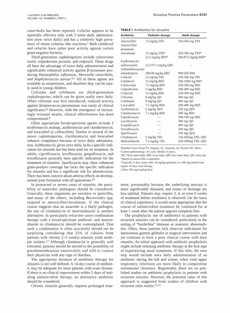
ment, presumably because the underlying mucosa ismore significantly diseased, and routes of drainage areless optimal. Patients may require 3, 4, or even 6 weeksof treatment before resolution is observed. On the basisof clinical experience, it would seem appropriate that thecourse of antimicrobial treatment be continued for atleast 1 week after the patient appears symptom free.
The prophylactic use of antibiotics in patients withrecurrent sinusitis can be considered, particularly in thesetting of “borderline” immune or anatomic abnormali-ties. Often, these patients lack clear-cut indications forintravenous gamma globulin or surgical intervention andyet continue to have a poor clinical course with theirsinusitis. An initial approach with antibiotic prophylaxismight include initiating antibiotic therapy at the first signof experiencing nasal symptoms. If this fails, the nextstep would include once daily administration of anantibiotic during the fall and winter, when viral upperrespiratory infections are more likely to compromiseostiomeatal clearance. Regrettably, there are no pub-lished studies on antibiotic prophylaxis in patients withrecurrent sinusitis. However, the potential value of thisapproach is suggested from studies of children withrecurrent otitis media.12,13
J ALLERGY CLIN IMMUNOLVOLUME 102, NUMBER 6, PART 2
Sinusitus Practice Parameters S137
catarrhalis has been reported. Cefaclor appears to beoptimally effective only with 3 times daily administra-tion (now twice daily) and has a relatively high preva-lence of serum sickness–like reactions.5 Both cefadroxiland cefaclor have rather poor activity against certaingram-negative bacteria.
Third-generation cephalosporins include cefuroximeaxetil, cefpodoxime proxetil, and cefprozil. These drugsall have the advantage of twice daily administration andsignificantly enhanced activity against β-lactamase–pro-ducing Haemophilus influenzae, Morexella catarrhalis,and Staphylococcus aureus.6,7 All of these agents areavailable as suspensions, and therefore they can be easi-ly used in young children.
Cefixime and ceftibuten are third-generationcephalosporins, which can be given orally once daily.When cefixime was first introduced, reduced activityagainst Streprococcus pneumoniae was rarely of clinicalsignificance.8 However, with the emergence of increas-ingly resistant strains, clinical effectiveness has beencompromised.9
Other appropriate broad-spectrum agents include 2erythromycin analogs, azithromycin and clarithromycin,and loracarbef (a carbacefem). Similar to several of thenewer cephalosporins, clarithromycin and loracarbefenhance compliance because of twice daily administra-tion. Azithromycin, given once daily, lacks a specific indi-cation for sinusitis but has been used for its treatment. Inadults, ciprofloxacin, levofloxacin, grepafloxacin, andtrovafloxacin presently have specific indications for thetreatment of sinusitis. Sparfloxacin may show enhancedgram-positive coverage but lacks the specific indicationfor sinusitis and has a significant risk for phototoxicity.There has been concern about adverse effects on develop-mental joint formation with all quinolones.10
In protracted or severe cases of sinusitis, the possi-bility of anaerobic pathogens should be considered.Generally, these organisms are sensitive to penicillin,and many of the others, including Bacteroides spp,respond to amoxicillin/clavulanate. If the clinicalcourse suggests that an anaerobe is a likely pathogen,the use of clindamycin or metronidazole is anotheralternative. In particularly refractive cases combinationtherapy with a broad-spectrum antibiotic and metron-idazole or clindamycin should be contemplated. Thatsuch a combination is often successful should not besurprising considering that 25% of cultures frompatients with chronic (>3 weeks) sinusitis yield multi-ple isolates.11 Although clindamycin is generally welltolerated, patients should be alerted to the possibility ofpseudomembranous enterocolitis and told to contacttheir physician with any sign of diarrhea.
The appropriate duration of antibiotic therapy forsinusitis is not well defined. A 14-day course of antibiot-ic may be adequate for most patients with acute disease.If there is no clinical improvement within 5 days of initi-ating antimicrobial therapy, an alternative antibioticshould be considered.
Chronic sinusitis generally requires prolonged treat-
TABLE I. Antibiotics for sinusitis
Antibiotic Pediatric dosage Adult dosage
Amoxicillin 13 mg/kg TID 250-500 mg TIDAmoxicillin/potassiumclavulanate 13 mg/kg TID* 250-500 mg TID*
22.5 mg/kg BID* 500-875 mg/kg BID*Erythromycin/sulfisoxazole 12.5/37.5 mg/kg QID –Sulfamethoxazole/trimethoprim 200/40 mg/kg BID 800/160 BIDCefaclor 13 mg/kg TID 250-500 mg TIDCefadroxil 15 mg/kg BID 500-1000 mg BIDCefuroxime 7.5 mg/kg BID 250-500 mg BIDCefpodoxime 5 mg/kg BID 200-400 mg BIDCefprozil 15 mg/kg BID 250-500 mg BIDCefixime 8 mg/kg QD 400 mg QDCeftibuten 9 mg/kg QD 400 mg QDLoracarbef 7.5 mg/kg BID 200-400 mg BIDAzithromycin 5 mg/kg QD† 250 mg QD†Clarithromycin 7.5 mg/kg BID 500 mg BIDCiprofloxacin — 500-700 mg BIDLevofloxicin — 500 mg QDGrepafloxacin — 400 mg QDTrovafloxacin — 200 mg QDSparfloxacin — 100 mg QD‡Clindamycin 5 mg/kg TID 150-450mg TID, QIDMetronidazole 7.5 mg/kg TID 250-500mg TID, QID
Modified from Virant FS, Shapiro GG. Sinusitis. In: Tierney DF, editor.Current pulmonology. St Louis: Mosby. In press.TID, Three times daily; BID, twice daily; QID, four times daily; QD, every day.*Based on amoxicillin component.†Typically 5-day course after 10 mg/kg (pediatric) or 500 mg (adult) loadequals 10 days total therapy.‡After 200 mg loading dose.
S138 Sinusitus Practice Parameters J ALLERGY CLIN IMMUNOLDECEMBER 1998
REFERENCES
1. Doern GV, Brueggemann A, Holley HP, Rauch AM. Antimicrobial resis-tance of Streptococcus pneumoniae recovered from outpatients in theUnited States during the winter months of 1994 to 1995: results of a 30-center national surveillance study. Antimicrob Agents Chemother1996;40:1208-13.
2. Wald ER, Reilly JS, Casselbrant M, et al. Treatment of acute maxillarysinusitis in childhood: a comparative study of amoxicillin and cefaclor. JPediatr 1984;104:297-302.
3. Wald ER, Chiponis D, Ledesma-Medina J. Comparative effectiveness ofamoxicillin and amoxicillin-clavulanate potassium in acute paranasalsinus infection in children: a double-blind, placebo-controlled trial. Pedi-atrics 1986;77:795-800.
4. Rachelefsky GS, Katz RM, Siegel SC. Chronic sinusitis in children withrespiratory allergy: the role of antimicrobials. J Allergy Clin Immunol1982;69:382-7.
5. Marchant CD, Shurin PA, Turczyk VA, et al. A randomized controlledtrial of cefaclor compared with trimethoprim sulfamethoxazole for treat-ment of acute otitis media. J Pediatr 1984;105:633-8.
6. Sydnor AJ, Gwaltney JM Jr, Cochetto DM, et al. comparative evaluationof cefuroxime axetil and cefaclor for treatment of acute bacterial maxil-lary sinusitis. Arch Otolaryngol Head Neck Surg 1989;115:1430-3.
7. Camacho AE, Cobo R, Otte J, et al. Clinical comparison of CefuroximeAxetil and amoxicillin/clavulanate in the treatment of patients with acutebacterial maxillary sinusitis. Am J Med 1992;93:271-6.
8. Howie VM, Owen MJ. Bacteriologic and clinical efficacy of cefiximecompared with amoxicillin and acute otitis media. Pediatr Infect Dis J1987;6:989-91.
9. Pichichero ME. Resistant respiratory pathogens and extended-spectrumantibiotics. Am Fam Physician 1995;52:1739-46.
10. File TM, Secreti J, Dunbar L, et al. A multicenter, randomized studycomparing the efficacy and safety of intravenous and/or oral Lev-ofloxacin versus Ceftriaxone and/or Cefuroxime Axetil in treatment ofadults with community-acquired pneumonia. Antimocrob AgentsChemother 1997;41:1965-72.
11. Wald ER, Byers C, Guerra N, Casselbrant M, Beste D. Subacute sinusi-tis in children. J Pediatr 1989;115:28-32.
12. Perrin JM, Charney E, MacWhinney JB Jr, et al. Sulfisoxazole as chemo-prophylaxis for recurrent otitis media: a double-blind crossover study inpediatric practice. N Engl J Med 1974;291:664.
13. Bluestone CD. Management of otitis media in infants and children: cur-rent role of old and new antimicrobial agents. Pediatr Infect Dis J1988;7(suppl):S129-36.
M. ANTIHISTAMINES
SUMMARY STATEMENTS
1. There are no data presently to recommend the use ofH1 antihistamines in acute bacterial sinusitis.
2. There may be a role for antihistamines in chronicsinusitis, especially in patients with allergic rhini-tis.
Both first- and second-generation H1 antihistamineshave a major role in the treatment of allergic rhinitis,allergic conjunctivitis, cutaneous allergic disorders, andanaphylaxis. A concise role for the use of these agents inthe treatment of sinusitis has not been elucidated. Thereare no recent studies evaluating the role of H1 antihista-mines in the treatment of sinusitis. Several review arti-cles and book chapters on treatment of acute and chron-ic sinusitis have been written recently, but rarely is theuse of antihistamines addressed.1-7 Despite these articles,antihistamines are widely sold for this condition.8,9
At the present time, there are no data to recommendthe use of H1 antihistamines in acute bacterial sinusitis.
There may be a role for these agents in chronic sinusitisin conjunction with rhinitis. In patients with allergicrhinitis, H1 antihistamines may help as adjuvant therapyfor chronic sinusitis and help prevent its occurrence byreducing seasonal edema. In general, second-generationantihistamines that are devoid of anticholinergic dryingeffects should be selected.
REFERENCES
1. Krajina Z. Clinical management of sinusitis. Rhinology 1977;15:141-7.2. Sotomayor JL, Scarpa N, Kolski GB. Sinusitis and asthma in childhood.
Hosp Pract 1986;21:129-30.3. Winther B, Gwaltney JM. Therapeutic approach to sinusitis: antiinfec-
tious therapy as the baseline of management. Otolaryngol Head NeckSurg 1990;103:876-9.
4. Wald ER. Sinusitis in children. Clin Ther 1988;10:33-44.5. Bolger WE, Kennedy DW. Current perspectives on sinusitis in adults. J
Respir Dis 1992;13:421-48.6. Slavin RG. Nasal polyps and sinusitis. In: Middleton E, Reed CE, Ellis
EF, et al, editors. Allergy: principles and practice. 4th ed. St Louis: CVMosby Co; 1993. p. 1455-70.
7. Goebel JA, Sessions DG, Thawley SE. Serious otitis media and sinusitis.In: Korenblat PE, Wedner JH, editors. Allergy: theory and practice. 2nded. Philadelphia: WB Saunders Co; 1992. p. 181-99.
8. Meltzer EM. Performance effects of antihistamines. J Allergy ClinImmunol 1990:86:613-9.
9. Kennedy DW. Overview. Otolaryngol Head Neck Surg 1990;103:847-54.
N. α-ADRENERGIC DECONGESTANTS
SUMMARY STATEMENTS
1. Both topical and oral decongestants are often used inthe therapy of acute or chronic sinusitis because it isthought that they may widen ostial patency andreduce turbinate swelling.
2. Prospective studies are lacking and are needed toassess the value of α-adrenergic agents in the treat-ment of sinusitis.
The basis for ancillary therapy of sinusitis with α-adrenergic decongestants derives from anecdotalaccounts and personal beliefs rather than definitive data.The recent appreciation that noninfectious inflammationin the nose and sinus predispose to infectious sinusitishas stimulated renewed interest in the development anddocumentation of efficacious ancillary therapies thatcould supplement or abrogate antibiotic use.1 It has beenthought that both topical and oral decongestants used inthe therapy of acute or chronic sinusitis widen ostiapotency and reduce turbinate swelling, thus promotingsinus and nasal ventilation.
Pharmacology
Decongestants can produce their effects by 2 broadmechanisms. First, direct-acting sympathomimetic drugsactivate α-adrenergic receptors resulting in vasoconstric-tion.2 Second, indirect-acting sympathomimetic agentsfunction by being taken up into the prejunctional nerveterminal where they displace norepinephrine from stor-age vesicles. The dispensed norepinephrine is thenreleased from the sympathetic nerve terminal to
J ALLERGY CLIN IMMUNOLVOLUME 102, NUMBER 6, PART 2
Sinusitus Practice Parameters S139
postjunctional α-adrenergic receptors producing vaso-constriction.
Side effects of decongestant
Topical agents used as nasal sprays act rapidly, usual-ly within minutes, and therapeutic doses have no sys-temic side effects. Rebound hyperemia or chemical rhini-tis, however, is a frequent side effect in patients who usethe drugs over an extended period of time.
Oral decongestants cause generalized constriction ofblood vessels, and increased arterial pressure is always ofconcern. Most available oral agents, however, cause bloodpressure elevations in normal persons only at doses thatsignificantly exceed the recommended dose. Other possi-ble adverse effects are reflex bradycardia, urinary reten-tion, mydriasis (with effects on glaucoma), and effects onendocrine and other regulators of metabolic function. Only3 drugs are commonly used as oral decongestants: phenyl-propanolamine, pseudoephedrine, and phenylephrine.Direct-acting sympathomimetics, such as phenylephrineand oxymetazoline, activate α1- and α2-adrenergic recep-tors, respectively, and both cause vasoconstriction and adecrease in nasal congestion. These drugs can be used top-ically during acute upper respiratory infections to reduceedema, and it is possible that they may help keep sinusostia patent.3 Oxymetazoline can be used to shrink thenasal mucosa followed by antiinflammatory steroid nasalpreparations to help treat chronic sinusitis.
Prospective studies are lacking and are needed toaccess the value of α-adrenergic agents in the treatmentof chronic sinusitis.4,5
REFERENCES
1. Zeiger RS. Prospects for ancillary treatment of sinusitis in the 1990s. JAllergy Clin Immunol 1992;90:478-95.
2. Johnson BA, Hricik JG. The pharmacology of α-adrenergic deconges-tants. Pharmacotherapy 1993;13:110S-5S.
3. Melen I, Friberg B, Andreasson L, et al. Effects of phenylpropanolamineon ostial and nasal patency in patients treated for chronic maxillarysinusitis. Acta Otolaryngol (Stockh) 1986:101:494-500.
4. Porta M, Jick H, Habakangas JAS. Follow-up study of pseudoephedrineusers. Ann Allergy 1986;57:340-2.
5. Connell JT, Linzmayer MI. Comparison of nasal airway patency changesafter treatment with oxymetazoline pseudoephedrine. Am J Rhinol1987;1:87-94.
O. GLUCOCORTICOSTEROIDS
SUMMARY STATEMENTS
1. The use of systemic steroid therapy for sinus diseasehas not been studied systematically in a well-con-trolled or blinded manner.
2. A few recent studies suggest that the addition ofintranasal steroids as an adjunct to antibiotic therapyis beneficial in the treatment of sinusitis.
3. The relative safety of directed therapy with inhaledsteroids makes their use a likely mode of treatmentfor sinusitis or at least for treatment of any underly-ing rhinitis.
Researchers and clinicians agree on the potential use-
fulness of corticosteroids as potent antiinflammatoryagents and the fact that sinusitis is an inflammatory dis-ease. The leap from this logical association to clinicalproof of the effectiveness of corticosteroids in managingsinus disease has been difficult.
As in asthma, the eosinophil plays a major role insinusitis. Harlin et al1 examined paranasal sinus tissuefrom 26 patients with chronic sinusitis with or withoutallergic rhinitis or asthma. Eosinophilic infiltration ofthe mucosa was a characteristic finding as was amarked association between the presence of extracellu-lar deposition of eosinophilic major basic protein anddamage to the mucosa. There was a notable similaritybetween the histopathology of the paranasal respirato-ry epithelium and that seen in asthma, in which the useof corticosteroids to decrease eosinophil-relatedinflammatory damage is well accepted. The authorsconclude that the eosinophil acts as an effector cell inchronic inflammatory disease of the paranasal respira-tory epithelium just as in the inflammation associatedwith asthma. Hamilos et al2 also confirmed the impor-tance of eosinophilic inflammation in chronic hyper-plastic sinusitis and observed a strong correlationbetween GM-CSF and IL-3 cytokines witheosinophilic accumulation.
In a clinical study3 aimed at developing a techniquefor assessing the severity of chronic sinusitis, 104patients undergoing surgery for chronic sinusitis wereevaluated with computed tomography, total serum IgEmeasurement, measurement of specific IgE concentra-tion for common allergens, and peripheral eosinophilcount. Extensive disease was present in 39% ofpatients and was correlated with asthma, specific IgEantibodies, and eosinophilia. The presence of peripher-al eosinophilia indicated a high likelihood of extensivesinus disease.
Increased numbers of activated eosinophils havebeen found in nasal polyposis, a risk factor for sinusi-tis. Although examination of the nasal mucosa of unaf-fected subjects revealed no activated eosinophils,examination of nasal polyps and the nasal mucosa ofpatients with polyps revealed that a significant portionof eosinophils that were present were activated. Thisportion declined after topical corticosteroid treatment,suggesting that the inflammatory reaction wasreduced.4-6
The antiinflammatory activities of corticosteroidsinclude decreased vascular permeability, inhibition ofrelease, and/or formation of mucous secretogogues,including histamine, leukotrienes, platelet-activatingfactor, and prostanoids, as well as inhibition of inflam-matory cell infiltration.
Topical corticosteroids have a clear effect on inflam-matory cell influx into the nasal mucosa after nasalantigen challenge.7 Pretreatment of nasal mucosa withinhaled steroid has been shown to modify both theimmediate- and late-phase response after antigen chal-lenge.8 Glucocorticosteroids also inhibit antigen-induced nasal hyperresponsiveness to histamine.9
S140 Sinusitus Practice Parameters J ALLERGY CLIN IMMUNOLDECEMBER 1998
Numerous clinical trials attest to the efficacy of topicalcorticosteroids in controlling symptoms of allergicrhinitis.10,11
The similarity of the respiratory epithelium in the noseand paranasal sinuses, as well as the contiguity of theseareas, would lead one to expect that sinusitis might also betreatable with inhaled corticosteroids. Yet anatomic barri-ers, including the narrow ostiomeatal complex and thewall-like ethmoid bulla, make access to the interior sinusmucosa unlikely, unpredictable, and unmeasurable.Whether nasal steroid therapy can sufficiently decreasenasal inflammation and improve mucociliary transport tothe point where the ostiomeatal complex becomes compe-tent is unknown.
The use of systemic steroid therapy for sinus diseasehas also not been studied. Nevertheless, it is not uncom-mon for physicians to use a course of prednisone in con-junction with antibiotics for persistent sinusitis. Thistherapy is based on the theory that inflammation, andnot just infection, participate in difficult-to-managesinus disease.
The relative safety of nasal steroids makes their use anattractive mode of treatment for sinusitis. French investi-gators12 compared sinus irrigation with the steroid tixo-cortol pivalate along with neomycin versus neomycinalone in a double-blind, randomized trial of 60 patientswith chronic sinusitis who performed intranasal lavageswith these medications daily for 11 days. Symptoms andmanometric readings were used to assess ostial obstruc-tion. The addition of tixocortol decreased obstruction from36% to 69%, apparently due to diminishing the inflamma-tory process.
Qvarnberg et al13 evaluated budesonide nasalaerosol versus placebo added to a regimen of sinuswashings and erythromycin for chronic or recurrentsinus disease in 40 patients. This was a double-blind,randomized evaluation. The budesonide group had lessnasal symptoms and facial pain. There were no othersignificant differences in outcome between the groups,with similar numbers in each group having positivesinus cultures and inflammatory cells in lavage fluidand requiring sinus surgery.
The utility of nasal flunisolide in conjunction withamoxicillin/clavulanate for treating acute sinusitis and forpreventing recurrences has been reported.14 In a multicen-ter, double-blind, randomized, parallel trial, patients aged14 years or older with radiograph-documented sinusitisreceived amoxicillin/clavulanate (500 mg 3 times daily)for 3 weeks. One half of the group received flunisolidenasal spray (100 µg), and the other half received placebo,3 times daily for 3 weeks. After finishing the antibiotic, theflunisolide or placebo spray therapy was continued foranother 4 weeks. Both groups improved significantly.Global assessment of effectiveness and turbinate swellingwere significantly better with flunisolide than placebo dur-ing the first 3 weeks of therapy. During the prophylacticphase of therapy, there was no difference in recurrences ofsinusitis between the 2 groups. The authors conclude thatthe addition of flunisolide nasal spray as an adjunct to
antibiotic for the treatment of sinusitis was judged themost effective treatment in the global evaluation and tend-ed to improve symptoms, to decrease inflammatory cellsin nasal secretions, to normalize ultrasound, and to aid inregression of radiographic abnormalities compared withplacebo spray. However, despite symptomatic improve-ment in many patients, many had continuing symptoms,radiographs remained abnormal in 80% of patients, andthere were frequent recurrences. In a more recent study 89children received 3 weeks of amoxicillin/clavulanate (40mg/kg 3 times daily) plus nasal budesonide 50 mg per nos-tril twice daily (n = 43) or placebo (n = 46). Cough andnasal discharge significantly improved only in the flu-nisolide group at the end of the second week, confirmingthe ancillary role of topical steroids.15
Currently, nasal steroid therapy has become an accept-ed adjunct in treating both acute and chronic sinusitis. Sev-eral intranasal steroids are now available in the US: flu-nisolide, beclomethasone, triamcinolone, fluticasone,budesonide, and mometasone. Each of these has beenproven to be effective in treatment of allergic rhinitis andmay be a useful addition in sinus disease.
REFERENCES
1. Harlin SL, Ansel DG, Lane SR, et al. A clinical and pathologic study ofchronic sinusitis: the role of the eosinophil. J Allergy Clin Immunol1988;81:867-75.
2. Hamilos DL, Leung DYM, Wood R, et al. Chronic hyperplastic sinusitis:association of tissue eosinophilia with mRNA expression of granulocyte-macrophage colony-stimulating factor and interleukin. J Allergy ClinImmunol 1993;92:39-48.
3. Newman LJ, Platts-Mills TA, Phillips CD, Hazen KC, Gross CW. Rela-tionship of computed tomographic findings to allergy, asthma andeosinophilia. JAMA 1994;271:363-7.
4. Stoop AE, van der Heijden HAMD, Biewenga J, van der Baan S.Eosinophils in nasal polyps and nasal mucosa: an immunohistochemicalstudy. J Allergy Clin Immunol 1993;91:616-22.
5. Schleimer RP. Mechanisms of glucocorticoid actions in asthma. Insightsin Allergy 1992;7:1-7.
6. Laitinen LA, Laitinen A, Haahtela T. A comparative study of the effectsof an inhaled corticosteroid, budesonide, and a β-2 agonist, terbutaline,on airway inflammation in newly diagnosed asthma: a randomized, dou-ble blind, parallel-group controlled trial. J Allergy Clin Immunol1992;90:32-42.
7. Bascom R, Wachs M, Naclerio RM, et al. Basophil influx occurs afternasal antigen challenge: effects of topical corticosteroid pretreatment. JAllergy Clin Immunol 1988;81:580-9.
8. Pipkorn U, Proud D, Lichtenstein LL, et al. Inhibition of mediator releasein allergic rhinitis by pretreatment with topical glucocorticosteroids. NEngl J Med 1987;316:1506-10.
9. Baroody FM, Cruz AA, Lichtenstein LL, et al. Intranasal beclomethasoneinhibits antigen-induced nasal hyperresponsiveness to histamine. J Aller-gy Clin Immunol 1992;90:373-6.
10. Seigel SC. Topical intranasal corticosteroid therapy in rhinitis. J AllergyClin Immunol 1988;81:984-91.
11. Juniper EF, Guyatt GH, O’Byrne PM, Viveiros M. Aqueous beclometha-sone dipropionate nasal spray: regular versus “as required” use n thetreatment of seasonal allergic rhinitis. J Allergy Clin Immunol1990;86:380-6.
12. Cuenant G, Stipon JP, Plante-Longchamp G, Baudoin C, Guerrier Y. Effi-cacy of endonasal neomycin-tixocortol pivalate irrigation in the treatmentof chronic allergic and bacterial sinusitis. ORL J Otorhinolaryngol RelatSpec 1986;48:226-32.
13. Qvarnberg Y, Kantola O, Salo J, Toivanen M, Valtonen H, Vuori E. Influ-ence of topical steroid treatment on maxillary sinusitis. Rhinology1992;30:103-12.
14. Meltzer EO, Orgel HA, Backhaus JW, et al. Intranasal flunisolide spray
J ALLERGY CLIN IMMUNOLVOLUME 102, NUMBER 6, PART 2
Sinusitus Practice Parameters S141
as an adjunct to oral antibiotic therapy for sinusitis. J Allergy ClinImmunol 1993;92:812-23.
15. Barlan IB, Erkan E, Bakir M, Berrak S, Basaran MM. Intranasal budes-onide spray as an adjunct to oral antibiotic therapy for acute sinusitus inchildren. Ann Allergy Asthma Immunol 1997;78:598-601.
P. ADJUNCTIVE THERAPIES INCLUDING
SALINE, MUCOLYTICS, AND EXPECTO-
RANTS
SUMMARY STATEMENTS
1. There are inadequate data to recommend use of wet-ting agents as individual therapy for sinusitis.
2. Clinical practice, as well as folklore, supports the useof wetting agents for symptomatic treatment as partof a pharmacologic regimen.
3. There are several scientific studies that imply, but donot directly confirm, a role for these agents in sinusi-tis.
4. The safety profile of each agent should be carefullyconsidered for individual patients.
5. Use of all these agents as prophylaxis for exacerba-tions of chronic sinusitis is empiric and not support-ed by clinical data.
Medical management of sinusitis is based on empiricgoals. Adjunctive agents are frequently prescribed inaddition to antibiotics to aid drainage of retained secre-tions through the sinus ostia into the nasal cavity. Use isbased on clinical experience.1-3
Rationale for adjunctive medication to antibiotics
Management of acute sinusitis usually includes an oralantibiotic. However, it has been estimated that about 45%of cases will resolve without the use of antibiotics. In onestudy of 50 patients with chronic anterior or posteriorpurulent nasal discharge, 20 patients were treated withtopical dexamethasone and decongestant sprays, 20patients with the same medications plus topicalneomycin, and 10 with matched placebo sprays or pro-pellants alone. Significantly more patients receiving theactive treatments demonstrated improvement in symp-toms compared with those receiving placebo. Theauthors concluded that improving sinus drainage permit-ted host mechanisms to recover and that topical antibi-otics provided no additional benefit. However, systemicantibiotics were not used.4 In another study of 80 allergicchildren with asthma 4 to 14 years of age, sinus abnor-malities were detected on x-ray film in 55 patients. Thefindings of mucosal thickening greater than 2 mm, opaci-fication, or air-fluid level were defined as abnormal radi-ographs. Those children with purulent postnasal drip (n =13) were treated with ampicillin, phenylephrine, andtripolidine. In 42 children without purulent drainage, theampicillin was replaced by intranasal beclomethasonespray for 1 month, together with phenylephrine andtripolidine. In both treatment groups sinus radiographsimproved with a decrease in severity of the asthma. How-ever, the presence of bacterial sinusitis was not ade-
quately documented, and all children received topicaldecongestants, and therefore that the role of theintranasal beclomethasone could not be adequatelyassessed.5
Both these studies suggest that systemic antibiotics arenot required. However, the serious complications ofsinusitis, such as intracranial extension of infection, havedecreased in frequency since the advent of antibiotics.Consequently, antibiotics are now generally prescribedas primary therapy.
Although treatment of sinusitis is associated with res-olution of radiographic abnormalities and improvementin the symptoms of asthma, scarce data exist on the effi-cacy of treatment for chronic sinusitis. On the basis ofour understanding of the pathophysiology of chronicsinusitis, medical management should be designed toeffectively treat infection in the sinuses, reduce tissueswelling in the region of the sinus ostia to facilitatedrainage of retained secretions, promote ciliary function,and maintain ostial patency both during and after thera-py. Combination treatment protocols are commonly usedon the basis of clinical experience rather than formal tri-als.6
Pharmacologic agents
GuaifenesinGuaifenesin (glyceryl guaicolate; 3-(2-methoxy-
phonoxy)-1, 2-propanediol) is a water- and alcohol-soluble substance that has been used as an expectorantto loosen phlegm and bronchial secretions in the symp-tomatic management of coughs associated with thecommon cold, bronchitis, laryngitis, pharyngitis,influenza, and measles, as well as sinusitis when theseconditions are complicated by tenacious mucus and/ormucus plugs and congestion. By reducing viscosity ofsecretions, guaifenesin increases the efficiency of thecough reflex and of ciliary action in removing accumu-lated secretions from the trachea and bronchi. There isclinical evidence that guaifenesin is an effective expec-torant in that it increases expectorated sputum volumeover the first 4 to 6 days of a productive cough,decreases sputum viscosity and difficulty in expectora-tion, and improves associated symptoms. There is cur-rently insufficient evidence to support efficacy of thedrug as an adjunct in sinusitis.7 High-dose guaifenesin(1200 mg twice daily) has been used empirically for itsability to thin tenacious respiratory secretions on thebasis of clinical efficacy in chronic bronchitis.8 Noclinical trials have been reported in sinusitis to demon-strate its efficacy. Guaifenesin is considered a pregnan-cy category C drug because animal reproduction stud-ies have not been conducted.9
IodineIodine-containing compounds, such as potassium
iodide or iodinated glycerol, might be expected to havesimilar effects as guaifenesin. However, they are of lim-ited clinical use, perhaps because of the potential adverse
S142 Sinusitus Practice Parameters J ALLERGY CLIN IMMUNOLDECEMBER 1998
effects of iodine hypersensitivity on chronic ingestion.Potassium iodide syrup is considered an expectorant inthe symptomatic treatment of chronic pulmonary dis-eases in which tenacious mucus complicates the prob-lem, including bronchial asthma, bronchitis, and pul-monary emphysema. Side effects may include gastroin-testinal upset, metallic taste, minor skin eruptions, nau-sea, vomiting, and epigastric pain.10 No studies in sinusi-tis have been reported.
Antibiotics as mucolyticsThe mucolytic properties of antibiotics have been
investigated. Norfloxacin reduced the elastic modulus(G’) of mucus in one study of patients with chronicsinusitis,11 and erythromycin reduces respiratory glyco-conjugate secretion in an in vitro preparation of humanairways.12 On the basis of the scarcity of current data,antibiotics should be selected primarily on their antimi-crobial profile.
Nonpharmacologic adjuncts
Many nonpharmacologic measures are advocated forsymptomatic relief of acute sinusitis. Because scientificdata on efficacy are lacking, physicians may dismisssome of these measures as folk medicine. For manypatients, however, 1 or more of these treatments may pro-vide effective relief of distressing symptoms while theinfection is resolving. Unfortunately, most of these mea-sures are short-lived in effectiveness, and they must berepeated as symptoms recur.
SalineSteam and saline prevent crusting of secretions in the
nasal cavity and especially in the region of the ostiomeatalcomplex. By liquefying secretions, they also help mucocil-iary clearance and reduce the feeling of facial pressure.Repetitive saline applications also act as a mild vasocon-strictor of nasal blood flow.13,14 Saline nebulizations wereshown to be beneficial in one study in children.15
Nasal instillation of saline spray 2 or 3 times a day,between steam treatments, provides a mild decongestantaction. Saline spray also helps to liquefy nasal secretionsand moisturize the nasal and sinus mucosa. Irrigationwith saline, or antral lavage, is a familiar procedure, butone which is best left to ear, nose, and throat surgeonsafter aspiration of purulent matter from the sinuses ofseverely congested patients.11
SteamThe traditional method of steam inhalation is to
instruct the patient to do the following:1. Pour boiling water in a pan or basin on a low table.2. Sit at the table with a towel draped over the head to
make a tent over the pan of water.3. Hold the face a few inches above the water and breathe
through the nose for approximately 10 minutes.
This procedure liquefies and softens crusts whilemoisturizing the dry, inflamed mucosa.
Many patients find that 2 such treatments a day pro-vide some effective symptomatic relief. If a patient is forsome reason unable to perform this simple procedure,using a vaporizer or a facial sauna or taking long, hotshowers may be beneficial, but none of these alternativesis a good substitute for the hot water and tent method.The ritual of boiling the kettle, preparing the tent, andrelaxing over the steamy brew probably has a good psy-chologic effect, enhancing the therapeutic benefit.Patients, however, should be reminded not to breathesteam directly from a boiling kettle.
Propylene glycolAlthough propylene/polyethylene glycol and saline
have been used in clinical studies as placebos, Spector etal16 investigated their possible therapeutic role as wettingagents. Eighteen patients with perennial rhinitis werestudied for 4 weeks of active treatment. Both agents pro-duced symptomatic improvement and objective improve-ments in airway obstruction at 2 and 4 weeks.
Hot airSome patients report benefit from breathing hot, dry
air. This procedure dries secretions and also generates orenhances a feeling of well being. Air at 41˚C has beenreported to be virucidal in vitro,19 and some commer-cially available devices provide heated air. However,maxillary sinusitis is usually bacterial, not viral, and onlyanecdotal evidence supports the premise that hot, dry airis virucidal in vivo. Several studies with different deviceshave yielded differing results.17-20
AstringentsAdding pine oil, mentholated preparations such as
Vicks VapoRub, oil of eucalyptus, or similar aromaticsmay add to the beneficial effect of the steam treatment.Such additions may help relieve stuffiness or at least givea subjective sensation of increased air flow. Again, thereare no scientific data to support this view, but patientsthink they work.
Spicy foods
Garlic has an active ingredient (n-allythiosulphinate)that provides short-lived decongestant effects. Eatingfoods highly seasoned with garlic has been consideredtherapeutic. Ziment21 included a recipe for his wife’sgarlic-and-chicken soup in his textbook on treatment ofrespiratory ailments. Encapsulated garlic powder issometimes recommended by patients who do not like theflavor of the garlic in their food. Chewing horseradishroot, which is available from many food markets, isanother home remedy reported effective in “clearing thesinuses” by some patients. Again, no scientific data sup-port its reported benefits.
REFERENCES
1. Slavin RG. Nasal polyps and sinusitis. In: Middleton E Jr, Reed CE,Ellis EF, Adkinson NF Jr, Yunginger JW, Busse WW, editors. Allergy:principles and practice. 4th ed. St Louis: Mosby; 1993.
J ALLERGY CLIN IMMUNOLVOLUME 102, NUMBER 6, PART 2
Sinusitus Practice Parameters S143
2. Druce HM, Slavin RG. Sinusitis: a critical need for further study. JAllergy Clin Immunol 1991;88:675-7.
3. Druce HM. Diagnostic and management of chronic sinusitis and itscomplications. Immunol Allergy Clin North Am 1987;7:117-32.
4. Sykes DA, Wilson R, Chan KL, Mackay IS, Cole PJ. Relative impor-tance of antibiotic and improved clearance in topical treatment ofchronic mucopurulent rhino sinusitis. A controlled study. Lancet1986;2:359-60.
5. Businco L, Fiore L, Frediani T, Artuso A, Di Fazio A, Bellioni P. Clin-ical and therapeutic aspects of sinusitis in children with bronchial asth-ma. Intern J Ped Otorhinol 1981;3:287-94.
6. Druce HM. Adjuncts to medical management of sinusitis. OtolaryngolHead Neck Surg 1990;103:880-3.
7. McEvoy GK, editor. AFHS Drug Information, 1992. Bethesda (MD):American Society of Hospital Pharmacists; 1992. p. 1600-1.
8. Petty TL. Results of a randomized, double-blind, placebo-controlledstudy of iodinated glycerol in chronic obstructive bronchitis. Chest1990;97:75-83.
9. Physicians desk reference. 47th ed. Montvale (NJ): Medical Econom-ics Co; 1993. p. 559.
10. Physicians desk reference. 47th ed. Montvale (NJ): Medical Econom-ics Co; 1993. p. 1023.
11. Slavin RG, Friedman WH. Nasal allergy: medical and surgical treat-ment. Adv Otolaryngol Head Neck Surg 1987;1:91-108.
12. Majima Y, Hirata K, Takeuchi K, Hattori M, Sakakura Y. Effects oforally administered drugs on dynamic viscoelasticity of human nasalmucus. Am Rev Respir Dis 1990;141:79-83.
13. Goswami S K, Kivity S, Marom Z. Erythromycin inhibits respiratoryglycoconjugate secretion from human airways in vitro. Am Rev RespirDis 1990;141:72-8.
14. Druce HM, Bonner RF, Patow C, et al. Response of nasal blood flowto neurohormones as measured by laser-Doppler velocimetry. J ApplPhysiol 1984;57:1276-83.
15. Van Beuer HPS, Bosmans J, Stevens WJ. Nebulization treatment withsaline compared to bromhexine in treating chronic sinusitis in asth-matic children. Allergy 1987;42:33-6.
16. Spector SL, Toshener D, Gay I, Rosenman E. Beneficial effects ofpropylene and polyethylene glycol and saline in the treatment ofperennial rhinitis. Clin Allergy 1982;12:187-96.
17. Yerushalmi A, Karman S, Lwoff A. Treatment of perennial allergicrhinitis by local hyperthermia. Proc Natl Acad Sci 1982;79:4766-9.
18. Oppenheimer J, Buchmeier A, Nelson HS. Double-blind trial of a heat-ed nasal aerosol in the treatment of perennial allergic rhinitis. J Aller-gy Clin Immunol 1993;92:56-60.
19. Johnston SL, Price JN, Lau LCK, Wells AF, Walters C, Feather IH, etal. The effect of local hyperthermia on allergen-induced nasal conges-tion and mediator release. J Allergy Clin Immunol 1993;92:850-6.
20. Salah B, Dinh Xuan AT, Fouilladieu JL, Lockhart A, Regnard J. Nasalmucociliary transport in healthy subjects is slower when breathing dryair. Eur Respir J 1988;1:852-5.
21. Ziment I. Respiratory pharmacology and therapeutics. 1st ed. Philadel-phia: WB Saunders Co; 1978.
Q. INTRAVENOUS IMMUNE GLOBULIN
SUMMARY STATEMENTS
1. Immunodeficiency is one of the underlying factors forthe development of chronic and recurrent sinusitis.
2. Intravenous immune globulin (IVIG) is indicated foruse in patients with impaired humoral immunity.
Immunodeficiency is one of the underlying factorsfor the development of chronic and recurrent sinusitis.At the present time, intravenous immune globulin(IVIG) is indicated in the treatment of antibody defi-ciency syndromes, including X-linked agammaglobu-linemia, common variable immunodeficiency, Wiskott-Aldrich syndrome, and hyper-IgM syndrome.1-7 The
indication for IVIG in other conditions, such as selec-tive antibody deficiency syndromes or IgG subclassdeficiencies, is more controversial.8-12 Both qualitativeand quantitative defects in the humoral immune systemmay result in recurrent or chronic (>3 months)sinopulmonary bacterial infections.13 In patients withimmunoglobulin deficiency, complications of chronicsinusitis include subperiosteal abscess and intracranialabscesses. Infected sinus cavities may also act as afocus for systemic infections because the response toantibiotic therapy is limited without an effectivehumoral immune system. In such situations replace-ment of deficient or dysfunctional gamma globulinwith IVIG is a potentially life-saving therapy.
The use of IVIG is indicated for use in patients withqualitative or quantitative humoral immune deficiencyin which chronic sinusitis may be a significant associ-ated disease. In those with specific antibody deficiencywith normal total immunoglobulins, chronic sinusitis isoften a major complication and at times may be theonly significant infectious process. Many immunodefi-cient patients undergo multiple surgical procedures andreceive chronic antibiotic therapy without significantresolution of their sinusitis. In these patients who failmedical and surgical therapies and have documentedfailure to respond to polysaccharide antigens, a trial ofIVIG therapy is warranted.1,7 Treatment is maintainedthroughout the fall and winter months and may be dis-continued during the summer if the patient is diseasefree.
REFERENCES
1. Buckley RH, Shiff RI. The use of intravenous immunoglobulin inimmune deficient diseases. N Engl J Med 1991;325:110-7.
2. Huston DP, Kavanaugh AF, Rohane TW, Huston MM. Immunoglobulindeficiency syndromes and therapy. J Allergy Clin Immunol 1991;87:1-17.
3. Berkman SA, Lee ML, Gale RP. Clinical uses of intravenousimmunoglobulins. Semin Hematol 1988;25:140-58.
4. Magilavy DB, Cassidy JT, Tubergen DA, et al. Intravenous gamma glob-ulin in the management of patients with hypogammaglobulinemia. JAllergy Clin Immunol 1978;61:378-83.
5. Cunningham-Rundles C, Siegal FP, Smithwick EN, et al. Efficacy ofintravenous immunoglobulin in primary humoral immune deficiency dis-ease. Ann Intern Med 1984;101:435-9.
6. Ammann AJ, Ashman RF, Buckely RH, et al. Use of intravenous gammaglobulin in antibody immunodeficiency. Results of a multi-center con-trolled trial. Clin Immunol Immunopathol 1982;22:60-7.
7. Intravenous immunoglobulin: prevention and treatment of disease. NIHConsensus Dev. Cont. Consensus Statement. 1990;8:21-3.
8. Silk HJ, Ambrosino D, Geha RS. Effect of intravenous gamma globulintherapy in IgG2 deficient and IgG2 sufficient children with recurrentinfections and poor response to immunization with Hemophilus influen-za Type b capsular polysaccharide antigen. Ann Allergy 1990;64:21-5.
9. Zara J, Silk H, Tinkelman D. An evaluation of post-immunization pneu-mococcal titers in children with recurrent infections and normal levels ofimmunoglobulin. Ann Allergy 1993;70:283-8.
10. Wasserman RL. Antibody deficiency: IgG subclass deficiency in vaccinenon-responded states. Pediatr Infect Dis J 1990;9:424-33.
11. Knutsen AP. Patients with IgG subclass and/or selective antibody defi-ciency to polysaccharide antigens: initiation of a controlled clinical trialof intravenous immune globulin. J Allergy Clin Immunol 1989;84:640-7.
12. Hanson LA, Soderstrom R, Nilssen DE, et al. IgG subclass deficiencywith or without IgA deficiency. Clin Immunol Immunopathol1991;61:S70-7.
S144 Sinusitus Practice Parameters J ALLERGY CLIN IMMUNOLDECEMBER 1998
13. Polmar SH. Sinusitis and immune deficiency. In: Lusk RP, editor. Pedi-atric sinusitis. New York: Levin Press Ltd; 1992. p. 53-8.
R. SURGICAL CONSIDERATIONS
SUMMARY STATEMENTS
1. Antral puncture and irrigation is an office procedurethat has a place in the management of acute ethmo-maxillary sinusitis refractory to medical therapy or inacute sinusitis in an immunosuppressed patient inwhom early identification of pathogenic organisms isparamount.
2. The term “functional endoscopic sinus surgery” isbased on the clinical and experimental findings thatostial obstruction is the final common pathway in thedevelopment of sinusitis.
The surgical approach to sinus disease has undergone adramatic transition, resulting from renewed insights intosinus physiology and the widespread use of rigid nasaltelescopes. Earlier in this century, several authors werethe first to note the importance of the ethmoid sinuses inthe etiology of frontal and maxillary sinusitis disease.1,2
Our current knowledge indicates that limited and local-ized inflammation in the anterior ethmoid sinus, and par-ticularly the ostiomeatal complex, is sufficient to causefrontal and/or maxillary sinusitis. Whereas past tradition-al sinus surgery often attempted to completely stripinflamed mucosa from the larger sinuses, believing it tobe irreversibly diseased, current surgical therapy aims toremove only the causative ethmoid disease and reestab-lish ventilation and drainage of the larger dependentsinuses. Modern endoscopic sinus surgery is thus func-tional and restorative because its goal is to restore normalsinus physiology with minimal removal of mucosa.
Nasal endoscopy aids in the diagnosis of ethmoid dis-ease. It is more sensitive than plain roentgenography andmore cost-effective than sinus computed tomography fordiagnosing ethmoid sinusitis, and repeat endoscopicevaluations make it possible to more objectively follow apatient’s response to medical and surgical therapy.3
Surgical approaches
The surgical approaches to sinusitis can be broadlycategorized into traditional and endoscopic methods.Traditional surgical approaches can be further classi-fied by the particular sinus on which they were per-formed and include traditional ethmoid, maxillary, andfrontal sinus surgery.4,5
The endoscopic approach in sinus surgery
The term “functional endoscopic sinus surgery” ispredicated on the clinical and experimental finding thatostial obstruction is the final common pathway in thedevelopment of sinusitis.6 When 2 mucosal surfacescome into firm contact, disruption of normal mucociliaryflow occurs at that point.3 The potential for obstructionof secretions and persistent local inflammation thenexists, and sinusitis may ensue. The key to the function-
al endoscopic approach is therefore the accurate identifi-cation of areas in which ventilation or mucociliary clear-ance may be obstructed. Recent advances in endoscopicsurgery involve computer-assisted navigation within thenaris and powered instrumentation.7-9
Operative intervention
Endoscopic sinus surgery can be performed undereither local or general anesthesia on an outpatient basis.Patients with asthma or other underlying medical condi-tions are usually kept overnight for observation. If theprocedure is performed under local anesthesia, intra-venous sedation is administered by the anesthesiologist,and the patient is monitored. The entire operation is car-ried out under endoscopic visualization through the nasalcavities and involves no external incisions. The extent ofsurgical dissection is dictated by the amount and locationof disease identified on preoperative computed tomogra-phy, as well as that encountered during surgery.10 Surgi-cal intervention for the treatment of recurrent sinusitiscarries a severe complication rate of 0.5% in 200,000cases per year.11
Conclusions
In summary, great advances in the diagnosis and sur-gical treatment of sinusitis have been made in the lastdecade, principally through the use of nasal endoscopyand coronal computed tomographic scanning. The rigidnasal telescope has dramatically improved our ability tovisualize the ostiomeatal complex, which is the criticalarea in the pathogenesis of chronic sinusitis. Functionalendoscopic surgery of the sinuses results in significantsubjective improvement in the great majority of cases.12
REFERENCES
1. Proctor DF. The nose, paranasal sinuses and pharynx. In: Walters W, edi-tor. Lewis-Walters’ practice of surgery. Vol 4. Hagerstown: WF Prior Co;1966. p. 1-37.
2. Naumann H. Pathologische Anatomie der chronischen Rhinitis undSinusitis. Proceedings VIII International Congress of Otorhinolaryngolo-gy; 1965; Amsterdam. p 80.
3. Messerklinger W. Endoscopy of the nose. Baltimore: Urban andSchwarzenberg, Inc; 1978.
4. Jahn AF. General principles. In: Blitzer A, Lawson W, Friedman WH,editors. Surgery of the paranasal sinuses. 2nd ed. Philadelphia: W.B.Saunders Co; 1991. p. 175-81.
5. Caldwell GW. The accessory sinuses of the nose, and an improvedmethod of treatment for suppuration of the maxillary antrum. N Y Med J1893;4:526-8.
6. Kennedy DW, Zinreich SJ, Rosenbaum AE, Johns ME. Functional endo-scopic sinus surgery: theory and diagnostic evaluation. Arch Otolaryngol1985;111:576-82.
7. Freysinger W, Gunkel AR, Thumfart WF. Image-guided endoscopic ENTsurgery. Eur Arch Otorhinolaryngol 1997;254:343-6.
8. Gunkel AR, Freysinger W, Thumfart WF. Computer-assisted surgery inthe frontal and maxillary sinus. Laryngoscope 1997;107:631-3.
9. Mendelsohn MG, Gross CW. Soft-tissue shavers in pediatric sinussurgery. Otolaryngol Clin North Am 1997;30:443-9.
10. Kennedy DW. Functional endoscopic sinus surgery: technique. Arch Oto-laryngol 1985;111:643-9.
11. Kaliner MA. Recurrent sinusitis: examining medical treatment options.Am J Rhinol 1997;11:123-32.
12. Kennedy DW. Prognostic factors, outcomes and staging in ethmoid sinussurgery. Laryngoscope 1992;102(suppl 57):1-18.

![Rhinitis, sinusitis and food disorders [Part 2] · Rhinitis medicamentosa is a syndrome of rebound nasal congestion - adrenergic decongestants - cocaine. Cerebral spinal fluid rhinorrhea-Refractory](https://static.fdocuments.net/doc/165x107/608cc99d7bd63111f57bca69/rhinitis-sinusitis-and-food-disorders-part-2-rhinitis-medicamentosa-is-a-syndrome.jpg)