SuperScript III Platinum CellsDirect Two-Step qRT- PCR Kit
32
User Manual SuperScript ® III Platinum ® CellsDirect Two-Step qRT- PCR Kit For two-step real-time quantitative RT-PCR from cell lysate Catalog Nos. 11737-030 and 11737-038 Rev. Date: 7 July 2010 Manual part no. 25-0750 MAN0000471
Transcript of SuperScript III Platinum CellsDirect Two-Step qRT- PCR Kit

User Manual
Corporate Headquarters5791 Van Allen WayCarlsbad, CA 92008T: 1 760 603 7200F: 1 760 602 6500E: [email protected]
For country-specific contact information visit our web site at www.invitrogen.com
SuperScript® III Platinum® CellsDirect Two-Step qRT-PCR Kit For two-step real-time quantitative RT-PCR from cell lysate
Catalog Nos. 11737-030 and 11737-038
Rev. Date: 7 July 2010 Manual part no. 25-0750 MAN0000471

ii

iii
Table of Contents
Kit Contents and Storage......................................................................................................v Introduction............................................................................................................................1 Lysing Cells ............................................................................................................................4 First-Strand cDNA Synthesis...............................................................................................8 qPCR — Guidelines and Recommendations.....................................................................9 qPCR — Instruments Using PCR Tubes/Plates .............................................................12 qPCR — Roche LightCycler® .............................................................................................14 Troubleshooting...................................................................................................................16 Related Products..................................................................................................................18 Technical Support................................................................................................................19 Purchaser Notification ........................................................................................................20 References .............................................................................................................................23

iv
v
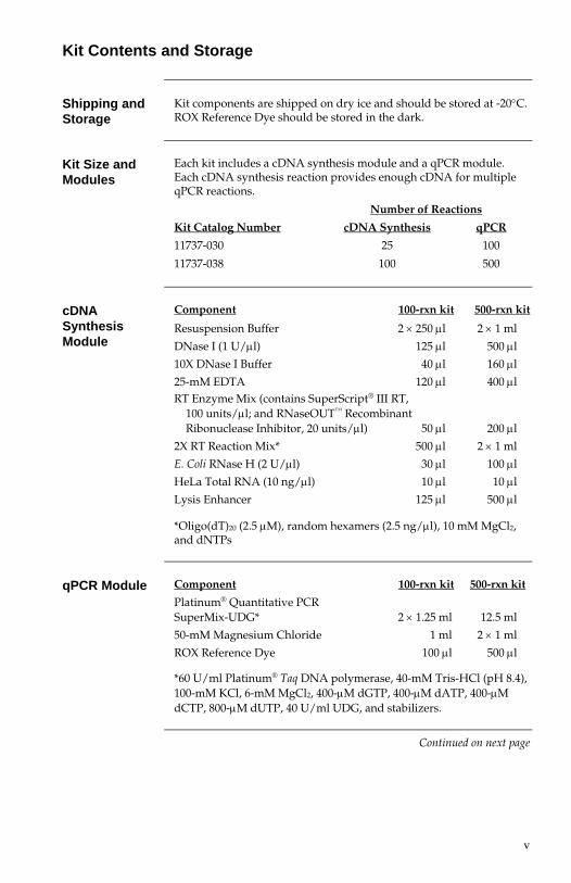
Kit Contents and Storage
Shipping and Storage
Kit components are shipped on dry ice and should be stored at -20°C. ROX Reference Dye should be stored in the dark.
Kit Size and Modules
Each kit includes a cDNA synthesis module and a qPCR module. Each cDNA synthesis reaction provides enough cDNA for multiple qPCR reactions.
Number of Reactions
Kit Catalog Number cDNA Synthesis qPCR
11737-030 25 100
11737-038 100 500
cDNA Synthesis Module
Component 100-rxn kit 500-rxn kit
Resuspension Buffer 2 × 250 μl 2 × 1 ml
DNase I (1 U/μl) 125 μl 500 μl
10X DNase I Buffer 40 μl 160 μl
25-mM EDTA 120 μl 400 μl RT Enzyme Mix (contains SuperScript® III RT,
100 units/μl; and RNaseOUT™ Recombinant Ribonuclease Inhibitor, 20 units/μl) 50 μl 200 μl
2X RT Reaction Mix* 500 μl 2 × 1 ml
E. Coli RNase H (2 U/μl) 30 μl 100 μl
HeLa Total RNA (10 ng/μl) 10 μl 10 μl
Lysis Enhancer 125 μl 500 μl
*Oligo(dT)20 (2.5 μM), random hexamers (2.5 ng/μl), 10 mM MgCl2, and dNTPs
qPCR Module Component 100-rxn kit 500-rxn kit
Platinum® Quantitative PCR SuperMix-UDG* 2 × 1.25 ml 12.5 ml
50-mM Magnesium Chloride 1 ml 2 × 1 ml
ROX Reference Dye 100 μl 500 μl
*60 U/ml Platinum® Taq DNA polymerase, 40-mM Tris-HCl (pH 8.4), 100-mM KCl, 6-mM MgCl2, 400-μM dGTP, 400-μM dATP, 400-μM dCTP, 800-μM dUTP, 40 U/ml UDG, and stabilizers.
Continued on next page

vi
Kit Contents and Storage, continued
Materials Supplied by the User
The following additional items are required for use with this kit:
• Coulter Counter or hemacytometer
• Microcentrifuge
• qPCR instrument
• Trypsin (for adherent cell cultures only)
• 1X cold phosphate-buffered saline PBS, without calcium or magnesium
• 0.2-ml thin-walled PCR tubes or 96-well PCR plates
• Bovine Serum Albumin (BSA), UltraPure (1 mg/ml)
• Ice
• Pipettes
• Disposable gloves

1
Introduction
System Overview
The SuperScript® III Platinum® CellsDirect Two-Step qRT-PCR Kit is an optimized kit for synthesizing first-strand cDNA directly from mammalian cell lysate without first isolating RNA, and then amplifying the cDNA in a real-time quantitative PCR (qPCR) reaction using Platinum® Quantitative PCR SuperMix-UDG.
In traditional qRT-PCR, RNA is first isolated from cells in a time-consuming procedure that can lead to a loss of material. Using the CellsDirect cDNA Synthesis System, the cells are lysed and the cDNA is generated from the lysate in a single tube with minimal handling and no sample loss. DNase I is added to eliminate genomic DNA prior to first-strand synthesis. After synthesis, the first-strand cDNA can be transferred directly to the qPCR reaction without intermediate organic extractions or ethanol precipitations.
This kit has been optimized for small cell samples, ranging from 10,000 cells down to a single cell. The use of SuperScript® III Reverse Transcriptase ensures high specificity and high yields of cDNA from small amounts of starting material—as little as 10 pg total RNA. The use of Platinum® Quantitative PCR SuperMix-UDG ensures optimal performance using a variety of qPCR detection technologies, including LUX™ Fluorogenic Primers and fluorescent hybridization probe technologies such as TaqMan® probes and molecular beacons.
Advantages of the Kit
This kit has the following advantages:
• Compatible with a wide range of mammalian cell types grown under different treatment conditions
• Cell lysis and first-strand cDNA synthesis in the same tube minimizes reagent loss, sample loss, and handling time
• Total lysate volume is used in the cDNA synthesis reaction, providing greater yields with a limited number of cells and allowing for detection of rare transcripts
• SuperScript® III Reverse Transcriptase, with reduced RNase H activity and higher thermal stability, produces high yields of cDNA in the first-strand synthesis reaction for greater sensitivity and enhanced detection of rare transcripts
• Platinum® Quantitative PCR SuperMix-UDG ensures optimal sensitivity and performance using a variety of qPCR detection technologies, with built-in carryover contamination protection and a linear dose response over a wide range of target concentrations
Continued on next page

2
Introduction, Continued
Diagram of cDNA Synthesis from Cell Lysate
������������ �����������
����������������
�������������
������������������ ������������������������ ��
������������������
������������� ��
����������
!�������������������"#����������! �
SuperScript®
III RT SuperScript® III Reverse Transcriptase is an engineered version of M-MLV RT with reduced RNase H activity and increased thermal stability. The enzyme can be used to synthesize first-strand cDNA at temperatures up to 55°C, providing increased specificity, higher yields of cDNA, and more full-length product than other reverse transcriptases.
Because SuperScript® III RT is not inhibited significantly by ribosomal and transfer RNA, it can effectively synthesize first-strand cDNA directly from total RNA. The concentration of SuperScript® III RT in this system has been optimized to synthesize first-strand cDNA from total RNA in cell lysate.
Continued on next page

3
Introduction, Continued
Platinum® Quantitative PCR SuperMix-UDG
Platinum® Quantitative PCR SuperMix-UDG is a ready-to-use reaction cocktail containing all components, except primers, for the amplification and detection of DNA in qPCR. It contains Platinum® Taq DNA polymerase, Mg++, uracil DNA glycosylase (UDG), proprietary stabilizers, and deoxyribonucleotide triphosphates, with dUTP instead of dTTP. The concentration of the SuperMix allows for the addition of primers, template, and (if necessary) probes or dyes.
Platinum® Taq DNA polymerase is precomplexed with specific monoclonal antibodies that inhibit polymerase activity during reaction assembly at room temperature. Full polymerase activity is restored after the denaturation step in PCR cycling, providing an automatic “hot start” in PCR and thereby increasing amplification efficiency, sensitivity, and yield.
UDG and dUTP are included in the mixture to prevent the reamplification of carryover PCR products between reactions. dUTP in the mix ensures that any amplified DNA will contain uracil. UDG, or uracil-N-glycosylase, removes uracil residues from single- or double-stranded DNA, preventing dU-containing DNA from serving as template in future PCRs. Incubation of subsequent PCRs with UDG before cycling destroys any contaminating dU-containing PCR product from previous reactions. After this decontamination step, UDG is inactivated by the high temperatures during normal PCR cycling, thereby allowing the amplification of genuine target sequence(s).
Control RNA HeLa Total RNA is included in the kit as a control. The concentration
of HeLa Total RNA provided (10 ng/μl) is equivalent to 1,000 cells.

4
Methods
Lysing Cells
Introduction In this step, you lyse your cells in Resuspension Buffer and Lysis
Enhancer and perform a DNase I digestion to remove genomic DNA from the sample.
Cell Types and Density
This kit has been optimized for small cell samples, ranging from 1 to 10,000 cells. This kit is compatible with several different mammalian cell lines, including HeLa, COS-7, 293, Jurkat, CV1, and K562. Cells may be grown under a variety of conditions and treatments. Any type of culture vessel can be used.
��������
• We recommend using a maximum of 10,000 cells per reaction. Higher numbers of cells may inhibit reverse transcription and result in reduced yields and/or truncated cDNA product.
• Make sure that all solutions and equipment that come in contact with the cells are sterile. Always use proper sterile technique and work in a laminar flow hood when handling cells.
Required Materials
The following materials are provided by the user:
• Mammalian cell cultures in growth media • Coulter Counter or hemacytometer • Centrifuge (for pelleting cells) • Incubator, water bath, or thermal cycler preheated to 75°C • Trypsin (for adherent cell cultures only) • 1X cold phosphate-buffered saline (PBS), without calcium or
magnesium • 0.2-ml thin-walled PCR tubes or 96-well PCR plates • Ice • Pipettes
The following materials are provided in the kit:
• Resuspension Buffer • Lysis Enhancer • DNase I, Amplification Grade (1 U/μl) • 10X DNase I Buffer • EDTA, 25 mM • Optional: Control HeLa Total RNA
All steps should be performed on ice, and reagents should be chilled and/or thawed immediately prior to use. The incubator should be preheated to 75°C.
Continued on next page

5
Lysing Cells, continued
Lysing Adherent Cells or Cells in Suspension
Use the following lysis procedure for adherent cell cultures in vessels larger than 24-well plates. For cells in suspension, skip Steps 1–4 and proceed to Step 5 below.
1. Add enough trypsin to cover the adherent cells in your tissue culture dish, plate, or flask (e.g., for a 10-cm dish, use ∼1 ml; for a T75 flask, use ∼3 ml).
2. Incubate for 5 minutes at room temperate or in a 37°C incubator.
3. Check for cell detachment under a microscope. If cells have not detached, gently tap the disk or flask to dislodge the cells, or let the cells incubate longer, checking them every minute under a microscope.
4. When all the cells have detached, add serum-containing media to a final volume of 10 ml (for 6- and 12-well plates, add a 1X–2X volume of media). Note that the media must contain serum to inactivate the trypsin.
5. Pipet the cells gently up and down to mix, and then transfer the cell suspension to a centrifuge tube.
6. Spin the cells at 200 × g for 5 minutes to pellet (or spin at the recommended speed and time for your cell line).
7. Aspirate the media and wash the cell pellet with 5–10 ml of 1X cold PBS.
8. Spin the cells at 200 × g for 5 minutes to pellet.
9. Aspirate the PBS and resuspend the pellet in 500 μl to 1 ml of 1X cold PBS. Mix the cell solution gently.
10. Collect a small aliquot to verify that the cells are at the desired concentration. Determine cell density electronically using a Coulter Counter or manually using a hemacytometer chamber.
11. Adjust the cell density using cold PBS so that it falls within the range of 1–10,000 cells/μl. Count the cells again to verify cell concentration.
12. To a 0.2-ml thin-walled PCR tube or plate well on ice, add 1 μl of Lysis Enhancer and 10 μl of Resuspension Buffer. Note: A master mix of Lysis Enhancer and Resuspension Buffer may be prepared for multiple reactions.
13. Transfer 1–2 μl of cells (<10,000 cells) to the PCR tube/well.
Control Reaction: For the control reaction, add 1 μl of Control HeLa Total RNA to the PCR tube or plate well instead of cell lysate.
14. Transfer the tube/plate to an incubator, water bath, or thermal cycler preheated to 75°C and incubate for 10 minutes.
Control Reaction: For the control reaction, incubate for 3 minutes.
15. After incubation, spin briefly to collect the condensation and proceed to DNase I Digestion, page 7.
Continued on next page

6
Lysing Cells, continued
For adherent cells grown in tissue culture wells, note the following:
• Seed cells in tissue culture wells so that 10 μl of resuspended cells will yield the desired concentration.
• Master mix: Before starting the following procedure, prepare a master mix of Lysis Enhancer and Resuspension buffer for multiple reactions. Add 1 μl of Lysis Enhancer for every 10 μl of Resuspension Buffer.
��������
You can order additional CellsDirect Resuspension Buffer and Lysis Enhancer from Invitrogen (Catalog no. 11739-010). Additional buffer and enhancer may be required if you are using 48-well or 24-well plates in your experiments.
Lysing Cells in Tissue Culture Wells
For adherent cells grown in tissue culture wells (i.e., in 24-well, 48-well, or 96-well plates), perform the following lysis procedure.
1. Aspirate the media in each well and wash each well with 1X cold PBS. Aspirate the PBS.
2. Add the Lysis Enhancer/Resuspension Buffer master mix (see Note above) to each well. For 96-well plates, add at least 11 μl of the buffer/enhancer mix to each well. For 24-well plates, add at least 110 μl of the buffer/enhancer mix to each well. The master mix should cover the cells in the well.
3. Incubate the plates on ice for up to 10 minutes. During that period, tap the plate periodically and check the cells under a microscope every 2–3 minutes to see whether they have detached or burst.
4. After 10 minutes, gently pipet the cells up and down to dislodge the remaining attached cells.
5. Transfer 10 μl of the cell suspension to a 0.2-ml thin-walled PCR tube or plate well.
Control Reaction: For the control reaction, add 10 μl of Resuspension Buffer and 1 μl of Lysis Enhancer to a PCR tube or plate well, and then add 1 μl of Control HeLa Total RNA.
6. Transfer the tube/plate to an incubator or thermal cycler preheated to 75°C and incubate for 10 minutes.
Control Reaction: For the control reaction, incubate for 3 minutes.
7. After incubation, spin briefly to collect the condensation, and proceed to DNase I Digestion, page 7.
Continued on next page

7
Lysing Cells, continued
DNase I Digestion
In this step, you treat the cell lysate with DNase I to degrade any contaminating DNA.
1. Place each tube/plate from Step 15, page 5, or Step 7, page 6, on ice, and add the following:
Component Amount
DNase I, Amplification Grade (1 U/μl) 5 μl 10X DNase I Buffer 1.6 μl
2. Mix by gently pipetting up and down or briefly vortexing, and spin briefly to collect the contents.
3. Incubate the tube/plate at 25°C (or room temperature) for 5 minutes. Note: A longer incubation time (up to 10 minutes) may be used for larger samples (>5,000 cells). However, incubation times exceeding 10 minutes can greatly reduce cDNA yield.
4. Spin briefly, and add 4 μl of 25-mM EDTA to each tube/well on ice. Mix by gently pipetting up and down, and spin briefly to collect the contents.
5. Incubate at 70°C for 10 minutes.
6. Spin briefly and proceed to First-Strand cDNA Synthesis, page 8.

8
First-Strand cDNA Synthesis
Required Materials
The following materials are provided by the user:
• Thermal cycler preheated to 25°C
• Ice
• Pipettes
The following materials are provided in the kit:
• 2X RT Reaction Mix
• RT Enzyme Mix (contains SuperScript® III RT, 100 units/μl; and RNaseOUT™ Recombinant Ribonuclease Inhibitor, 20 units/μl)
• RNase H (2 U/μl)
First-Strand cDNA Synthesis
1. To each tube/plate from DNase I Digestion, Step 6, page 7, add the following:
Component Amount
2X RT Reaction Mix 20 μl RT Enzyme Mix* 2 μl
*For negative RT controls, use 1 μl of sterile, distilled water and 1 μl of RNaseOUT™ Recombinant Ribonuclease Inhibitor instead of the RT Enzyme Mix.
2. Spin the tube/plate briefly to collect the contents.
3. Transfer the tube/plate to a thermal cycler preheated to 25°C and incubate for 10 minutes.
4. Incubate at 50°C for 20 minutes.
5. Inactivate the reaction at 85°C for 5 minutes.
6. Add 1 μl of RNase H (2 U/μl) to each tube/well and incubate at 37°C for 20 minutes.
7. Chill the reaction on ice, and store at –20°C or proceed directly to qPCR.

9
qPCR — Guidelines and Recommendations
Introduction After first-strand cDNA synthesis, you can proceed directly to qPCR
without additional purification.
Required Materials
The following materials are provided by the user:
• qPCR instrument
• Appropriate PCR plates/tubes for instrument
• Primers/probes
• Pipettes
The following materials are provided in the kit:
• Components of the qPCR module
Instrument Settings
qPCR can be performed with a variety of instruments, including the ABI PRISM® 7000/7300/7500/7700/7900 and GeneAmp® 5700, Bio-Rad iCycler™, Stratagene Mx4000® and Mx3000P™, Corbett Research Rotor-Gene™, MJ Research DNA Engine Opticon® and Opticon® 2, Cepheid Smart Cycler®, and Roche LightCycler®. Please refer to your instrument user manual for operating instructions. Optimal cycling conditions will vary with different machines.
The protocols on the following pages have been optimized for the ABI PRISM® 7700 and the Roche LightCycler®.
ROX Reference Dye
ROX Reference Dye can be used to adjust for non-PCR-related fluctuations in fluorescence between reactions, and provides a stable baseline in multiplex reactions. Its use is optional. It is composed of a glycine conjugate of 5-carboxy-X-rhodamine, succinimidyl ester (25 μM) in 20 mM Tris-HCl (pH 8.4), 0.1 mM EDTA, and 0.01% Tween® 20.
ROX is supplied at 50X concentration. Add 1 μl of ROX for every 50 μl of reaction volume. For convenience and to reduce pipetting errors, you can premix a solution of ROX and Platinum® Quantitative PCR SuperMix-UDG. To prepare a master mix:
1. Add ROX Reference Dye to Platinum® Quantitative PCR SuperMix-UDG, at a ratio of 1 μl of ROX for every 25 μl of SuperMix-UDG.
2. Mix by vortexing for 10 seconds.
3. Store mixture at either –20°C or 4°C in the dark. Use 26 μl of a ROX/SuperMix-UDG mixture per 50 μl of reaction volume.
Note: Use of ROX Reference Dye is not supported on the LightCycler®. The iCycler™ typically uses fluorescein as the reference dye; see the iCycler™ manual for more information. ROX Reference Dye is not required on the ABI PRISM® 7900.
Continued on next page

10
qPCR — Guidelines and Recommendations, continued
Detection Methods
LUX™ Primers
LUX™ Primers are fluorogenic primers for qPCR and qRT-PCR. Each LUX™ Primer set includes one primer labeled with single fluorophore (FAM or JOE) and one corresponding unlabeled primer. The labeled primer is designed with a hairpin structure that provides built-in fluorescent quenching, enabling detection of your gene of interest without a separate quencher or probe.
To design and order custom LUX™ Primers, visit www.invitrogen.com/LUX. Predesigned and functionally validated Certified LUX™ Primer Sets for Housekeeping Genes are also available from Invitrogen.
Dual-Labeled Probes
Fluorescent dual-labeled probe technology such as TaqMan® probes requires two PCR primers as well as a probe that hybridizes to the internal portion of the amplicon. The probe sequence should be free of secondary structure and should not hybridize to itself or to primer 3´ ends. The optimal concentration of probe may vary between 50 and 800 nM, with a recommended starting concentration of 100 nM.
PCR primers used with probes should be designed according to standard PCR guidelines. Optimal results may require a titration of primer concentrations between 100 and 500 nM. A final concentration of 200 nM per primer is effective for most reactions.
Fluorescent Dyes
This kit has been developed and optimized for fluorogenic primer- or probe-based detection technology. We do not recommend using it with fluorescent binding dyes, such as SYBR® Green I.
Melting Curve Analysis
If possible, melting curve analysis should be performed during qPCR to identify the presence of primer dimers and analyze the specificity of the reaction. Melting curve analysis can identify primer dimers by their lower annealing temperature compared to that of the amplicon. The presence of primer dimers in samples containing template decreases PCR efficiency and obscures analysis and determination of cycle thresholds.
The formation of primer dimers most often occurs in no-template controls, where the polymerase enzyme is essentially idle, and in this case the quantitative analysis of the template samples is not affected. Melting curve analysis of no-template controls can discriminate between primer dimers and spurious amplification due to contaminating nucleic acids in reagent components.
Continued on next page
11
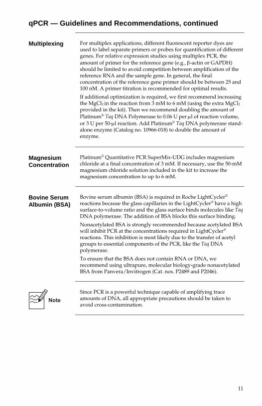
qPCR — Guidelines and Recommendations, continued
Multiplexing For multiplex applications, different fluorescent reporter dyes are
used to label separate primers or probes for quantification of different genes. For relative expression studies using multiplex PCR, the amount of primer for the reference gene (e.g., β-actin or GAPDH) should be limited to avoid competition between amplification of the reference RNA and the sample gene. In general, the final concentration of the reference gene primer should be between 25 and 100 nM. A primer titration is recommended for optimal results.
If additional optimization is required, we first recommend increasing the MgCl2 in the reaction from 3 mM to 6 mM (using the extra MgCl2
provided in the kit). Then we recommend doubling the amount of Platinum® Taq DNA Polymerase to 0.06 U per μl of reaction volume, or 3 U per 50-μl reaction. Add Platinum® Taq DNA polymerase stand-alone enzyme (Catalog no. 10966-018) to double the amount of enzyme.
Magnesium Concentration
Platinum® Quantitative PCR SuperMix-UDG includes magnesium chloride at a final concentration of 3 mM. If necessary, use the 50-mM magnesium chloride solution included in the kit to increase the magnesium concentration to up to 6 mM.
Bovine Serum Albumin (BSA)
Bovine serum albumin (BSA) is required in Roche LightCycler® reactions because the glass capillaries in the LightCycler® have a high surface-to-volume ratio and the glass surface binds molecules like Taq DNA polymerase. The addition of BSA blocks this surface binding.
Nonacetylated BSA is strongly recommended because acetylated BSA will inhibit PCR at the concentrations required in LightCycler® reactions. This inhibition is most likely due to the transfer of acetyl groups to essential components of the PCR, like the Taq DNA polymerase.
To ensure that the BSA does not contain RNA or DNA, we recommend using ultrapure, molecular biology-grade nonacetylated BSA from Panvera/Invitrogen (Cat. nos. P2489 and P2046).
Since PCR is a powerful technique capable of amplifying trace amounts of DNA, all appropriate precautions should be taken to avoid cross-contamination.

12
qPCR — Instruments Using PCR Tubes/Plates
Introduction This section provides a cycling program, reaction mixture, and
protocol for qPCR instruments that use PCR tubes/plates (e.g., ABI PRISM®, Stratagene Mx4000® and Mx3000P™, Corbett Research Rotor-Gene™, MJ Opticon®). For a protocol using the Roche LightCycler®, see page 14.
The tables below summarize the cycling conditions and reaction components for LUX™ Primers and dual-labeled probes. After programming the instrument and preparing the reaction mix, follow the protocol on the following page to perform the reaction.
You can use 1–8 μl of cDNA template in the following protocol, depending on the concentration of the template. Adjust the volume of water in the master mix accordingly for a final reaction volume of 50 μl. We recommend 4 μl of template as a general starting point.
Cycling Program
Program the qPCR instrument as follows:
50°C for 2 minutes hold (UDG incubation) 95°C for 2 minutes hold 50 cycles of: 95°C, 15 seconds 60°C, 30 seconds
Melting Curve Analysis: Refer to instrument documentation. (Melting curve analysis is not available with TaqMan® Probes.)
Master Mix Use the following table to prepare a master mix of all components
except template and water. Note: Preparation of a master mix is crucial in qPCR to minimize pipetting errors.
Component 1 rxn 50 rxns
Platinum® Quantitative PCR SuperMix-UDG1 25 μl 1250 μl
ROX Reference Dye (optional) 1 μl 50 μl Forward primer, 10 μM 1 μl 50 μl Reverse primer, 10 μM 1 μl 50 μl TaqMan® only: Probe, 10 μM 0.5 μl 25 μl Autoclaved, distilled water2 to 42–49 μl to 2100–2550 μl
1Final concentration: 0.06 U/μl Platinum® Taq DNA polymerase, 20-mM Tris-HCl (pH 8.4), 50-mM KCl, 3-mM MgCl2, 200-μM dGTP, 200-μM dATP, 200-μM dCTP, 400-μM dUTP, 1 U UDG 2Volume of water used in the master mix will depend on template volume used in the reaction (see steps 3–4 next page).
Continued on next page

13
qPCR — Instruments Using PCR Tubes/Plates, continued
Protocol 1. Program the qPCR instrument to perform a brief UDG
incubation immediately followed by PCR amplification, as shown on the previous page. Optimal cycling temperatures and times may vary for different target sequences, primer sets, and instruments.
2. Prepare a master mix of all components except template as specified on the previous page.
3. For each reaction, add 42–49 μl of the master mix (depending on template volume) to a 0.2-ml microcentrifuge tube or each well of a 96-well PCR plate.
4. Add 1–8 μl of cDNA template from the first-strand synthesis reaction (Step 7, page 8) to each reaction vessel, for a final reaction volume of 50 μl. (Use 4 μl of template as a general starting point.) Cap or seal the tube/plate.
5. Gently mix and make sure that all components are at the bottom of the reaction vessel. Centrifuge briefly if needed.
6. Place reactions in a thermal cycler programmed as described above. After cycling, hold the reaction at 4°C until further analysis. Collect and analyze the results.

14
qPCR — Roche LightCycler®
Introduction This section provides a cycling program, reaction mixture, and
protocol for the Roche LightCycler®.
The tables below summarize the cycling conditions and reaction components for LUX™ Primers and dual-labeled probes. After programming the instrument and preparing the reaction mix, follow the protocol on the following page to perform the reaction.
Cycling Program
Program the qPCR instrument as follows:
Program choice: Amplification Analysis mode: Quantification 50°C for 2 minutes hold (UDG incubation) 92°C for 1 minute hold 50 cycles of: 92°C, 5 seconds 60oC, 30 seconds (single acquire)
Melting Curve Analysis: Refer to instrument documentation. (Melting curve analysis is not available with TaqMan® Probes.)
Master Mix Use the following table to prepare a master mix of all components
except template. Note: Preparation of a master mix is crucial in qPCR to minimize pipetting errors.
Component 1 rxn 34 rxns Platinum® Quantitative
PCR SuperMix-UDG1 10 μl 340 μl BSA, UltraPure (1 mg/ml)2 1 μl 34 μl Forward primer, 10 μM 1 μl 34 μl Reverse primer, 10 μM 1 μl 34 μl TaqMan® only: Probe, 10 μM 0.5 μl 17 μl Autoclaved, distilled water to 18 μl to 612 μl
1Final concentration: 0.06 U/μl Platinum® Taq DNA polymerase, 20-mM Tris-HCl (pH 8.4), 50-mM KCl, 3-mM MgCl2, 200-μM dGTP, 200-μM dATP, 200-μM dCTP, 400-μM dUTP, 1 U UDG 2Validated with non-acetylated Ultrapure BSA (10% solution) from Invitrogen/Panvera (Cat. nos. P2489 and P2046).
Continued on next page

15
qPCR — Roche LightCycler®, continued
Protocol 1. Program the LightCycler® to perform a brief UDG incubation
immediately followed by PCR amplification, as shown on the previous page. Optimal cycling temperatures and times may vary for different target sequences and primer sets.
2. Set the fluorescence on the LightCycler® to the F1 channel.
3. Prepare a master mix of all components except template as specified on the previous page.
4. For each reaction, add 18 μl of the master mix to each capillary tube.
5. Add 2 μl of the cDNA from the first-strand synthesis reaction (Step 7, page 8) to each capillary tube for a final reaction volume of 20 μl, and cap the tube.
6. Centrifuge tubes at 700 × g for 5 seconds.
7. Place reaction tubes in the rotor of the LightCycler® and run the program. After cycling, hold the reaction at 4°C until further analysis. Collect and analyze the results.

16
Troubleshooting
Problem Possible Cause Suggested Solution
Cells in tissue-culture wells do not detach/burst
Incubation temperature of lysis reaction is too low
Incubate lysis reaction at room temperature instead of on ice.
No amplification curve appears on the qPCR graph
There is no PCR product
Run the PCR product on a gel to determine whether PCR worked. Then proceed to the troubleshooting steps below.
No PCR product is evident, either in the qPCR graph or on a gel
Procedural error Confirm that all steps were followed. Use the Control RNA to verify the efficiency of the first-strand reaction (see the next page on troubleshooting with the Control RNA).
RNA is degraded Add control total HeLa RNA to sample to determine if RNase is present in the first-strand reaction.
A longer DNase I digestion can hydrolyze the RNA in the sample. Use a digestion time of <10 minutes.
Maintain aseptic conditions to prevent RNase contamination.
Fluorescent probe not functional
Validate probe design and presence of fluorophore and quencher: Treat TaqMan® probe with DNase, and check for increase in fluorescence. Redesign and/or resynthesize probe if necessary.
Target mRNA contains strong transcriptional pauses
Maintain an elevated temperature after the annealing step.
Increase the temperature of first-strand reaction (up to 55°C).
PCR product is evident in the gel, but not on the qPCR graph
qPCR instrument settings are incorrect
Confirm that you are using the correct instrument settings (dye selection, reference dye, filters, acquisition points, etc.).
Problems with your specific qPCR instrument
For instrument-specific tips and troubleshooting using LUX™ Primers, see the instrument protocols at www.invitrogen.com/lux. For instrument-specific information about probes, consult your instrument documentation and/or your probe technology documentation.
Poor sensitivity Not enough starting template RNA
Increase the number of cells used
Higher than expected signal
Too much first-strand product was used in qPCR
Decrease amount of the first-strand product in qPCR.
Continued on next page

17
Troubleshooting, continued
Problem Possible Cause Suggested Solution
Signals are present in no-template controls, and/or multiple peaks are present in the melting curve graph
Template or reagents are contaminated by nucleic acids (DNA, cDNA)
Use melting curve analysis if possible, and/or run the PCR products on a 4% agarose gel after the reaction to identify contaminants. To reduce the risk of contamination, take standard precautions when preparing your PCR reactions. Ideally, amplification reactions should be assembled in a DNA-free environment. We recommend using aerosol-resistant barrier tips.
Primer dimers or other primer artifacts are present
Use melting curve analysis to identify primer dimers by their lower melting temperature if possible. We recommend using validated pre-designed primer sets or designing primers or primer/probe combinations using dedicated software programs or primer databases. Primer contamination or truncated or degraded primers can also lead to artifacts. Check the purity of your primers by gel electrophoresis. If agarose gels are used, we recommend cooling the gels before visualization with intercalating dyes.
Product detected at higher than expected cycle number
RNA is degraded Add control total HeLa RNA to sample to determine if RNase is present in the first-strand reaction.
A longer DNase I digestion can hydrolyze the RNA in the sample. Use a digestion time of <10 minutes.
Maintain aseptic conditions to prevent RNase contamination.
Product detected at lower-than-expected cycle number, and/or positive signal from no-template controls
Template or PCR carry-over contamination
Isolate source of contamination and replace reagent(s). Use separate dedicated pipettors for reaction assembly and post-PCR analysis. Assemble reactions (except for target addition) in a DNA-free area. Use aerosol-resistant pipet tips or positive displacement pipettors.
Unexpected bands after electrophoretic analysis
Contamination by genomic DNA
Do not omit the DNase Digestion step on page 7. For larger samples (>1,000 cells), use a longer DNase I incubation time, i.e., up to 10 minutes.
Design primers that anneal to sequence in exons on both sides of an intron or exon/exon boundary of the mRNA to allow differentiation between amplification of cDNA and products potential contaminating genomic DNA.
To test if products were derived from DNA, prepare a negative RT control.
Nonspecific annealing of qPCR primers
Vary the annealing conditions.
Optimize magnesium concentration for each template and primer combination.

18
Related Products Product Size Cat. No.
CellsDirect Resuspension and Lysis Buffer 10 ml Resuspension Buffer and 1 ml Lysis Enhancer
11739-010
E-Gel® Pre-cast Agarose Gels 0.8% Starter Pak 1.2% Starter Pak 2% Starter Pak 4% Starter Pak
9 gels and base 9 gels and base 9 gels and base 9 gels and base
G5000-08 G5000-01 G5000-02 G5000-04
UltraPure™ Agarose 100 g 500 g
15510-019 15510-027
UltraPure™ Agarose 1000 100 g 10975-035
100-bp DNA Ladder 50 μg 15628-019
123-bp DNA Ladder 100 μg 250 μg
15613-011 15613-029
1-Kb Plus DNA Ladder 250 μg 1,000 μg
10787-018 10787-026
19
Technical Support
Web Resources
Visit the Invitrogen website at www.invitrogen.com for:
• Technical resources, including manuals, vector maps and sequences, application notes, SDSs, FAQs, formulations, citations, handbooks, etc.
• Complete technical support contact information
• Access to the Invitrogen Online Catalog
• Additional product information and special offers
Contact Us For more information or technical assistance, call, write, fax, or email.
Additional international offices are listed on our website (www.invitrogen.com).
Corporate Headquarters: 5791 Van Allen Way Carlsbad, CA 92008 USA Tel: 1 760 603 7200 Tel (Toll Free): 1 800 955 6288 Fax: 1 760 602 6500 E-mail: [email protected]
Japanese Headquarters: LOOP-X Bldg. 6F 3-9-15, Kaigan Minato-ku, Tokyo 108-0022 Tel: 81 3 5730 6509 Fax: 81 3 5730 6519 E-mail: [email protected]
European Headquarters: Inchinnan Business Park 3 Fountain Drive Paisley PA4 9RF, UK Tel: +44 (0) 141 814 6100 Tech Fax: +44 (0) 141 814 6117 E-mail: [email protected]
SDS Safety Data Sheets (SDSs) are available at www.invitrogen.com/sds.
Certificate of Analysis
The Certificate of Analysis provides detailed quality control and product qualification information for each product. Certificates of Analysis are available on our website. Go to www.invitrogen.com/support and search for the Certificate of Analysis by product lot number, which is printed on the box.
Limited Warranty
Invitrogen (a part of Life Technologies Corporation) is committed to providing our customers with high-quality goods and services. Our goal is to ensure that every customer is 100% satisfied with our products and our service. If you should have any questions or concerns about an Invitrogen product or service, contact our Technical Support Representatives. All Invitrogen products are warranted to perform according to specifications stated on the certificate of analysis. The Company will replace, free of charge, any product that does not meet those specifications. This warranty limits the Company’s liability to only the price of the product. No warranty is granted for products beyond their listed expiration date. No warranty is applicable unless all product components are stored in accordance with instructions. The Company reserves the right to select the method(s) used to analyze a product unless the Company agrees to a specified method in writing prior to acceptance of the order. Invitrogen makes every effort to ensure the accuracy of its publications, but realizes that the occasional typographical or other error is inevitable. Therefore the Company makes no warranty of any kind regarding the contents of any publications or documentation. If you discover an error in any of our publications, report it to our Technical Support Representatives. Life Technologies Corporation shall have no responsibility or liability for any special, incidental, indirect or consequential loss or damage whatsoever. The above limited warranty is sole and exclusive. No other warranty is made, whether expressed or implied, including any warranty of merchantability or fitness for a particular purpose.

20
Purchaser Notification
Limited Use Label License No. 1: Thermostable Polymerases
Use of this product is covered by one or more of the following US patents and corresponding patent claims outside the US: 5,789,224, 5,618,711, and 6,127,155. The purchase of this product includes a limited, non-transferable immunity from suit under the foregoing patent claims for using only this amount of product for the purchaser’s own internal research. No right under any other patent claim, no right to perform any patented method, and no right to perform commercial services of any kind, including without limitation reporting the results of purchaser's activities for a fee or other commercial consideration, is conveyed expressly, by implication, or by estoppel. This product is for research use only. Diagnostic uses under Roche patents require a separate license from Roche. Further information on purchasing licenses may be obtained by contacting the Director of Licensing, Applied Biosystems, 850 Lincoln Centre Drive, Foster City, California 94404, USA.
Continued on next page

21
Purchaser Notification, continued
Limited Use Label License No. 5: Invitrogen Technology
The purchase of this product conveys to the buyer the non-transferable right to use the purchased amount of the product and components of the product in research conducted by the buyer (whether the buyer is an academic or for-profit entity). The buyer cannot sell or otherwise transfer (a) this product (b) its components or (c) materials made using this product or its components to a third party or otherwise use this product or its components or materials made using this product or its components for Commercial Purposes. The buyer may transfer information or materials made through the use of this product to a scientific collaborator, provided that such transfer is not for any Commercial Purpose, and that such collaborator agrees in writing (a) not to transfer such materials to any third party, and (b) to use such transferred materials and/or information solely for research and not for Commercial Purposes. Commercial Purposes means any activity by a party for consideration and may include, but is not limited to: (1) use of the product or its components in manufacturing; (2) use of the product or its components to provide a service, information, or data; (3) use of the product or its components for therapeutic, diagnostic or prophylactic purposes; or (4) resale of the product or its components, whether or not such product or its components are resold for use in research. For products that are subject to multiple limited use label licenses, the terms of the most restrictive limited use label license shall control. Life Technologies Corporation will not assert a claim against the buyer of infringement of patents owned or controlled by Life Technologies Corporation which cover this product based upon the manufacture, use or sale of a therapeutic, clinical diagnostic, vaccine or prophylactic product developed in research by the buyer in which this product or its components was employed, provided that neither this product nor any of its components was used in the manufacture of such product. If the purchaser is not willing to accept the limitations of this limited use statement, Life Technologies is willing to accept return of the product with a full refund. For information about purchasing a license to use this product or the technology embedded in it for any use other than for research use please contact Out Licensing, Life Technologies, 5791 Van Allen Way, Carlsbad, California 92008; Phone (760) 603-7200 or e-mail: [email protected].
Limited Use Label License No. 14: Direct Inhibition by Anti-Polymerase Antibodies
Licensed to Life Technologies Corporation, under U.S. Patent Nos. 5,338,671; 5,587,287; and foreign equivalents for use in research only.
Continued on next page

22
Purchaser Notification, continued
Limited Use Label License No. 274: 5' Nuclease Process
A license to perform the 5' nuclease process for research requires the use of a Licensed 5' Nuclease Kit (containing Licensed Probe), or the combination of an Authorized Core Kit plus Licensed Probe, or license rights that may be purchased from Applied Biosystems. This product is an Authorized Core Kit without Licensed Probe. Its purchase price includes a limited, non-transferable immunity from suit under U.S. Patents and corresponding patent claims outside the United States, owned by Roche Molecular Systems, Inc. or F. Hoffmann-La Roche Ltd (“Roche”), for using only this amount of the product in the practice of the 5' nuclease process solely for the purchaser's own internal research and development activities. This product is also an Authorized Core Kit for use with service sublicenses available from Applied Biosystems. This product conveys no rights under U.S. Patents Nos. 5,804,375, 6,214,979, 5,538,848, 5,723,591, 5,876,930, 6,030,787, or 6,258,569, or corresponding patent claims outside the United States, expressly, by implication, or by estoppel. No right under any other patent claims (such as apparatus or system claims) and no right to perform commercial services of any kind, including without limitation reporting the results of purchaser's activities for a fee or other commercial consideration, is hereby granted expressly, by implication, or by estoppel. This product is for research purposes only. Diagnostic uses require a separate license from Roche. Further information regarding the 5' nuclease licensing program may be obtained from the Director of Licensing, Applied Biosystems, 850 Lincoln Centre Drive, Foster City, California 94404, USA.

23
References
Higuchi, R., Fockler, C., Dollinger, G., and Watson, R. (1993) Kinetic PCR analysis: Real-
time monitoring of DNA amplification reactions. Bio/Technology 11, 1026-1030.
Higuchi, R., Fockler, C., Walsh, P.S., Griffith, R. (1992) Simultaneous amplification and detection of specific DNA sequences. Bio/Technology 10, 413-417.
Holland P.M., Abramson R.D., Watson R., and Gelfand D.H. (1991) Detection of specific polymerase chain reaction products by utilizing the 5′-3′ exonuclease activity of DNA polymerase. Proc. Natl. Acad. Sci. 88, 7276-7280.
Lowe, B., H. A. Avila, F. Bloom, M. Gleeson and W. Kusser. (2003) Quantitation of gene expression in neural precursors by RT-PCR using self-quenched, fluorogenic LUX primers. Anal. Biochem. 315, 95-105
Nazarenko, I.A., Bhatnagar, S.K., and Hohman, R.J. (1997) A closed tube format for amplification and detection of DNA based on energy transfer. Nucleic Acids Res. 25, 2516-2521.
Nazarenko, I., Pires, R., Lowe, B., Obaidy, M., and Rashtchian, A. (2002a) Effect of primary and secondary structure of oligodeoxyribonucleotides on the fluorescent properties of conjugated dyes. Nucleic Acids Res. 30, 2089-2195
Nazarenko I., Lowe B., Darfler M., Ikonomi P., Schuster D., and Rashtchian A. (2002b) Multiplex quantitative PCR using self-quenched primers labeled with a single fluorophore. Nucleic Acids Res. 30, e37.
Ririe, K.M., Rasmussen, R.P., and Wittwer, C.T. (1997) Product differentiation by analysis of DNA melting curves during the polymerase chain reaction. Anal. Biochem. 245, 154-160.
Tyagi, S. and Kramer, F.R. (1996) Molecular beacons: Probes that fluoresce upon hybridization. Nat. Biotechnol. 14, 303-308.
Wittwer, C.T., Herrmann, M.G., Moss, A.A., and Rasmussen, R.P. (1997) Continuous fluorescence monitoring of rapid cycle DNA amplification. BioTechniques 22, 130-138.
Potter, J, Zheng, W, and Lee, J (2003) Focus® 25, 17-21
Sambrook, J., Fritsch, E.F., Maniatis, T. (2nd ed.) (1989) Molecular cloning. Cold Spring Harbor Laboratory Press.
Orth, H.D. (1976) Kontakte 3, 35.
ABI PRISM® and GeneAmp® are registered trademarks of Applera Corporation. TaqMan® is a registered trademark of Roche Molecular Systems, Inc. iCycler™, Mx4000™, Rotor-Gene™, DNA Engine Opticon™, and Smart Cycler® are trademarks of their respective companies. LightCycler® is a registered trademark of Idaho Technologies, Inc.
©2010 Life Technologies Corporation. All rights reserved. For research use only. Not intended for any animal or human therapeutic or diagnostic use. The trademarks mentioned herein are the property of Life Technologies Corporation or their respective owners.

24
Notes


User Manual
Corporate Headquarters5791 Van Allen WayCarlsbad, CA 92008T: 1 760 603 7200F: 1 760 602 6500E: [email protected]
For country-specific contact information visit our web site at www.invitrogen.com