Superparamagnetic Iron Oxide Nanoparticles: Promises for
24
Introduction In 2008, it has been recognized that more than 1 million of Americans and more than 10 million people worldwide were expected to be diag- nosed with cancer [1]; however, in 2010 it was expected that the average percentage of people who suffer from some sorts of cancer would be 31% (according to the recent U.N. organized surveys performed in multiple countries). This considerable increase in cancer incidence may relate to the fact that determination of the can- cer is strongly dependent upon the develop- ment of new diagnostic technologies. However, what is of great importance is that cancers are going to be a significant cause of human death in near future. Thus, intense scientific research was focused on the diagnosis and treatment of cancer. Due to the dysregulated cell growth in the hu- man body, large groups of different diseases occurred which became recognized as cancer. The uncontrollable cell-cycles not only can form malignant tumors but also can be spread to other parts of the body through the lymphatic system or bloodstream, which is called “metastasis”. Although there are numerous available anti-cancer drugs, the main problems currently associated with systemic drug admini- stration include the general systemic distribu- tion of therapeutic drugs, the lack of drug speci- ficity towards a pathological site, the necessity of a large dose to achieve high local concentra- tion, non-specific toxicity and other adverse side effects [2]. Therefore, using modern drug deliv- ery systems are essential for high-yield cancer therapy. The term “drug delivery” refers to the pharma- ceutical agents of interest which are entrapped within, or attached to, an organic polymer matrix or inorganic particles, and in that case, drug safety and efficacy can be greatly improved and new targeted therapies are possible [2]. The strategy for drug delivery can be approached by many methods: (1) drug modification by chemi- cal means [3, 4]; (2) drug encapsulated in vesi- Int J Mol Epidemiol Genet 2011;2(4):367-390 www.ijmeg.org /ISSN1948-1756/IJMEG1111002 Review Article Superparamagnetic iron oxide nanoparticles: promises for diagnosis and treatment of cancer Sophie Laurent 1 , Morteza Mahmoudi 2,3 1 Department of General, Organic, and Biomedical Chemistry, NMR and Molecular Imaging Laboratory, University of Mons, Avenue Maistriau, 19, B-7000 Mons, Belgium; 2 National Cell Bank, Pasteur Institute of Iran, Tehran, Iran; 3 Nanotechnology Research Center, Faculty of Pharmacy, Tehran University of Medical Sciences, Tehran, Iran Received November 14, 2011; accepted November 22, 2011; Epub November 25, 2011; Published December 15, 2011 Abstract: During the last decade, significant scientific research efforts have led to a significant growth in understand- ing of cancer at the genetic, molecular, and cellular levels providing great opportunities for diagnosis and treatment of cancer diseases. The hopes for fast cancer diagnosis and treatment were significantly increased by the entrance of nanoparticles to the medical sciences. Nanoparticles are attractive due to their unique opportunities together with negligible side effects not only in cancer therapy but also in the treatment of other ailments. Among all types of nanoparticles, surface-engineered superparamagnetic iron oxide nanoparticles (SPIONs) have been attracted a great attention for cancer therapy applications. This review covers the recent advances in the development of SPIONs to- gether with their opportunities and challenges, as theranosis agents, in cancer treatment. Keywords: Cancer, tumor, nanoscience, nanotechnology, nanoparticle, superparamagnetic iron oxide nanoparticles, SPIONs, coatings, surfaces, targeted drug delivery, controlled drug release, anticancer drugs, chemotherapy, radio- therapy, thermotherapy
Transcript of Superparamagnetic Iron Oxide Nanoparticles: Promises for
Introduction In 2008, it has been recognized that more than 1 million of Americans and more than 10 million people worldwide were expected to be diag-nosed with cancer [1]; however, in 2010 it was expected that the average percentage of people who suffer from some sorts of cancer would be 31% (according to the recent U.N. organized surveys performed in multiple countries). This considerable increase in cancer incidence may relate to the fact that determination of the can-cer is strongly dependent upon the develop-ment of new diagnostic technologies. However, what is of great importance is that cancers are going to be a significant cause of human death in near future. Thus, intense scientific research was focused on the diagnosis and treatment of cancer. Due to the dysregulated cell growth in the hu-man body, large groups of different diseases occurred which became recognized as cancer. The uncontrollable cell-cycles not only can form
malignant tumors but also can be spread to other parts of the body through the lymphatic system or bloodstream, which is called “metastasis”. Although there are numerous available anti-cancer drugs, the main problems currently associated with systemic drug admini-stration include the general systemic distribu-tion of therapeutic drugs, the lack of drug speci-ficity towards a pathological site, the necessity of a large dose to achieve high local concentra-tion, non-specific toxicity and other adverse side effects [2]. Therefore, using modern drug deliv-ery systems are essential for high-yield cancer therapy. The term “drug delivery” refers to the pharma-ceutical agents of interest which are entrapped within, or attached to, an organic polymer matrix or inorganic particles, and in that case, drug safety and efficacy can be greatly improved and new targeted therapies are possible [2]. The strategy for drug delivery can be approached by many methods: (1) drug modification by chemi-cal means [3, 4]; (2) drug encapsulated in vesi-
Int J Mol Epidemiol Genet 2011;2(4):367-390 www.ijmeg.org /ISSN1948-1756/IJMEG1111002
Review Article Superparamagnetic iron oxide nanoparticles: promises for diagnosis and treatment of cancer Sophie Laurent1, Morteza Mahmoudi2,3
1Department of General, Organic, and Biomedical Chemistry, NMR and Molecular Imaging Laboratory, University of Mons, Avenue Maistriau, 19, B-7000 Mons, Belgium; 2National Cell Bank, Pasteur Institute of Iran, Tehran, Iran; 3Nanotechnology Research Center, Faculty of Pharmacy, Tehran University of Medical Sciences, Tehran, Iran Received November 14, 2011; accepted November 22, 2011; Epub November 25, 2011; Published December 15, 2011 Abstract: During the last decade, significant scientific research efforts have led to a significant growth in understand-ing of cancer at the genetic, molecular, and cellular levels providing great opportunities for diagnosis and treatment of cancer diseases. The hopes for fast cancer diagnosis and treatment were significantly increased by the entrance of nanoparticles to the medical sciences. Nanoparticles are attractive due to their unique opportunities together with negligible side effects not only in cancer therapy but also in the treatment of other ailments. Among all types of nanoparticles, surface-engineered superparamagnetic iron oxide nanoparticles (SPIONs) have been attracted a great attention for cancer therapy applications. This review covers the recent advances in the development of SPIONs to-gether with their opportunities and challenges, as theranosis agents, in cancer treatment. Keywords: Cancer, tumor, nanoscience, nanotechnology, nanoparticle, superparamagnetic iron oxide nanoparticles, SPIONs, coatings, surfaces, targeted drug delivery, controlled drug release, anticancer drugs, chemotherapy, radio-therapy, thermotherapy
Superparamagnetic iron oxide nanoparticles and cancer
368 Int J Mol Epidemiol Genet 2011:2(4):367-390
cles [5-7] or inorganic hollow nanoshells [8-10]; (3) controlled release system such as polymer-based [11-13] and hydrogel-based vectors [14-16], and (4) small-scale integrated systems such as microchip systems [17-20]. The delivery routes are also an important issue for systemic delivery of drugs due to the systemic circulation. Intravenous [21, 22], intraperitoneal [23], pul-monary [24], transdermal [25-27], nose [28, 29], vaginal [30] and eye [31, 32] routes of ad-ministration are the approaches designed to improve the selectivity of chemotherapy. Although there are significant improvements in targeted drug delivery systems, there is a great deal of commonly pursued, desirable qualities for such efficient drug delivery devices, which are not necessarily guidelines. The crucial areas of potential that powerful drug delivery systems should have are: (1) long circulation (i.e. long half-life), (2) high levels of bioavailability and specific targeting, (3) intracellular/organelle delivery, (4) stimuli responsiveness (i.e. effi-cacy), (5) reporting/imaging, and (6) biodegra-dation. In recent years, the fabrication of nanoparticles and exploration of their unusual properties have attracted the attention of physicists, chemists, biologists and engineers. Interest in nanoparti-cles arises from the fact that the mechanical, chemical, electrical, optical, magnetic, electro-optical and magneto-optical properties of these particles are different from their bulk properties and depend on the particle size. Entrance of nanoparticles in the medical field caused signifi-cant hopes for early diagnosis and treatment of catastrophic diseases such as cancer. Nanopar-ticles are typically referred to as microscopic particles that are between 1 nm and 100 nm in size. Using nanoparticles, delivery systems have the potential to target drugs to specific sites of the body or precisely control drug release rates for prolonged times according to body reaction of chemicals microenvironment [2]. More spe-cifically, nanoparticles as drug carriers in tar-geted delivery systems in cancer therapies can provide desired and precise penetration of therapeutic and diagnostic substances within for example tumor sites/tissues/cells while their corresponding side effects are much lower in comparison to conventional cancer therapies [2]. Nanoparticles, as smart drug delivery systems,
have strong capability to overcome numerous challenges that still exist including the targeting of drugs to specific cells, the creation of novel vaccine delivery approaches, and the develop-ment of cell-based delivery systems. Nanoscale drug delivery devices that are stimuli responsive respond dynamically to changes in the environ-ment. These stimuli could be in the form of changes in temperature [33], light [16], pH [15, 34, 35], ultrasound [35-37], or magnetic fields [38, 39]. Disease processes which upset ho-meostasis can lead to environmental changes that can be exploited by stimuli-responsive therapeutics. Thus, in recent years, significant efforts have been devoted to develop nanotech-nology for drug delivery since it offers a suitable means of delivering small molecular weight drugs [40], as well as macromolecules such as proteins, peptides [41] or genes [42, 43] by either localized or targeted delivery to the tissue of interest. It is also worthwhile to mention that nanoparticles may have promise for biomolecu-lar imaging [44]; thus, another potentially impor-tant facet of nanoparticles and its application in cancer research could be its capability to probe underlying mechanisms in cancer. In order to increase the efficiency of drug deliv-ery, magnetic nanoparticles are defined as a promising candidate not only due to capacity of antibodies to attach to their surfaces, but also due to achieve targeting ability by using external magnetic guidance [45, 46]. Among different types of magnetic particles, superparamagnetic iron oxide nanoparticles (SPIONs), with a mean diameter as low as 10 nm and superior mag-netic properties, have proven to be among the most capable candidates [47]. In the field of drug delivery, SPIONs particles are considered as small, thermally agitated magnets in carrier liquids, which are called “ferrofluids”. A distin-guishing feature of SPIONs for drug delivery is their applicability for both alternatives (i.e. mag-netic properties and antibody attachment) and consequently developing a targeting capability [2]. Superparamagnetism essentially acts as an activation mechanism because once the exter-nal magnetic field is removed, the magnetiza-tion disappears, and thus the agglomeration, and hence the possible embolization of the cap-illary vessels can be avoided [48]. SPIONs also have capability to be used as multi-functional agents [49-51]. The surfaces of SPI-ONs are engineered to have several simultane-
Superparamagnetic iron oxide nanoparticles and cancer
369 Int J Mol Epidemiol Genet 2011:2(4):367-390
ous biomedical-related functions such as drug carrier properties, magnetic resonance imaging (MRI) contrast agents, and local heat induction (hyperthermia) capacity. More recently, a field called “molecular imaging” has appeared, this technique allows the in vivo visualization of mo-lecular events occurring at the cellular level and needs the development of high affinity ligands and their grafting to SPIONs [44]. SPIONs are also good substrates for bioconjugation, they are used as reporters for many physiologic proc-esses and have a lot of clinical applications such as liver and spleen imaging, inflammation, apoptosis, and cardiovascular disease [45, 52, 53]. SPIONs can be accumulated in a biological site by passive or active targeting mechanism. In passive targeting, contrast agents are concen-trated in the phagocytic cells (e.g. Kupffer’s cells and macrophages) and corresponding or-gans (e.g. liver, spleen, and lymph nodes) that are responsible for clearance from the body. For example, SPIONs coated with dextran, carboxy-dextran have been used for passive targeting [54-56]. In active targeting, contrast agents need conjugation to a specific ligand to target a site of interest. Typical ligands include antibod-ies, peptides, sugars, aptamers, etc. which are molecules that can be linked to SPIONs cova-lently or non-covalently [52, 57, 58]. For biological applications, nanoparticles must be highly stable in aqueous ionic solutions at physiological pH. Vectors grafted on their sur-face must be able to recognize the target cells or tissues. Particles must be non-toxic and re-main in the circulation for a sufficient amount of time to allow targets to be reached. Intensive research are currently undertaken to develop specific contrast agents in targeted cancer im-aging. For example, αvβ3 integrin targeted SPI-ONs have been used for the specific MRI detec-
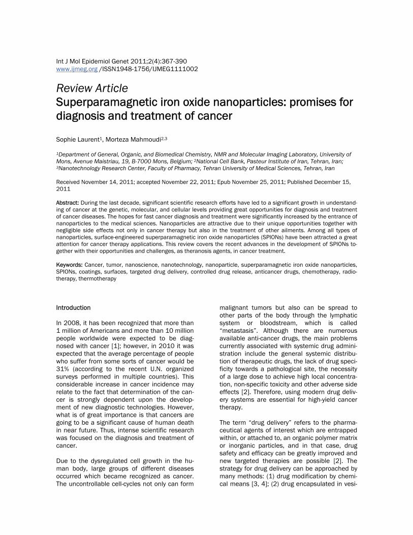
tion of small regions of angiogenesis associated with solid tumors [59, 60]. In summary, SPIONs can be used as theranosis agent in the context of cancer, in other words it can serve as both a diagnostic and therapeutic agent. In this review, the main protocols for the grafting of vectors to SPIONs are reported and some applications are described. In addition, we discuss various in vitro and in vivo studies using surface engineered SPIONs for diagnosis and treatment of cancer diseases. Synthesis and stabilization of SPIONs Several methods for chemical synthesis of SPI-ONs have been described. The most commonly used are summarized in Table 1. Amongst these methods, co-precipitation of Fe2+ and Fe3+ ions in a basic aqueous media (e.g. NaOH or NH4OH solutions) is the simplest way, but usually nanoparticles are polydispersed and poorly crys-tallized [48]. To avoid these disadvantages, thermal decomposition methods have been employed to produce SPIONs with monodisper-sity and uniform crystalline [61]. Subsequently, the hydrophobic iron oxide nanoparticles can be coated with phospholipids, silica, or amphiphilic polymers as shells to display good solubility and biocompatibility in vivo (Figure 1). SPIONs can be coated in situ during the nuclea-tion and growth of the magnetic core (this simul-taneous process is often referred to as the “one-pot” method) or after the synthesis following the final application. Monomeric organic stabi-lizers, polymers and inorganic coatings can also be used to stabilize nanoparticles in aqueous solutions [46, 69]. Organic surfactants are fre-quently used for the stabilization and coating of magnetic nanoparticles. Fatty acids can stabi-lize the aqueous fluids by the formation of a surface bilayer with a chemisorbed fatty acid
Table 1. Principal preparation methods of SPIONs Synthetic method Advantages Disadvantages References
Coprecipitation Rapid synthesis with high yield Problem of oxidation and aggregation [48, 62]
Hydrothermal reac-tions
Narrow size distribution and good control, scalable
Long reaction times [63, 64]
High temperature decomposition
Good control of size and shape, High yield
Furthers steps needed to obtain water stable suspension
[61, 65, 66]
microemulsion Control of particle size Poor yield and large amounts of solvent required, excess of surfactant to elimi-nate
[51, 67, 68]
Superparamagnetic iron oxide nanoparticles and cancer
370 Int J Mol Epidemiol Genet 2011:2(4):367-390
primary layer and an interpenetrating second layer, the latter is physisorbed onto the primary layer with the hydrophilic head-groups pointing outwards [70]. Coating agents which are physically adsorbed (by electrostatic interactions or hydrogen bind-ing) show limited stability in comparison to coat-ing agents which are chemically adsorbed. The stability of the coating grafting also depends on the quantity of the chemical interaction that each molecule or macromolecule can establish with the SPION surface. The most common coat-ings for biocompatible iron oxide suspensions are polymers [71] such as derivatives of dextran [72, 73] (dextran, carboxymethylated dextran, carboxydextran), arabinogalactan, glycosami-noglycan, starch, polyethyleneglycol, siloxane, sulphonated styrene-divinylbenzene, poly(lactic acid), poly(ε-caprolactone) or polyalkyl-cyanoacrylate [74-76]. Due to their good solubility in water and biocom-patibility, polysaccharides are among the most
commonly used coating for the stabilisation of SPIONs. Dextran-SPION can be prepared using co-precipitation method with in situ coating by polysaccharide[77]. Molday and MacKenzie used ferrous and ferric chloride under basic condition in the presence of dextran. Dextran-coated SPIONs with surface functionalities were also devel-oped for specific applications: carboxydextran for cell labeling [78] or aminodextran for graft-ing with DNA [79]. The chemical modifications of dextran have showed that reduction of termi-nal sugars can have a signifi-cant effect on particle size and coating stability [80]. Particles prepared with carboxydextran yielded a more stable coating [71, 81]. Considering the dex-tran-coated particles, no evi-dence of strong chemical ad-sorption was observed by FTIR (Fourier transform infrared spectroscopy) and SSIMS (statistic secondary ion mass spectra) analysis [82]. The na-ture of the interactions between the dextran and the SPION sur-
face and its evolution with temperature has been investigated by thermogravimetric and differential thermal analyses and by coupling these data with FTIR analysis [83]. Noteworthy, these dextran-coated iron oxide particles do not show any toxicity [84-87]. Other surface-modifying agents have been ex-plored to increase stability of magnetic nanopar-ticles. In order to obtain a strong conjugation of dextran to the maghemite surface, Mornet et al. [88] have described a synthetic route consisting of surface modification of SPIONs by silanation of the iron core with aminopropylsilane groups and covalent conjugation with partially oxidized dextran and subsequent reduction of the shiff base [89]. The bonding nature of organosilanes to iron surfaces can be analyzed by FTIR and secon-dary ion mass spectroscopy (ToF-SIMS). The Si-O-Fe bond is commonly described as covalent [90]. Such systems are ordered molecular as-semblies formed by the adsorption of an active
Figure 1. Scheme showing representative groups that can be used to stabi-lize the SPIONs.
Superparamagnetic iron oxide nanoparticles and cancer
371 Int J Mol Epidemiol Genet 2011:2(4):367-390
molecule (e.g. siloxane, carboxylates, thiolates, and phosphate) on a solid surface (as iron ox-ide) with different terminal groups (usually -OH, -COOH, and -NH) [91, 92]. This coating can pre-sent terminal groups allowing further function-alization by chemical reactions. These function-alities are also frequently incorporated in vari-ous polymers where attachment of species on the surface is desired. The stability depends basically on the affinity of the active molecule for the substrate (solid surface), pH and ionic strength of the environment. Dextran-coated superparamagnetic iron oxide particles can also form stable complexes with transfection agents. Moreover, such complexes can be internalized by endosomes/lysosomes, and have been utilized for tell labeling and in vivo MRI tell tracking [93]. Alginate, another polysaccharide, has also been used for the coating and stabilization of magnetic nanoparti-cles [51]. Starch-coated SPIONs, obtained via co-precipitation in the presence of starch were also investigated for targeting of brain tumors in rats [94]. Chitosan, a biocompatible and bio-degradable polymer, is of particular interest for coating magnetic nanoparticles [95, 96]. It has been reported that oleic acid-coated SPI-ONs can be easily dispersed in chitosan, pro-ducing stable ferrofluids with a typical hydrody-namic diameter of approximately 65 nm [97]. Polylactic acid, another biodegradable poly-mer, has been used to prepare stable biocom-patible ferrofluids with different ferromagnetic particle sizes, ranging from 10 to 180 nm [98]. Polylactic acid-coated nanoparticles can also be loaded with anticancer drugs (e.g., tamoxifen), which allows their use in simulta-neous tumor imaging, drug delivery and the real-time monitoring of therapeutic effects [99]. Similar biocompatible nanoparticles have also been prepared via an in situ controlled co-precipitation of magnetite from aqueous solutions containing suitable Fe2+ and Fe3+ salts, in a polymeric starch matrix. This proc-ess resulted in starch-coated SPIONs that demonstrated good potential for the imaging of nerve tells and the brain [94]. One very successful strategy for the prepara-tion of stable and biocompatible nanoparticles is to graft polyethylene glycol (PEG) onto the sur-face (a process known as PEGylation). PEG is not only biocompatible but also has favorable chemi-
cal properties and solubility. In this situation, the stabilization is due primarily to steric inter-actions, while PEGylation can be used to fur-ther enhance the pharmacokinetic properties and improve the blood circulation times [100, 101]. For example, an increased image contrast in MRI was achieved by using polymeric mi-celles formed from SPIONs encapsulated in biocompatible, biodegradable poly(ε-caprolactone)-β-PEG copolymers. These materi-als have demonstrated significantly improved r2 relaxivities and a good sensitive MRI detection [102]. Copolymers of PEG were also used to stabilize the SPIONs [51, 62]. Aside from the extended half-life that it can pro-vide, one of the great advantages of PEG coat-ing is that it can also be easily conjugated to antibodies or to peptides to achieve a specific targeted delivery. For example, in a recent re-port, biocompatible water-soluble magnetite nanocrystals were fabricated via the thermal decomposition of ferric triacetylacetonate in 2-pyrrolidone in the presence of monocarboxyl-terminated PEG (MPEG-COOH) [103]. The car-boxylic acid groups on the surface of the parti-cles were conjugated with a cancer-targeting anti-carcinoembryonic antigen (CEA) mono-clonal antibody, via a carbodiimide coupling reaction. The resultant materials were as-sessed for their ability to label cancer tissues in vivo, for subsequent MRI detection [2]. PEG-coated iron oxide nanoparticles may also be conjugated to specific targeting peptides and receptors such as chlorotoxin [104], transac-tivator protein (Tat) of HIV-1 [105], and in-tegrins [59]. The high temperature decomposition process, known to give nanoparticles a better control of particle size and monodispersity, was described as a one-pot method by decomposition of Fe(acac)3 in 2-pyrolididinone in the presence of MPEG-COOH [103]. Another way to introduce PEG onto SPIONs is to use PEG-silane [106]. This involves reaction of the triethoxysilane group with the hydroxyl group of the SPION sur-face. Sun et al showed that SPIONs can be treated successively with a bifunctional silane PEG trifluoroethylester linker, ethylenediamine and folic acid (FA). The specificity of these vec-torized SPIONs was demonstrated by an in-creased nanoparticle uptake and a significant contrast enhancement of tumor cells. An alter-native protocol to prepare SPION-PEG-FA in-
Superparamagnetic iron oxide nanoparticles and cancer
372 Int J Mol Epidemiol Genet 2011:2(4):367-390
volved the reaction of FA with tertbutyl-oxycarbonyl (t-BOC) and N-hydroxysuccinimide (NHS), followed by the reaction of (t-boc)folate-NHS with amino-PEG-carboxyl [107]. The resul-tant (t-boc)folate-PEG-carboxyl was then reacted with SPION with have NH2 groups obtained by reaction with 3-aminopropyl-trimethoxysilane. Finally, the t-boc protective group was removed. Recently, another anchor, dopamine (DPA) has been proposed due to its high affinity for the iron oxide nanoparticle surface and the possibil-ity of functionalization with other molecules through amide bonds [108]. Other polymers and copolymers, which have been used to coat magnetic nanoparticles, include PVP), polyethylenimine (PEI), polyvinyl alcohol (PVA), polysodium-4-styrene sulfonate, poly(trimethylammonium ethylacrylate methyl sulfate)-poly-(acrylamide), polyvinylbenzyl-O-beta-D-galactopyranosyl D-gluconamide (PVLA), poly-caprolactone, and gummic acid [51, 62]. In addition, several stable and biocompatible magnetic fluids have been prepared by coating magnetic nanoparticles with proteins, such as human serum albumin (HSA), avidin, and Annexin AS (anxA5)-VSOP [109, 110]. Dicarboxylic or tricarboxylic acids (citric, tartaric or dimercaptosuccinic acids) [111, 112] are also used for the surface functionalization and stabilization of SPIONs. Some of the functional groups can bind to the surface of the iron oxide, while the remaining carboxylate groups provide negative charges (depending on the pH) and improve the hydrophilicity of the SPION surface. Several studies are reported and demonstrated that phosphonates and phosphates bind effi-ciently to iron oxide particle surfaces and can serve as potential alternatives to fatty acids [92, 113, 114]. Functionalized phosphonate and phosphate seems to have an acceptable bio-compatibility [115] and it is possible to suggest their utilization as coating agents of magnetic nanoparticles in medical applications [116]. Phosphonates are molecules that contain one or more R-PO(OH)2 Lewis acid groups. The P-C bond is very stable toward oxidation or hydroly-sis. These compounds possess a very high abil-ity to form strong complexes with transition met-als in aqueous solution and show a large affinity for the metal oxide surfaces [117]. The mechanism of adsorption of dimercapto-succinic acid (DMSA) has been studied by con-
ductimetric measurements and adsorption iso-therms curves. DMSA is oxidized during the coating process in tetrameric polysulfide chains [DMSAox]4 which are absorbed by the carboxy-late moiety on the particles after alkalisation and neutralisation. The obtained particles are stable particles at pH = 7 [118, 119]. Among the inorganic coatings, silica, carbon, precious metals (e.g., Ag and Au), or metal ox-ides are the most frequently used [69]. The silica coating significantly improves the stability of magnetic nanoparticles, protecting them from oxidation, and reduces any potential toxic effects of the nanoparticles [62]. Silica coat-ing can be achieved by using several different approaches; the most popular is the sol-gel process with tetraethyl orthosilicate (TEOS) (known as the Stober method) [120, 121]. Sil-ica shell formation is achieved by the hydrolysis of TEOS in the presence of ammonia and SPI-ONs, the thickness of the silica coating can be controlled by varying the concentration of am-monium and the ratio of TEOS to water. Amino-silane coatings were activated using glutaralde-hyde, which served as a linker for the binding of a monoclonal antibody directed against can-cer. This process resulted in new immu-nomagnetic nanoparticles for the targeted MRI of cancer [122]. Strategies of vectorization of magnetic nanoma-terials for targeted imaging Targeted cellular labeling and molecular imag-ing require further functionalization in order to provide molecular recognition for specific biologi-cal sites. Vectorization is also critical for the further stabilization of nanoparticles, to im-prove their biocompatibility, and to reduce their potential toxicity. The main vectorization strate-gies include: (i) the noncovalent grafting of bio-molecules (e.g., antibodies or proteins) via ionic bonding or adsorption; and (ii) the cova-lent conjugation of biomolecules via strong chemical bonding [123]. Typical examples of the noncovalent approach include the prepa-ration of streptavidin-coated iron oxide nanoparticles [124-126]. Although the nonco-valent methods are relatively easy to undertake, the results are very often not reproducible and the response of the materials may be very difficult to control. In addition, noncovalently functionalized nanocomposites are sometimes unstable in variable biological media, and may
Superparamagnetic iron oxide nanoparticles and cancer
373 Int J Mol Epidemiol Genet 2011:2(4):367-390
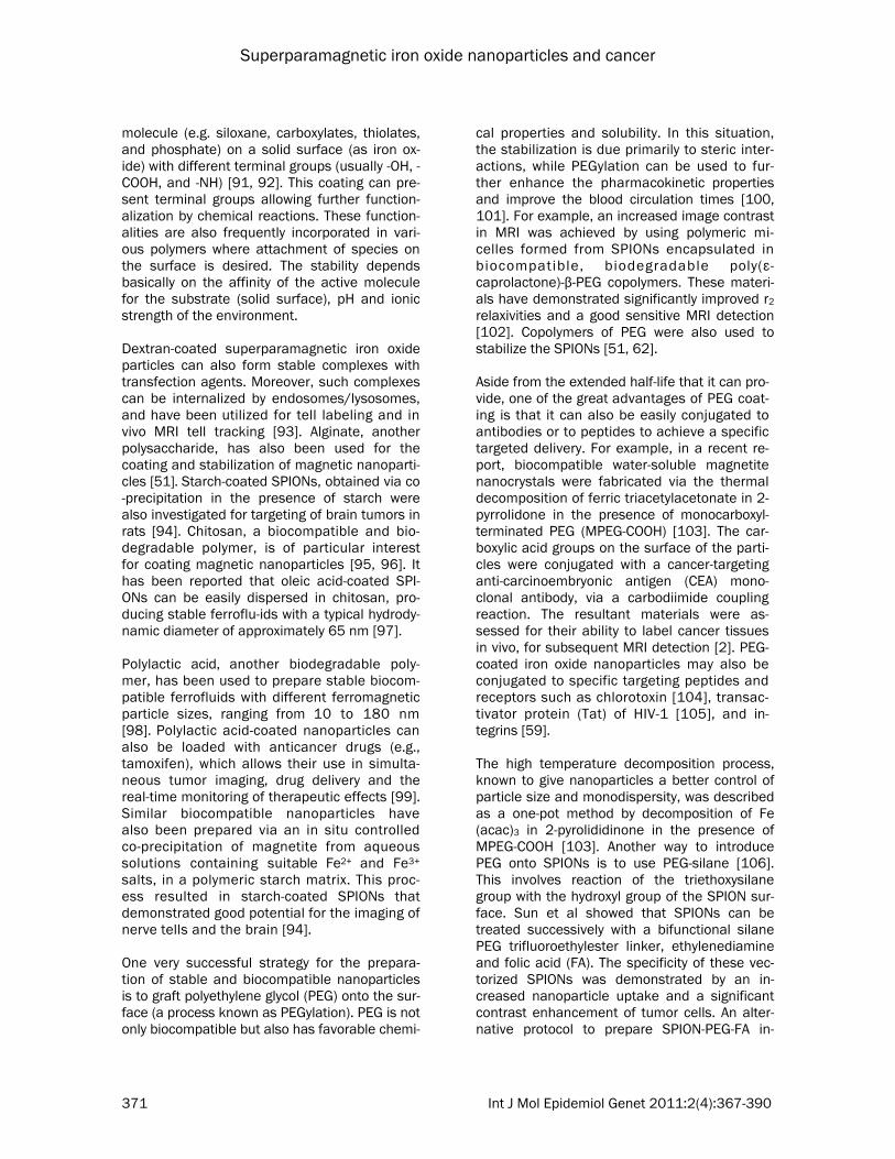
lose their biological coating and undergo pre-cipitation. Therefore the development of a co-valent approach has attracted much more at-tention during recent years. This involves the formation of linkages between vectors and nanoparticles (see Figure 2). The linkage must be stable (it should be resis-tant to hydrolysis, oxidizing and reducing condi-tions) and performed in mild conditions to avoid vector degradation. Typical vector are peptides, proteins, antibodies, mimetic molecules, carbo-hydrates, lipids, etc. The usual reacting group is an amine or a carboxylic function in the pres-ence of carbodiimine as activator of the carbox-ylic group but reactions with hydroxyl, thiol or phenol residues are also possible. An oxidative conjugation strategy has been used in previous studies. This method is based on the periodate oxidation of a carbohydrate poly-mer like dextran or carboxydextran to aldehydes, which may then be linked to biomolecules through the formation of a Schiff base. This strategy has been used for the covalent conju-gation of dextran coated magnetic nanoparti-cles with peptides [127], proteins [128, 129], monoclonal antibodies [130-132] or agglutinin [133]. A substantial loss of the biological activ-ity of the protein has been observed. To mini-mize this effect, Hogemann et al. [134] have linked proteins and iron oxide particles via a linker. Their results suggest that the oxidative
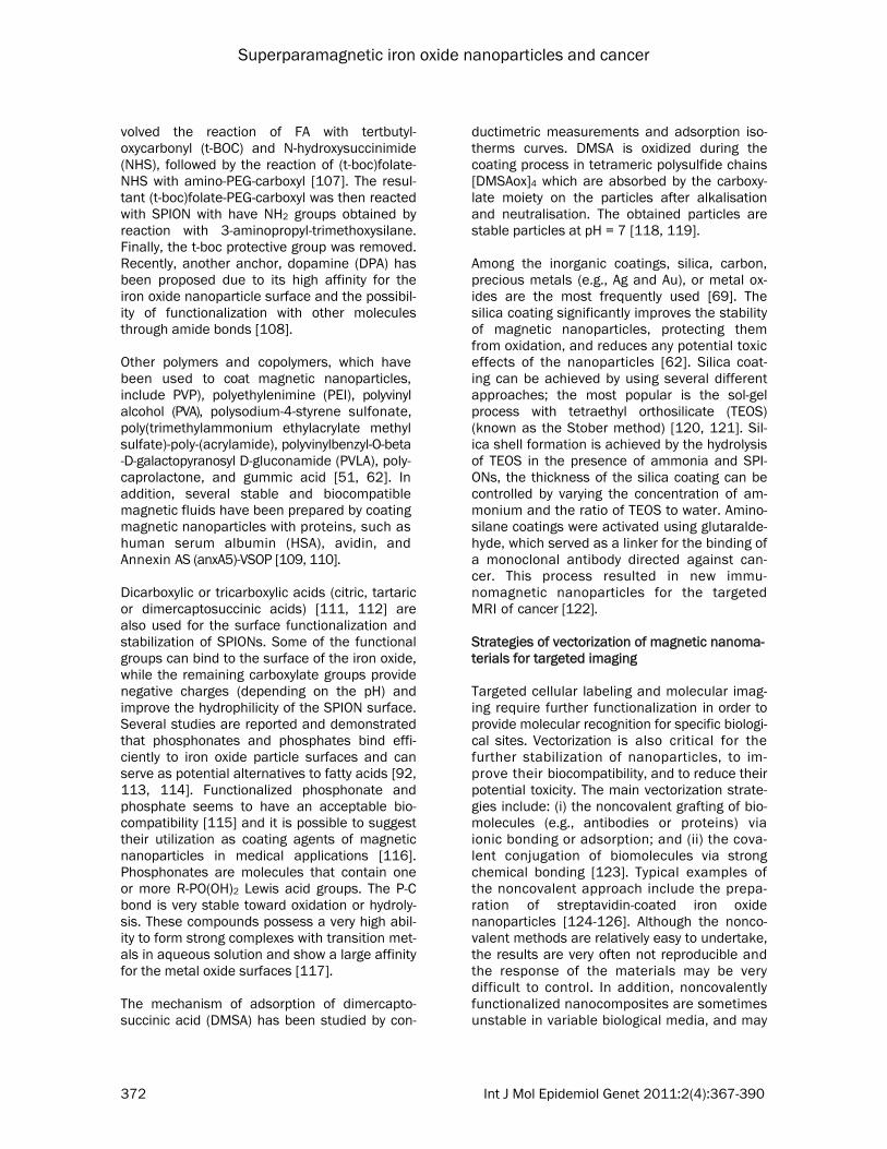
conjugation chemistry significantly interferes with the binding of the conjugates of the recep-tor. Current efforts are devoted in the direction of non oxidative strategies. The target mole-cules can be covalently linked through a 3 step-reaction sequence described by Josephson et al. [135]. This approach is based on amine-terminated CLIO nanoparticles, which can be obtained from dextran-coated nanoparticles by cross-linking using epichlorohydrin and then ammonia. A peptide was attached to the amino group of a cross linked dextran iron oxide (CLIO-NH2) using SPDP through a disulfide exchange reaction. A range of target biomolecules can be conjugated by using standard organic chemis-try methods: formation of disulfide, carbon-thiol, and amide bonds [105, 136-139]. In the so-called “DMSA techniques” 2,3-dimercaptosuccinic acid (DMSA) -coated mag-netic nanoparticles can be covalently linked to a variety of biomolecules via S-S bonds using N-succinimidyl 3-(2-pyridyldithio) propionate (SPDP) as a coupling agent (see Figure 3) [118]. This approach has been used to couple antibod-ies, lectins and annexin V to DMSA-coated mag-netic nanoparticles [119, 140, 141]. Magnetite nanoparticles coated with silica were prepared, after surface modification with an amino-silane coupling agent, SG-Si900, amine was covalently linked using glutaraldehyde as a cross-linker [142]. Alternatively, vectors with carboxylic functions can be directly grafted on the silica coated particles using EDC to activate the carboxyl groups [143]. The silane coupling materials (like 3-aminopropyltrimethoxysilane or p-aminophenyl trimethoxysilane) [144] are able to adsorptively or covalently bind to the metal oxide and are able to form covalent bonds with bioaffinity adsorbents through organo-functionalities. The mechanism of the silane coupling agents reaction according to Arkles
Figure 2. Covalent grafting of vectors (V) onto the nanoparticles (P) such as for a molecular imaging probe.
Figure 3. Formation of grafted particles via S-S bridge: the pyridyl sulfide moiety of SPDP grafted on the vector is sub-stituted by the SH group on the nanoparticles. Reprinted with permission from (reference 52). Copyright 2010 Ameri-can Chemical Society.
Superparamagnetic iron oxide nanoparticles and cancer
374 Int J Mol Epidemiol Genet 2011:2(4):367-390
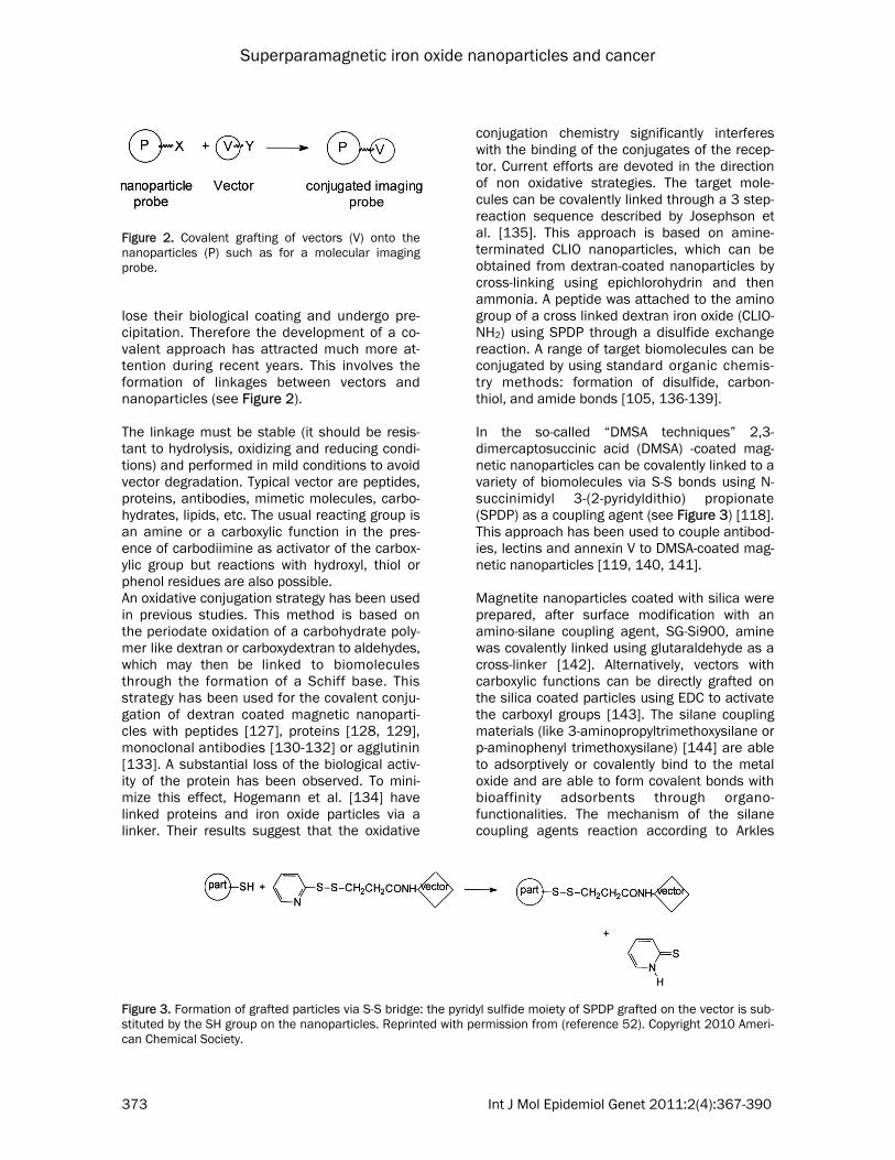
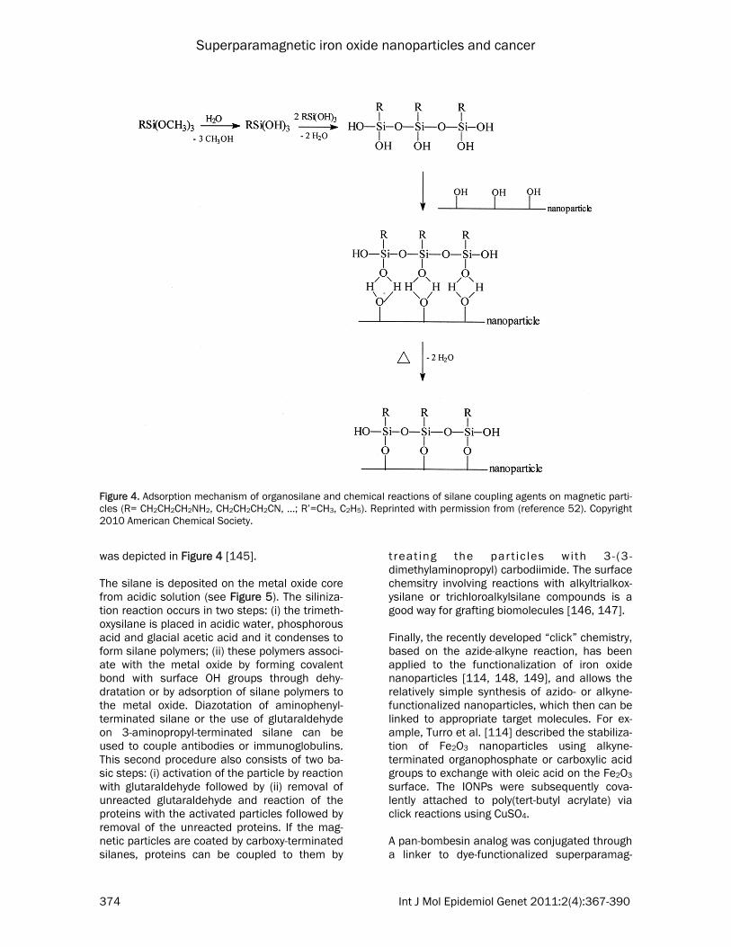
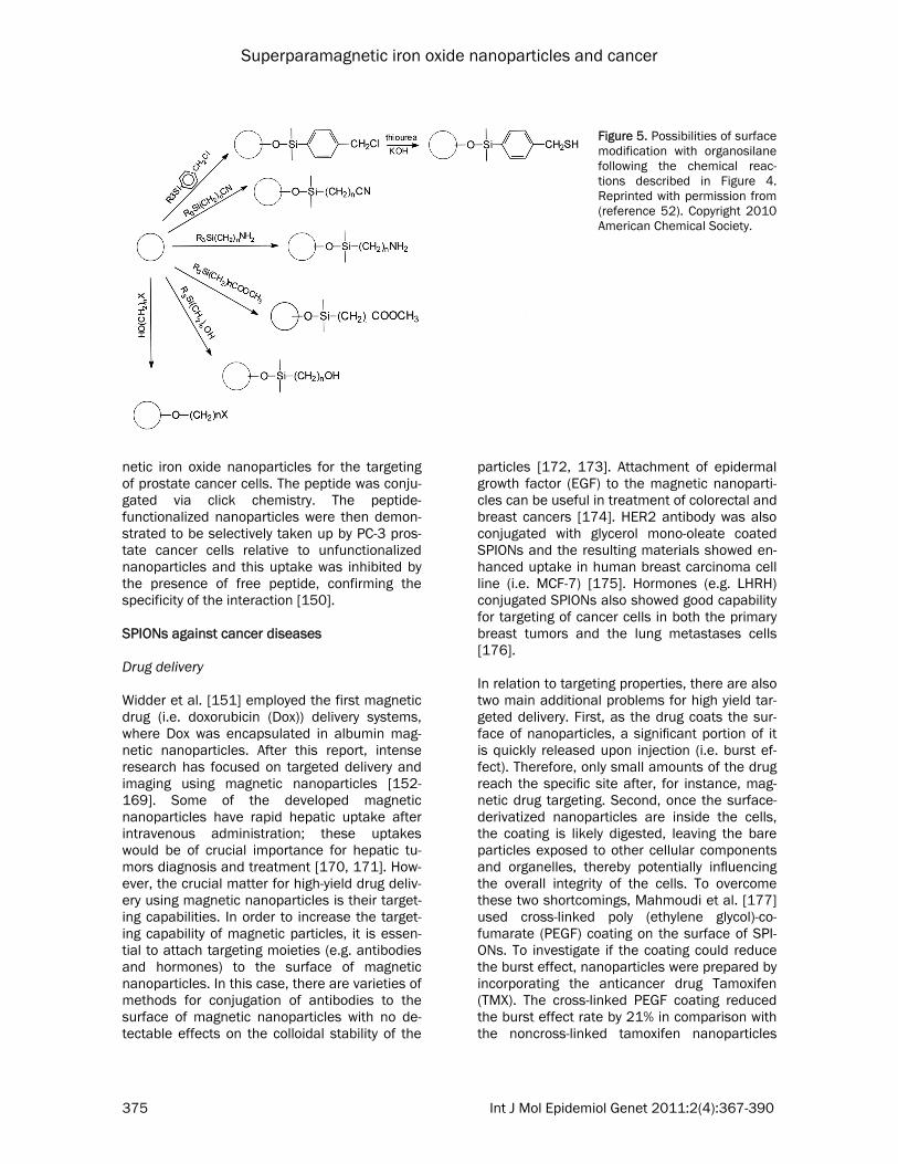
was depicted in Figure 4 [145]. The silane is deposited on the metal oxide core from acidic solution (see Figure 5). The siliniza-tion reaction occurs in two steps: (i) the trimeth-oxysilane is placed in acidic water, phosphorous acid and glacial acetic acid and it condenses to form silane polymers; (ii) these polymers associ-ate with the metal oxide by forming covalent bond with surface OH groups through dehy-dratation or by adsorption of silane polymers to the metal oxide. Diazotation of aminophenyl- terminated silane or the use of glutaraldehyde on 3-aminopropyl-terminated silane can be used to couple antibodies or immunoglobulins. This second procedure also consists of two ba-sic steps: (i) activation of the particle by reaction with glutaraldehyde followed by (ii) removal of unreacted glutaraldehyde and reaction of the proteins with the activated particles followed by removal of the unreacted proteins. If the mag-netic particles are coated by carboxy-terminated silanes, proteins can be coupled to them by
t reat ing the part ic les wi th 3- (3-dimethylaminopropyl) carbodiimide. The surface chemsitry involving reactions with alkyltrialkox-ysilane or trichloroalkylsilane compounds is a good way for grafting biomolecules [146, 147]. Finally, the recently developed “click” chemistry, based on the azide-alkyne reaction, has been applied to the functionalization of iron oxide nanoparticles [114, 148, 149], and allows the relatively simple synthesis of azido- or alkyne-functionalized nanoparticles, which then can be linked to appropriate target molecules. For ex-ample, Turro et al. [114] described the stabiliza-tion of Fe2O3 nanoparticles using alkyne-terminated organophosphate or carboxylic acid groups to exchange with oleic acid on the Fe2O3 surface. The IONPs were subsequently cova-lently attached to poly(tert-butyl acrylate) via click reactions using CuSO4. A pan-bombesin analog was conjugated through a linker to dye-functionalized superparamag-
Figure 4. Adsorption mechanism of organosilane and chemical reactions of silane coupling agents on magnetic parti-cles (R= CH2CH2CH2NH2, CH2CH2CH2CN, …; R’=CH3, C2H5). Reprinted with permission from (reference 52). Copyright 2010 American Chemical Society.
Superparamagnetic iron oxide nanoparticles and cancer
375 Int J Mol Epidemiol Genet 2011:2(4):367-390
netic iron oxide nanoparticles for the targeting of prostate cancer cells. The peptide was conju-gated via click chemistry. The peptide-functionalized nanoparticles were then demon-strated to be selectively taken up by PC-3 pros-tate cancer cells relative to unfunctionalized nanoparticles and this uptake was inhibited by the presence of free peptide, confirming the specificity of the interaction [150]. SPIONs against cancer diseases Drug delivery Widder et al. [151] employed the first magnetic drug (i.e. doxorubicin (Dox)) delivery systems, where Dox was encapsulated in albumin mag-netic nanoparticles. After this report, intense research has focused on targeted delivery and imaging using magnetic nanoparticles [152-169]. Some of the developed magnetic nanoparticles have rapid hepatic uptake after intravenous administration; these uptakes would be of crucial importance for hepatic tu-mors diagnosis and treatment [170, 171]. How-ever, the crucial matter for high-yield drug deliv-ery using magnetic nanoparticles is their target-ing capabilities. In order to increase the target-ing capability of magnetic particles, it is essen-tial to attach targeting moieties (e.g. antibodies and hormones) to the surface of magnetic nanoparticles. In this case, there are varieties of methods for conjugation of antibodies to the surface of magnetic nanoparticles with no de-tectable effects on the colloidal stability of the
particles [172, 173]. Attachment of epidermal growth factor (EGF) to the magnetic nanoparti-cles can be useful in treatment of colorectal and breast cancers [174]. HER2 antibody was also conjugated with glycerol mono-oleate coated SPIONs and the resulting materials showed en-hanced uptake in human breast carcinoma cell line (i.e. MCF-7) [175]. Hormones (e.g. LHRH) conjugated SPIONs also showed good capability for targeting of cancer cells in both the primary breast tumors and the lung metastases cells [176]. In relation to targeting properties, there are also two main additional problems for high yield tar-geted delivery. First, as the drug coats the sur-face of nanoparticles, a significant portion of it is quickly released upon injection (i.e. burst ef-fect). Therefore, only small amounts of the drug reach the specific site after, for instance, mag-netic drug targeting. Second, once the surface-derivatized nanoparticles are inside the cells, the coating is likely digested, leaving the bare particles exposed to other cellular components and organelles, thereby potentially influencing the overall integrity of the cells. To overcome these two shortcomings, Mahmoudi et al. [177] used cross-linked poly (ethylene glycol)-co-fumarate (PEGF) coating on the surface of SPI-ONs. To investigate if the coating could reduce the burst effect, nanoparticles were prepared by incorporating the anticancer drug Tamoxifen (TMX). The cross-linked PEGF coating reduced the burst effect rate by 21% in comparison with the noncross-linked tamoxifen nanoparticles
Figure 5. Possibilities of surface modification with organosilane following the chemical reac-tions described in Figure 4. Reprinted with permission from (reference 52). Copyright 2010 American Chemical Society.
Superparamagnetic iron oxide nanoparticles and cancer
376 Int J Mol Epidemiol Genet 2011:2(4):367-390
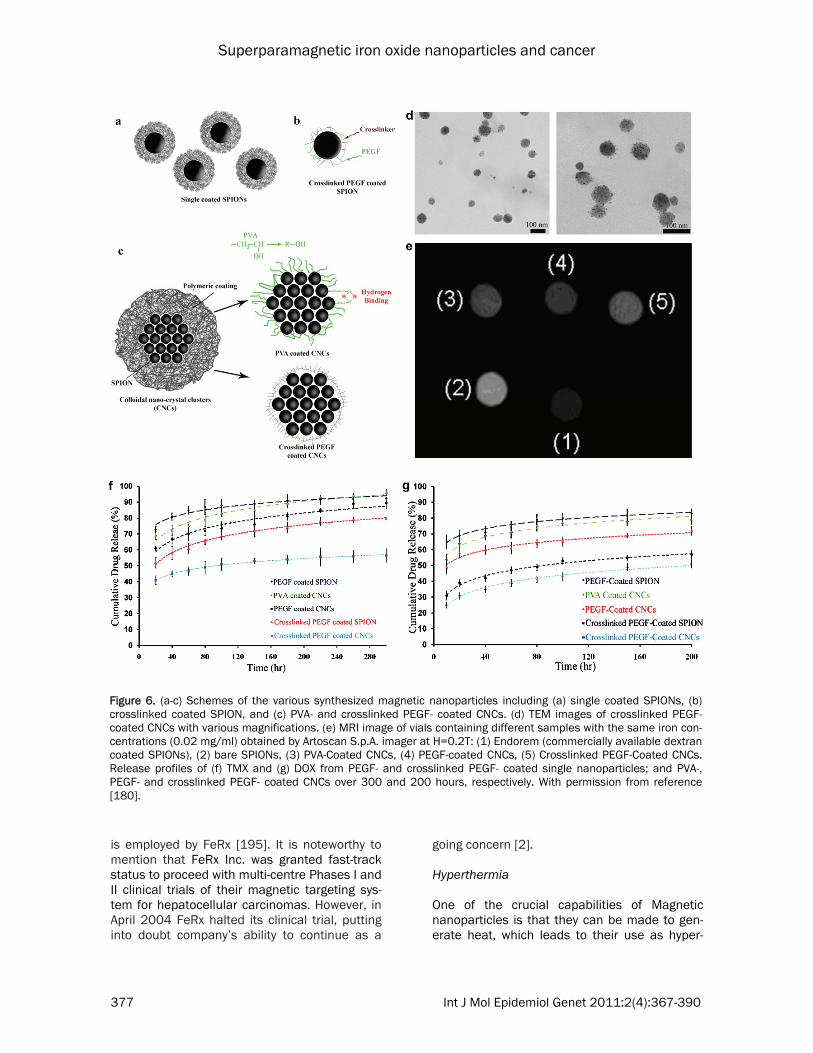
[177]. Thus, the authors claimed that the SPI-ONs with coating based on crosslinked unsatu-rated aliphatic polyesters are potentially useful to develop novel carriers for drug and gene de-livery applications [177]. Another crucial matter is the multifunctional capabilities of the engineered SPIONs, which are a key focus in bionanotechnology and will have profound impact on molecular diagnostics, imaging and therapeutic of cancer diseases. Gao et al. [178] prepared FePt coated SPIONs and claimed that their core-shell particles have good capability to be functionalized by various targeting molecules (e.g. antibodies). These multifunctional nanoparticles can enhance the capability of magnetic particles for simultane-ous detection and monitoring of the transforma-tion of tumors by noninvasive MRI during che-motherapeutic treatment by nanoparticles. In order to combine multiple components on a nanometer scale for creation of new imaging modalities, which are unavailable from individ-ual components, Jin et al. [179] prepared multi-functional nanoprobes. These not only offered contrast for electron microscopy, MRI and scat-tering-based imaging but also, more impor-tantly, enabled a new imaging mode, magneto-motive photoacoustic imaging. This form of im-aging with remarkable contrast enhancement compared with photoacoustic images using con-ventional nanoparticle contrast agents; these particles were composed of thin gold coated SPIONs by creating a gap between the core and the shell. Very recently, Mahmoudi et al. [50] synthesized a multi-component system made of gold-coupled core-shell SPIONs, as a new nano-probe with signal enhancement in surface Ra-man spectroscopy, due to its jagged-shaped gold shell coating. These new classes of multi-functional magnetic nanoparticles [50, 179] may have great impact in the future in relation to cancer diagnosis and therapy. Amiri et al. [180] reported cell endocytosis, drug release, NMR relaxometry and in vitro MRI stud-ies on a novel class of superparamagnetic col-loidal nano-crystal clusters (CNCs) with various biocompatible coatings. They claimed that the transverse relaxivity r2, the parameter repre-senting the MRI efficiency in negative contrast agents, for the polyvinyl alcohol (PVA)-coated, polyethylene glycol-co-fumarate (PEGF)-coated, and crosslinked PEGF-coated CNCs was effi-cient enough to contrast suitably the MRI (see
Figure 6). In addition, their prepared samples have been shown to facilitate controlled drug release (particularly the crosslinked PEGF-coated compound), thereby finally allowing them to propose this class of compounds for future applications in cancer, as theranostics agents. Besides in vitro evaluations of drug loaded sur-face engineered magnetic nanoparticles, there are numerous in vivo targeting assessments. For instance SPIONs have been injected very close to tumor sites in animal models and hu-man clinical studies for targeting delivery of anticancer drugs [181-188]. SPIONs have the capability to enhance MRI in combination with diffusion weighted MRI, which can be a novel, accurate, and fast method for detecting pelvic lymph node metastases even in normal-sized nodes of patients with bladder or prostate can-cer [189]. The surface engineered SPIONs can be also employed in diagnosis and treatment of colorectal cancer [190]. In order to increase the yield of magnetic target-ing, Widder et al. [191, 192] employed intra-arterial injection proximal to the tumor site (Dox filled magnetic particles). Their results demon-strated a 200-fold increased targeting yield in comparison with intravenous injection [193]. Since this study, success in cytotoxic drug deliv-ery and tumor remission has been reported by several groups using animals models including swine [194, 195], rabbits [196] and rats [197-199]. In order to increase the targeting yield of magnetic nanoparticles, permanent magnets can be implanted in a targeted site; for exam-ple, permanent magnets were implanted at solid osteosarcoma sites in hamsters and anti-cancer drugs subsequently delivered to the tar-geted site using magnetic liposomes [200]. Us-ing magnetic implantation methods, the yield of targeting were enhanced four-fold in compari-son with normal intravenous (non-magnetic) delivery. Results also showed significant in-crease in anti-tumor activity and the elimination of weight-loss as a side effect [201]. This tech-nique has also been employed to target cyto-toxic drugs to brain tumors, where passing the drugs through the blood-brain barrier is difficult [199]. Preliminary successful animal trials have lead to the development of magnetic nanoparti-cles for use in human trials. For example, mag-netic nanoparticles (metallic Fe coated with activated carbon) which carried the Dox as drug
Superparamagnetic iron oxide nanoparticles and cancer
377 Int J Mol Epidemiol Genet 2011:2(4):367-390
is employed by FeRx [195]. It is noteworthy to mention that FeRx Inc. was granted fast-track status to proceed with multi-centre Phases I and II clinical trials of their magnetic targeting sys-tem for hepatocellular carcinomas. However, in April 2004 FeRx halted its clinical trial, putting into doubt company’s ability to continue as a
going concern [2]. Hyperthermia One of the crucial capabilities of Magnetic nanoparticles is that they can be made to gen-erate heat, which leads to their use as hyper-
Figure 6. (a-c) Schemes of the various synthesized magnetic nanoparticles including (a) single coated SPIONs, (b) crosslinked coated SPION, and (c) PVA- and crosslinked PEGF- coated CNCs. (d) TEM images of crosslinked PEGF-coated CNCs with various magnifications. (e) MRI image of vials containing different samples with the same iron con-centrations (0.02 mg/ml) obtained by Artoscan S.p.A. imager at H=0.2T: (1) Endorem (commercially available dextran coated SPIONs), (2) bare SPIONs, (3) PVA-Coated CNCs, (4) PEGF-coated CNCs, (5) Crosslinked PEGF-Coated CNCs. Release profiles of (f) TMX and (g) DOX from PEGF- and crosslinked PEGF- coated single nanoparticles; and PVA-, PEGF- and crosslinked PEGF- coated CNCs over 300 and 200 hours, respectively. With permission from reference [180].
Superparamagnetic iron oxide nanoparticles and cancer
378 Int J Mol Epidemiol Genet 2011:2(4):367-390
thermia agents, delivering toxic amounts of thermal energy to cancerous tumors where a moderate degree of tissue warming results in more effective cell destruction [202, 203]. The production of local heat in magnetic nanoparti-cles is due to the fact that the magnetic anisot-ropy of magnetic nanoparticles can be much greater than those of a bulk materials, while differences in the Curie or Néel temperatures, i.e., the temperatures of spontaneous parallel or antiparallel orientation of spins between magnetic nanoparticles and the corresponding microscopic phases, reach hundreds of degrees [203, 204]. Magnetic nanoparticles were first employed in hyperthermia application in the work of Chan et al. [205] and Jordan et al. [206] in 1993, where they proved that superparamag-netic crystal suspension had great capability to absorb the energy of an alternating magnetic field; this absorbed energy can in turn be con-verted into heat. Given that tumor cells are more sensitive to a temperature increase than healthy ones [207, 208], this property can be used in vivo to increase the temperature of can-cerous tissue and to destroy the pathological cells by hyperthermia. During the last 2 decades, intense efforts were focused on improvement of hyperthermia tech-niques for clinical applications. Advances in the area of nanotechnology have contributed to the development of superparamagnetic fluid hyper-thermia, which is well recognized as promising method for cancer treatment because of the ease of targeting the cancerous tissue and hence having fewer side effects than chemo-therapy and radiotherapy, as proven by the re-sults of current/ongoing clinical trials [209]. Hergt et al. [210] injected 100 mg dextran coated magnetic nanoparticles into the tail vein of Sprague Dawley rats, treated with AC mag-netic field (12 min, 450 kHz, unknown field and SAR). According to authors’ considerable the tumor shrinkage and tissue necrosis was ob-served. Jordan and co-workers [211-225] did the first clinical patient trials [226] with mag-netic nanoparticles. In this case, a special hy-perthermia-generating prototype instrument was developed which is able to generate vari-able magnetic fields in the range of 0 - 15 kA/m at a frequency of 100 kHz. At the same time, the machine allows for real-time patient tem-perature measurements to ensure that neither the upper limit of the therapeutic temperature
threshold is exceeded, thus preventing thermal ablation, nor the lower, ineffective limit is crossed. This prototype is capable of treating tumors placed in any region of the body (e.g., prostate cancer, brain tumors). Maier-Hauff et al. [227] injected neuro-navigationally controlled intra-tumoral instilla-tion of an aqueous dispersion of iron-oxide nanoparticles in 66 patients (59 with recurrent glioblastoma) and subsequently the particles were heated up using an alternating magnetic field. Treatment was combined with fractionated stereotactic radiotherapy. A median dose of 30 grays (Gy) using a fractionation of 5 × 2 Gy/week was applied. The primary study endpoint was overall survival following diagnosis of first tumor recurrence (OS-2), while the secondary endpoint was overall survival after primary tu-mor diagnosis (OS-1). The median overall sur-vival from diagnosis of the first tumor recur-rence among the 59 patients with recurrent glioblastoma was 13.4 months (95% CI: 10.6-16.2 months). Median OS-1 was 23.2 months while the median time interval between primary diagnosis and first tumor recurrence was 8.0 months. The authors claimed that the side ef-fects of their new therapeutic approach were moderate, and no serious complications were observed [227]. According to these results, one can conclude that thermotherapy using mag-netic nanoparticles in conjunction with a re-duced radiation dose is reasonably safe and effective and leads to longer OS-2 compared to conventional therapies in the treatment of re-current glioblastoma [227]. Till now, only local hyperthermia is applicable for magnetic fluid hyperthermia; in this case, surface-engineered magnetic nanoparticles, which are dispersed in a carrier fluid, are placed inside the cancerous tumor through direct injection or tumor specific antibody targeting. This specific antibody target-ing cause the attachments of nanoparticles to the cancerous cells and the labeled-cells are exposed to an alternating magnetic field. This field makes the magnetic nanoparticles gener-ate heat by magnetic relaxation mechanisms; these local heats can induce apoptosis to the labeled cells. Simultaneous drug delivery and hyperthermia One of the crucial problems with magnetic tar-geted hyperthermia is that a limited dose of nanoparticles reached the tumor tissue. This
Superparamagnetic iron oxide nanoparticles and cancer
379 Int J Mol Epidemiol Genet 2011:2(4):367-390
resulted in insufficient temperature enhance-ment in the cancerous sites; thus there is a risk of proliferation of cancer cells that survived dur-ing thermotherapy [229]. In order to overcome the problem, several specific tumor receptor targeting moieties together with anticancer drugs can be attached to the surface of parti-cles. These employed targeting moieties to-gether with anti-cancerous drugs may induce cell apoptosis. In this case, the hyperthermia treatment was used as a driving force for simul-taneous drug delivery purposes (e.g. using ther-mosensitive polymers [203], as nanoparticle’s coating) [230] For example, β-cyclodextrin (CD) was used as a drug container for hydrophilic (paclitaxel) or lipophilic (doxorubicin) structures. Drugs incorporated in the CD can thus be re-leased through the use of induction heating, or hyperthermic effects, by applying a high-frequency magnetic field. In this case, folic acid (FA) and CD-functionalized magnetic nanoparti-cles were synthesized and it was found that by induction of heating, drug release was triggered from the CD cavity on the particle - a behavior that was controlled by switching the high-frequency magnetic field on and off. Another drug delivery system, based on cova-lently attaching genistein onto SPIONs coated by cross-linked carboxymethylated chitosan (CMCH), has been developed [231] and the re-sults confirmed that the nanosystem could sig-nificantly enhanced cancer cell apoptosis. Importance of protein-nanoparticle interactions It has been long recognized that proteins were associated with nanoparticles, upon entrance of nano-objects into a biological fluid [232-235]. The amount and types of the associated pro-teins, which is called a protein “corona”, on the surface of the nanoparticles leads to an in vivo response [232]. More specifically, composition of the obtained protein corona can be used for prediction of the way cells interact with, recog-nize and process the nanoparticles [236]. It also has been shown that the composition of the hard corona protein profiles can be signifi-cantly different due to the changes in surface chemistries of the same materials [237]. Thus, decoration of the surface of magnetic nanopar-ticles would be very useful for the efficient de-sign of targeted delivery systems where the tar-geting moieties are covered on the surface of SPIONs. More specifically, inappropriate surface chemistry of nanoparticles have great capability
to be severely covered by the proteins which may causing the elimination of targeting moie-ties; in contrast, the targeting species of the well surface decorated particles are active due to their lower protein affinity (lowest thickness of protein corona in comparison with inappropri-ate surface) to proteins causing the highest tar-geting yields (see Figure 7). Clearly there is a significant amount of work to be done to con-firm these issues. Conclusions and future perspectives Multifunctional SPIONs play an important role in the development of simultaneous targeted de-livery, imaging, and hyperthermia for diagnosis and treatment of cancerous tumors in vivo. Deeper understanding on the protein corona compositions at the surface of nanoparticles are essential for development of nanoparticle specific uptake by desired cells in vivo. In addi-tion, control of the protein corona which is formed at the surface of magnetic particles, which are decorated by cancer-specific binding agents, would make targeted delivery and mag-netic fluid hyperthermia treatment much more selective than traditional chemotherapy and even conventional hyperthermia. Furthermore, multifunctional magnetic particles can be mag-netically targeted and concentrated in the target tissue, and drug release together with heating are then only induced to significant burst effects and temperatures where the magnetic nanopar-ticles have been deposited. In addition, tissue-deposited magnetic particles will generally stay where they were initially deposited, thus allow-ing for repeated and concentrated drug release and hyperthermia treatments in the same area. At the moment the amount of particles deliv-ered to the cancerous tissues/cells by means of antibody targeting is too low for a sufficient drug release and temperature increase. Thus, the main challenges in this field will be the design of stealth nanoparticles able to circulate in the blood compartment for a long time and the sur-face grafting of ligands able to facilitate their specific internalization in cancerous cells. Al-though the results obtained from the first clini-cal trials of magnetic nanoparticles are very promising, it would be premature to claim that these molecules contribute therapeutic advan-tages because survival and disease progression benefits were not defined endpoints of the fea-sibility studies.

Superparamagnetic iron oxide nanoparticles and cancer
380 Int J Mol Epidemiol Genet 2011:2(4):367-390
There are several matters that should be con-sidered to enhance the targeted drug delivery, targeted imaging and hyperthermia yield of SPI-ONs. These include the exact definition of cell membrane composition; in vivo control of drug release; in vivo control of heat distribution; man-agement surface of the nanoparticles’ for for-mation of desired protein corona composition followed by fast internalization by the target cells; and optimization of the biophysicochemi-cal properties of the particles. A non homogene-ous particle distribution in the tissue may lead to the occurrence of uncontrollable targeting, drug release, and hot spots where the high drug amounts/temperature could cause non-specific necrosis of the tissue. In contrast, in regions with a low particle concentration the drug and/or temperature would not be sufficiently high enough to trigger the onset of apoptosis and a proliferation of surviving cancerous cells can still occur. Both effects should be prevented and thus more even distribution of particles in the tissue as well as the monitoring of the spa-tial heat distribution should form the focus of future research. Acknowledgement In memory of Baba Ardeshir.
Address correspondence to: Dr. Morteza Mahmoudi, National Cell Bank, Pasteur Institute of Iran, Tehran, Iran; Nanotechnology Research Center, Faculty of Pharmacy, Tehran University of Medical Sciences, Tehran, Iran Tel: +98 (21) 66492595; Fax: +98 (21) 66492595; E-mail: [email protected] References [1] Anand P, Kunnumakara AB, Sundaram C,
Harikumar KB, Tharakan ST, Lai OS, Sung B, Aggarwal BB. Cancer is a Preventable Disease that Requires Major Lifestyle Changes. Pharm Res 2008; 25: 2097-116.
[2] Mahmoudi M, Sant S, Wang B, Laurent S, Sen T. Superparamagnetic iron oxide nanoparti-cles (SPIONs): Development, surface modifica-tion and applications in chemotherapy. Ad-vanced Drug Delivery Reviews 2011; 63: 24-46.
[3] Means GE, Feeney RE. Chemical Modifications of Proteins: History and Applications. Bioconju-gate Chemistry 1990; 1: 2-12.
[4] Chau Y, Dang NM, Tan FE, Langer R. Investiga-tion of targeting mechanism of new dextran-peptide-methotrexate conjugates using biodis-tribution study in matrix-metalloproteinase-overexpressing tumor xenograft model. Jour-nal of Pharmaceutical Sciences 2006; 95: 542-51.
[5] Gregoriadis G. Engineering liposomes for drug delivery: Progress and problems. Trends in Biotechnology 1995; 13: 527-37.
Figure 7. Cartoon showing the importance of surface chemistries in targeted delivery/imaging applications.
Superparamagnetic iron oxide nanoparticles and cancer
381 Int J Mol Epidemiol Genet 2011:2(4):367-390
[6] Adams ML, Lavasanifar A, Kwon GS. Amphi-philic block copolymers for drug delivery. Jour-nal of Pharmaceutical Sciences 2003; 92: 1343-55.
[7] Rosler A, Vandermeulen GWM, Klok HA. Ad-vanced drug delivery devices via self-assembly of amphiphilic block copolymers. Advanced Drug Delivery Reviews 2001; 53: 95-108.
[8] Wu GH, Milkhailovsky A, Khant HA, Fu C, Chiu W, Zasadzinski JA. Remotely triggered lipo-some release by near-infrared light absorption via hollow gold nanoshells. Journal of the American Chemical Society 2008; 130: 8175-7.
[9] Son SJ, Bai X, Lee SB. Inorganic hollow nanoparticles and nanotubes in nanomedi-cine. Part 1. Drug/gene delivery applications. Drug Discovery Today 2007; 12: 650-6.
[10] Akerman ME, Chan WCW, Laakkonen P, Bhatia SN, Ruoslahti E. Nanocrystal targeting in vivo. Proceedings of the National Academy of Sciences of the United States of America 2002; 99: 12617-21.
[11] Uhrich KE, Cannizzaro SM, Langer RS, Shakesheff KM. Polymeric systems for con-trolled drug release. Chemical Reviews 1999; 99: 3181-98.
[12] Veronese FM, Pasut G. PEGylation, successful approach to drug delivery. Drug Discovery Today 2005; 10: 1451-8.
[13] Richardson TP, Peters MC, Ennett AB, Mooney DJ. Polymeric system for dual growth factor delivery. Nature Biotechnology 2001; 19: 1029-34.
[14] Vinogradov SV, Bronich TK, Kabanov AV. Nanosized cationic hydrogels for drug delivery: preparation, properties and interactions with cells. Advanced Drug Delivery Reviews 2002; 54: 135-47.
[15] Gupta P, Vermani K, Garg S. Hydrogels: from controlled release to pH-responsive drug deliv-ery. Drug Discovery Today 2002; 7: 569-79.
[16] Qiu Y, Park K. Environment-sensitive hydrogels for drug delivery. Advanced Drug Delivery Re-views 2001; 53: 321-39.
[17] Santini JT, Cima MJ, Langer R. A controlled-release microchip. Nature 1999; 397: 335-8.
[18] LaVan DA, McGuire T, Langer R. Small-scale systems for in vivo drug delivery. Nature Bio-technology 2003; 21: 1184-91.
[19] Grayson ACR, Choi IS, Tyler BM, Wang PP, Brem H, Cima MJ, Langer R. Multi-pulse drug delivery from a resorbable polymeric micro-chip device. Nature Materials 2003; 2: 767-72.
[20] Razzacki SZ, Thwar PK, Yang M, Ugaz VM, Burns MA. Integrated microsystems for con-trolled drug delivery. Advanced Drug Delivery Reviews 2004; 56: 185-98.
[21] Collins JM. PHARMACOLOGIC RATIONALE FOR REGIONAL DRUG DELIVERY. Journal of Clinical Oncology 1984; 2: 498-504.
[22] Egan TD. Intravenous drug delivery systems: Toward an intravenous ''vaporizer''. Journal of Clinical Anesthesia 1996; 8: S8-S14.
[23] Markman M. Intraperitoneal antineoplastic drug delivery: rationale and results. Lancet Oncology 2003; 4: 277-83.
[24] Edwards DA, Hanes J, Caponetti G, Hrkach J, BenJebria A, Eskew ML, Mintzes J, Deaver D, Lotan N, Langer R. Large porous particles for pulmonary drug delivery. Science 1997; 276: 1868-71.
[25] Prausnitz MR, Bose VG, Langer R, Weaver JC. ELECTROPORATION OF MAMMALIAN SKIN - A MECHANISM TO ENHANCE TRANSDERMAL DRUG-DELIVERY. Proceedings of the National Academy of Sciences of the United States of America 1993; 90: 10504-8.
[26] Henry S, McAllister DV, Allen MG, Prausnitz MR. Microfabricated microneedles: A novel approach to transdermal drug delivery. Jour-nal of Pharmaceutical Sciences 1998; 87: 922-5.
[27] Prausnitz MR, Mitragotri S, Langer R. Current status and future potential of transdermal drug delivery. Nature Reviews Drug Discovery 2004; 3: 115-24.
[28] Illum L. Nasal drug delivery: new develop-ments and strategies. Drug Discovery Today 2002; 7: 1184-9.
[29] Illum L. Nasal drug delivery - possibilities, problems and solutions. J Control Release. 2003; 87: 187-98.
[30] Brannonpeppas L. NOVEL VAGINAL DRUG-RELEASE APPLICATIONS. Advanced Drug De-livery Reviews 1993; 11: 169-77.
[31] Loftssona T, Jarvinen T. Cyclodextrins in oph-thalmic drug delivery. Advanced Drug Delivery Reviews 1999; 36: 59-79.
[32] Le Bourlais C, Acar L, Zia H, Sado PA, Needham T, Leverge R. Ophthalmic drug deliv-ery systems - Recent advances. Progress in Retinal and Eye Research 1998; 17: 33-58.
[33] Jeong B, Bae YH, Lee DS, Kim SW. Biodegrad-able block copolymers as injectable drug-delivery systems. Nature 1997; 388: 860-2.
[34] Li F, Liu WG, De Yao K. Preparation of oxidized glucose-crosslinked N-alkylated chitosan membrane and in vitro studies of pH-sensitive drug delivery behaviour. Biomaterials 2002; 23: 343-7.
[35] Mitragotri S. Innovation - Healing sound: the use of ultrasound in drug delivery and other therapeutic applications. Nature Reviews Drug Discovery 2005; 4: 255-60.
[36] Liu YY, Miyoshi H, Nakamura M. Encapsulated ultrasound microbubbles: Therapeutic applica-tion in drug/gene delivery. Journal of Con-trolled Release 2006; 114: 89-99.
[37] Ng KY, Liu Y. Therapeutic ultrasound: Its appli-cation in drug delivery. Medicinal Research Reviews 2002; 22: 204-23.
[38] Lubbe AS, Alexiou C, Bergemann C. Clinical
Superparamagnetic iron oxide nanoparticles and cancer
382 Int J Mol Epidemiol Genet 2011:2(4):367-390
applications of magnetic drug targeting. Jour-nal of Surgical Research 2001; 95: 200-6.
[39] Polyak B, Friedman G. Magnetic targeting for site-specific drug delivery: applications and clinical potential. Expert Opinion on Drug De-livery 2009; 6: 53-70.
[40] Sahoo SK, Labhasetwar V. Nanotech ap-proaches to delivery and imaging drug. Drug Discovery Today 2003; 8: 1112-20.
[41] Torchilin VP, Lukyanov AN. Peptide and protein drug delivery to and into tumors: challenges and solutions. Drug Discovery Today 2003; 8: 259-66.
[42] Naldini L, Blomer U, Gallay P, Ory D, Mulligan R, Gage FH, Verma IM, Trono D. In vivo gene delivery and stable transduction of nondivid-ing cells by a lentiviral vector. Science 1996; 272: 263-7.
[43] Zufferey R, Nagy D, Mandel RJ, Naldini L, Trono D. Multiply attenuated lentiviral vector achieves efficient gene delivery in vivo. Nature Biotechnology 1997; 15: 871-5.
[44] Mahmoudi M, Serpooshan V, Laurent S. Engi-neered nanoparticles for biomolecular imag-ing. Nanoscale 2011; 3: 3007-29.
[45] Mahmoudi M, Hosseinkhani H, Hosseinkhani M, Boutry S, Simchi A, Shane Journeay Ws, Subramani K, Laurent S. Magnetic resonance imaging tracking of stem cells in vivo using iron oxide nanoparticles as a tool for the ad-vancement of clinical regenerative medicine. Chemical Reviews 2011; 111: 253-80.
[46] Gupta AK, Gupta M. Synthesis and surface engineering of iron oxide nanoparticles for biomedical applications. Biomaterials 2005; 26: 3995-4021.
[47] Mahmoudi M, Milani AS, Stroeve P, Arbab SA. Superparamagnetic Iron Oxide Nanoparticles: Synthesis, Surface Engineering, Cytotoxicity and Biomedical Applications; Nova Science Publisher; New York, 2011; ISBN: 978-1-61668-964-3.
[48] Mahmoudi M, Simchi A, Imani M, Milani AS, Stroeve P. Optimal design and characteriza-tion of superparamagnetic iron oxide nanopar-ticles coated with polyvinyl alcohol for tar-geted delivery and imaging. Journal of Physical Chemistry B 2008; 112: 14470-81.
[49] Mahmoudi M, Sahraian MA, Shokrgozar MA, Laurent S. Superparamagnetic iron oxide nanoparticles: Promises for diagnosis and treatment of multiple sclerosis. ACS Chemical Neuroscience 2011; 2: 118-40.
[50] Mahmoudi M, Amiri H, Shokrgozar MA, Sasan-pour P, Rashidian B, Laurent S, Casula MF, Lascialfari A. Raman active jagged-shaped gold-coated magnetic particles as a novel multimodal nanoprobe. Chemical Communica-tions 2011; 47: 10404-6.
[51] Mahmoudi M, Simchi A, Imani M. Recent ad-vances in surface engineering of superpara-magnetic iron oxide nanoparticles for biomedi-
cal applications. Journal of the Iranian Chemi-cal Society 2010; 7: S1-S27.
[52] Laurent S, Forge D, Port M, Roch A, Robic C, Vander Elst L, Muller RN. Magnetic Iron Oxide Nanoparticles: Synthesis, Stabilization, Vec-torization, Physicochemical Characterizations, and Biological Applications. Chemical Reviews 2008; 108: 2064-110.
[53] Laurent S, Boutry S, Mahieu I, Vander Elst L, Muller RN. Iron Oxide Based MR Contrast Agents: from Chemistry to Cell Labeling. Cur-rent Medicinal Chemistry 2009; 16: 4712-27.
[54] Fukukura Y, Kamiyama T, Takumi K, Shindo T, Higashi R, Nakajo M. Comparison of ferucar-botran-enhanced fluid-attenuated inversion-recovery echo-planar, T2-weighted turbo spin-echo, T2*-weighted gradient-echo, and diffu-sion-weighted echo-planar imaging for detec-tion of malignant liver lesions. Journal of Mag-netic Resonance Imaging 2010; 31: 607-16.
[55] Saito K, Sugimoto K, Nishio R, Araki Y, Mori-yasu F, Kakizaki D, Tokuuye K. Perfusion study of liver lesions with superparamagnetic iron oxide: distinguishing hepatocellular carcinoma from focal nodular hyperplasia. Clinical imag-ing 2009; 33: 447-53.
[56] Santoro L, Grazioli L, Filippone A, Grassedonio E, Belli G, Colagrande S. Resovist enhanced MR imaging of the liver: Does quantitative assessment help in focal lesion classification and characterization? Journal of Magnetic Resonance Imaging 2009; 30: 1012-20.
[57] Meier R, Henning TD, Boddington S, Tavri S, Arora S, Piontek G, Rudelius M, Corot C, Daldrup-Link HE. Breast Cancers: MR Imaging of Folate-Receptor Expression with the Folate-Specific Nanoparticle P1133. Radiology 2010; 255: 527-35.
[58] Radermacher KA, Boutry S, Laurent S, Elst LV, Mahieu I, Bouzin C, Magat J, Gregoire V, Feron O, Muller RN, Jordan BF, Gallez B. Iron oxide particles covered with hexapeptides targeted at phosphatidylserine as MR biomarkers of tumor cell death. Contrast Media & Molecular Imaging 2010; 5: 258-67.
[59] Nasongkla N, Bey E, Ren J, Ai H, Khemtong C, Guthi JS, Chin SF, Sherry AD, Boothman DA, Gao J. Multifunctional Polymeric Micelles as Cancer-Targeted, MRI-Ultrasensitive Drug De-livery Systems. Nano Letters 2006; 6: 2427-30.
[60] Winter PM, Caruthers SD, Allen JS, Cai K, Wil-liams TA, Lanza GM, Wickline SA. Molecular imaging of angiogenic therapy in peripheral vascular disease with ανβ3-integrin-targeted nanoparticles. Magnetic Resonance in Medi-cine 2010; 64: 369-76.
[61] Park J, An K, Hwang Y, Park JG, Noh HJ, Kim JY, Park JH, Hwang NM, Hyeon T. Ultra-large-scale syntheses of monodisperse nanocrys-tals. Nat Mater 2004; 3: 891-5.
[62] Mahmoudi M, Milani AS, Stroeve P. Synthesis,
Superparamagnetic iron oxide nanoparticles and cancer
383 Int J Mol Epidemiol Genet 2011:2(4):367-390
surface architecture and biological response of superparamagnetic iron oxide nanoparticles for application in drug delivery: a review. Inter-national Journal of Biomedical Nanoscience and Nanotechnology 2010; 1: 164-201.
[63] Wang X, Zhuang J, Peng Q, Li Y. A general strategy for nanocrystal synthesis. Nature 2005; 437: 121-4.
[64] Khan Y, Durrani SK, Siddique M, Mehmood M. Hydrothermal synthesis of alpha Fe2O3 nanoparticles capped by Tween-80. Materials Letters 2011; 65: 2224-7.
[65] Sun S, Zeng H, Robinson DB, Raoux S, Rice PM, Wang SX, Li G. Monodisperse MFe2O4 (M = Fe, Co, Mn) Nanoparticles. Journal of the American Chemical Society 2004; 14: 273-9.
[66] Chikate RC, Jun KW, Rode CV. Nonaqueous synthesis and characterization of capped خ±-Fe2O3 nanoparticles from iron(III) hydroxy-oleate precursor. Polyhedron 2008; 27: 933-8.
[67] Dresco PA, Zaitsev VS, Gambino RJ, Chu B. Preparation and Properties of Magnetite and Polymer Magnetite Nanoparticles. Langmuir. 1999; 15: 1945-51.
[68] Narita A, Naka K, Chujo Y. Facile control of silica shell layer thickness on hydrophilic iron oxide nanoparticles via reverse micelle method. Colloids and Surfaces A: Physico-chemical and Engineering Aspects 2009; 336: 46-56.
[69] Lu A-H, Salabas EL, Schüth F. Magnetic Nanoparticles: Synthesis, Protection, Function-alization, and Application. Angewandte Che-mie International Edition 2007; 46: 1222-44.
[70] Shen L, Stachowiak A, Fateen S-EK, Laibinis PE, Hatton TA. Structure of Alkanoic Acid Sta-bilized Magnetic Fluids. A Small-Angle Neutron and Light Scattering Analysis. Langmuir 2000; 17: 288-99.
[71] Boyer C, Whittaker MR, Bulmus V , Liu J, Davis TP. The design and utility of polymer-stabilized iron-oxide nanoparticles for nanomedicine applications. NPG Asia Materials 2010; 2: 23-30.
[72] Hong RY, Feng B, Chen LL, Liu GH, Li HZ, Zheng Y, Wei DG. Synthesis, characterization and MRI application of dextran-coated Fe3O4 magnetic nanoparticles. Biochemical Engi-neering Journal 2008; 42: 290-300.
[73] Saboktakin MR, Tabatabaie RM, Maharramov A, Ramazanov MA. A synthetic macromolecule as MRI detectable drug carriers: Aminodextran-coated iron oxide nanoparticles. Carbohy-drate Polymers 2010; 80: 695-8.
[74] Corot C, Robert P, Idee JM, Port M. Recent advances in iron oxide nanocrystal technology for medical imaging. Advanced Drug Delivery Reviews 2006; 58: 1471-504.
[75] Gomez-Lopera SA, Arias JL, Gallardo V, Delgado AV. Colloidal Stability of Magnetite/Poly(lactic acid) Core/Shell Nanoparticles.
Langmuir 2006; 22: 2816-21. [76] Arias JL, Gallardo V, oacute, mez-Lopera SA,
Delgado AV. Loading of 5-Fluorouracil to Poly(ethyl-2-cyanoacrylate) Nanoparticles with a Magnetic Core. Journal of Biomedical Nanotechnology 2005; 1: 214-23.
[77] Molday RS, Mackenzie D. Immunospecific ferromagnetic iron-dextran reagents for the labeling and magnetic separation of cells. Journal of Immunological Methods 1982; 52: 353-67.
[78] Lunov O, Syrovets T, Büchele B, Jiang X, Röcker C, Tron K, Nienhaus GU, Walther P, Mailänder V, Landfester K, Simmet T. The effect of carboxydextran-coated superpara-magnetic iron oxide nanoparticles on c-Jun N-terminal kinase-mediated apoptosis in human macrophages. Biomaterials 2010; 31: 5063-71.
[79] Mouaziz H, Veyret R, Theretz A, Ginot F, ric, Elaissari A. Aminodextran Containing Magnet-ite Nanoparticles for Molecular Biology Appli-cations: Preparation and Evaluation. Journal of Biomedical Nanotechnology 2009; 5: 172-81.
[80] Paul KG, Frigo TB, Groman JY, Groman EV. Synthesis of Ultrasmall Superparamagnetic Iron Oxides Using Reduced Polysaccharides. Bioconjugate Chemistry 2004; 15: 394-401.
[81] Kampert E, Janssen FFBJ, Boukhvalov DW, Russcher JC, Smits JMM, de Gelder R, de Bruin B, Christianen PC, Zeitler U, Katsnelson MI, Maan JC, Rowan AE. Ligand-Controlled Magnetic Interactions in Mn4 Clusters. Inor-ganic Chemistry. 2009; 48: 11903-8.
[82] Chu WJ. Surface properties of superparamag-netic iron oxide MR contrast agents: Ferumox-ides, ferumoxtran, ferumoxsil. Magnetic Reso-nance Imaging 1995; 13: 675-91.
[83] Carmen Bautista M, Bomati-Miguel O, del Puerto Morales Ma, Serna CJ, Veintemillas-Verdaguer S. Surface characterisation of dex-tran-coated iron oxide nanoparticles prepared by laser pyrolysis and coprecipitation. Journal of Magnetism and Magnetic Materials 2005; 293: 20-7.
[84] Anzai Y, McLachlan S, Morris M, Saxton R, Lufkin RB. Dextran-coated superparamagnetic iron oxide, an MR contrast agent for assessing lymph nodes in the head and neck. American Journal of Neuroradiology 1994; 15: 87-94.
[85] Bourrinet P, Bengele HH, Bonnemain B, Den-causse A, Idee JM, Jacobs PM, Lewis JM. Pre-clinical Safety and Pharmacokinetic Profile of Ferumoxtran-10, an Ultrasmall Superparamag-netic Iron Oxide Magnetic Resonance Contrast Agent. Investigative Radiology 2006; 41: 313-24.
[86] Bellin MF, Beigelman C, Precetti-Morel S. Iron oxide-enhanced MR lymphography: initial ex-perience. European Journal of Radiology 2000; 34: 257-64.
Superparamagnetic iron oxide nanoparticles and cancer
384 Int J Mol Epidemiol Genet 2011:2(4):367-390
[87] Clement O, Siauve N, Cuénod CA, Frija G. Liver Imaging With Ferumoxides (Feridex(R)): Fun-damentals, Controversies, and Practical As-pects. Topics in Magnetic Resonance Imaging 1998; 9: 167-82.
[88] Mornet Sp, Portier J, Duguet E. A method for synthesis and functionalization of ultrasmall superparamagnetic covalent carriers based on maghemite and dextran. Journal of Magnet-ism and Magnetic Materials 2005; 293: 127-34.
[89] Catherine CB, Adam SGC. Functionalisation of magnetic nanoparticles for applications in biomedicine. Journal of Physics D: Applied Physics 2003; 36: R198.
[90] Wapner K, Grundmeier G. Spectroscopic analysis of the interface chemistry of ultra-thin plasma polymer films on iron. Surface and Coatings Technology 2005; 200: 100-3.
[91] Love JC, Estroff LA, Kriebel JK, Nuzzo RG, Whitesides GM. Self-Assembled Monolayers of Thiolates on Metals as a Form of Nanotech-nology. Chemical Reviews 2005; 105: 1103-70.
[92] Chen Y, Liu W, Ye C, Yu L, Qi S. Preparation and characterization of self-assembled alkan-ephosphate monolayers on glass substrate coated with nano-TiO2 thin film. Materials Research Bulletin 2001; 36: 2605-12.
[93] Arbab AS, Wilson LB, Ashari P, Jordan EK, Lewis BK, Frank JA. A model of lysosomal me-tabolism of dextran coated superparamag-netic iron oxide (SPIO) nanoparticles: implica-tions for cellular magnetic resonance imaging. NMR in Biomedicine 2005; 18: 383-9.
[94] Kim DK, Mikhaylova M, Wang FH, Kehr J, Bjelke Br, Zhang Y, Tsakalakos T, Muhammed Mamoun. Starch-Coated Superparamagnetic Nanoparticles as MR Contrast Agents. Chemis-try of Materials 2003; 15: 4343-51.
[95] Lee HS, Hee Kim E, Shao H, Kook Kwak B. Synthesis of SPIO-chitosan microspheres for MRI-detectable embolotherapy. Journal of Magnetism and Magnetic Materials 2005; 293: 102-5.
[96] Huang HY, Shieh YT, Shih CM, Twu YK. Mag-netic chitosan/iron (II, III) oxide nanoparticles prepared by spray-drying. Carbohydrate Poly-mers 2010; 81: 906-10.
[97] Reimer P, Marx C, Rummeny EJ, Müller M, Lentschig M, Balzer T, Dietl KH, Sulkowski U, Berns T, Shamsi K, Peters PE. SPIO-enhanced 2D-TOF MR angiography of the portal venous system: Results of an intraindividual compari-son. Journal of Magnetic Resonance Imaging 1997; 7: 945-9.
[98] Holgado MA, Alvarez-Fuentes J, Fernández-Arévalo M, Arias JL. Possibilities of Poly(D,L-lactide-co-glycolide) in the Formulation of Nanomedicines Against Cancer. Current Drug Targets 2011; 12: 1096-111.
[99] Hu FX, Neoh KG, Kang ET. Synthesis and in
vitro anti-cancer evaluation of tamoxifen-loaded magnetite/PLLA composite nanoparti-cles. Biomaterials 2006; 27: 5725-33.
[100] Bulte JWM, Cuyper Md, Despres D, Frank JA. Preparation, relaxometry, and biokinetics of PEGylated magnetoliposomes as MR contrast agent. Journal of Magnetism and Magnetic Materials 1999; 194: 204-9.
[101] Shultz MD, Calvin S, Fatouros PP, Morrison SA, Carpenter EE. Enhanced ferrite nanoparti-cles as MRI contrast agents. Journal of Mag-netism and Magnetic Materials 2007; 311: 464-8.
[102] Ai H, Flask C, Weinberg B, Shuai XT, Pagel MD, Farrell D, Duerk J, Gao J. Magnetite-Loaded Polymeric Micelles as Ultrasensitive Magnetic-Resonance Probes. Advanced Materials 2005; 17: 1949-52.
[103] Li Z, Wei L, Gao MY, Lei H. One-Pot Reaction to Synthesize Biocompatible Magnetite Nanopar-ticles. Advanced Materials 2005; 17: 1001-5.
[104] Sun C, Veiseh O, Gunn J, Fang C, Hansen S, Lee D, Sze R, Ellenbogen RG, Olson J, Zhang M. In Vivo MRI Detection of Gliomas by Chloro-toxin-Conjugated Superparamagnetic Nano-probes Small 2008; 4: 372-9.
[105] Dodd CH, Hsu HC, Chu WJ, Yang P, Zhang HG, Mountz Jr JD, Zinn K, Forder J, Josephson L, Weissleder R, Mountz JM, Mountz JD. Normal T-cell response and in vivo magnetic reso-nance imaging of T cells loaded with HIV trans-activator-peptide-derived superparamagnetic nanoparticles. Journal of Immunological Meth-ods 2001; 256: 89-105.
[106] Sun C, Sze R, Zhang M. Folic acid-PEG conju-gated superparamagnetic nanoparticles for targeted cellular uptake and detection by MRI. Journal of Biomedical Materials Research Part A 2006; 78: 550-7.
[107] Zhang Y, Zhang J. Surface modification of monodisperse magnetite nanoparticles for improved intracellular uptake to breast cancer cells. Journal of Colloid and Interface Science 2005; 283: 352-7.
[108] Xu C, Xu K, Gu H, Zheng R, Liu H, Zhang X, Guo Z, Xu B. Dopamine as A Robust Anchor to Immobilize Functional Molecules on the Iron Oxide Shell of Magnetic Nanoparticles. Journal of the American Chemical Society 2004; 126: 9938-9.
[109] Raty JK, Liimatainen T, Wirth T, Airenne KJ, Ihalainen TO, Huhtala T, Hamerlynck E, Vihi-nen-Ranta M, Närvänen A, Ylä-Herttuala S, Hakumäki JM. Magnetic resonance imaging of viral particle biodistribution in vivo. Gene Ther 2006; 13: 1440-6.
[110] Schellenberger E, Schnorr J, Reutelingsperger C, Ungethüm L, Meyer W, Taupitz M, Hamm B. Linking Proteins with Anionic Nanoparticles via Protamine: Ultrasmall Protein-Coupled Probes for Magnetic Resonance Imaging of Apoptosis. Small 2008; 4: 225-30.
Superparamagnetic iron oxide nanoparticles and cancer
385 Int J Mol Epidemiol Genet 2011:2(4):367-390
[111] Hodenius MAJ, Niendorf T, Krombach GA, Richtering W, Eckert T, Lueken H, Speldrich M, Günther RW, Baumann M, Soenen SJ, De Cuyper M, Schmitz-Rode T. Synthesis, Physico-chemical Characterization and MR Relaxome-try of Aqueous Ferrofluids. Journal of Nanoscience and Nanotechnology 2008; 8: 2399-409.
[112] Racuciu M, Creanga D, Airinei A. Citric-acid-coated magnetite nanoparticles for biological applications. The European Physical Journal E: Soft Matter and Biological Physics 2006; 21: 117-21.
[113] Sahoo Y, Pizem H, Fried T, Golodnitsky D, Burstein L, Sukenik CN, Markovich G. Alkyl Phosphonate/Phosphate Coating on Magnet-ite Nanoparticles: A Comparison with Fatty Acids. Langmuir 2001; 17: 7907-11.
[114] White MA, Johnson JA, Koberstein JT, Turro NJ. Toward the Syntheses of Universal Ligands for Metal Oxide Surfaces: Controlling Surface Functionality through Click Chemistry. Journal of the American Chemical Society 2006; 128: 11356-7.
[115] Auernheimer J, Zukowski D, Dahmen C, Kantlehner M, Enderle A, Goodman SL, Kessler H. Titanium Implant Materials with Improved Biocompatibility through Coating with Phosphonate-Anchored Cyclic RGD Pep-tides. ChemBioChem 2005; 6: 2034-40.
[116] Mohapatra S, Pramanik P. Synthesis and sta-bility of functionalized iron oxide nanoparticles using organophosphorus coupling agents. Colloids and Surfaces A: Physicochemical and Engineering Aspects 2009; 339: 35-42.
[117] Barja BC, Herszage J, dos Santos Afonso M. Iron(III)-phosphonate complexes. Polyhedron 2001; 20: 1821-30.
[118] Fauconnier N, Pons JN, Roger J, Bee A. Thiola-tion of Maghemite Nanoparticles by Dimercap-tosuccinic Acid. Journal of Colloid and Inter-face Science 1997; 194: 427-33.
[119] Halbreich A, Roger J, Pons JN, Geldwerth D, Da Silva MF, Roudier M, Bacri JC. Biomedical applications of maghemite ferrofluid. Bio-chimie 1998; 80: 379-90.
[120] Barnakov YA, Yu MH, Rosenzweig Z. Manipula-tion of the Magnetic Properties of Magnetite-Silica Nanocomposite Materials by Controlled Stober Synthesis. Langmuir 2005; 21: 7524-7.
[121] Im SH, Herricks T, Lee YT, Xia Y. Synthesis and characterization of monodisperse silica col-loids loaded with superparamagnetic iron oxide nanoparticles. Chemical Physics Letters 2005; 401: 19-23.
[122] Galvin P, Thompson D, Ryan K, McCarthy A, Moore AC, Burke CS, Dyson M, Maccraith BD, Gun'ko YK, Byrne MT, Volkov Y, Keely C, Kee-han E, Howe M, Duffy C, Macloughlin R. Nanoparticle-based drug delivery: case stud-ies for cancer and cardiovascular applications.
Cellular and Molecular Life Sciences 2011; [Epub ahead of print].
[123] Sun EY, Josephson L, Kelly KA, Weissleder R. Development of Nanoparticle Libraries for Biosensing. Bioconjugate Chemistry 2005; 17: 109-13.
[124] Jung HI, Kettunen MI, Davletov B, Brindle KM. Detection of Apoptosis Using the C2A Domain of Synaptotagmin I. Bioconjugate Chemistry 2004; 15: 983-7.
[125] Artemov D, Mori N, Okollie B, Bhujwalla ZM. MR molecular imaging of the Her-2/neu recep-tor in breast cancer cells using targeted iron oxide nanoparticles. Magnetic Resonance in Medicine 2003; 49: 403-8.
[126] Go KG, Bulte JWM, de Ley L, The TH, Kamman RL, Hulstaert CE, Blaauw EH, Ma LD. Our ap-proach towards developing a specific tumour-targeted MRI contrast agent for the brain. European Journal of Radiology 1993; 16: 171-5.
[127] Johansson LO, Bjørnerud A, Ahlström HK, Ladd DL, Fujii DK. A targeted contrast agent for magnetic resonance imaging of thrombus: Implications of spatial resolution. Journal of Magnetic Resonance Imaging 2001; 13: 615-8.
[128] Kresse M, Wagner S, Pfefferer D, Lawaczeck R, Elste V, Semmler W. Targeting of ultrasmall superparamagnetic iron oxide (USPIO) parti-cles to tumor cells in Vivo by using transferrin receptor pathways. Magnetic Resonance in Medicine 1998; 40: 236-42.
[129] Zhao M, Beauregard DA, Loizou L, Davletov B, Brindle KM. Non-invasive detection of apop-tosis using magnetic resonance imaging and a targeted contrast agent. Nat Med 2001; 7: 1241-4.
[130] Weissleder R, Lee AS, Fischman AJ, Reimer P, Shen T, Wilkinson R, Callahan RJ, Brady TJ. Polyclonal human immunoglobulin G labeled with polymeric iron oxide: antibody MR imag-ing. Radiology 1991; 181: 245-9.
[131] Toma A, Otsuji E, Kuriu Y, Okamoto K, Ichi-kawa D, Hagiwara A, Ito H, Nishimura T, Yamagishi H. Monoclonal antibody A7-superparamagnetic iron oxide as contrast agent of MR imaging of rectal carcinoma. Br J Cancer 2005; 93: 131-6.
[132] Bulte JWM, Zhang SC, van Gelderen P, Hery-nek V, Jordan EK, Duncan ID, Frank JA. Neuro-transplantation of magnetically labeled oli-godendrocyte progenitors: Magnetic reso-nance tracking of cell migration and myelina-tion. Proceedings of the National Academy of Sciences 1999; 96: 15256-61.
[133] van Everdingen KJ, Enochs WS, Bhide PG, Nossiff N, Papisov M, Bogdanov A Jr, Brady TJ, Weissleder R. Determinants of in vivo MR imaging of slow axonal transport. Radiology 1994; 193: 485-91.
[134] Hogemann D, Josephson L, Weissleder R,
Superparamagnetic iron oxide nanoparticles and cancer
386 Int J Mol Epidemiol Genet 2011:2(4):367-390
Basilion JP. Improvement of MRI Probes To Allow Efficient Detection of Gene Expression. Bioconjugate Chemistry 2000; 11: 941-6.
[135] Josephson L, Tung CH, Moore A, Weissleder R. High-Efficiency Intracellular Magnetic Labeling with Novel Superparamagnetic-Tat Peptide Conjugates. Bioconjugate Chemistry 1999; 10: 186-91.
[136] Montet X, Weissleder R, Josephson L. Imaging Pancreatic Cancer with a Peptide Nanoparticle Conjugate Targeted to Normal Pancreas. Bio-conjugate Chemistry 2006; 17: 905-11.
[137] Tsourkas A, Shinde-Patil VR, Kelly KA, Patel P, Wolley A, Allport JR, Weissleder R. In Vivo Im-aging of Activated Endothelium Using an Anti-VCAM-1 Magnetooptical Probe. Bioconjugate Chemistry 2005; 16: 576-81.
[138] Montet X, Funovics M, Montet-Abou K, Weissleder R, Josephson L. Multivalent Effects of RGD Peptides Obtained by Nanoparticle Display. Journal of Medicinal Chemistry 2006; 49: 6087-93.
[139] Kelly KA, Allport JR, Tsourkas A, Shinde-Patil VR, Josephson L, Weissleder R. Detection of Vascular Adhesion Molecule-1 Expression Using a Novel Multimodal Nanoparticle. Circu-lation Research 2005; 96: 327-36.
[140] Huh YM, Jun Yw, Song HT, Kim S, Choi Js, Lee JH, Yoon S, Kim KS, Shin JS, Suh JS, Cheon J. In Vivo Magnetic Resonance Detection of Can-cer by Using Multifunctional Magnetic Nanocrystals. Journal of the American Chemi-cal Society 2005; 127: 12387-91.
[141] Chen ZP, Zhang Y, Zhang S, Xia JG, Liu JW, Xu K, Gu N. Preparation and characterization of water-soluble monodisperse magnetic iron oxide nanoparticles via surface double-exchange with DMSA. Colloids and Surfaces A: Physicochemical and Engineering Aspects 2008; 316: 210-6.
[142] Cao JQ, Wang YX, Yu JF, Xia JY, Zhang CF, Yin DZ, Häfeli, Urs O. Preparation and radiolabel-ing of surface-modified magnetic nanoparti-cles with rhenium-188 for magnetic targeted radiotherapy. Journal of Magnetism and Mag-netic Materials 2004; 277: 165-74.
[143] Daou TJ, Pourroy G, Greneche JM, Bertin A, Felder-Flesch D, Begin-Colin S. Water soluble dendronized iron oxide nanoparticles. Dalton Trans 2009: 4442-9.
[144] Whitehead RA, Chagnon MS, Groman EV, Jo-sephson L. Magnetic particles for use in sepa-rations, US Patent 4,695,392, 1987.
[145] Arkles B. Tailoring surfaces with silanes. Chem Tech 1977; 7: 766.
[146] Brandriss S, Margel S. Synthesis and charac-terization of self-assembled hydrophobic monolayer coatings on silica colloids. Lang-muir 1993; 9: 1232-40.
[147] Zablotskaya A, Segal I, Maiorov M, Zablotsky D, Mishnev A, Lukevics E, Shestakova I, Dom-racheva I. Synthesis and characterization of
nanoparticles with an iron oxide magnetic core and a biologically active trialkylsilylated aliphatic alkanolamine shell. Journal of Mag-netism and Magnetic Materials 2007; 311: 135-9.
[148] Sun EY, Josephson L, Weissleder R. "Clickable" Nanoparticles for Targeted Imaging. Molecular Imaging 2006; 5: 1535-3508.
[149] Shukoor MI, Natalio F, Therese HA, Tahir MN, Ksenofontov V, Panthofer M, et al. Fabrication of a Silica Coating on Magnetic γ-Fe2O3 Nanoparticles by an Immobilized Enzyme. Chemistry of Materials 2008; 20: 3567-73.
[150] Martin A, Hickey J, Ablack A, Lewis J, Luyt L, Gillies E. Synthesis of bombesin-functionalized iron oxide nanoparticles and their specific uptake in prostate cancer cells. Journal of Nanoparticle Research 2010; 12: 1599-608.
[151] Widder KJ, Senyei AE, Ranney DF. Magneti-cally responsive microspheres and other carri-ers for the biophysical targeting of antitumor agents. Advances in Pharmacology and Che-motherapy 1979; 16: 213-71.
[152] Devineni D, Blanton CD, Gallo JM. Preparation and in vitro Evaluation of Magnetic Micro-sphere-Methotrexate Conjugate Drug Delivery Systems. Bioconjugate Chemistry 1995; 6: 203-10.
[153] Febvay S, Marini DM, Belcher AM, Clapham DE. Targeted Cytosolic Delivery of Cell-Impermeable Compounds by Nanoparticle-Mediated, Light-Triggered Endosome Disrup-tion. Nano Letters 2010; 10: 2211-9.
[154] Gao W, Chan JM, Farokhzad OC. pH-Responsive Nanoparticles for Drug Delivery. Molecular Pharmaceutics 2010; 7: 1913-20.
[155] Hammond PT. Virtual Issue on Nanomaterials for Drug Delivery. ACS Nano 2011; 5: 681-4.
[156] Hu SH, Liu TY, Huang HY, Liu DM, Chen SY. Magnetic-Sensitive Silica Nanospheres for Controlled Drug Release. Langmuir 2007; 24: 239-44.
[157] Jin Q, Mitschang F, Agarwal S. Biocompatible Drug Delivery System for Photo-Triggered Con-trolled Release of 5-Fluorouracil. Biomacro-molecules 2011; 12: 3684-91.
[158] Kamaly N, Kalber T, Thanou M, Bell JD, Miller AD. Folate Receptor Targeted Bimodal Lipo-somes for Tumor Magnetic Resonance Imag-ing. Bioconjugate Chemistry 2009; 20: 648-55.
[159] Kang H, Trondoli AC, Zhu G, Chen Y, Chang YJ, Liu H, Huang YF, Zhang X, Tan W. Near-Infrared Light-Responsive Core-Shell Nanogels for Targeted Drug Delivery. ACS Nano 2011; 5: 5094-9.
[160] Park J, Gao W, Whiston R, Strom TB, Metcalfe S, Fahmy TM. Modulation of CD4+ T Lympho-cyte Lineage Outcomes with Targeted, Nanoparticle-Mediated Cytokine Delivery. Mo-lecular Pharmaceutics 2010; 8: 143-52.
[161] Shi J, Votruba AR, Farokhzad OC, Langer R.

Superparamagnetic iron oxide nanoparticles and cancer
387 Int J Mol Epidemiol Genet 2011:2(4):367-390
Nanotechnology in Drug Delivery and Tissue Engineering: From Discovery to Applications. Nano Letters 2010; 10: 3223-30.
[162] Shi J, Xiao Z, Kamaly N, Farokhzad OC. Self-Assembled Targeted Nanoparticles: Evolution of Technologies and Bench to Bedside Trans-lation. Accounts of Chemical Research 2011; 44: 1123-34.
[163] Sigot V, Arndt-Jovin DJ, Jovin TM. Targeted Cellular Delivery of Quantum Dots Loaded on and in Biotinylated Liposomes. Bioconjugate Chemistry 2010; 21: 1465-72.
[164] Singh A, Dilnawaz F, Mewar S, Sharma U, Ja-gannathan NR, Sahoo SK. Composite Poly-meric Magnetic Nanoparticles for Co-Delivery of Hydrophobic and Hydrophilic Anticancer Drugs and MRI Imaging for Cancer Therapy. ACS Applied Materials & Interfaces 2011; 3: 842-56.
[165] Singh P, Gupta U, Asthana A, Jain NK. Folate and Folate-PEG-PAMAM Dendrimers: Synthe-sis, Characterization, and Targeted Anticancer Drug Delivery Potential in Tumor Bearing Mice. Bioconjugate Chemistry 2008; 19: 2239-52.
[166] Yu X, Pishko MV. Nanoparticle-Based Biocom-patible and Targeted Drug Delivery: Charac-terization and in Vitro Studies. Biomacro-molecules 2011; 12: 3205-12.
[167] Zeng F, Lee H, Allen C. Epidermal Growth Fac-tor-Conjugated Poly(ethylene glycol)-block- Poly(delta-valerolactone) Copolymer Micelles for Targeted Delivery of Chemotherapeutics. Bioconjugate Chemistry 2006; 17: 399-409.
[168] Zhu Y, Fang Y, Kaskel S. Folate-Conjugated Fe3O4@SiO2 Hollow Mesoporous Spheres for Targeted Anticancer Drug Delivery. The Jour-nal of Physical Chemistry C 2010; 114: 16382-8.
[169] Zou P, Yu Y, Wang YA, Zhong Y, Welton A, Gal-ban C, Wang S, Sun D. Superparamagnetic Iron Oxide Nanotheranostics for Targeted Can-cer Cell Imaging and pH-Dependent Intracellu-lar Drug Release. Molecular Pharmaceutics 2010; 7: 1974-84.
[170] Cuenca AG, Jiang H, Hochwald SN, Delano M, Cance WG, Grobmyer SR. Emerging implica-tions of nanotechnology on cancer diagnostics and therapeutics. Cancer 2006; 107: 459-66.
[171] Brigger In, Dubernet C, Couvreur P. Nanoparti-cles in cancer therapy and diagnosis. Ad-vanced Drug Delivery Reviews 2002; 54: 631-51.
[172] Gruttner C, Muller K, Teller J, Westphal F, Foreman A, Ivkov R. Synthesis and antibody conjugation of magnetic nanoparticles with improved specific power absorption rates for alternating magnetic field cancer therapy. Journal of Magnetism and Magnetic Materials 2007; 311: 181-6.
[173] Pantic I. Magnetic nanoparticles in cancer diagnosis and treatment: novel approaches. Reviews on Advanced Materials Science
2010; 26: 67-73. [174] Creixell M, Herrera AP, Ayala V, Latorre-
Esteves M, Perez-Torres M, Torres-Lugo M, Rinaldi C. Preparation of epidermal growth factor (EGF) conjugated iron oxide nanoparti-cles and their internalization into colon cancer cells. Journal of Magnetism and Magnetic Materials 2010; 322: 2244-50.
[175] Dilnawaz F, Singh A, Mohanty C, Sahoo SK. Dual drug loaded superparamagnetic iron oxide nanoparticles for targeted cancer ther-apy. Biomaterials 2010; 31: 3694-706.
[176] Zhou J, Leuschner C, Kumar C, Hormes JF, Soboyejo WO. Sub-cellular accumulation of magnetic nanoparticles in breast tumors and metastases. Biomaterials 2006; 27: 2001-8.
[177] Mahmoudi M, Shokrgozar MA, Simchi A, Imani M, Milani AS, Stroeve P, Vali H, Hafeli UO, Bonakdar S. Multiphysics flow modeling and in vitro toxicity of iron oxide nanoparticles coated with poly(vinyl alcohol). Journal of Physical Chemistry C 2009; 113) :2322-31.
[178] Gao J, Liang G, Cheung JS, Pan Y, Kuang Y, Zhao F, Zhang B, Zhang X, Wu EX, Xu B. Multi-functional Yolk-Shell Nanoparticles: A Poten-tial MRI Contrast and Anticancer Agent. Jour-nal of the American Chemical Society 2008; 130: 11828-33.
[179] Jin Y, Jia C, Huang SW, O'Donnell M, Gao X. Multifunctional nanoparticles as coupled con-trast agents. Nat Commun 2010; 1: 41.
[180] Amiri H, Mahmoudi M, Lascialfari A. Super-paramagnetic colloidal nanocrystal clusters coated with polyethylene glycol fumarate: A possible novel theranostic agent. Nanoscale 2011; 3: 1022-30.
[181] Lubbe AS, Bergemann C, Huhnt W, Fricke T, Riess H, Brock JW, Huhn D. Preclinical experi-ences with magnetic drug targeting: Tolerance and efficacy. Cancer Research 1996; 56: 4694-701.
[182] Lubbe AS, Bergemann C, Riess H, Schriever F, Reichardt P, Possinger K, Matthias M, Dörken B, Herrmann F, Gürtler R, Hohenberger P, Haas N, Sohr R, Sander B, Lemke AJ, Ohlen-dorf D, Huhnt W, Huhn D. Clinical experiences with magnetic drug targeting: A phase I study with 4'-epidoxorubicin in 14 patients with ad-vanced solid tumors. Cancer Research 1996; 56: 4686-93.
[183] Koo H, Huh MS, Sun IC, Yuk SH, Choi K, Kim K, Kwon IC. In Vivo Targeted Delivery of Nanoparticles for Theranosis. Accounts of Chemical Research 2011; 44: 1018-28.
[184] Lee JE, Lee N, Kim T, Kim J, Hyeon T. Multi-functional Mesoporous Silica Nanocomposite Nanoparticles for Theranostic Applications. Accounts of Chemical Research 2011; 44: 893-902.
[185] Foy SP, Manthe RL, Foy ST, Dimitrijevic S, Krishnamurthy N, Labhasetwar V. Optical Im-aging and Magnetic Field Targeting of Mag-
Superparamagnetic iron oxide nanoparticles and cancer
388 Int J Mol Epidemiol Genet 2011:2(4):367-390
netic Nanoparticles in Tumors. ACS Nano 2010; 4: 5217-24.
[186] He X, Na MH, Kim JS, Lee GY, Park JY, Hoff-man AS, Nam JO, Han SE, Sim GY, Oh YK, Kim IS, Lee BH. A Novel Peptide Probe for Imaging and Targeted Delivery of Liposomal Doxorubi-cin to Lung Tumor. Molecular Pharmaceutics 2011; 8: 430-8.
[187] Hultman KL, Raffo AJ, Grzenda AL, Harris PE, Brown TR, O'Brien S. Magnetic Resonance Imaging of Major Histocompatibility Class II Expression in the Renal Medulla Using Immu-notargeted Superparamagnetic Iron Oxide Nanoparticles. ACS Nano 2008; 2: 477-84.
[188] Li C, Penet MF, Wildes F, Takagi T, Chen Z, Winnard PT, Artemov D, Bhujwalla ZM. Nano-plex Delivery of siRNA and Prodrug Enzyme for Multimodality Image-Guided Molecular Path-way Targeted Cancer Therapy. ACS Nano 2010; 4: 6707-16.
[189] Thoeny HC, Triantafyllou M, Birkhaeuser FD, Froehlich JM, Tshering DW, Binser T, Fleisch-mann A, Vermathen P, Studer UE. Combined Ultrasmall Superparamagnetic Particles of Iron Oxide-Enhanced and Diffusion-Weighted Magnetic Resonance Imaging Reliably Detect Pelvic Lymph Node Metastases in Normal-Sized Nodes of Bladder and Prostate Cancer Patients. European Urology 2009; 55: 761-9.
[190] Qiang Y, Antony J, Sharma A, Nutting J, Sikes D, Meyer D. Iron/iron oxide core-shell nano-clusters for biomedical applications. Journal of Nanoparticle Research 2006; 8: 489-96.
[191] Widder KJ, Morris RM, Howard DP, Senyei AE. Tumor remission in Yoshida sarcoma-bearing rats by selective targeting of magnetic albu-min microspheres containing doxorubicin. Proceedings of the National Academy of Sci-ences of the United States of America 1981; 78: 579-81.
[192] Widder KJ, Morris RM, Poore GA, Howard DP, Senyei AE. Selective targeting of magnetic albumin microspheres containing lowdose doxorubicin: total remission in Yoshida sar-coma-bearing rats. European Journal of Can-cer and Clinical Oncology 1983; 19: 135-9.
[193] Senyei AE, Reich SD, Gonczy C, Widder KJ. In vivo kinetics of magnetically targeted low-dose doxorubicin. Journal of Pharmaceutical Sci-ences 1981; 70: 39-41.
[194] Goodwin S, Peterson C, Hoh C, Bittner C. Tar-geting and retention of magnetic targeted carriers (MTCs) enhancing intra-arterial che-motherapy. Journal of Magnetism and Mag-netic Meterials 1999; 194: 132-9.
[195] Goodwin SC, Bittner CA, Peterson CL, Wong G. Single-dose toxicity study of hepatic intra-arterial infusion of doxorubicin coupled to a novel magnetically targeted drug carrier. Toxi-cological Sciences 2001; 60: 177-83.
[196] Alexiou C, Arnold W, Klein RJ, Parak FG, Hulin P, Bergemann C, Erhardt W, Wagenpfeil S,
Andreas S Lübbe. Locoregional cancer treat-ment with magnetic drug targeting. Cancer Research 2000; 60: 6641-8.
[197] Lubbe AS, Bergemann C, Brock J, McClure DG. Physiological aspects in magnetic drug-targeting. Journal of Magnetism and Magnetic Meterials 1999; 194: 149-55.
[198] Pulfer SK, Gallo JM. Enhanced brain tumor selectivity of cationic magnetic polysaccharide microspheres. Journal of Drug Targeting 1998; 6: 215-27.
[199] Pulfer SK, Ciccotto SL, Gallo JM. Distribution of small magnetic particles in brain tumor-bearing rats. Journal of Neuro-Oncology 1999; 41: 99-105.
[200] Kubo T, Sugita T, Shimose S, Nitta Y, Ikuta Y, Murakami T. Targeted delivery of anticancer drugs with intravenously administered mag-netic liposomes in osteosarcoma-bearing hamsters. International Journal of Oncology 2000; 17: 309-15.
[201] Kubo T, Sugita T, Shimose S, Nitta Y, Ikuta Y, Murakami T. Targeted systemic chemotherapy using magnetic liposomes with incorporated adriamycin for osteosarcoma in hamsters. International Journal of Oncology 2001; 18: 121-5.
[202] Pankhurst QA, Connolly J, Jones SK, Dobson J. Applications of magnetic nanoparticles in bio-medicine. Journal of Physics D: Applied Phys-ics 2003; 36: R167-R81.
[203] Laurent S, Dutz S, Häfeli UO, Mahmoudi M. Magnetic fluid hyperthermia: Focus on super-paramagnetic iron oxide nanoparticles. Ad-vances in Colloid and Interface Science 2011; 166: 8-23.
[204] Gubin SP, Koksharov YA, Khomutov GB, Yurkov GY. Magnetic nanoparticles: prepara-tion, structure and properties. Russian Chem Reviews 2005; 74: 489-520
[205] Chan DCF, Kirpotin DB, Bunn PA. Synthesis and evaluation of colloidal magnetic iron-oxides for the sitespecific radiofrequency-induced hyperthermia of cancer. Journal of Magnetism and Magnetic Materials 1993; 122: 374-8.
[206] Jordan A, Wust P, Fahling H, John W, Hinz A, Felix R. Inductive heating of ferrimagnetic particles and magnetic fluids: Physical evalua-tion of their potential for hyperthermia. Inter-national Journal of Hyperthermia 1993; 9: 51-68.
[207] Jordan A, Scholz R, Wust P, Schirra H, Thomas Schiestel, Schmidt H, Felix R. Endocytosis of dextran and silan-coated magnetite nanoparti-cles and the effect of intracellular hyperther-mia on human mammary carcinoma cells in vitro. Journal of Magnetism and Magnetic Materials 1999; 194: 185-96.
[208] Moroz P, Jones SK, Gray BN. Magnetically mediated hyperthermia: current status and future directions. Int J Hyperthermia 2002;
Superparamagnetic iron oxide nanoparticles and cancer
389 Int J Mol Epidemiol Genet 2011:2(4):367-390
18: 267-84. [209] Maier-Hauff K, Ulrich F, Nestler D, Niehoff H,
Wust P, Thiesen B, Orawa H, Budach V, Jordan A. Efficacy and safety of intratumoral ther-motherapy using magnetic iron-oxide nanopar-ticles combined with external beam radiother-apy on patients with recurrent glioblastoma multiforme. Journal of Neuro-Oncology 2011; 103: 317-24.
[210] Hergt R, Andra W, D'Ambly CG, Hilger I, Kaiser WA, Richter U, Schmidt, HG. Physical limits of hyperthermia using magnetite fine particles. IEEE Transactions on Magnetics 1998; 34: 3745-3754.
[211] Dudeck O, Bogusiewicz K, Pinkernelle J, Gaf-fke G, Pech M, Wieners G, Bruhn H, Jordan A, Ricke J. Local arterial infusion of superpara-magnetic iron oxide particles in hepatocellular carcinoma: A feasibility and 3.0 T MRI study. Investigative Radiology 2006; 41: 527-35.
[212] Johannsen M, Gneveckow U, Eckelt L, Feuss-ner A, Waldfner N, Scholz R, Deger S, Wust P, Loening SA, Jordan A. Clinical hyperthermia of prostate cancer using magnetic nanoparticles: Presentation of a new interstitial technique. International Journal of Hyperthermia 2005; 21: 637-47.
[213] Johannsen M, Gneveckow U, Taymoorian K, Cho CH, Thiesen B, Scholz R, Waldöfner N, Loening SA, Wust P, Jordan A. Thermal therapy of prostate cancer using magnetic nanoparti-cles. Actas Urol Esp 2007; 31: 660-7.
[214] Johannsen M, Gneveckow U, Taymoorian K, Thiesen B, Waldofner N, Scholz R, Jung K, Jordan A, Wust P, Loening SA. Morbidity and quality of life during thermotherapy using mag-netic nanoparticles in locally recurrent pros-tate cancer: Results of a prospective phase I trial. International Journal of Hyperthermia 2007; 23: 315-23.
[215] Johannsen M, Gneveckow U, Thiesen B, Tay-moorian K, Cho CH, Waldöfner N, Scholz R, Jordan A, Loening SA, Wust P. Thermotherapy of Prostate Cancer Using Magnetic Nanoparti-cles: Feasibility, Imaging, and Three-Dimensional Temperature Distribution. Euro-pean Urology 2007; 52: 1653-62.
[216] Johannsen M, Thiesen B, Gneveckow U, Tay-moorian K, Waldufner N, Scholz R, Deger S, Jung K, Loening SA, Jordan A. Thermotherapy using magnetic nanoparticles combined with external radiation in an orthotopic rat model of prostate cancer. Prostate 2006; 66: 97-104.
[217] Johannsen M, Thiesen B, Jordan A, Tay-moorian K, Gneveckow U, Waldöfner N, Scholz R, Koch M, Lein M, Jung K, Loening SA. Mag-netic fluid hyperthermia (MFH) reduces pros-tate cancer growth in the orthotopic Dunning R3327 rat model. Prostate 2005; 64: 283-92.
[218] Jordan A. MagForce® Nanotherapy: With tu-mor-specific nanoparticles against cancer. VDI Berichte 2005: 111-6.
[219] Jordan A, Maier-Hauff K. Magnetic nanoparti-cles for intracranial thermotherapy. Journal of Nanoscience and Nanotechnology 2007; 7: 4604-6.
[220] Jordan A, Maier-Hauff K, Wust P, Rau B, Jo-hannsen M. Thermotherapy using magnetic nanoparticles. Thermotherapie mit mag-netischen nanopartikeln 2007; 13: 894-902.
[221] Jordan A, Scholz R, Maier-Hauff K, van Landeghem FKH, Waldoefner N, Teichgraeber U, Pinkernelle J, Bruhn H, Neumann F, Thiesen B, von Deimling A, Felix R. The effect of ther-motherapy using magnetic nanoparticles on rat malignant glioma. Journal of Neuro-Oncology 2006; 78: 7-14.
[222] Maier-Hauff K, Rothe R, Scholz R, Gneveckow U, Wust P, Thiesen B, Feussner A, von Deim-ling A, Waldoefner N, Felix R, Jordan A. Intrac-ranial thermotherapy using magnetic nanopar-ticles combined with external beam radiother-apy: Results of a feasibility study on patients with glioblastoma multiforme. Journal of Neuro-Oncology 2007; 81: 53-60.
[223] Morgul MH, Raschzok N, Schwartlander R, Vondran FW, Michel R, Stelter L, Pinkernelle J, Jordan A, Teichgraber U, Sauer IM. Tracking of primary human hepatocytes with clinical MRI: Initial results with Tat-peptide modified super-paramagnetic iron oxide particles. Interna-tional Journal of Artificial Organs 2008; 31: 252-7.
[224] Thiesen B, Jordan A. Clinical applications of magnetic nanoparticles for hyperthermia. International Journal of Hyperthermia 2008; 24: 467-74.
[225] Wust P, Gneveckow U, Johannsen M, Buhmer D, Henkel T, Kahmann F, Sehouli J, Felix R, Ricke J, Jordan A. Magnetic nanoparticles for interstitial thermotherapy - Feasibility, toler-ance and achieved temperatures. Interna-tional Journal of Hyperthermia 2006; 22: 673-85.
[226] Maier-Hauff K, Ulrich F, Nestler D, Niehoff H, Wust P, Thiesen B, Orawa H, Budach V, Jordan A. Efficacy and safety of intratumoral ther-motherapy using magnetic iron-oxide nanopar-ticles combined with external beam radiother-apy on patients with recurrent glioblastoma multiforme. Journal of Neuro-Oncology 2011; 103: 317-24.
[227] Maier-Hauff K, Ulrich F, Nestler D, Niehoff H, Wust P, Thiesen B, Orawa H, Budach V, Jordan A. Efficacy and safety of intratumoral ther-motherapy using magnetic iron-oxide nanopar-ticles combined with external beam radiother-apy on patients with recurrent glioblastoma multiforme. Journal of Neuro-Oncology 2011; 103: 317-24.
[228] Johannsen M, Thiesen B, Wust P, Jordan A. Magnetic nanoparticle hyperthermia for pros-tate cancer. International Journal of Hyper-thermia 2010; 26: 790-5.
Superparamagnetic iron oxide nanoparticles and cancer
390 Int J Mol Epidemiol Genet 2011:2(4):367-390
[229] Hergt R, Dutz S. Magnetic particle hyperther-mia-biophysical limitations of a visionary tu-mour therapy. Journal of Magnetism and Mag-netic Materials 2007; 311: 187-92.
[230] Hayashi K, Ono K, Suzuki H, Sawada M, Mo-riya M, Sakamoto W, Yogo T. High-frequency, magnetic-field-responsive drug release from magnetic nanoparticle/organic hybrid based on hyperthermic effect. ACS Appl Mater Inter-faces 2010; 2: 1903-11.
[231] Si HY, Li DP, Wang TM, Zhang HL, Ren FY, Xu ZG, Zhao YY. Improving the anti-tumor effect of genistein with a biocompatible superpara-magnetic drug delivery system. J Nanosci Nanotechnol 2010; 10: 2325-31.
[232] Cedervall T, Lynch I, Lindman S, Berggård T, Thulin E, Nilsson H, Dawson KA, Linse S. Un-derstanding the nanoparticle-protein corona using methods to quantify exchange rates and affinities of proteins for nanoparticles. Pro-ceedings of the National Academy of Sciences 2007; 104: 2050-5.
[233] Lundqvist M, Stigler J, Elia G, Lynch I, Ceder-vall T, Dawson KA. Nanoparticle size and sur-face properties determine the protein corona with possible implications for biological im-pacts. Proceedings of the National Academy of Sciences 2008; 105: 14265-70.
[234] Cedervall T, Lynch I, Foy M, Berggård T, Don-nelly SC, Cagney G, Linse S, Dawson KA. De-tailed Identification of Plasma Proteins Ad-sorbed on Copolymer Nanoparticles. Ange-wandte Chemie 2007; 119: 5856-8.
[235] Mahmoudi M, Shokrgozar MA, Sardari S, Moghadam MK, Vali H, Laurent S, Stroeve P. Irreversible changes in protein conformation due to interaction with superparamagnetic iron oxide nanoparticles. Nanoscale 2011; 3: 1127-38.
[236] Mahmoudi M, Azadmanesh K, Shokrgozar MA, Journeay WS, Laurent S. Effect of nanoparti-cles on the cell life cycle. Chemical Reviews 2011; 111: 3407-32.
[237] Mahmoudi M, Lynch I, Ejtehadi MR, Monopoli MP, Bombelli FB, Laurent S. Protein-nanoparticle interactions: Opportunities and challenges. Chemical Reviews 2011; 111: 5610-37.