Superoxide dismutase 1 (SOD1) is essential for H2O2 ... · Superoxide dismutase 1 (SOD1) is...
6
Superoxide dismutase 1 (SOD1) is essential for H 2 O 2 -mediated oxidation and inactivation of phosphatases in growth factor signaling Jose C. Juarez*, Mari Manuia*, Mark E. Burnett*, Oscar Betancourt*, Benoit Boivin † , David E. Shaw ‡ , Nicholas K. Tonks † , Andrew P. Mazar*, and Fernando Don ˜ ate* § *Attenuon, LLC, San Diego, CA 92121; † Cold Spring Harbor Laboratory, Cold Spring Harbor, NY 11724; and ‡ D. E. Shaw Research, New York, NY 10036 Edited by Webster K. Cavenee, University of California at San Diego School of Medicine, La Jolla, CA, and approved March 20, 2008 (received for review October 8, 2007) Superoxide dismutase 1 (SOD1) is an abundant copper/zinc enzyme found in the cytoplasm that converts superoxide into hydrogen peroxide and molecular oxygen. Tetrathiomolybdate (ATN-224) has been recently identified as an inhibitor of SOD1 that attenuates FGF-2- and VEGF-mediated phosphorylation of ERK1/2 in endothe- lial cells. However, the mechanism for this inhibition was not elucidated. Growth factor (GF) signaling elicits an increase in reactive oxygen species (ROS), which inactivates protein tyrosine phosphatases (PTP) by oxidizing an essential cysteine residue in the active site. ATN-224-mediated inhibition of SOD1 in tumor and endothelial cells prevents the formation of sufficiently high levels of H2 O 2 , resulting in the protection of PTPs from H 2 O 2 -mediated oxidation. This, in turn, leads to the inhibition of EGF-, IGF-1-, and FGF-2-mediated phosphorylation of ERK1/2. Pretreatment with exogenous H2O2 or with the phosphatase inhibitor vanadate abrogates the inhibition of ERK1/2 phosphorylation induced by ATN-224 or SOD1 siRNA treatments. Furthermore, ATN-224- mediated SOD1 inhibition causes the down-regulation of the PDGF receptor. SOD1 inhibition also increases the steady-state levels of superoxide, which induces protein oxidation in A431 cells but, surprisingly, does not oxidize phosphatases. Thus, SOD1 inhibition in A431 tumor cells results in both prooxidant effects caused by the increase in the levels of superoxide and antioxidant effects caused by lowering the levels of H2 O 2 . These results identify SOD1 as a master regulator of GF signaling and as a therapeutic target for the inhibition of angiogenesis and tumor growth. angiogenesis cancer redox tetrathiomolybdate ATN-224 T etrathiomolybdate (TM) is a copper-binding drug that has been shown to have efficacy as an anti-angiogenic and anti-tumor agent in several mouse models of cancer (1–8) and has been tested in several oncology clinical trials (9–11). ATN-224 is a second- generation analogue of TM, which is currently being evaluated in several phase II trials in cancer patients (11). The copper dependent enzyme superoxide dismutase 1 (SOD1) has recently been identi- fied as the main target for the anti-angiogenic activity of ATN-224 (1). SOD1 belongs to the SOD family of enzymes, which catalyze the dismutation of superoxide into H 2 O 2 and oxygen, thus main- taining low steady-state levels of superoxide (10 10 M) (12–14). Because excess superoxide is toxic and superoxide has very poor membrane permeability, SOD is ubiquitously present in different organelles within the cell (12). SOD1 is present in the cytosol, nucleus and the intermembrane space of mitochondria; SOD2, a manganese containing enzyme, is present in the mitochondrial matrix, and ecSOD (SOD3), a secreted copper containing protein, is found in the extracellular matrix of tissues (12). The SOD family of enzymes has been extensively studied. Although the SOD2 knock-out is lethal in mice (15), the SOD1 and ecSOD knock out phenotypes are generally considered mild (16–20). CuZnSOD (SOD1) knock out mice are usually healthy although female mice may have decreased fertility (17). More recently, SOD1 / mice were found to have an elevated susceptibility to liver tumors (18) as a consequence of a high rate of DNA mutations that occur at an early age (2– 6 months) (19). SOD1 knock-down by siRNA has been shown to induce senescence in fibroblasts (21). We showed that ATN-224 inhibits proliferation in human um- bilical vein endothelial cells (HUVEC) and induces apoptosis in MM1S multiple myeloma tumor cells (1). SOD1 inhibition by ATN-224 increased the steady-state levels of superoxide in HUVEC (1). Furthermore, ATN-224 treatment of HUVEC abol- ished FGF-2 and VEGF-mediated ERK1/2 phosphorylation (1). We hypothesized that the inhibition of ERK phosphorylation was due to an imbalance in ROS, which have been known to affect multiple signaling pathways (22–26). However, the precise mech- anism by which inhibition of SOD1 abolishes ERK phosphorylation was not determined. Signaling regulation by ROS generally involves the reversible oxidation of Cys residues in target proteins leading to subsequent alteration of activity (22–26). The activity of several growth factors (GF) e.g., EGF, IGF-1, PDGF, and VEGF is redox regulated by a mechanism in which GF binding to its receptor tyrosine kinase activates production of ROS, which in turn inacti- vates phosphatases, shifting the balance within cells toward phos- phorylation and allowing kinase cascades to propagate (22–26). All protein tyrosine phosphatases (PTPs) have an essential cysteine (Cys) residue in their active sites that is readily oxidized because of a lower pK a than normally found for the Cys thiol (23, 24). The source and nature of the ROS involved in this process are not clearly defined; however, it is suspected that either superoxide or H 2 O 2 , produced by superoxide dismutation, or both oxidize PTPs. Here, we present evidence showing that SOD1 is essential for growth factor signaling in endothelial and tumor cells. Inhibition of SOD1 protects PTPs from oxidation by preventing the formation of H 2 O 2 . This, in turn, inhibits ERK phosphorylation in cells stimulated with EGF, FGF-2, or IGF-1. Paradoxically, SOD1 inhibition induces both prooxidant effects mediated by an excess superoxide and antioxidant effects caused by a decrease in H 2 O 2 . These results further validate SOD1 as a therapeutic target for the inhibition of angiogenesis and tumor growth. Results SOD1 Is the Target for ATN-224, and Its Inhibition Attenuates EGF- and IGF-1-Mediated ERK1/2 Phosphorylation in Cells. The effects of ATN- 224 on SOD1 inhibition, proliferation, and signaling in HUVEC and MM1S cells have been demonstrated to be maximal after 48 h Author contributions: F.D. designed research; J.C.J., M.M., M.E.B., O.B., B.B., and F.D. performed research; N.K.T. contributed new reagents/analytic tools; A.P.M. and F.D. ana- lyzed data; and D.E.S., N.K.T., A.P.M., and F.D. wrote the paper. The authors declare no conflict of interest. This article is a PNAS Direct Submission. § To whom correspondence should be addressed. E-mail: [email protected]. This article contains supporting information online at www.pnas.org/cgi/content/full/ 0709451105/DCSupplemental. © 2008 by The National Academy of Sciences of the USA www.pnas.orgcgidoi10.1073pnas.0709451105 PNAS May 20, 2008 vol. 105 no. 20 7147–7152 BIOCHEMISTRY Downloaded by guest on February 7, 2020
Transcript of Superoxide dismutase 1 (SOD1) is essential for H2O2 ... · Superoxide dismutase 1 (SOD1) is...

Superoxide dismutase 1 (SOD1) is essential forH2O2-mediated oxidation and inactivation ofphosphatases in growth factor signalingJose C. Juarez*, Mari Manuia*, Mark E. Burnett*, Oscar Betancourt*, Benoit Boivin†, David E. Shaw‡, Nicholas K. Tonks†,Andrew P. Mazar*, and Fernando Donate*§
*Attenuon, LLC, San Diego, CA 92121; †Cold Spring Harbor Laboratory, Cold Spring Harbor, NY 11724; and ‡D. E. Shaw Research, New York, NY 10036
Edited by Webster K. Cavenee, University of California at San Diego School of Medicine, La Jolla, CA, and approved March 20, 2008 (received for reviewOctober 8, 2007)
Superoxide dismutase 1 (SOD1) is an abundant copper/zinc enzymefound in the cytoplasm that converts superoxide into hydrogenperoxide and molecular oxygen. Tetrathiomolybdate (ATN-224)has been recently identified as an inhibitor of SOD1 that attenuatesFGF-2- and VEGF-mediated phosphorylation of ERK1/2 in endothe-lial cells. However, the mechanism for this inhibition was notelucidated. Growth factor (GF) signaling elicits an increase inreactive oxygen species (ROS), which inactivates protein tyrosinephosphatases (PTP) by oxidizing an essential cysteine residue in theactive site. ATN-224-mediated inhibition of SOD1 in tumor andendothelial cells prevents the formation of sufficiently high levelsof H2O2, resulting in the protection of PTPs from H2O2-mediatedoxidation. This, in turn, leads to the inhibition of EGF-, IGF-1-, andFGF-2-mediated phosphorylation of ERK1/2. Pretreatment withexogenous H2O2 or with the phosphatase inhibitor vanadateabrogates the inhibition of ERK1/2 phosphorylation induced byATN-224 or SOD1 siRNA treatments. Furthermore, ATN-224-mediated SOD1 inhibition causes the down-regulation of the PDGFreceptor. SOD1 inhibition also increases the steady-state levels ofsuperoxide, which induces protein oxidation in A431 cells but,surprisingly, does not oxidize phosphatases. Thus, SOD1 inhibitionin A431 tumor cells results in both prooxidant effects caused by theincrease in the levels of superoxide and antioxidant effects causedby lowering the levels of H2O2. These results identify SOD1 as amaster regulator of GF signaling and as a therapeutic target for theinhibition of angiogenesis and tumor growth.
angiogenesis � cancer � redox � tetrathiomolybdate � ATN-224
Tetrathiomolybdate (TM) is a copper-binding drug that has beenshown to have efficacy as an anti-angiogenic and anti-tumor
agent in several mouse models of cancer (1–8) and has been testedin several oncology clinical trials (9–11). ATN-224 is a second-generation analogue of TM, which is currently being evaluated inseveral phase II trials in cancer patients (11). The copper dependentenzyme superoxide dismutase 1 (SOD1) has recently been identi-fied as the main target for the anti-angiogenic activity of ATN-224(1). SOD1 belongs to the SOD family of enzymes, which catalyzethe dismutation of superoxide into H2O2 and oxygen, thus main-taining low steady-state levels of superoxide (�10�10 M) (12–14).Because excess superoxide is toxic and superoxide has very poormembrane permeability, SOD is ubiquitously present in differentorganelles within the cell (12). SOD1 is present in the cytosol,nucleus and the intermembrane space of mitochondria; SOD2, amanganese containing enzyme, is present in the mitochondrialmatrix, and ecSOD (SOD3), a secreted copper containing protein,is found in the extracellular matrix of tissues (12). The SOD familyof enzymes has been extensively studied. Although the SOD2knock-out is lethal in mice (15), the SOD1 and ecSOD knock outphenotypes are generally considered mild (16–20). CuZnSOD(SOD1) knock out mice are usually healthy although female micemay have decreased fertility (17). More recently, SOD1�/� micewere found to have an elevated susceptibility to liver tumors (18) as
a consequence of a high rate of DNA mutations that occur at anearly age (2–6 months) (19). SOD1 knock-down by siRNA has beenshown to induce senescence in fibroblasts (21).
We showed that ATN-224 inhibits proliferation in human um-bilical vein endothelial cells (HUVEC) and induces apoptosis inMM1S multiple myeloma tumor cells (1). SOD1 inhibition byATN-224 increased the steady-state levels of superoxide inHUVEC (1). Furthermore, ATN-224 treatment of HUVEC abol-ished FGF-2 and VEGF-mediated ERK1/2 phosphorylation (1).We hypothesized that the inhibition of ERK phosphorylation wasdue to an imbalance in ROS, which have been known to affectmultiple signaling pathways (22–26). However, the precise mech-anism by which inhibition of SOD1 abolishes ERK phosphorylationwas not determined. Signaling regulation by ROS generally involvesthe reversible oxidation of Cys residues in target proteins leading tosubsequent alteration of activity (22–26). The activity of severalgrowth factors (GF) e.g., EGF, IGF-1, PDGF, and VEGF is redoxregulated by a mechanism in which GF binding to its receptortyrosine kinase activates production of ROS, which in turn inacti-vates phosphatases, shifting the balance within cells toward phos-phorylation and allowing kinase cascades to propagate (22–26). Allprotein tyrosine phosphatases (PTPs) have an essential cysteine(Cys) residue in their active sites that is readily oxidized because ofa lower pKa than normally found for the Cys thiol (23, 24). Thesource and nature of the ROS involved in this process are not clearlydefined; however, it is suspected that either superoxide or H2O2,produced by superoxide dismutation, or both oxidize PTPs.
Here, we present evidence showing that SOD1 is essential forgrowth factor signaling in endothelial and tumor cells. Inhibition ofSOD1 protects PTPs from oxidation by preventing the formationof H2O2. This, in turn, inhibits ERK phosphorylation in cellsstimulated with EGF, FGF-2, or IGF-1. Paradoxically, SOD1inhibition induces both prooxidant effects mediated by an excesssuperoxide and antioxidant effects caused by a decrease in H2O2.These results further validate SOD1 as a therapeutic target for theinhibition of angiogenesis and tumor growth.
ResultsSOD1 Is the Target for ATN-224, and Its Inhibition Attenuates EGF- andIGF-1-Mediated ERK1/2 Phosphorylation in Cells. The effects of ATN-224 on SOD1 inhibition, proliferation, and signaling in HUVECand MM1S cells have been demonstrated to be maximal after 48 h
Author contributions: F.D. designed research; J.C.J., M.M., M.E.B., O.B., B.B., and F.D.performed research; N.K.T. contributed new reagents/analytic tools; A.P.M. and F.D. ana-lyzed data; and D.E.S., N.K.T., A.P.M., and F.D. wrote the paper.
The authors declare no conflict of interest.
This article is a PNAS Direct Submission.
§To whom correspondence should be addressed. E-mail: [email protected].
This article contains supporting information online at www.pnas.org/cgi/content/full/0709451105/DCSupplemental.
© 2008 by The National Academy of Sciences of the USA
www.pnas.org�cgi�doi�10.1073�pnas.0709451105 PNAS � May 20, 2008 � vol. 105 � no. 20 � 7147–7152
BIO
CHEM
ISTR
Y
Dow
nloa
ded
by g
uest
on
Feb
ruar
y 7,
202
0

of incubation (1). Thus, the standard treatment of cells withATN-224 in signaling studies is for 48 h in full growth media. TheIC50 for ATN-224 inhibition of SOD1 in A431 cells was 185 � 65nM (n � 5), and, for the inhibition of proliferation, it was 4.5 � 0.40�M (n � 6) (Fig. 1A), indicating that SOD1 must be completelyinhibited to achieve antiproliferative effects as has already beenshown for HUVEC (1). As expected, SOD1 siRNA did not haveantiproliferative effects by itself, because it did not inhibit SOD1activity completely (�70% inhibition). However, it had additiveeffects with ATN-224 (Fig. 1B). As shown for HUVEC (1), theSOD mimetic Mn(III)tetrakis(4-benzoic acid) porphyrin (MnT-BAP) abrogated the antiproliferative effects of ATN-224 in A431cells (Fig. 1C). Altogether, these results (Fig. 1 B and C) confirmthat SOD1 is the target of ATN-224’s antiproliferative activity.Because ATN-224 treatment of HUVEC abolished FGF-2, andVEGF mediated ERK1/2 phosphorylation (1), we evaluatedwhether ATN-224 treatment would inhibit ERK1/2 phosphoryla-tion in tumor cells. We chose GF that are known to be redoxregulated, such as EGF and IGF-1 (22, 23, 25, 27), and cell linesknown to be sensitive to the particular GF. ATN-224 treatmentdecreased phosphorylation of ERK1/2 (pERK) mediated by EGFin A431, but not in A549 cells (Fig. 1D). ATN-224 treatment at theconcentrations used in this study completely inhibits SOD activityin all cell types evaluated (data not shown). Supporting information(SI) Fig. S1A shows decreases in pERK with increasing concen-trations of ATN-224 in EGF-stimulated A431 cells. ATN-224treatment also inhibited pERK in HT-29 cells stimulated withIGF-1 (Fig. 1E) and triggered apoptosis in both A431 and HT-29(Fig. S1B). Some tumor cells, such as A549 cells, were resistant toATN-224-mediated inhibition of pERK (Fig. 1D), and apoptosiswas not induced in these cells. However, ATN-224 treatment didinhibit proliferation in these cells (data not shown). If the signalingeffects mediated by ATN-224 were due to the inhibition of SOD1,then SOD1 siRNA should replicate the results. Treatment with
SOD1 siRNAs, but not control siRNA, attenuated ERK1/2 phos-phorylation in A431 cells stimulated with EGF (Fig. 1F), suggestingthat ATN-224 effects on signaling are the result of SOD1 inhibition.ATN-224-mediated inhibition of pERK was also observed in IGF-1-induced MM1S or EGF-stimulated fibroblasts (HDFa) (Fig. S1 Cand D). Furthermore, ATN-224 also inhibited the high basal levelsof pERK in U87 glioblastoma cells (Fig. S1E). Therefore, inter-ference with SOD1 has antiproliferative/antiproapoptotic activityand inhibits EGF and IGF-1 stimulation of pERK in several celllines in vitro.
SOD1 Inhibition Attenuates Phosphorylation of the EGF Receptor(EGFR) and the IGF-1 Receptor (IGF-1R) and Down-Regulates thePDGF� Receptor (PDGFR). To investigate at which level the inhibitionof the MAPK pathway was taking place, the phosphorylation of theEGFR at Tyr 1173 and of IGF-1R at Tyr-1135/1136 were examinedafter ATN-224 treatment in EGF or IGF-1-treated cells. Maximalphosphorylation of the receptors occurred at 100 ng/ml for EGFand 20 ng/ml for IGF-1. Phosphorylation of the EGFR (Fig. 2 A andB) and IGF-1R (Fig. 2C) after stimulation with EGF and IGF-1,respectively, were decreased by ATN-224 treatment. Furthermore,ATN-224 treatment of A431 cells attenuated EGF-mediated phos-phorylation of a subset of Tyr residues (992, 1045, 1068, and 1173)in the EGFR (Fig. 2B), some of which have been known to mediatesignaling in the MAPK pathway (28) and to be activated by ROS(29). These results suggest that the down-regulation of pERKthrough SOD1 inhibition occurs at the level of the receptor tyrosinekinase, although other points of inhibition in the cascade cannot beruled out. The PDGF pathway is also known to be regulated byROS (30, 31). The possible effects of ATN-224 on PDGF signalingwere investigated in U87 glioblastoma cells, which overexpress thePDGF� receptor (PDGFR). ATN-224 treatment of U87 cellscaused the down-regulation of PDGFR protein expression but notof the EGFR protein (Fig. 2D), indicating the potential for signif-
Fig. 1. SOD1 is the target for ATN-224 and SOD1 inhibition attenuated the phosphorylation of ERK1/2 upon GF stimulation. (A) A431 cells were treated withATN-224 for 48 h, and proliferation (MTT assay) (inverted filled triangles) and SOD1 activity (filled circles) were determined. (B) A431 cells were transfected withSOD1 siRNA (74) (filled circles), control siRNA (inverted filled triangles), or nontransfected (open circles) and plated on a 96-well plate, and proliferation wasdetermined as in A. (C) A431 proliferation (MTT assay) was measured in cells treated with MnTBAP either alone (filled bars) or with 30 �M ATN-224 (empty bars).(D) A431 or A549 cells were treated with ATN-224 for 48 h. Then, cells were stimulated with 10 ng/ml of EGF for 10 min and lysed, and Western blot analyseswere carried out. (E) HT-29 cells were incubated with ATN-224 for 48 h. Then, cells were stimulated with IGF-1 for 10 min and lysed, and ELISAs were carried out.(F) A431 cells were transfected with different SOD1 siRNA [74, 75, 76, and all three (comb.)] or control siRNA (C) for 48 h. Then, the cells were stimulated with10 ng/ml of EGF for 10 min and lysed, and Western blot analyses were carried out.
7148 � www.pnas.org�cgi�doi�10.1073�pnas.0709451105 Juarez et al.
Dow
nloa
ded
by g
uest
on
Feb
ruar
y 7,
202
0
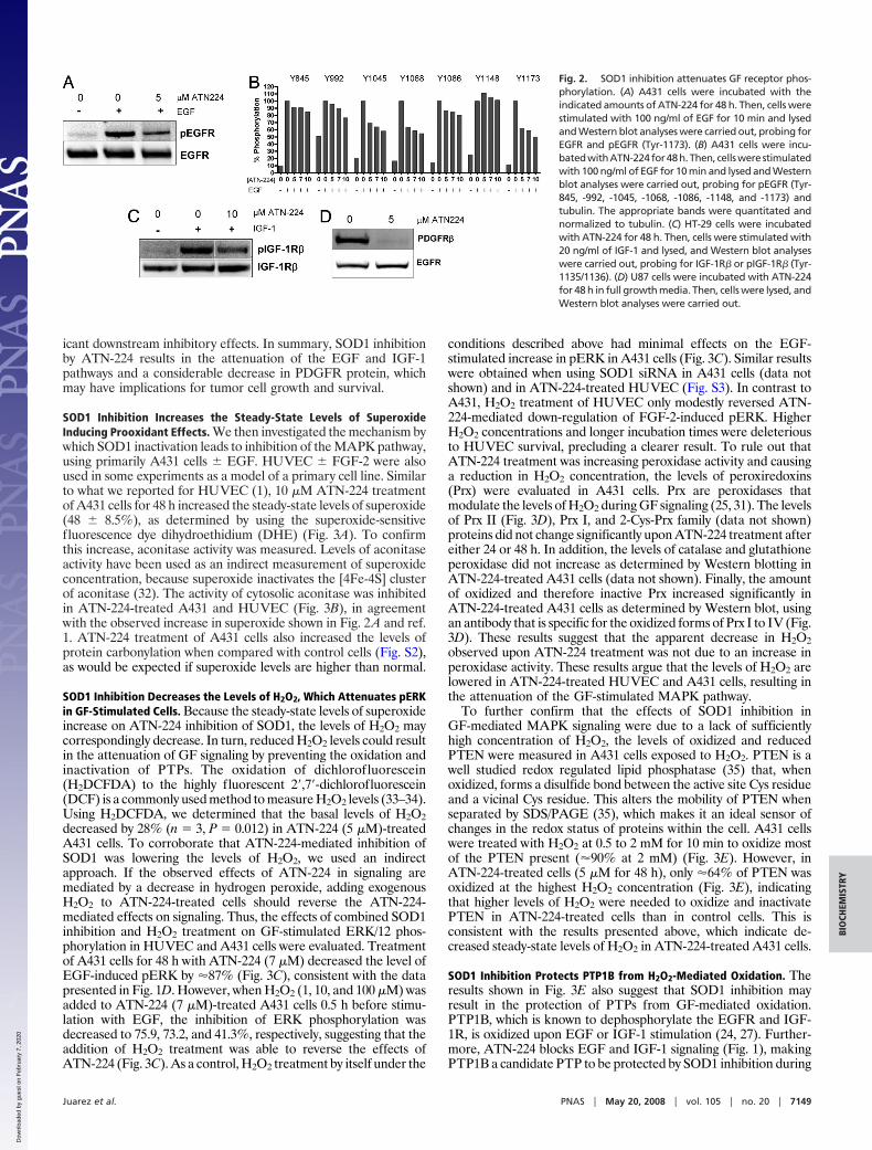
icant downstream inhibitory effects. In summary, SOD1 inhibitionby ATN-224 results in the attenuation of the EGF and IGF-1pathways and a considerable decrease in PDGFR protein, whichmay have implications for tumor cell growth and survival.
SOD1 Inhibition Increases the Steady-State Levels of SuperoxideInducing Prooxidant Effects. We then investigated the mechanism bywhich SOD1 inactivation leads to inhibition of the MAPK pathway,using primarily A431 cells � EGF. HUVEC � FGF-2 were alsoused in some experiments as a model of a primary cell line. Similarto what we reported for HUVEC (1), 10 �M ATN-224 treatmentof A431 cells for 48 h increased the steady-state levels of superoxide(48 � 8.5%), as determined by using the superoxide-sensitivefluorescence dye dihydroethidium (DHE) (Fig. 3A). To confirmthis increase, aconitase activity was measured. Levels of aconitaseactivity have been used as an indirect measurement of superoxideconcentration, because superoxide inactivates the [4Fe-4S] clusterof aconitase (32). The activity of cytosolic aconitase was inhibitedin ATN-224-treated A431 and HUVEC (Fig. 3B), in agreementwith the observed increase in superoxide shown in Fig. 2A and ref.1. ATN-224 treatment of A431 cells also increased the levels ofprotein carbonylation when compared with control cells (Fig. S2),as would be expected if superoxide levels are higher than normal.
SOD1 Inhibition Decreases the Levels of H2O2, Which Attenuates pERKin GF-Stimulated Cells. Because the steady-state levels of superoxideincrease on ATN-224 inhibition of SOD1, the levels of H2O2 maycorrespondingly decrease. In turn, reduced H2O2 levels could resultin the attenuation of GF signaling by preventing the oxidation andinactivation of PTPs. The oxidation of dichlorofluorescein(H2DCFDA) to the highly fluorescent 2�,7�-dichlorofluorescein(DCF) is a commonly used method to measure H2O2 levels (33–34).Using H2DCFDA, we determined that the basal levels of H2O2decreased by 28% (n � 3, P � 0.012) in ATN-224 (5 �M)-treatedA431 cells. To corroborate that ATN-224-mediated inhibition ofSOD1 was lowering the levels of H2O2, we used an indirectapproach. If the observed effects of ATN-224 in signaling aremediated by a decrease in hydrogen peroxide, adding exogenousH2O2 to ATN-224-treated cells should reverse the ATN-224-mediated effects on signaling. Thus, the effects of combined SOD1inhibition and H2O2 treatment on GF-stimulated ERK/12 phos-phorylation in HUVEC and A431 cells were evaluated. Treatmentof A431 cells for 48 h with ATN-224 (7 �M) decreased the level ofEGF-induced pERK by �87% (Fig. 3C), consistent with the datapresented in Fig. 1D. However, when H2O2 (1, 10, and 100 �M) wasadded to ATN-224 (7 �M)-treated A431 cells 0.5 h before stimu-lation with EGF, the inhibition of ERK phosphorylation wasdecreased to 75.9, 73.2, and 41.3%, respectively, suggesting that theaddition of H2O2 treatment was able to reverse the effects ofATN-224 (Fig. 3C). As a control, H2O2 treatment by itself under the
conditions described above had minimal effects on the EGF-stimulated increase in pERK in A431 cells (Fig. 3C). Similar resultswere obtained when using SOD1 siRNA in A431 cells (data notshown) and in ATN-224-treated HUVEC (Fig. S3). In contrast toA431, H2O2 treatment of HUVEC only modestly reversed ATN-224-mediated down-regulation of FGF-2-induced pERK. HigherH2O2 concentrations and longer incubation times were deleteriousto HUVEC survival, precluding a clearer result. To rule out thatATN-224 treatment was increasing peroxidase activity and causinga reduction in H2O2 concentration, the levels of peroxiredoxins(Prx) were evaluated in A431 cells. Prx are peroxidases thatmodulate the levels of H2O2 during GF signaling (25, 31). The levelsof Prx II (Fig. 3D), Prx I, and 2-Cys-Prx family (data not shown)proteins did not change significantly upon ATN-224 treatment aftereither 24 or 48 h. In addition, the levels of catalase and glutathioneperoxidase did not increase as determined by Western blotting inATN-224-treated A431 cells (data not shown). Finally, the amountof oxidized and therefore inactive Prx increased significantly inATN-224-treated A431 cells as determined by Western blot, usingan antibody that is specific for the oxidized forms of Prx I to IV (Fig.3D). These results suggest that the apparent decrease in H2O2observed upon ATN-224 treatment was not due to an increase inperoxidase activity. These results argue that the levels of H2O2 arelowered in ATN-224-treated HUVEC and A431 cells, resulting inthe attenuation of the GF-stimulated MAPK pathway.
To further confirm that the effects of SOD1 inhibition inGF-mediated MAPK signaling were due to a lack of sufficientlyhigh concentration of H2O2, the levels of oxidized and reducedPTEN were measured in A431 cells exposed to H2O2. PTEN is awell studied redox regulated lipid phosphatase (35) that, whenoxidized, forms a disulfide bond between the active site Cys residueand a vicinal Cys residue. This alters the mobility of PTEN whenseparated by SDS/PAGE (35), which makes it an ideal sensor ofchanges in the redox status of proteins within the cell. A431 cellswere treated with H2O2 at 0.5 to 2 mM for 10 min to oxidize mostof the PTEN present (�90% at 2 mM) (Fig. 3E). However, inATN-224-treated cells (5 �M for 48 h), only �64% of PTEN wasoxidized at the highest H2O2 concentration (Fig. 3E), indicatingthat higher levels of H2O2 were needed to oxidize and inactivatePTEN in ATN-224-treated cells than in control cells. This isconsistent with the results presented above, which indicate de-creased steady-state levels of H2O2 in ATN-224-treated A431 cells.
SOD1 Inhibition Protects PTP1B from H2O2-Mediated Oxidation. Theresults shown in Fig. 3E also suggest that SOD1 inhibition mayresult in the protection of PTPs from GF-mediated oxidation.PTP1B, which is known to dephosphorylate the EGFR and IGF-1R, is oxidized upon EGF or IGF-1 stimulation (24, 27). Further-more, ATN-224 blocks EGF and IGF-1 signaling (Fig. 1), makingPTP1B a candidate PTP to be protected by SOD1 inhibition during
Fig. 2. SOD1 inhibition attenuates GF receptor phos-phorylation. (A) A431 cells were incubated with theindicated amounts of ATN-224 for 48 h. Then, cells werestimulated with 100 ng/ml of EGF for 10 min and lysedand Western blot analyses were carried out, probing forEGFR and pEGFR (Tyr-1173). (B) A431 cells were incu-batedwithATN-224for48h.Then, cellswere stimulatedwith 100 ng/ml of EGF for 10 min and lysed and Westernblot analyses were carried out, probing for pEGFR (Tyr-845, -992, -1045, -1068, -1086, -1148, and -1173) andtubulin. The appropriate bands were quantitated andnormalized to tubulin. (C) HT-29 cells were incubatedwith ATN-224 for 48 h. Then, cells were stimulated with20 ng/ml of IGF-1 and lysed, and Western blot analyseswere carried out, probing for IGF-1R� or pIGF-1R� (Tyr-1135/1136). (D) U87 cells were incubated with ATN-224for 48 h in full growth media. Then, cells were lysed, andWestern blot analyses were carried out.
Juarez et al. PNAS � May 20, 2008 � vol. 105 � no. 20 � 7149
BIO
CHEM
ISTR
Y
Dow
nloa
ded
by g
uest
on
Feb
ruar
y 7,
202
0

GF signaling. Thus, two different methods were used to interrogatethe redox status of PTP1B in control versus ATN-224-treated A431cells (Fig. 4 and Fig. S4). First, using an experimental approach inwhich oxidized Cys incorporate a biotin moiety, several proteins inA431 cells (bands 1, 2, and 5) were found to be protected fromoxidation by ATN-224 (decreased signal), whereas others (bands 3,4, 6, and 7) were not (Fig. 4A). Stimulation of A431 cells with EGFoxidizes PTP1B, reaching a maximum after 2–5 min (Fig. S4A).After 5 min of EGF stimulation, ATN-224 treatment protectedPTP1B from EGF-mediated oxidation in A431 cells (Fig. 4B).Furthermore, when a maleimide-biotin reagent that selectivelyreacts with free sulfhydryl groups was used, more biotin wasincorporated in the PTP1B band in ATN-224-treated A431 cells(69 � 39% increase, n � 3, P � 0.01) than in control cells, indicating
the presence of higher levels of reduced PTP1B (Fig. S4 B and C).Likewise, the levels of reduced PTP1B in ATN-224-treatedHUVEC were 86 � 50% (n � 3, P � 0.05) higher than control (Fig.S4C). On the contrary, in A549, in which ATN-224 treatment doesnot reduce EGF-stimulated pERK (Fig. 1D), there was no increasein biotin incorporation (data not shown). Thus, paradoxically,SOD1 inhibition results in antioxidant effects on PTPs likely due toa decrease in H2O2 and prooxidant effects possibly due to anincrease in superoxide (Fig. 3 A and B and Fig. S2).
PTP Inhibitors Antagonize the Attenuation of pERK Caused by SOD1Inhibition in GF-Stimulated Cells. Small-molecule inhibitors of PTPswere used to confirm the hypothesis that ATN-224 effects on GFsignaling are mediated through the protection of PTPs fromoxidation. Orthovanadate is a well characterized broad spectruminhibitor of PTPs that has been widely used to probe the role ofPTPs in signaling (36, 37). HUVEC were treated with orthovana-date 15 or 30 min before stimulation with FGF-2. Both orthovana-date pretreatments abrogated the ATN-224-mediated inhibition ofERK1/2 phosphorylation in FGF-2-treated HUVEC while havingno effect in combination with FGF-2 in control cells (Fig. 5A).Likewise, orthovanadate pretreatment antagonized the inhibitoryeffects of SOD1 siRNA on ERK1/2 phosphorylation (Fig. 5B) inEGF-stimulated A431 cells. Orthovanadate pretreatment increasedthe levels of pERK in both SOD1 siRNA and control (Fig. 5B).However, the change for the control siRNA was only 21%, whereas,for SOD1, siRNA the increase was 56% (Fig. 5B). Similar resultswere obtained in repeat experiments (n � 3). Consistent with thehypothesis that PTPs, and specifically PTP1B, are a target of SOD1inhibition, addition of a specific inhibitor of PTP1B (PTP1Bi) inATN-224-treated A431 cells 3 h before GF stimulation abrogatedthe effects of ATN-224 on EGF-stimulated pERK (Fig. 5C). Thisinhibitor has been shown to be specific for PTP1B (38). However,it cannot be ruled out that other phosphatases were also inhibited.Taken together, these findings suggest that SOD1 inhibition pro-vides protection against oxidation of PTPs during GF signalingresulting in the attenuation of ERK1/2 phosphorylation.
DiscussionThe main findings of this work are (Fig. S5) that (i) SOD1 inhibitioncauses both prooxidant and antioxidant effects, (ii) excess super-
Fig. 3. ATN-224 treatment of A431 cells alters the redox state, which seemsresponsible for the inhibition of ERK1/2 phosphorylation. (A) A431 cells wereincubated with ATN-224 for 48 h in full growth media. Then, cells were treatedwith dihydroethidium (DHE) at 1.6 �M in PBS for 15 min followed by extensivewashing and analyzed by flow cytometry to determine the levels of superox-ide (n � 3; *, P � 0.001). (B) A431 cells were incubated with ATN-224 for 48 hin full growth media. Then, cytosolic aconitase activity was measured (averageof n � 3 � standard deviation) as described in Materials and Methods. (C) A431cells were incubated with 7 �M ATN-224 for 48 h in full growth media. Then,the cells were treated with 1, 10, or 100 �M of H2O2 for 0.5 h in cell mediacontaining 0.5% FBS, washed, and then stimulated with 10 ng/ml of EGF. Thenumbers under the bands [relative intensity (RI)] represent the intensity of thepERK bands normalized to tubulin. (D) A431 cells were treated with ATN-224for 24 or 48 h, cells were lysed, and Western blot analyses were carried out,probing with antibodies against Prx II and Prx-SO3. The later recognizes theoxidized forms of Prx I to IV. (E) A431 were incubated with ATN-224 for 48 hin full growth media. Then, cells were treated with H2O2 in cell media con-taining 0.5% FBS for 10 min. After that, cells were washed, lysed in thepresence of NEM, run on a nonreducing SDS/PAGE, transferred, and probedwith antibodies against PTEN. When PTEN is oxidized by H2O2, it runs as afaster species than reduced PTEN in a SDS/PAGE. The numbers under the bands(RI) represent the intensity of reduced PTEN bands normalized to tubulin.
Fig. 4. Inhibition of SOD1 by ATN-224 reduces the levels of oxidized PTP1Band other proteins. (A) A431 cells treated with 5 �M ATN-224 for 48 h werelysed in the presence of iodoacetic acid to block reduced Cys residues, bufferexchanged, reduced with DTT and reacted with maleimide-biotin so thatoriginally oxidized proteins would acquire a biotin label. Proteins were thenIP with streptavidin–agarose (SA) followed by Western blot analysis, whichwas probed for biotin. (B) A431 cells were treated with 5 �M ATN-224 for 48 hand then stimulated with 100 ng/ml of EGF for 5 min and treated as in B, andbiotinylated proteins were then immunoprecipitated with SA followed byWestern blot analysis, which was probed for PTP1B.
7150 � www.pnas.org�cgi�doi�10.1073�pnas.0709451105 Juarez et al.
Dow
nloa
ded
by g
uest
on
Feb
ruar
y 7,
202
0

oxide does not oxidize PTP1B, and (iii) SOD1 plays an essentialrole in GF-mediated MAPK signaling by mediating the transientoxidation and inactivation of PTPs. These conclusions are drawnfrom studies performed with several tumor cell lines, primaryendothelial cells and fibroblasts. In A549 cells, SOD1 inhibition didnot attenuate GF signaling. This cell line contains a mutatedconstitutively active form of Ras that is known to regulate ROS (26)and, as such, may overcome the effects of SOD1 inhibition.Moreover, mutated Ras constitutively activates the MAPK path-way, which may also oppose the effects of SOD1 inhibition of ERKphosphorylation. Finally, it is presently unclear how ATN-224treatment down-regulates the levels of the PDGF receptor in U87cells (Fig. 2D).
Although the type and origin of ROS that inactivates PTPsduring GF signaling has not been unequivocally identified, it isgenerally accepted that GF binding to its receptor increases levelsof superoxide, which is then dismutated into H2O2 (22, 23, 26).Because superoxide can dismutate non-enzymatically at a fast rate(5 � 105 M�1�s�1) (12, 14) and because superoxide can potentially
oxidize PTPs (39), the possible contribution of SOD1 to thisprocess has been neglected. However, the results presented heredemonstrate that superoxide does not oxidize PTP1B and that thespontaneous dismutation of superoxide is insufficient to generatethe levels of H2O2 needed for PTP inactivation. Therefore, enzy-matically active SOD1 is required for PTP oxidation during GFsignaling. Our results show that SOD1 inhibition protects PTP1Bfrom oxidation in A431 cells and HUVEC. This protection oc-curred under basal conditions (Fig. S4) and immediately after EGFstimulation (Fig. 4B). Furthermore, the effects of SOD1 inactiva-tion on ERK phosphorylation were dependent on having activePTPs. This was demonstrated by reversing the effects of ATN-224or SOD1 siRNA on GF-stimulated ERK1/2 phosphorylation bytwo different approaches: by adding H2O2 exogenously, whichshould oxidize and inactivate PTPs, and by treating with twodifferent PTP inhibitors, orthovanadate and a specific PTP1Binhibitor. PTP1B is known to dephosphorylate EGFR and IGF-1R(24, 27, 28), which are also targets for SOD1 inhibition in A431 andHT-29 cells. Therefore, PTP1B was a candidate PTP responsiblefor the SOD1-mediated effects on the GF-stimulated MAPKpathway. We demonstrated (Fig. 5C) that a pharmacological in-hibitor of PTP1B blocked the effects of ATN-224 on ERK phos-phorylation in response to EGF stimulation. Although it cannot beruled out that the PTP1B inhibitor cross-reacted with other PTPs,this result suggested an important role for PTP1B as an effector ofSOD1 inhibition, which is in agreement with the partial inhibitionobserved at the level of the GF tyrosine kinase receptor byATN-224 treatment (Fig. 2 A–C). Consistent with this hypothesis,a number of unidentified redox regulated proteins, some of whichcould be PTPs, were also protected from oxidation during GFstimulation by SOD1 inhibition (Fig. 4A). Other possible PTPs thatare affected by SOD1 inhibition must still be identified.
As we have shown in Fig. 3E, the levels of oxidized and inactivePrx increased upon ATN-224-mediated SOD1 inhibition poten-tially due to the excess superoxide generated. This result raises thepossibility that the reversible inactivation of Prx that occurs duringGF signaling (25, 31), allowing the accumulation of sufficiently highH2O2 concentrations, may be initiated by superoxide. SOD1 inhi-bition increased the steady-state levels of superoxide considerablyas determined by direct measurements of superoxide, using thesuperoxide sensitive fluorescent dye DHE and other methods.Superoxide is short lived and reactive but did not spontaneouslyconvert into H2O2 to any appreciable level, as previously discussed.Instead, the excess superoxide was involved in other reactions, suchas with Prx and aconitase. The increase in the level of superoxidethat occurs after a 50% inhibition in the activity of SOD1 inactivates�15–30% of total aconitase (32). At 500 nM ATN-224, a �50%inhibition of cytosolic aconitase activity in A431 cells was observed(Fig. 3B), suggesting that significantly high levels of superoxide aregenerated at those concentrations of ATN-224. Besides aconitaseand Prx, our results show that superoxide reacted with a number ofunidentified proteins, determined by measuring total protein car-bonylation in ATN-224-treated A431 cells. Thus, these reactionsaccount at least partially for the excess superoxide. Surprisingly,superoxide cannot oxidize PTP1B in vivo, but it does so in vitro withhigh efficiency (39). This suggests that either competing reactionsscavenge superoxide very efficiently or that some type of compart-mentalization prevents superoxide from reaching PTP1B, or both.
The data presented here indicate that SOD1 is essential forGF-mediated ERK1/2 phosphorylation. It is unclear although howthis inhibition contributes to the antiproliferative activity of ATN-224, because partial SOD1 inhibition with siRNA did not have aneffect on proliferation but was sufficient to inhibit pERK upon EGFstimulation in A431 cells. It is reasonable to hypothesize that SOD1inhibition will also affect other signaling pathways that are regu-lated by H2O2. Finally, the data presented here support the hy-pothesis that SOD1 is a therapeutic target for the treatment ofcancer and that inhibiting SOD1 results in the down-regulation of
Fig. 5. PTP inhibitors block ATN-224 and SOD1 siRNA-mediated attenuation ofERK1/2 phosphorylation. (A) HUVEC were incubated with ATN-224 for 48 h in fullgrowth media. Cells were then treated with orthovanadate for either 15 or 30min followed by stimulation with FGF-2 and lysed, and Western blot analyseswere carried out. The numbers under the bands (relative intensity) represent theintensity of the pERK bands normalized to tubulin. (B) A431 cells were treatedeitherwith7�MATN-224(224)for48hortransfectedwithSOD1siRNAorcontrolsiRNA for 48 h. Cells were then treated with 1 mM orthovanadate (V) for 15 minor not treated before stimulation with 10 ng/ml of EGF for 10 min. Cells werelysed, and Western blot analyses were carried out. V, orthovanadate treatment;224, ATN-224 treatment. (C) A431 cells were treated with ATN-224 for 48 h andexposed to a PTP1B inhibitor 1 h before being stimulated with 10 ng/ml of EGF for10 min. Cells were lysed, and ELISAs were carried out, probing for ERK1/2, pERK.The graph shows means � SD (n � 3) of pERK normalized to ERK1/2.
Juarez et al. PNAS � May 20, 2008 � vol. 105 � no. 20 � 7151
BIO
CHEM
ISTR
Y
Dow
nloa
ded
by g
uest
on
Feb
ruar
y 7,
202
0

multiple signaling pathways important for endothelial and tumorcell function.
Materials and MethodsCells and Antibodies. A431, HT-29, A549, and U87 were obtained from AmericanType Culture Collection (ATCC), and HUVEC and HDFa were from Cascade Bio-logics. FGF-2, EGF, IGF-1 and PDGF were from R&D Systems. ATN-224 (cholinetetrathiomolybdate) was manufactured under cGMP, using a proprietary man-ufacturing process. The antibodies against ERK1/2, pERK1/2, EGF, pEGF (Tyr 845,992, 1045, 1068, 1148, and 1173), IGF-1R�, pIGF-1R� (Tyr 1135/1136), cleavedPARP, and PTEN were from Cell Signaling. Antibodies against pEGFR (Tyr 1086),Prx I, PrxII, Prx-2Cys, and Prx-SO3 were from Abcam. The antibody against PDGFR�
was from Upstate Biotechnology. The antibody against �-tubulin was fromSanta Cruz Biotechnology, and the antibody against SOD1 was from BiodesignInternational.
SOD Activity and MTT Assay. SOD activity and MTT [3-(4,5-dimethylthiazol-2-yl)-2,5-diphenyltetrazolium bromide] assays were performed as described in ref. 1.
Western Blot Analysis and Immunoprecipitations. Cells were plated in fullgrowth media containing 10% FBS and incubated in ATN-224. Cells were thenstimulated with the proper growth factor in the presence of 0.5% FBS media,harvested, and lysed in RIPA [1% Nonidet P-40, 0.25% sodium deoxycholate, 150mM NaCl, and 50 mM Tris (pH 7.4)] with protease inhibitors (Roche), 1 mM AEBSF(Calbiochem), and phosphatase inhibitors Set I and II (Calbiochem). Lysates weresubjected to Western blot analysis, using an antibody specific with appropriateloading controls. Bands were quantitated using a Kodak Image Station 400.Immunoprecipitations were carried out by using 200–500 �g of cell extractincubated overnight with 20 �l of a protein G–agarose slurry (Roche) or strepta-vidin–agarose (Pierce).
SOD siRNA Transfection Experiments. SOD1 Stealth siRNA (Invitrogen) or me-dium GC% Negative control siRNA (Invitrogen; catalog no. 12935-300) was di-luted with OptiMEM I (Invitrogen). Lipofectamine RNAiMAX (Invitrogen) wasdiluted 1:100 with siRNA and incubated for 0.5 h at ambient temperature. A431cells were plated at 100,000 cells per well in a 12-well plate containing siRNA/RNAiMAX solution. After overnight incubation, the media was replaced with
10% FBS DMEM � 7 �M ATN-224 and incubated for another 48 h. Cells werestimulated with 0.5% FBS and DMEM � 100 ng/ml EGF for 10 min. Extracts wereprepared with RIPA, 1 mM AEBSF, and phosphatase inhibitors Set I and II(Calbiochem).
Aconitase assay. Aconitase was assayed according to the manufacturer’s proto-col (OXIS International), using a 96-well assay plate in a SpectraMax plate reader.
Detection of Superoxide or H2O2. A431 cells were incubated with the indicatedamounts of ATN-224 for 48 h. Cells were then trypsinized, washed with PBS,counted, and resuspended in PBS. For superoxide measurements, dihydro-ethidium (DHE) (Invitrogen) was added to the cells at 1.6 �M for 15 min. For H2O2
measurements, H2DCFDA (Invitrogen) at 20 �M was added to the cells for 15 min.After incubation with the respective dyes, the cells were extensively washed andanalyzed by flow cytometry.
Determination of Redox Status of PTP. To determine the redox status of PTEN,A431 cells that had been treated with ATN-224 for 48 h were exposed toincreasing concentrations of H2O2 for 10 min, lysed in the presence of N-ethylmaleimide (NEM), and analyzed by western blot with an antibody againstPTEN. To determine the redox status of PTP1B, A431 cells were treated for 48 hwith ATN-224 and lysed after treatment with EGF. The lysis buffer was degassed,supplemented with freshly prepared iodoacetic acid (10 mM), catalase (100�g/ml), and superoxide dismutase (100 �g/ml) and placed on ice. Cell plates weremoved into an anaerobic chamber and rapidly lysed. Alkylation of free thiols wasallowed to occur for a 1 h. Cell lysate was then applied to desalting columns(Pierce) containing 1 mM DTT to allow reversibly oxidized PTPs to be reducedback. Samples were centrifuged, and the eluate was incubated with a biotin-conjugated PEO-iodoacetyl probe (5 mM) (Pierce) for 1 h. Biotinylated proteinswere purified by streptavidin–Sepharose pulldown, and Western blot analysiswas carried out by probing with a PTP1B antibody or streptavidin–HRP.
Statistical Analysis. GraphPad software was used for all statistical analysis. Dataare presented as mean � SD. Data were analyzed by using unpaired, two-tailedt tests when comparing two variables.
Supporting Information. Apoptosis studies, protein carbonylation, and the de-termination of redox status of PTP1B are described in SI Materials and Methods.
1. Juarez JC, et al. (2006) Copper binding by tetrathiomolybdate attenuates angiogenesisand tumor cell proliferation through the inhibition of superoxide dismutase 1. ClinCancer Res 12:4974–4982.
2. Donate F, et al. (2008) Identification of biomarkers for the anti-angiogenic andanti-tumor activity of the Superoxide Dismutase 1 (SOD1) inhibitor Tetrathiomolyb-date (ATN-224). Br J Cancer 98:776–783.
3. Hassouneh B, et al. (2007) Tetrathiomolybdate promotes tumor necrosis and preventsdistant metastases by suppressing angiogenesis in head and neck cancer. Mol CancerTher 6:1039–1045.
4. Pan Q, Bao LW, Kleer CG, Brewer GJ, Merajver SD (2003) Antiangiogenic tetrathiomo-lybdate enhances the efficacy of doxorubicin against breast carcinoma. Mol CancerTher 2:617–622.
5. Pan Q, Bao LW, Merajver SD (2003) Tetrathiomolybdate inhibits angiogenesis andmetastasis through suppression of the NFkappaB signaling cascade. Mol Cancer Res1:701–706.
6. Pan Q, et al. (2002) Copper deficiency induced by tetrathiomolybdate suppresses tumorgrowth and angiogenesis. Cancer Res 62:4854–4859.
7. Goodman VL, Brewer GJ, Merajver SD (2005) Control of copper status for cancertherapy. Curr Cancer Drug Targets 5:543–549.
8. Lowndes SA, Harris AL (2004) Copper chelation as an antiangiogenic therapy. OncolRes 14:529–539.
9. Redman BG, et al. (2003) Phase II trial of tetrathiomolybdate in patients with advancedkidney cancer. Clin Cancer Res 9:1666–1672.
10. Brewer GJ, et al. (2006) Treatment of metastatic cancer with tetrathiomolybdate, ananticopper, antiangiogenic agent: Phase I study. Clin Cancer Res 6:1–10.
11. Lowndes SA, et al. (2007) Phase I Study of ATN-224 (Choline Tetrathiomolybdate) inMetastatic Cancer. Clin Cancer Res, in press.
12. Fridovich I (1995) Superoxide radical and superoxide dismutases. Annu Rev Biochem64:97–112.
13. Imlay JA, Fridovich I (1991) Assay of metabolic superoxide production in Escherichiacoli. J Biol Chem 266:6957–6965.
14. Fridovich I (1978) The biology of oxygen radicals. Science 201:875–880.15. Li Y, et al. (1995) Dilated cardiomyopathy and neonatal lethality in mutant mice lacking
manganese superoxide dismutase. Nat Genet 11:376–381.16. Reaume AG, et al. (1996) Motor neurons in Cu/Zn superoxide dismutase-deficient mice
develop normally but exhibit enhanced cell death after axonal injury. Nat Genet13:43–47.
17. Ho YS, et al. (1998) Reduced fertility in female mice lacking copper-zinc superoxidedismutase. J Biol Chem 273:7765–7769.
18. Elchuri S, et al. (2005) CuZnSOD deficiency leads to persistent and widespread oxidativedamage and hepatocarcinogenesis later in life. Oncogene 24:367–380.
19. Busuttil RA, et al. (2005) Organ-specific increase in mutation accumulation and apo-ptosis rate in CuZn-superoxide dismutase-deficient mice. Cancer Res 65:11271–11275.
20. Sentman ML, et al. (2006) Phenotypes of mice lacking extracellular superoxide dis-mutase and copper- and zinc-containing superoxide dismutase. J Biol Chem 281:6904–6909.
21. Blander G, de Oliveira RM, Conboy CM, Haigis M, Guarente L (2003) Superoxidedismutase 1 knock-down induces senescence in human fibroblasts. J Biol Chem278:38966–38969.
22. Rhee SG (2006) Cell signaling: H2O2, a necessary evil for cell signaling. Science312:1882–1883.
23. Tonks NK (2005) Redox redux: Revisiting PTPs and the control of cell signaling. Cell121:667–670.
24. Tonks NK (2006) Protein tyrosine phosphatases: From genes, to function, to disease.Nat Rev Mol Cell Biol 7:833–846.
25. Rhee SG, et al. (2005) Intracellular messenger function of hydrogen peroxide and itsregulation by peroxiredoxins. Curr Opin Cell Biol 17:183–189.
26. Ushio-Fukai M (2006) Redox signaling in angiogenesis: Role of NADPH oxidase. Car-diovasc Res 71:226–235.
27. Lee SR, Kwon KS, Kim SR, Rhee SG (1998) Reversible inactivation of protein-tyrosinephosphatase 1B in A431 cells stimulated with epidermal growth factor. J Biol Chem273:15366–15372.
28. Zwick E, Hackel PO, Prenzel N, Ullrich A (1999) The EGF receptor as central transducerof heterologous signalling systems. Trends Pharmacol Sci 20:408–412.
29. Sato K, Nagao T, Iwasaki T, Nishihira Y, Fukami Y (2003) Src-dependent phosphoryla-tion of the EGF receptor Tyr-845 mediates Stat-p21waf1 pathway in A431 cells. GenesCells 8:995–1003.
30. Sundaresan M, Yu ZX, Ferrans VJ, Irani K, Finkel T (1995) Requirement for generationof H2O2 for platelet-derived growth factor signal transduction. Science 270:296–299.
31. Choi MH, et al. (2005) Regulation of PDGF signalling and vascular remodelling byperoxiredoxin II. Nature 435:347–353.
32. Gardner PR, Fridovich I (1992) Inactivation-reactivation of aconitase in Escherichia coli:A sensitive measure of superoxide radical. J Biol Chem 267:8757–8763.
33. Tarpey MM, Wink DA, Grisham MB (2004) Methods for detection of reactive metab-olites of oxygen and nitrogen: In vitro and in vivo considerations. Am J Physiol RegulIntegr Comp Physiol 286:R431–44.
34. Wardman P (2007) Fluorescent and luminescent probes for measurement of oxidativeand nitrosative species in cells and tissues: Progress, pitfalls, and prospects. Free RadicBiol Med 43:995–1022.
35. Lee SR, et al. (2002) Reversible inactivation of the tumor suppressor PTEN by H2O2. J BiolChem 277:20336–20342.
36. Seo DW, et al. (2003) TIMP-2 mediated inhibition of angiogenesis: An MMP-independent mechanism. Cell 114:171–180.
37. Bhutani M, et al. (2007) Capsaicin is a novel blocker of constitutive and interleukin-6-inducible STAT3 activation. Clin Cancer Res 13:3024–3032.
38. Wiesmann C, et al. (2004) Allosteric inhibition of protein tyrosine phosphatase 1B. NatStruct Mol Biol 11:730–737.
39. Barrett WC, et al. (1999) Roles of superoxide radical anion in signal transductionmediated by reversible regulation of protein-tyrosine phosphatase 1B. J Biol Chem274:34543–34546.
7152 � www.pnas.org�cgi�doi�10.1073�pnas.0709451105 Juarez et al.
Dow
nloa
ded
by g
uest
on
Feb
ruar
y 7,
202
0