Sulphated and unsulphated serum, - Gutgut.bmj.com/content/gutjnl/17/11/861.full.pdf · CHOLESTASIS:...
9
Gut, 1976, 17, 861-869 Sulphated and unsulphated bile acids in serum, bile, and urine of patients with cholestasis1 G. P. VAN BERGE HENEGOUWEN2, K-H. BRANDT, H. EYSSEN, AND G. PARMENTIER From the Department of Internal Medicine, Municipal Hospital, Arnhem, The Netherlands; Rega Institute, Louvain, Belgium SUMMARY Samples of serum, bile, and urine were collected simultaneously from patients with cholestasis of varying aetiology and from patients with cirrhosis; their bile acid composition was determined by gas/liquid chromatography and mass spectrometry In cholestasis, the patterns in all three body fluids differed consistently and strikingly. In serum, cholic acid was the major bile acid and most bile acids (> 93 %) were unsulphated, whereas, in urine, chenodeoxycholic was the major bile acid, and the majority of bile acids (> 60 %) were sulphated. Secondary bile acids were virtually absent in bile, serum, and urine. The total amount of bile acids excreted for 24 hours correlated highly with the concentration of serum bile acids; in patients with complete obstruction, urinary excretion averaged 71 6 mg/24 h. In cirrhotic patients, serum bile acids were less raised, and chenodeoxy- cholic acid was the predominant acid. In healthy controls, serum bile acids were consistently richer in chenodeoxycholic acid than biliary bile acids, and no bile acids were present in urine. No unusual monohydroxy bile acids were present in patients with primary biliary cirrhosis, but, in several patients, there was a considerable amount of hyocholic acid present in the urinary bile acids. The analyses of individual bile acids in serum and urine did not appear to provide helpful information in the differential diagnosis of cholestasis. Thus, in cholestasis, conjugation of chenodeoxycholic acid with sulphate becomes a major biochemical pathway, urine becomes a major route of bile acid excretion, and abnormal bile acids are formed. In cholestasis the enterohepatic circulation of bile acids is interrupted and retention of bile acids in the liver occurs with a rise in serum bile acid levels. Renal excretion of bile acids becomes a significant route of elimination (Gregg, 1967) and sulphation of bile acids seems to play an important role in the rate of renal excretion (Palmer, 1967; Admirand et al., 'Part of this work was presented at the Third Bile Acid Meeting, 13-15 June 1974, Freiburg, West Germany, and at the 76th Annual Meeting of the American Gastroentero- logical Association, 17-22 May 1975, San Antonio, Texas, USA, and published in abstract form (Gastroenterology, 68: 1005, 1975). 2Present addresss and for reprints: Nijmegen University School of Medicine, Department of Internal Medicine, Division of Gastroenterology, St. Radboud Hospital, Nijmegen, Netherlands. Received for publication 28 June 1976 1972; Stiehl, 1974). Older studies using methods of low precision and sensitivity suggested that cholic acid became the major bile acid in biliary obstruc- tion (Rudman and Kendall, 1957; Carey, 1958), a finding generally confirmed by more recent studies (Makino et al., 1969; Murphy et al., 1972; Greim et al., 1972). More recently, unsaturated monohydroxy bile acids have been detected in urine and serum of cholestatic patients (Makino et al., 1971; Back, 1973). The aim of our study was to define the bile acid pattern of simultaneously obtained serum, bile, and urine samples in patients with cholestasis in order to gain insight into the changes occurring in bile acid metabolism in this condition. We report here such analyses in a well-defined group of patients, as well as similar analyses in samples from patients with cir- rhosis and healthy control subjects. After this work had been completed, analysis of serum and urine bile 861 on 5 June 2018 by guest. Protected by copyright. http://gut.bmj.com/ Gut: first published as 10.1136/gut.17.11.861 on 1 November 1976. Downloaded from
Transcript of Sulphated and unsulphated serum, - Gutgut.bmj.com/content/gutjnl/17/11/861.full.pdf · CHOLESTASIS:...

Gut, 1976, 17, 861-869
Sulphated and unsulphated bile acids in serum, bile,and urine of patients with cholestasis1G. P. VAN BERGE HENEGOUWEN2, K-H. BRANDT, H. EYSSEN, ANDG. PARMENTIER
From the Department of Internal Medicine, Municipal Hospital, Arnhem, The Netherlands; Rega Institute,Louvain, Belgium
SUMMARY Samples of serum, bile, and urine were collected simultaneously from patients withcholestasis of varying aetiology and from patients with cirrhosis; their bile acid composition was
determined by gas/liquid chromatography and mass spectrometry In cholestasis, the patterns in allthree body fluids differed consistently and strikingly. In serum, cholic acid was the major bile acidand most bile acids (> 93 %) were unsulphated, whereas, in urine, chenodeoxycholic was the majorbile acid, and the majority of bile acids (> 60 %) were sulphated. Secondary bile acids were virtuallyabsent in bile, serum, and urine. The total amount of bile acids excreted for 24 hours correlated highlywith the concentration of serum bile acids; in patients with complete obstruction, urinary excretionaveraged 71 6 mg/24 h. In cirrhotic patients, serum bile acids were less raised, and chenodeoxy-cholic acid was the predominant acid. In healthy controls, serum bile acids were consistently richer inchenodeoxycholic acid than biliary bile acids, and no bile acids were present in urine. No unusualmonohydroxy bile acids were present in patients with primary biliary cirrhosis, but, in severalpatients, there was a considerable amount of hyocholic acid present in the urinary bile acids. Theanalyses of individual bile acids in serum and urine did not appear to provide helpful information inthe differential diagnosis of cholestasis. Thus, in cholestasis, conjugation of chenodeoxycholic acidwith sulphate becomes a major biochemical pathway, urine becomes a major route of bile acidexcretion, and abnormal bile acids are formed.
In cholestasis the enterohepatic circulation of bileacids is interrupted and retention of bile acids in theliver occurs with a rise in serum bile acid levels.Renal excretion of bile acids becomes a significantroute of elimination (Gregg, 1967) and sulphation ofbile acids seems to play an important role in the rateof renal excretion (Palmer, 1967; Admirand et al.,
'Part of this work was presented at the Third Bile AcidMeeting, 13-15 June 1974, Freiburg, West Germany, and atthe 76th Annual Meeting of the American Gastroentero-logical Association, 17-22 May 1975, San Antonio, Texas,USA, and published in abstract form (Gastroenterology, 68:1005, 1975).
2Present addresss and for reprints: Nijmegen UniversitySchool of Medicine, Department of Internal Medicine,Division of Gastroenterology, St. Radboud Hospital,Nijmegen, Netherlands.
Received for publication 28 June 1976
1972; Stiehl, 1974). Older studies using methods oflow precision and sensitivity suggested that cholicacid became the major bile acid in biliary obstruc-tion (Rudman and Kendall, 1957; Carey, 1958), afinding generally confirmed by more recent studies(Makino et al., 1969; Murphy et al., 1972; Greim etal., 1972). More recently, unsaturated monohydroxybile acids have been detected in urine and serum ofcholestatic patients (Makino et al., 1971; Back,1973).The aim of our study was to define the bile acid
pattern of simultaneously obtained serum, bile, andurine samples in patients with cholestasis in order togain insight into the changes occurring in bile acidmetabolism in this condition. We report here suchanalyses in a well-defined group of patients, as well assimilar analyses in samples from patients with cir-rhosis and healthy control subjects. After this workhad been completed, analysis of serum and urine bile
861
on 5 June 2018 by guest. Protected by copyright.
http://gut.bmj.com
/G
ut: first published as 10.1136/gut.17.11.861 on 1 Novem
ber 1976. Dow
nloaded from
G. P. van Berge Henegouwen, K-H. Brandt, H. Eyssen, and G. Parmentier
acids in patients with cirrhosis was reported byStiehl et al. (1975). Makino etal. (1975) also reportedresults of bile acid analyses in samples of serum,urine, and bile of patients with cirrhosis.
Methods
PATIENTS
Thirty-two patients with cholestasis of varyingaetiology were studied as well as 18 patients withcirrhosis (Table 1) and healthy control subjects.Diagnosis was established by liver biopsy. Thediagnosis of total obstruction of the biliary tractwas made before laparotomy and cholangiographyonthe basis of negative urinary and faecal urobilin ex-cretion and confirmed at laparotomy. All patientswith complete obstructions were in a steady-statecondition when samples were collected, as repeatedliver function tests-for example, bilirubin, SGOT,SGPT, alkaline phosphatase, 5'-nucleotidase,cholesterol, and protein electrophoresis-revealedno significant changes over a two week period.
Pruritus was graded on a scale from 1 to 4 by pre-defined criteria.
Collection ofsamplesSerum samples were collected during the fastingstate. In 26 (13 cirrhosis and 13 primary biliarycirrhosis) patients and in 15 control subjects, serumand bile samples were collected simultaneously. Bilerich duodenal fluid was obtained, under standardisedconditions, five to 15 minutes after cholecystokinin(Cecekin, Vitrum, Stockholm) stimulation using adouble lumen Dreiling tube. Samples were stored at
200C.Urine analyses were carried out on 24-hour collec-
tions from 23 patients with cholestasis and sixnormal controls; serum of these patients was
obtained in the fasting state on the same day that the24-hour urine collection was done. All patients had anormal renal function, as assessed by serum crea-tinine. During collection urine was stored at 4°Cand thereafter at - 20°C.
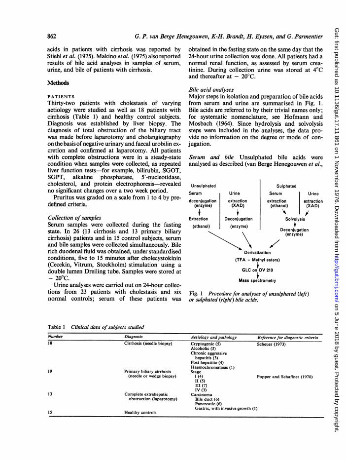
Bile acid analysesMajor steps in isolation and preparation of bile acidsfrom serum and urine are summarised in Fig. 1.Bile acids are referred to by their trivial names only;for systematic nomenclature, see Hofmann andMosbach (1964). Since hydrolysis and solvolysissteps were included in the analyses, the data pro-vide no information on the degree or mode of con-jugation.
Serum and bile Unsulphated bile acids wereanalysed as described (van Berge Henegouwen et al.,
Unsulphated
Serum Urine
deconjugation extraction(enzyme) (XAD)i i
Extraction Deconjugation
(ethanol) (enzyme)
SulphatedSerum Urine
extraction extraction(ethanol) (XAD)
Solvolysis
Deconjugation(enzyme)
/7Derivatization
(TFA - Methyl esters)
GLC on OV 210
Mass spectrometry
Fig. 1 Procedure for analyses of unsulphated (left)or sulphated (right) bile acids.
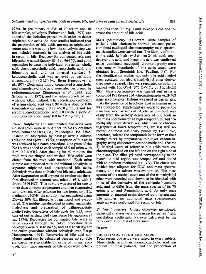
Table 1 Clinical data of subjects studiedNumber Diagnosis Aetiology and pathology Reference for diagnostic criteria18 Cirrhosis (needle biopsy) Cryptogenic (5) Scheuer (1973)
Alcoholic (5)Chronic aggressive
hepatitis (3)Post hepatitic (4)Haemochromatosis (1)
19 Primary biliary cirrhosis Stage(needle or wedge biopsy) I (4) Popper and Schaffner (1970)
It (5)III (7)IV (3)
13 Complete extrahepatic Carcinomaobstruction (laparotomy) Bile duct (6)
Pancreatic (6)Gastric, with invasive growth (1)
15 Healthy controls
862
on 5 June 2018 by guest. Protected by copyright.
http://gut.bmj.com
/G
ut: first published as 10.1136/gut.17.11.861 on 1 Novem
ber 1976. Dow
nloaded from
Sulphated and unsulphated bile acids in serum, bile, and urine ofpatients with cholestasis
1974). In preliminary studies of 10 serum and 10bile samples, solvolysis (Palmer and Bolt, 1971) wasadded to the isolation procedure in order to detectsulphated bile acids. As these studies indicated thatthe proportion of bile acids present as sulphates inserum and bile was quite low, the solvolysis step wasnot included routinely in the analysis of bile acidsin serum or bile. Recovery for conjugated referencebile acids was satisfactory (84 5 to 96-3 %), and goodseparation between the individual bile acids-cholicacid, chenodeoxycholic acid, deoxycholic acid, andlithocholic acid-and the internal standard 7-ketodeoxycholic acid was achieved by gas/liquidchromatography (GLC) (van Berge Henegouwen etal., 1974). Determinations ofconjugated serum cholicand chenodeoxycholic acid were also performed byradioimmunoassay (Simmonds et al., 1973; andSchalm et al., 1975), and the values correlated wellwith our GLC method. The correlation coefficientof serum cholic acid was 0 99 with a slope of 0-84(concentration range 7 0 to 456 0 Mmol/l) and forserum chenodeoxycholic acid 100 with a slope of1 20 (concentration range 0-8 to 235-2 p-mol/l).
Urine Sulphated and unsulphated bile acids wereisolated from urine with Amberlite-XAD2 obtainedfromRohm and Haas, Co., Philadelphia, PA, USA.Instead of adsorption by passage over a column(Makino and Sjovall, 1972), adsorption of bile acidswas achieved by a batch procedure. One gram of theXAD2 was added to each sample of 5 ml urine with5 ml 0-2 NaOH. After shaking for 60 minutes, theresin was centrifuged and washed. Bile acids wereeluted from the resin with methanol. Each urinesample was processed with and without solvolysis toseparate sulphated and unsulphated bile acids.Solvolysis was done to hydrolyse bile acid sulphates.After evaporation until dryness the residue was there-fore dissolved in acetone and ethanol (9:1, with 1drop of 6 N HCI). This mixture was stored for one tothree days at room temperature and then evaporateduntil dryness. After refluxing for two hours with 2%methanolic KOH, the solution was neutralised usingDowex 50W-X4, filtered with methanol and evapo-rated. The residue was dissolved in water; enzymatichydrolysis and preparation of trifluoroacetatemethyl ester derivatives of bile acids for GLC werecarried out as described (van Berge Henegouwen etal., 1974). Recoveries for conjugated bile acids inurine carried through the entire procedure withsolvolysis were 80-0 to 84-5 % and 84-0 to 90 0% forthe entire procedure without solvolysis (van BergeHenegouwen, 1974). Recoveries of bile acid sul-phates could not be calculated because no suitablestandards were available. In urine of normal con-trols, only trace amounts of bile acids were detect-
able (less than 0-2 mg/l) and solvolysis did not in-crease the amount of bile acids.
Mass spectrometry As several urine samples ofpatients with cholestasis showed unusual peaks,combined gas/liquid chromatography-mass spectro-metric studies were carried out. The identity of litho-cholic acid, 3p-hydroxy-5-cholen-24-oic acid, urso-deoxycholic acid, and hyocholic acid was confirmedusing combined gas/liquid chromatography-massspectrometry (standards of bile acids tested wereobtained from Steroloids Inc. (Pawling, NY). Forthe identification studies not only bile acid methylester acetates, but also trimethylsilyl ether deriva-tives were prepared. They were separated on columnspacked with 3% OV-1, 3% OV-17,; or 3% Hi-Eff8BP. Mass spectrometry was carried out using acombined Pye (Series 104) chromatography-AEI-MSmass spectrometer. Helium was used as carrier gas.As the presence of hyocholic acid in human urine
was unexpected, supplementary work to prove thestructure was carried out. Acetic acid is lost veryeasily from the acetoxy derivatives of bile acids inthe mass spectrometer at high temperature, but tri-methylsilyl ether derivatives, which can be chroma-tographed at lower temperatures, are less well re-moved on most stationary phases by GLC. We,therefore, isolated the compounds in the form of theirmethyl esters by preparative thin-layer chromato-graphy using chloroform-acetone-methanol (70:25:5). Methyl esters of reference bile acids were co-chromatographed on the left and on the right side ofthe plate. The silica gel band corresponding to thehyocholic acid region was scraped off and elutedwith chloroform:methanol (1:1, v/v). The eluate wasdivided into aliquots for GLC and mass spectro-metry, and the solvent was evaporated. The massspectra of the methyl esters and of the trimethylsilylether were recorded and shown to be identical withthose of the derivative of the authentic hyocholicacid and to differ from the mass spectra of its 7lpepimers, w- and /-muricholic acid. As only traceamounts of unusual peaks showed up in serum andbile samples, no additional mass spectrometricanalyses were performed for serum or bile.
Statistical analyses Unless otherwise mentioned,statistical analyses were done using the paired t test;correlation coefficients (r) were calculated by therank correlation test of Spearman.
Results
CHOLESTASIS: SERUM BILE ACIDSTotal serum bile acids were raised in every subject.More cholic acid than chenodeoxycholic acid waspresent in most patients, and the proportion of
2*
863
on 5 June 2018 by guest. Protected by copyright.
http://gut.bmj.com
/G
ut: first published as 10.1136/gut.17.11.861 on 1 Novem
ber 1976. Dow
nloaded from

864 G. P. van Berge He
cholic was greatest in patients with the highestlevels (Fig. 2). The proportion present as deoxycholicand lithocholic was extremely low. Analyses beforeand after solvolysis indicated that sulphates com-prised less than 7% of the serum bile acids and con-sisted mainly of sulphated chenodeoxycholic acid.
CHOLESTASIS: URINE BILE ACIDSUrinary bile acids were consistently raised, and thedaily excretions correlated highly (r = 0 85) withserum concentrations. In the 23 patients studied,urinary bile acids ranged from 3-1 to 79-8 mg/?4 hwith mean (± SD) of 31-4 mg/24 h (± 21 -0). In these23 patients, serum bile acids ranged from 9 0 to218A4 ,umol/l with a mean (± SD) of 120-5 ,mol/l(± 41 1). In patients with complete obstruction, theaverage daily excretion was 71-6 mg.The pattern of urine bile acids differed markedly
from that of serum bile acids in that sulphatedchenodeoxycholic acid was the major bile acid pre-sent in urine (Figs. 3 and 4). Clearances of sulphatedand unsulphated bile acids were calculated in 10patients from the 24-hour urine excretions andserum bile acid concentrations (Fig. 5). The renalclearance of sulphated bile acids was 10 to 100 timesgreater than that of unsulphated bile acids. This
negouwen, K-H. Brandt, H. Eyssen, and G. Parmentier
calculation assumes that all sulphated bile acids inurine originated from plasma. If there is sulphationand secretion into urine of bile acids by the renaltubule, the calculated values are an overestimate.
CHOLESTASIS: BILIARY BILE ACIDSMean biliary bile acid composition and bile acidconcentration for the different diseases is given inTable 2. In patients with primary biliary cirrhosis,the proportion of cholic acid was increased. Noabnormal bileacidswere detected in the bile of patientswith primary biliary cirrhosis, and the total litho-cholic concentrations were always below 3 % of thetotal biliary bile acid concentration. The lithocholicpeak increased after solvolysis, indicating that amajor part was present as a sulphate (> 60%).
In 13 patients with primary biliary cirrhosis, bilesamples and fasting serum samples were obtainedsimultaneously. In all but one patient, serum con-tained more chenodeoxycholic acid than bile.
CHOLESTASIS: NOVEL BILE ACIDSSix of seven urine samples contained considerableamounts (2 5 to 21-3 ,umol/24 h) of a bile acid havingthe retention time on gas/liquid chromatography ofhyocholic acid. By mass spectrometry, the fragmen-
1Q0
,}.90
-.50
,6 40
2.020
1C.:RA
Li-ver cirrhoSsn 48)*s Pmary itory cirhosis('.. ..'Qpt sit,.., r
.bt,ucIs: _ , .. . .
AL
A40
£
4
A
A
.A
A.0..
a .'
.'
4-0a0
0 0
00. 00 o
0 00..vi
0,
.. ......... -Al C0.i.4.-...... . .::, .
,:.100% 0%cChodoycoc
275
250
225 Fig. 2 Serum bile acidQ-5 composition (abscissa) and
concentration (ordinate) inMO patients with liver cirrhosis
8 (0), patients with primary175 biliary cirrhosis (0), and
patients with completeextrahepatic obstruction (A).
150 Since lithocholic and deoxy-cholic acids compose less
125 l than 5% of total bile acids,only cholic acid and
aS chenodeoxycholic acid100 3 concentrations are shown.
Total bile acid concentration75 in healthy subjects was less
than 6-3 FM (2.5 [±g/ml),and the upper limits of
50 normal values are indicatedby the hatched area.
25
100%Cholic-
ic.-D
.8Q,. .i I
on 5 June 2018 by guest. Protected by copyright.
http://gut.bmj.com
/G
ut: first published as 10.1136/gut.17.11.861 on 1 Novem
ber 1976. Dow
nloaded from

Sulphated and unsuiphated bile acids in serum, bile, and urine ofpatients with cholestasis
Serum Urine
Fig. 3 Bile acid composition ofserum and urine(%, normalised to 100%) from 23 patients withcholestasis shown using circular coordinates. Only cholicand chenodeoxycholic acids are shown, as other bileacids constituted less than 5% of the total bile acids inany sample. Abbreviations; cholic acid (CHO), sulphatedcholic acid (CHO-S), chenodeoxycholic acid (CDC),and sulphated chenodeoxycholic acid (CDC-S).
0
UCc
c0to
C)0
Serum Urine
80-
60F
40F
20h
Cholic acid
P<0.001
Serum Urine
Chenodeoxycholic acid
P< 0.001
Fig. 4 Bile acid composition ofserum samples and24-hour urine collections in 23 patients with cholestasis.The serum sample was obtained on the day of the urinecollection. The percentage represents the sum of
sulphated and unsulphated species.
Fig. 5 Renal clearances for sulphated and unsulphatedbile acids for 10 patients. The mean and SEM areshown. Renal clearance on the ordinate is expressed inl/day.
tation pattern of the trimethylsilyl ether was con-
sistent with that of the 7a epimer of a 3,6,7-tri-hydroxy-5p-cholanoic acid. A base peak was foundat m/e 458. In contrast, the 7fl epimers, w- andmuricholic acid gave a base peak at m/e 285, which isthe result of fission between carbon atoms 6 and 7.
The second prominent peak was present at m/e 369,which is typical for vicinal trimethylsiloxy groups.
No peaks were observed at m/e 343 and 253, whichare conspicuous in the spectrum of the trimethyl-silyl derivatives of cholic acid. Trace amounts of an
unsaturated monohydroxy bile acid (3/-hydroxy-5-cholen-24-oic acid) were found in three patients, andtrace amounts of ursodeoxycholic acid were foundin two patients. The structure of these bile acids was
also confirmed by mass spectrometry.
Table 2 Biliary bile acid composition and concentration in duodenal bile samples obtained after CCK stimulation
Group Acid
Cholic Chenodeoxycholic Deoxycholic Lithocholic
I. Mean composition (% ± SEM)1. Primary biliary cirrhosis (N = 13) 65-0* (18-7) 32-5t (9 4) 2-4* (0 6) 0.1* (0-03)2. Liver cirrhosis (N = 13) 43 0 (15 1) 44-6 (9 9) 9-8 (2 5) 2-6 (0 8)3. Normal controls (N = 15) 42-0 (49) 445 (37) 11-4 (2-0) 21 (02)
I1. Mean concentration (,umol/I + SEM)1. Liver cirrhosis (N = 13) 6 15* (2 45) 6.35* (1-43) 1 40* (0 35) 0-33* (0-13)2. Normal controls (N = 15) 32-28 (4-18) 33-28 (3 25) 7 30 (1-25) 1 50 (015)
*Significantly different from normal (p <001) (Wilcoxon's rank sum test).tSignificantly different from normal (p < 0-05) (Wilcoxon's rank sum test).
to5(46
865
on 5 June 2018 by guest. Protected by copyright.
http://gut.bmj.com
/G
ut: first published as 10.1136/gut.17.11.861 on 1 Novem
ber 1976. Dow
nloaded from

G. P. van Berge Henegouwen, K-H. Brandt, H. Eyssen, and G. Parmentier
CIRRHOSISTotal serum bile acids were raised in most patients,but in contrast with cholestasis, the major serumbile acid was chenodeoxycholic acid (Fig. 2). Urinarybile acids were increased, and as in the patients withcholestasis, the total bile acid excretion correlatedhighly with the total serum concentration. Themajor urinary bile acid was chenodeoxycholic acid,and the majority of this was presen. as the sulphate.Indeed, most of the sulphated bile acid fraction ofurine was composed of chenodeoxycholic acid. Thebiliary bile acid pattern was similar to that of thehealthy controls (Table 2). Cirrhotic patients tendedto contain less deoxycholic acid in biliary bile acidsthan healthy controls, but the proportion was iden-tical in serum and bile. As in the patients with choles-tasis, the serum contained more chenodeoxycholicacid than bile (Fig. 6). Bile from cirrhotic patientswas much more dilute than from healthy controls(Table 2). This has been described previously byTumberg and Grahame (1970).
HEALTHY CONTROL SUBJECTSValues for total fasting serum bile acids in healthycontrol subjects were less than 6-3 ,umol/l. Serum
50%
cholic acid levels varied between 05 and 1 8 ,umol/l,and serum chenodeoxycholic acid levels between0 7 and 3 5 ,umol/l. These values are in agreementwith other reports (Sandberg et al., 1965; Makinoet al., 1969). Serum bile acids contained more cheno-deoxycholic acid than the biliary bile acids (Fig.7).No bile acids were detected inthe urine of the healthycontrol subjects.
CLINICAL CORRELATIONThe score of pruritus correlated highly with totalserum bile acids (r = 0 78). There was an equallyhigh correlation between the level of the majorserum bile acid, cholic acid, and pruritus (r = 0-81).
In patients with primary biliary cirrhosis, therewas no significant relationship between the patternof serum bile acids and the stage of the disease,although the relative concentration of cholate washigher in the second and third stage of the disease. Inthe patients with complete extrahepatic obstructionthere was no correlation between serum bilirubin andtotal bile acids or between total bile acids andalkaline phosphatase. In patients without completeextrahepatic obstruction, there was good correlationbetween serum bilirubin and total bile acids (r =
Deoxycholic0 A
-ooDo0
o\0C01110
Cholic0
0Chenodeoxycholic
100%
Fig. 6 Bile acid composition ofsamples offasting-state serum and bile (obtained by CCK stimulation) frompatients with cirrhosis; the data are plotted using triangular coordinates and with the assumption that total bile acidsare composed solely of cholic, chenodeoxycholic, and deoxycholic acids. Lines connect samples from one patient.The greater proportion ofchenodeoxycholic acid in the serum sample is statistically significant.
866
on 5 June 2018 by guest. Protected by copyright.
http://gut.bmj.com
/G
ut: first published as 10.1136/gut.17.11.861 on 1 Novem
ber 1976. Dow
nloaded from

Sulphated and unsulphated bile acids in serum, bile, and urine ofpatients with cholestasis
Deoxycholic0 A
wo0
0 50%
\ oChenodeoxycholic
100%
Fig. 7 Bile acid composition offasting-state serum and bile (obtained by CKK stimulation) from healthy controlsubjects plotted as described in Fig. 6. The greater proportion of chenodeoxycholic acid in the serum sample isstatistically significant.
0-82) as well as between total bile acids and alkalinephosphatase (r = 0 66). In patients with primarybiliary cirrhosis, good correlation exists betweenSGOT and total serum bile acids (r = 0-72). Therewas a very high correlation between alkaline phos-phatase and 5'-nucleotidase activities in all patients(r = 0 90).
Discussion
These data, which define the bile acid pattern of thesulphated and unsulphated fractions in serum, urine,and bile of patients with cholestasis, cirrhosis, andhealthy controls, provide the first comprehensivedescription of the alterations in bile acid metabolismoccurring in patients with obstructive hepatobiliarydisease. The most important finding is the great in-crease in the formation of primary bile acid sul-phates, especially chenodeoxycholic acid, which isassociated with a striking increase in the excretion ofbile acids in the urine. The site of bile acid sulpha-tion is not known, but it can occur in the liver(Palmer, 1967; Liersch and Stiehl, 1974). A secondpredictable change is the virtual disappearance ofsecondary bile acids from serum, urine, and bile dueto decreased passage of bile acids into the distal
intestine, as first described by Sj6vall (1960). Aminor change is the appearance of the novel bileacid, hyocholic acid, which indicates increased 6-hydroxylation. 6-Hydroxylation of bile acids hasbeen reported in isolated subcellular fractions ofhuman liver, but doesnotoccurappreciablyinhealthysubjects (Triilzsch et al., 1974). Evidence that 6-hydroxylation of bile acids in man occurs duringcholestasis has recently been presented bySummerfield et al. (1976). The presence of otherunusual bile acids like 3f3-hydroxy-5-cholen-24-oicand ursodeoxycholic acid in urine of patients withcholestasis has been described by Makino et al.(1971) and Back (1973) and was confirmed in the pre-sent study.
Faecal bile acids were not measured in our sub-jects, but are thought to be quite low in patients withcholestasis (Miettinen, 1973). The average urinaryexcretion in our subjects with complete extrahepaticobstruction was 71 6 mg/day, compared with pub-lished figures for daily bile acid synthesis of 250 mg/day, based on chemical measurements of faecal bileacids in normal subjects eating a solid diet(Miettinen, 1973). If urinary bile acid excretion canbe equated with bile acid synthesis, then synthesisappears to be diminished in patients with cholestasis.
0\0C0
Cholic 2
867
on 5 June 2018 by guest. Protected by copyright.
http://gut.bmj.com
/G
ut: first published as 10.1136/gut.17.11.861 on 1 Novem
ber 1976. Dow
nloaded from

868 G. P. van Berge Henegouwen, K-H. Brandt, H. Eyssen, and G. Parmentier
In the cirrhotic patient, the pattern of serum bileacids differed from that in cholestasis in containingrelatively more chenodeoxycholic acid. Since bileacid kinetics were not carried out, we cannot definethe mechanism responsible for this difference. Amarkedly reduced cholic acid synthesis, in cirrhosis,as shown by McCormick et at. (1973), could con-tribute.
Finally, in all patients it is clear that the bile acidpattern of serum, urine, and bile show consistentdifferences both in health and disease. This findingwas first reported by Erb et al. (1972, 1973), and hasnow been confirmed independently by radioimmuno-assay (Schalm et al., 1975). There is considerableevidence suggesting that chenodeoxycholic acid maybe absorbed passively from the intestine (Hislop etal., 1967; Schiff ct al., 1972), and is cleared lessefficiently by the liver (Cowen et al., 1975; Hoffmanet al., 1975). Thus, bile always contains morecholic acid, and in serum, chenodeoxycholic acid isthe dominant bile acid. The pattern of urinary bileacids reflects the product of the renal clearance ofeach individual bile acid and its average serumconcentration. Thus, the dominant urinary bileacids are those with the highest clearance-namely,the bile acid sulphates.
CLINICAL CORRELATIONSOur studies do not lend support to the proposal thatthe pattern of serum bile acids provides usefuldiagnostic information in the patient with cholestasis.In our patients, the presence of deoxycholic acid ap-peared to exclude the diagnosis of complete biliaryobstruction, but it is doubtful if this association willprove to be clinically useful. The absence of deoxy-cholic acid does not provide useful diagnostic in-formation, as it may reflect either decreased forma-tion or decreased substrate availability. Thus, thedata presented here support the suggestion ofLaRusso et al. (1975) that the clinical value of serumbile acid measurements is likely to be greatest inpatients with anicteric liver disease.
We are indebted to Dr A. F. Hofmann for manyhelpful suggestions in the preparation of this manu-script and for carrying out control determinations ofserum bile acids with specific radioimmunoassays andto Miss A. T. Ruben for assistance in the bile acidanalyses. This study was supported by grants fromthe Dutch Health Organisation TNO and 'de DrieLichten'.
References
Admirand, W. H.. Stiehl, A., and Thaler, M. M. (1972).Sulfation of bile salts: an important metabolic pathway incholestasis (Abstract). Gastroenterology, 62, 190.
Back, P. (1973). Identification and quantitative determina-tion of urinary bile acids excreted in cholestasis. ClinicaChimica Acta, 44, 199-207.
Carey, J. B., Jr (1958). The serum trihydroxy-dihydroxy bileacid ratio in liver and biliary tract disease. Journal ofClinical Investigation, 37, 1494-1503.
Cowen, A. E., Korman, M. G., Hofmann, A. F., andThomas, P. J. (1975). Plasma disappearance of radio-activity after intravenous injection of labelled bile acids inman. Gastroenterology, 68, 1567-1573.
Erb, W., Schreiber, G., and Walczak, M. (1972). Gas-chromatographische Untersuchungen der Serumgallen-sauren: Methodik sowie Ergebnisse bei Patienten mitakuter Hepatitis. Zeitschrift fdr Gastroenterologie, 10, 349-358.
Erb, W., Schreiber, G., and Walczak, M. (1973). Unter-suchungen der Gallensauren im Serum von Patienten mitExtrahepatischer Cholostase und mit Chronische Leberer-krankungen. Zeitschrift fur Gastroenterologie, 11, 279-288.
Gregg, J. A. (1967). Presence of bile acids in jaundiced humanurine. Nature, 214, 29-31.
Greim, H., Trulzsch, D., Czygan, P., Rudick, J., Hutterer, F.,Schaffner, F., and Popper, H. (1972). Mechanism ofcholestasis. 6. Bile acids in human livers with or withoutbiliary obstruction. Gastroenterology, 63, 846-850.
Hislop, I. G., Hofmann, A. F., and Schoenfield, L. J. (1967).Determinants of the rate and site of bile acid absorption inman (Abstract). Journal of Clinical Investigation, 46, 1070.
Hoffman, N. E., Iser, J. H., and Smallwood, R. A. (1974).Hepatic bile acid transport: effect of conjugation andposition of hydroxyl groups. Amnerican Journal of Physi-ology, 229, 298-302.
Hofmann, A. F., and Mosbach, E. H. (1964). Identificationof allodeoxycholic acid as the major component of gall-stones induced in the rabbit by 5a-cholestan-3p-ol.Journal of Biological Chemistry, 239, 2813-2821.
LaRusso, N. F., Hoffman, N. E., Hofmann, A. F., andKorman, M. G. (1975). Validity and sensitivity of an intra-venous bile acid tolerance test in patients with liverdisease. New England Journal of Medicine, 292, 1209-1214.
Liersch, M., and Stiehl, A. (1974). Bildung von Gallensaure-sulfatester in perfundierten Rattenlebern nach Gall-engangverschluss. Zeitschrift fidr Gastroenterologie, 12,131-134.
McCormick, W. C. Iff, Bell, C. C. Jr, Swell, L., andVlahcevic, Z. R. (1973). Cholic acid synthesis as an indexof the severity of liver disease in man. Gut, 14, 895-902.
Makino, I., Hashimoto, H., Shinozaki, K., Yoshino, K., andNakagawa, S. (1975). Sulphated and nonsulphated bileacids in urine, serum, and bile of patients with hepato-biliary diseases. Gastroenterology, 68, 545-553.
Makino, I., Nakagawa, S., and Mashimo, K. (1969). Con-jugated and unconjugated serum bile acid levels in patientswith hepatobiliary diseases. Gastroenterology, 56, 1033-1039.
Makino, I., and Sjovall, J. (1972). A versatile method foranalysis of bile acids in plasma. Analytical Letters, 5, 341-349.
Makino, I., Sjovall, J., Norman, A., and Strandvik, B. (1971).Excretion of 3f3-hydroxy-z15-cholenoic and 3o-hydroxy-5a-cholanoic acids in urine of infants with biliary atresia.FEBS Letters, 15, 161-164.
Miettinen, T. A. (1973). Clinical implications of bile acidmetabolism in man. In The Bile Acids, vol. 2, pp. 191-247.Edited by P. P. Nair and D. Kritchevsky. Plenum Press:New York-London.
Murphy, G. M., Ross, A., and Billing, B. H. (1972). Serumbile acids in primary biliary cirrhosis. Gut, 13, 201-206.
Palmer, R. H. (1967). The formation of bile acid sulfates: a
on 5 June 2018 by guest. Protected by copyright.
http://gut.bmj.com
/G
ut: first published as 10.1136/gut.17.11.861 on 1 Novem
ber 1976. Dow
nloaded from
Sulphated and unsulphated bile acids in serum, bile and urine ofpatients with cholestasis 869
new pathway of bile acid metabolism in humans. Pro-ceedings of the National Academy of Sciences of the UnitedStates of America, 58, 1047-1050.
Palmer, R. H., and Bolt, M. G. (1971). Bile acid sulfates. I.Synthesis of lithocholic acid sulfates and their identifica-tion in human bile. Journal ofLipid Research, 12, 671-679.
Popper, H., and Schaffner, F. (1970). Non-suppurativedestructive chronic cholangitis and chronic hepatitis. InProgress in Liver Disease, vol. 3, pp. 336-354. Edited byH. Popper and F. Schaffner. Grune and Stratton: NewYork.
Rudman, D., and Kendall, F. E. (1957). Bile acid content ofhuman serum. I. Serum bile acids in patients with hepaticdisease. Journal of Clinical Investigation, 36, 530-537.
Sandberg, D. M., Sjovall, J., Sjovall, K., and Turner, D. A.(1965). Measurement of human serum bile acids by gas-liquid chromatography. Journal of Lipid Research, 6, 182-192.
Schalm, S. W., Turcotte, J., Hofmann, A. F., and Cowen,A. E. (1975). A new bile acid radioimmunoassay: develop-ment, validation and preliminary application of an assayfor chenodeoxycholyl conjugates (Abstract). Gastro-enterology, 68, 913.
Scheuer, P. (1973). Liver biopsy interpretation. 2nd edn.Bailliere, Tindall: London.
Schiff, E. R., Small, N. C., and Dietschy, J. M. (1972).Characterisation of the kinetics of the passive and activetransport mechanisms for bile acid absorption in the smallintestine and colon of the rat. Journal of Clinical Investiga-
tion, 51, 1351-1362.Simmonds, W. J., Korman, M. G., Go, V. L. W., andHofmann, A. F. (1973). Radioimmunoassay of conjugatedcholyl bile acids in serum. Gastroenterology, 65, 705-711.
Sjovall, J. (1960). Bile acids in man under normal andpathological conditions. Bile acids and steroids 73.Clinica Chimica Acta, 5, 33-41.
Stiehl, A. (1974). Bile salt sulphates in cholestasis. EuropeanJournal of Clinical Investigation, 4, 59-63.
Stiehl, A., Earnest, D. L., and Admirand, W. H. (1975).Sulfation and renal excretion of bile salts in patients withcirrhosis of the liver. Gastroenterology, 68, 534-544.
Summerfield, J. A., Billing, B. H., and Shackleton, C. H. L.(1976). Identification of bile acids in the serum and urine incholestasis. Biochemnical Journal, 154, 507-516.
Trulzsch, D., Roboz, J., Greim, H., Czygan, P., Rudick, J.,Hutterer, F., Schaffner, F., and Popper, H. (1974). Hy-droxylation of taurolithocholate by isolated human livermicrosomes. I. Identification of metabolic product. Bio-chemical Medicine, 9, 158-166.
Turnberg, L. A., and Grahame, G. (1970). Bile salt secretionin cirrhosis of the liver. Gutt, 11, 126-133.
van Berge Henegouwen, G. P. (1974). Bile Acids and Cholest-asis. Thesis, Nijmegen, The Netherlands.
van Berge Henegouwen, G. P., Ruben, A. T., and Brandt,K. H. (1974). Quantitative analysis of bile acids in serumand bile, using gas-liquid chromatography. ClinicaChimica Acta, 54, 249-261.
on 5 June 2018 by guest. Protected by copyright.
http://gut.bmj.com
/G
ut: first published as 10.1136/gut.17.11.861 on 1 Novem
ber 1976. Dow
nloaded from