Sulfur and iron surface states on fractured pyrite surfaces - RRuff
10
American Mineralogist, Volume 83, pages 1067-1076, 1998 Sulfur and iron surface states on fractured pyrite surfaces H.W. NEsnrlrrrt'. G.M. BlNcnonrr'A.R. PRATTT2 rNp M.J. ScnrNr' rDepartment of Earth Sciences, University of WesternOntario, London, Ontario N6A 5B7, Canada ,Department of Chemistry, University of WesternOntario, London, Ontario N6A 5B7, Canada Ansrn.lcr Pyrite has a poor {001} cleavage. Unlike most other minerals with a rocksalt-type structure, pyrite typically fractures conchoidally, demonstratingthat parting surfacesare not constrained to the {001 } crystallographicplane. Cleavagealong {001 } require rupture of only Fe-S bonds, but pyrite consists of both Fe-S and S-S bonds. Analysis of bond energiesindicates that S-S bonds are the weaker bonds and they are likely to be ruptured when pyrite is fractured. With each ruptured S-S bond, two mononuclearspecies (formally S, ) ari produced,one bound to one fracture surfaceand the second to the oppositefracture surface.This monomer is reducedto 52- (monosulfide)during relaxation through oxidation of surface Fe2* ions to Fe3*. These surfacerelaxation processes explain the surface states observed in S(2p) and Fe(2p.,,) X-ray photoelecfton specffa (XPS) of pyrite. The S(2p) XPS spectrum is interpreted to include bulk disulfide contributions at 162.6 eV and two surface state conffibutions at 162.0 and 161.3 eV. The monosulfide (S' ) emission is near 161.3 eY as observed in S(2p) spectra of pynhotite, and the 162 eY peak is interpreted to result from the surface-mostsulfur atom of surface disulfide ions. The Fe(2p.,') XPS specffum includes three contributions, a bulk Fe'z*emission near 1O7 eV and emissions fiom two Fe surface states. One surface state is interpreted to be Fe'?* surface ions. Their coordination is changed from octahedral before fracture to square pyramidal after fracture. The consequent stabilization of the antibonding Fe @ orbital yields unpaired electrons in the valence band resulting in multiplet peak structure in the Fe(2p.,r)spectrum.Similarly, each surface Fe3* ion, having contributed a non-bonding 3d electron to the valence band (bonding orbital), contains unpaired 3d electrons, resulting in multiplet splitting of its Fe(Zp.,,) signal. The high-energy tail observed in the Fe(2p.,,) spectrum of pyrite is the product of emissionsfrom both surface states with Fe'z* multiplet peaks centered near 708 eV and the surface Fe3* multiplets spanning the binding energies from 708.75 to about 712 eY. INrnooucrroN Mineral fracture surfaces are commonly exposed to natural solutions in sedimentary environments and during mining operations. Transport of sediment within fluvial and coastal marine sedimentary environments results in innumerable grain-grain collisions, and production of fracture surfaces through abrasion. Similarly, glaciation exposes fresh fracture surfacesto natural weathering so- lutions. Much of the Northern Hemisphere now is blan- keted in glacially derived detritus. From an industrial per- spective, fresh fracture surfacesare produced by milling in preparationfor flotation. Pyrite is the most common of sulfide minerals, and the chemical state of its fracture surfaces is the focus of this study. Fractured pyrite surfaces react with aerated solutions of sedimentary environments, generally to produce Fe- oxyhydroxides and sulphuric acid. Pyrite is an abundant mineral in mine wasteswhere it again reactswith aerated x E-mail: [email protected] solutions to producehigh concentrations of sulphuric acid observed in acidic mine waste waters. The chemical states of pyrite and other sulflde mineral fracture surfaces are vitally important to efficient separationof ore min- erals from gangue (e.g., pyrite) during flotation. A com- plete understanding of natural weatheringprocesses, min- eral processing, and treatmentof mine wastes necessarily begins with documentationof pristine fracture surfaces, thus motivating detailed study of such surfaces. Recentinterestin the natureof sulfur species at pristine pyrite surfaces is substantial. Hyland and Bancroft (1989) and Nesbitt and Muir (1994) noted a major disulfide con- tribution to the S(2p) X-ray photoelectron spectra(XPS) spectrum of a fractured, unreacted pyrite surface,but re- quired a small contribution to both the low and high en- ergy sides of the disulfide peak to properly fit the spec- trum. Bronold et al. (1994) recognized a distinct peak on the low energy side of the disulfide peak and ascribedit to a surface disulfide species.Termes et al. (1987) and Buckley et al. (1988) documentedthe S(2p) spectrum of 0003-004x/98/09 1 0-l 067$05.00 t067
Transcript of Sulfur and iron surface states on fractured pyrite surfaces - RRuff

American Mineralogist, Volume 83, pages 1067-1076, 1998
Sulfur and iron surface states on fractured pyrite surfaces
H.W. NEsnrlrrrt'. G.M. BlNcnonrr'A.R. PRATTT2 rNp M.J. ScnrNr'
rDepartment of Earth Sciences, University of Western Ontario, London, Ontario N6A 5B7, Canada,Department of Chemistry, University of Western Ontario, London, Ontario N6A 5B7, Canada
Ansrn.lcr
Pyrite has a poor {001} cleavage. Unlike most other minerals with a rocksalt-typestructure, pyrite typically fractures conchoidally, demonstrating that parting surfaces are
not constrained to the {001 } crystallographic plane. Cleavage along {001 } require rupture
of only Fe-S bonds, but pyrite consists of both Fe-S and S-S bonds. Analysis of bond
energies indicates that S-S bonds are the weaker bonds and they are likely to be rupturedwhen pyrite is fractured. With each ruptured S-S bond, two mononuclear species (formally
S, ) ari produced, one bound to one fracture surface and the second to the opposite fracture
surface. This monomer is reduced to 52- (monosulfide) during relaxation through oxidationof surface Fe2* ions to Fe3*. These surface relaxation processes explain the surface states
observed in S(2p) and Fe(2p.,,) X-ray photoelecfton specffa (XPS) of pyrite. The S(2p)XPS spectrum is interpreted to include bulk disulfide contributions at 162.6 eV and two
surface state conffibutions at 162.0 and 161.3 eV. The monosulfide (S' ) emission is near
161.3 eY as observed in S(2p) spectra of pynhotite, and the 162 eY peak is interpretedto result from the surface-most sulfur atom of surface disulfide ions. The Fe(2p.,') XPS
specffum includes three contributions, a bulk Fe'z* emission near 1O7 eV and emissions
fiom two Fe surface states. One surface state is interpreted to be Fe'?* surface ions. Their
coordination is changed from octahedral before fracture to square pyramidal after fracture.
The consequent stabilization of the antibonding Fe @ orbital yields unpaired electrons in
the valence band resulting in multiplet peak structure in the Fe(2p.,r) spectrum. Similarly,
each surface Fe3* ion, having contributed a non-bonding 3d electron to the valence band(bonding orbital), contains unpaired 3d electrons, resulting in multiplet splitting of its
Fe(Zp.,,) signal. The high-energy tail observed in the Fe(2p.,,) spectrum of pyrite is theproduct of emissions from both surface states with Fe'z* multiplet peaks centered near 708
eV and the surface Fe3* multiplets spanning the binding energies from 708.75 to about712 eY.
INrnooucrroN
Mineral fracture surfaces are commonly exposed tonatural solutions in sedimentary environments and duringmining operations. Transport of sediment within fluvialand coastal marine sedimentary environments results ininnumerable grain-grain collisions, and production offracture surfaces through abrasion. Similarly, glaciationexposes fresh fracture surfaces to natural weathering so-lutions. Much of the Northern Hemisphere now is blan-keted in glacially derived detritus. From an industrial per-spective, fresh fracture surfaces are produced by millingin preparation for flotation. Pyrite is the most common ofsulfide minerals, and the chemical state of its fracturesurfaces is the focus of this study.
Fractured pyrite surfaces react with aerated solutionsof sedimentary environments, generally to produce Fe-oxyhydroxides and sulphuric acid. Pyrite is an abundantmineral in mine wastes where it again reacts with aerated
x E-mail: [email protected]
solutions to produce high concentrations of sulphuric acid
observed in acidic mine waste waters. The chemical
states of pyrite and other sulflde mineral fracture surfaces
are vitally important to efficient separation of ore min-
erals from gangue (e.g., pyrite) during flotation. A com-
plete understanding of natural weathering processes, min-
eral processing, and treatment of mine wastes necessarilybegins with documentation of pristine fracture surfaces,
thus motivating detailed study of such surfaces.Recent interest in the nature of sulfur species at pristine
pyrite surfaces is substantial. Hyland and Bancroft (1989)
and Nesbitt and Muir (1994) noted a major disulfide con-
tribution to the S(2p) X-ray photoelectron spectra (XPS)
spectrum of a fractured, unreacted pyrite surface, but re-
quired a small contribution to both the low and high en-
ergy sides of the disulfide peak to properly fit the spec-
trum. Bronold et al. (1994) recognized a distinct peak on
the low energy side of the disulfide peak and ascribed it
to a surface disulfide species. Termes et al. (1987) and
Buckley et al. (1988) documented the S(2p) spectrum of
0003-004x/98/09 1 0-l 067$05.00 t067

r068 NESBITT ET AL.: S AND FE ON PYRITE SURFACES
transition metal polysufides and established that polysul-fide binding energies are at slightly higher binding energythan the disulflde peak, and intermediate between disul-fide and elemental sulfur (end-members of the polysulfideseries). Mycroft et al. (1990) observed abundant polysul-fide species on reacted pyrite surfaces by both in-situ Ra-man and XPS techniques. Their polysulfide contributionwas observed on the high energy side of the disulfidepeak, and within the range ofbinding energies establishedby Termes et al. (1987) and Buckley et al. (1988). Onthis basis, Nesbitt and Muir (1994) assigned rhe high-binding energy peak to polysulfide and interpreted theS(2p) spectrum to include 85% disulfide (Sl ), about l07oS'? (monosulfide) and approximately 57o polysulfide(Si-). An interpretation of the S(2p) XPS spectrum is hereoffered, which resolves previous differences in interpre-tation and provides an explanation for unusual featuresof the pyrite Fe(2p.,r) specffum.
The Fe(2p.,r) spectrum of pyrite contains a strong, near-symmetrical peak in the region of '701
eV but also con-tains a weak but distinct high-energy tail that extends toabout 7l2 eV. Numerous explanations for the tail havebeen offered (Nesbitt and Muir 1994), but none are com-pletely satisfactory. The elecffonic configuration of bulkand surface Fe2* ions is considered here with emphasisplaced on elecffonic states in the valence band. Fer* ofbulk pyrite exists as a low-spin state, but ligand field con-siderations indicate that surface Fe ions have unpairedelectrons in the valence band. These unpaired electronscan result in multiplet splitting of the Fe(2p.,r) spectralpeaks, and this aspect receives detailed consideration.
Pnopnnrrns oF pyRrrE
Structure and bondingPyrite can be considered a derivative of the rocksalt
structure with lower symmetry (Pa3) because the dianionSl- is elongate (dumbbell shaped). Its long axis is "tilt-ed" relative to the crystallographic axes, and resides intwo (opposed) orientations in the pyrite unit cell, thuslowering its symmetry (Fig. la). Minerals with rocksalt-like structures commonly have perfect {001} cleavage,primarily because a minimum number of cation-anionbonds have to be broken in this plane, and because thiscleavage surface is autocompensated and displays a sub-stantially lower surface energy than other surfaces suchas {110} or {111} (Henrich and Cox 1994). Pyrite, incontrast, displays poor {001 } cleavage (Deer et al. 1992)and commonly a conchoidal fracture (Eggleston et al.1996). It differs in another respect; there is one type ofbond in halite, the Na-Cl bond, but two in pyrite, Fe-Sand S-S bonds (Figs. lb,2a and2b).
XPS S(2p) and Fe(2p.,,) spectraNumerous studies have reported the XPS S(2p) spectral
properties of pyrite parting surfaces. Nesbitt and Muir(1994) observed a sulfur surface state in the XPS S(2p)spectrum with a binding energy similar to the monosul-fide anion (S,-) of pyrrhotite. Bronold et al. (1994) con-
Plon view of {001}Cleovoge Surfoce
FrcunE 1. (a) Fe and Sl ions at the pyrite {001} surface.The Fe ions are represented by shaded circles and the dianionby elongate "dumbbells". The shaded end of the dumbbells ex-tends slightly above the plane containing the face-centered andcorner-shared cations, and the other end of the dumbbells arebelow the plane. The square outlines the unit cell. The ellipses,with long axis shown, illustrate Fe-S bonds. The face-centeredFe ion is bonded to a disulfide beneath the plane shown, and toanother disulfide above the plane drawn to achieve octahedralcoordination. S-S bonds are not shown but extend from the centerof the shaded end to the center of the other end. (b) A portionof the Fe-S, cluster in pyrite, and the configuration of Fe-S andS-S bonds. One S atom of the dianion is shown bonded to threeFe ions. The second S atom is similarly bonded to three Fe ionsbut to preserve clarity these are not shown.
ducted a synchrotron experiment in which S(2p) XPSspectra were collected at different photon energies. Theyobtained unequivocal evidence for two sulfur surfacestates at 161.3 and 162.0 eY (Fie. 2) that are 0.6 and 1.3eV lower than the bulk disulfide binding energy (162.6
NESBITT ET AL.: S AND FE ON PYRITE SURFACES
4 i 7i2 71o 708 706 704
I etNOtl.lC ENERGY (eV)
r069
1 6 8 1 6 6 1 6 4 1 6 2 1 6 0 1 5 8
BINDING ENERGY (eV)
aFz)oo
7 1
3000
(/) 2500Fzl 2000
o tsoo
1000
500
1 1000
1 0000
9000
8000
7000
6000
4000
3000
2000
1 000
0
S-S BondRuptured
Fe-S BondRuptured
S-S BondRuptured
Fe-S BondRuptured
166 lU 162 ]60
Frcunn 2. Structural and bonding relations in the near-sur-face region of a pyrite fracture surface, and a XPS S(2p) spec-trum of a fractured pyrite surface (modified after Bronold et al.(199a) (a) Arrangements of S and Fe ions exposed on an atom-ically rough surface approximately parallel to the [001) plane.Black dots - Fe2* ions Shaded circles - S atoms of disullidesituated in a plane immediately "above" the plane containingthe Fe ions. Large open circles - S atoms of disulfide locatedin a plane beneath the plane containing the Fe ions. (b) A balland stick equivalent of (a). Dots : Fe2* ions Disulfide, pairs ofpatterned and open circles connected by a wedge-shaped line.Patterned circles are situated "above" the Fe plane, and opencircles below it. The thick end of the connecting line indicatesthe "tilt" on the disullide. Thin straight lines represent Fe-Sbonds. The large circle labeled "a" represents the surface statesof monosulfide; ("b"), the surface-most S atom of the surfacedisulfide; ("c"), fully coordinated near-surface S atoms of disul-fides; and "c*" S atoms of bulk disulfide. A polysulfide surfacestate (Sl ) is also noted. (c) An S(2p) XPS spectrum of a frac-tured pyrite surface at the bottom of the diagram illustrates thevarious contributions to the spectrum by the letters, which cor-respond to the various surface and bulk states of the above ball-and-stick diagram
eV). These large binding energy shifts are unexpected foran autocompensated surface, and indicate that the surfacestates differ substantially from the bulk disulfide.
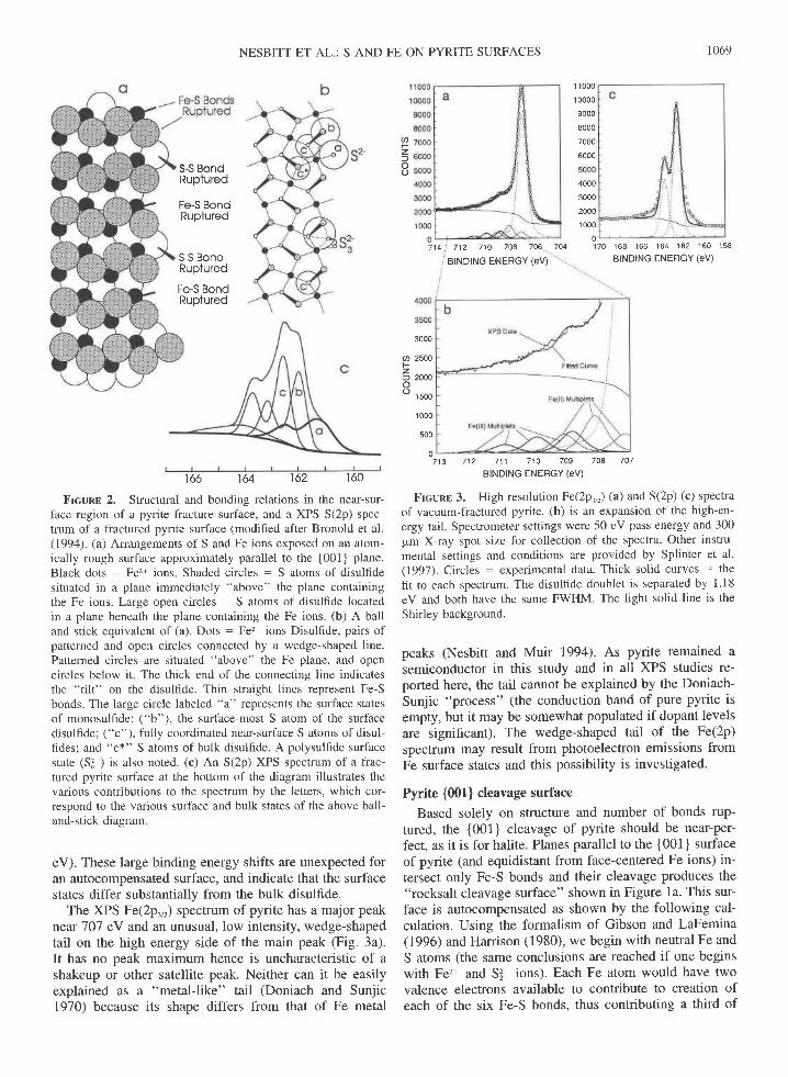
The XPS Fe(2p.,,) spectrum of pyrite has a major peak
near'101 eV and an unusual, low intensity, wedge-shapedtail on the high energy side of the main peak (Fig. 3a).It has no peak maximum hence is uncharacteristic of ashakeup or other satellite peak. Neither can it be easilyexplained as a "metal-like" tail (Doniach and Sunjic1970) because its shape differs from that of Fe metal
170
0713 712 711 710 709 708 707
BINDING ENEBGY (eV)
Frcunt 3. High resolution Fe(.2p^,r) (a) and S(2p) (c) spectraof vacuum-fractured pyrite. (b) is an expansion of the high-en-ergy tail Spectrometer settings were 50 eV pass energy and 300pm X-ray spot size for collection of the spectra. Other instru-mental settings and conditions are provided by Splinter et al.(1997). Circles - experimental data. Thick solid curves - thefit to each spectrum. The disulfide doublet is separated by 1.18eV and both have the same FWHM. The light solid line is theShirley background.
peaks (Nesbitt and Muir 1994). As pyrite remained asemiconductor in this study and in all XPS studies re-ported here, the tail cannot be explained by the Doniach-Sunjic "process" (the conduction band of pure pyrite isempty, but it may be somewhat populated if dopant levelsare significant). The wedge-shaped tail of the Fe(2p)spectrum may result from photoelectron emissions fromFe surface states and this possibility is investigated.
Pyrite {001} cleavage surface
Based solely on structure and number of bonds rup-tured, the {001} cleavage of pyrite should be near-per-fect, as it is for halite. Planes parallel to the {001} surfaceof pyrite (and equidistant from face-centered Fe ions) in-tersect only Fe-S bonds and their cleavage produces the"rocksalt cleavage surface" shown in Figure 1a. This sur-face is autocompensated as shown by the following cal-culation. Using the formalism of Gibson and LaFemina(1996) and Harrison (1980), we begin with neutral Fe andS atoms (the same conclusions are reached if one beginswith Fe'?* and Sl ions). Each Fe atom would have twovalence electrons available to contribute to creation ofeach of the six Fe-S bonds, thus contributing a third of

1070 NESBITT ET AL: S AND FE ON PYRITE SURFACES
an electron for each bond to be formed. Two sulfur atomshave six valence electrons each, for a total of twelve.Each sulfur atom is tetrahedrally coordinated in pyrite,three apices directed toward Fe atoms and the fourth to-ward the second sulfur atom. Two of the twelve valenceelectrons are consequently shared by the two sulfur atomsto form the S-S bond (and S, dimers). The remaining tenare available to be shared among the six equidistant Featoms, thus there are % electrons from S, available forcreation of each Fe-S bond. The same electronic contri-butions are available from each Fe atom and S, dimerlocated at pyrite cleavage surfaces. Some conffibutions,however, would be to the dangling bonds extending fromthe surface, and surface relaxation likely would involvetransfer of electrons from some dangling bonds to othersin an attempt to achieve a more stable surface. Specifi-cally, the % electrons of dangling bonds associated withsurface Fe atoms may be transferred to dangling bondsof surface S, dimers containing s/. electrons per danglingbond. The results are completely empty cation danglingbonds, and completely filled anion dangling bonds (twoelectrons per bonding orbital). Such a transfer produceselecffonically stable cation and anion surface states,which stabilizes the entire surface. The surface is stableand charge neutral, therefore autocompensated (Gibsonand LaFemina 1996).
However, pyrite displays irregular fracture surfaces thatare obviously not restricted to {001}, or any other crys-tallographic plane. Pyrite contains cation-anion bonds(Fe'z+-Sj- bond is equivalent to the Na-Cl bond) as wellas S-S bonds (no equivalent in halite). The presence ofthe S-S bond may have a substantial affect on cleavageproperties if the energy required to rupture it is less thanthe energy needed to rupture the Fer*-Sj- bond. Fe-Cl,Fe-Br, and Fe-I bond energies in FeClr, FeBr, and FeI,are respectively 400, 340, and 280 kJ/mol (Huheey 1978,Appendix F). The ionic radii of the iodide and disulfldeanions are almost identical (2.06 and 2.08 A, respective-ly). The disulfide is, however, doubly charged so that theFe'zt-Sl bond energy should be approximately twice thatof the Fe-I bond according to the Born-Mayer equation.The Fe'z*-Sl bond energy is clearly greater than 300 kJ/mol and is likely to be appreciably grearer rhan 400 kJ/mol. The S-S bond energy is lower, 245 -r 20 kJ/mol(Huheey 1978, Appendix F), suggesting rhat rhe weakerS-S bond should be ruptured during fracture. Althoughproduction of irregular fracture surfaces requires ruptureof more bonds than does production of the {001} cleav-age surface, the energy saved by breaking S-S bonds(rather than Fe,*-S3- bonds) could compensate for thegreater number of bonds broken.
The above bond energy considerations strongly suggestthat a realistic understanding of pyrite surface propertiesmust include consideration of irregular surfaces. Many ofthese surfaces will be polar, non-autocompensated, unsta-ble, and likely to have associated, reactive surface states.
Pyrite fracture sur-faces
Although planes parallel to {001} intersect only Fe-Sbonds, most other planes intersect both Fe-S and S-Sbonds. Where an S-S bond is intersected (Fig. 1b), thebond will remain intact only if three Fe-S bonds at eitherend of the dianion are broken instead of the S-S bond.The energetics of such are not favorable. The conse-quence is that S-S bonds are likely to be ruptured duringfracture of pyrite, producing unique surface sulfur states.
Rupture of an S-S bond leaves one sulfur monomer(nominally S ) on one fracture surface and the other onthe opposite surface. Their presence leads to a high prob-ability of producing "local," noncompensated, and ther-modynamically unstable regions surrounding the S mono-mer. Using the formalism of Gibson and LaFemina(1996),Vt of an elecffon is associated with each danglingbond of surface Fe atoms. If a surface sulfur monomerwere produced upon fracture (an S-S bond severed), therewould be six electrons available to contribute to fourbonds (one a dangling bond), averaging 1.5 elecffons perbond. Ttansfer of the l/s electron to the surface S- dan-gling bond would yield 11/6 electrons per dangling bondon the sulfur monomer, a value short of the two electronsper bonding orbital needed to stabilize the surface stateand to achieve charge neutrality (to be autocompensated).Autocompensation around the sulfur monomer can be at-tained by oxidizing Fe2* to Fe3* and reducing the S'-monomer to 52 , as now discussed.
Sunrlcn RELAxATToN AND suRFACE sTATES
Relaxation
Sulfur monomers with a formal valence of 1- 15r ;represent an enigmatic aspect associated with relaxationof pyrite fracture surfaces. Mineralogical studies indicateS'- and disulfide (S3-) are commonly present in naturallyoccurring sulfide minerals (Pratt et al. 1994; Buckley andWoods 1985). Transition metal polysulfides (Sl-, 2 < n< 8) are also well established (Termes et al. 1987; Buck-ley et al. 1988). Although many states of sulfur, includingelemental sulfur (S!), have been reported for minerals, S'has not been observed in nature. If produced at pyritefracture surfaces, it may undergo signiflcant modificationduring relaxation.
Modification to bond angles or lengths, or migration ofspecies to or from the surface does little to address theformal oxidation state of the Sr- monomer. The simplestand perhaps the most reasonable means to stabilize themonomer is to fill the 3p orbitals to attain a filled octet,hence a stable "Ar" configuration. In effect, the unstableSr monomer may "relax" to the more stable monosul-fide (S' ) found in minerals such as pyrrhotite. The S,-surface species may acquire the additional electron fromadjacent Fe2* ions to produce, in effect, surface Fe3* andSt- ions:
Fe"'z"]r*" + S;.,"." -+ Felj,""" + Sl;".. (1)
From a band theory perspective, an unoccupied S sur-

NESBITT ET AL.: S AND FE ON PYRITE SURFACES 1071
face electronic state is produced by fracture, but is sub-sequently filled during relaxation by acquisition of anFe(3d) electron (top-of-valence band is depleted). Themechanism for transfer is uncertain, but Goodenough(1982) shows that there can be strong overlap of theFe(3d) density of states with the S(2p) density of statesthus allowing electron transfer to the anion with minimalenergy input. As well, the energy associated with frac-turing may promote temporarily an Fe(3d) electron to theconduction band where it migrates to, and becomes lo-calized on, a S' site to produce 52 . Reaction I is con-sistent with the principles governing surface relaxation(Gibson and LaFemina 1996) where electrons of anti-bonding metal orbitals are transferred to adjacent anionsto fill their bonding orbitals. The transfer leads to an au-tocompensated region surrounding the S' and Fe3* sur-face states, as noted previously.
The second possibility for production of S'? surfacestates is acquisition of electrons from other S' mono-mers. Electrons from some S' sites may become delo-calized, perhaps promoted to the conduction band by en-ergy derived from fracturing the mineral. Oncedelocalized they migrate to other S' sites where theyagain become localized to produce stable S'? (filled oc-tet). This "disproportionation" results in production of S0and St monomers at the surface and may be representedformally by:
2S1,"... -+ S"o,d""" + S3,,r".". (2)
The S0 species may remain a monomer or may react witha subtending disulfide to produce polysulfide (Si ) in thenear-surface as shown in Figure 2b.
Evidence from arsenopyrite surfaces
Arsenic in arsenopyrite is bonded to S to yield an As-S dianion akin to disulfide of pyrite. The formal chargeon each of As and S is I - (as for pyrite). XPS study ofpristine arsenopyrite surface (Nesbitt et al. 1995) dem-onstrates that about 85Vo of As is present as the dimer(As-S) and about l5%o as AsO. About l5%o of sulfur ispresent as 52 (monosulfide) thus allowing for chargeneutrality (Nesbitt et al. 1995). Production of a partingsurface may cause surface As-S bonds to be severed. Re-laxation then occurs with electrons being localized pref-erentially on the more electronegative S atom rather thanon the As atom. The consequence is production of S'?and Aso at arsenopyrite fracture surfaces. The reactionmay be represented by:
As-S""*"". -+ Asgn"". + S"'""".". (3)
The XPS spectra of Nesbitt et al. (1995) provide evi-dence for the production of surface monosulfide (S' ) atfractured arsenopyrite surfaces, and considering the sim-ilarities between pyrite and arsenopyrite, the data providecircumstantial support for the presence of S'z at fracturedpyrite surfaces.
S(2p) lNn Fo(2yr,r) XPS spBcrRA RETNTERPRETED
S(2p) spectrum
Bulk states. By decreasing the photon excitation en-ergy, thus increasing surface sensitivity, Bronold et al.(1994) demonstrated that the peak at 162.6 eY repte-sented a bulk emission (Fig. 2c). The peaks at 162.0 and161.3 eV became more intense as surface sensitivity in-creased, demonsffating that these two peaks representedsurface states. Bronold et al. (1994) based their interpre-tation of sulfur surface states on a perfect {001} cleavagesurface, that is only Fe-S bonds were considered to havebeen severed during fracture, leaving all S-S bonds intact.They consequently considered disulfide (S3-) to be theonly anionic species present on pyrite fracture surfaces,and sulfur surface states were interpreted to arise solelyfrom disulfide. As did Bronold et al. (1994), we interpretthe peak at 162.6 eV (Fig. 2c) to represent emissions fromS atoms of bulk disulfide (e.g., states c, c*, and deeper Satoms of Fig. 2b).
Si- and Sr- surface states. The pyrite fracture surfaceof Figure 2 shows the effects of ruptured Fe-S and S-Sbonds, and the consequent presence of surface disulfide(S3 ) and monosulfide (S'-) ions (Figs. 2a and 2b). Thesurface-most disulfide ion contains S atoms labeled "b"and "c" (Fig. 2b). The atom labeled "b" is not fullycoordinated because at least one Fe-S bond has been sev-ered with the Fe ion residing on the opposite face. The Satom labeled "c" is fully coordinated (fourfold). The sur-face-most atom "b" is more likely to produce a surfacestate (low coordination). Bronold et al. (1994) appealedto an electric "double layer" within the near-surface toargue that atom "b" contributed to the peak at 161.3 eVand assigned the emission from atom "c" to the peak at162.0 eV. These assignments would, however, yield apeak at 161.3 eV that was more intense than the peak at162.0 eY (considering attenuation), contrary to observa-tion (Fig. 2c). To address the inconsistency, Bronold etal. (1994) assigned the S atom labeled "sx" (pig. 2b) tothe peak at 162.0 eV (Fig. 2c). This partially overcomesthe intensity problem but produces another. The S atoms"c" and "c*" are located at different positions withinthe electric "double layer", and should give rise to adifferent binding energy shift for each type of atom. If,as proposed by Bronold et al. (1994), the peak at 162.0eV includes contributions from S atoms "c" and "c*",the peak should be broad because the two contributionshave different binding energies. The peak is, however,nanow and provides no indication of being a composite.Assignment of the "c*" emission to the 162.0 eV peakis therefore questioned. This atom is located at the ex-treme lower boundary of the electric double layer and isfully coordinated, hence is likely to be bulk-like and con-tribute to the 162.6 eV bulk peak rather than the surfacestate peak at 162.0 eY.
Although the electric "double layer" should causeshifts in binding energy of near-surface S atoms, this ef-fect may not be the only conffibution to such shifts. Spe-

r072
cifically, binding energy shifts resulting from S atoms ofdifferent coordination number have not been included inthe considerations of Bronold et al. (1994), although co-ordination is known to affect binding sulfur energies. Satoms in CuS and pentlandite, for example, display twocoordinations. In both minerals, S of low coordination hassomewhat lower binding energies than the more highlycoordinated S atoms (Legrand et al. 1998; Laajalehto etal. 1996). S atoms at pyrite fracture surfaces are neces-sarily of lower coordination (due to bond scission) thanbulk S atoms, and a peak shift to lower binding energyis expected, just as observed for low coordinate S atomsin these minerals. A simple interpretation of the S(2p)surface states is presented that focuses on the chemicalstate of S atoms.
Monosulfide (Fig. 2b, state "a") is produced by ruptureof a S-S bond but it necessarily remains bonded to Fe.After relaxation to S, it should have spectral propertiesakin to S,- of pyrrhotite. The monosulfide (S,-) peak ofpyrrhotite is situated at 161.25 (*0.1) eV (Pratr et al.1994; Buckley and Woods 1985), and the pyrire S(2p)surface state at 161.3 eV (Fig. 2c) is consequently inter-preted to represent a S, surface state (Figs. 2b and 2c,"a"). The monosulfide (Fig. 2b, state "a") alone is con-sidered to conffibute to the peak at 161.3 eV. The low-coordinate S atom "b" of the surface disulfide ion (Fig.2b) is assigned to the peak at 162.0 eY, whereas the fullycoordinated S atoms "c" , "s*", and all S atoms deeperthan these are assigned to the bulk contribution at 162.6eV. By this interpretation, the relative intensity of the sur-face state peaks (161.3 and 162.O eV) is a direct measureof the number of S-S and Fe-S bonds broken duringfracture.
A second interpretation combines elements of theBronold proposals and ours. Sulfur atoms "a" and "b"contribute to the 161.3 eV peak, "c" and "c*" contributeto the peak at 162.0 eV, and all deeper atoms contributeto the bulk peak at 162.6 eY. The ambiguities associatedwith this interpretation already have been discussed. Itassumes that "c" yield photoemissions of iden-tical binding energy although they are located at differentdepths from the surface. It also assumes that S atoms ofsubstantially different chemical state give rise to surfacestates of similar binding energies. Most striking is thatthe same binding energy must be assigned to the mono-sulfide (S, ) and the "b" atom of the surface disulfideion. Their chemical states are much different and the as-signment seems unlikely.
Sl- surface states. An additional surface state, Sl , isshown in Figure 2b. Although its existence is not certain,the state may arise from rupture of an S-S bond, withsubsequent transfer of an elecffon to another S monomeras discussed previously (reaction 2). The resulting "So"species may react with the immediately underling disul-fide to produce polysulfide (S3 ) by analogy with reac-tions in aqueous solutions; little energy is required to pro-duce Sl from S0 (solid) and aqueous disulfide (Langmuir1997; Johnson 1982). The abundance of polysulfides on
NESBITT ET AL.: S AND FE ON PYRITE SURFACES
obOdoherolv
Coordlndfed(l0Dq ' 2 sy1
| ,/-\| , / \_ - .
., , ̂ .,^, ---t'-oa'
ttFemllevot __E-
i*t+/' ' oi :f3l** 5
)+:4" +; +II
^ ?9s - -. , Surfoce Fe(ll) Surfoce Fe(lt) Surfoce Fe(lll)Densltyofsiotesond 3dstotes 3dStotes
- 3dSiotes'
-Bulk Fe(ll) 3d Siotes (Low Sph)
Frcunn 4. The Fe(3d) ligand field splitting due to reducedcoordination resulting from fracture. Fe'?* is octahedrally coor-dinated before fracture and square pyramidal after fracture. (a)represents the bulk density of states (DOS) for pyrite, (b) theelectronic levels for Fe'?* in bulk pyrite in octahedral coordina-tion, (c) electronic levels of surface Fe2* in square pyramidalcoordination (low-spin state), (d) electronic levels of surface Fe2*in square pyramidal coordination with one unpaired electron inthe d., level, and (e) electronic levels of surface Fe3* in squarepyramidal coordination with one unpaired electron in the d,, lev-el. The ordinate is not drawn to scale. CB denotes conductionband and VB denotes the valence band. Primes indicate unpairedelectrons. The energy separating a, and b, in (c) and (d) is 0.35eV and the electron pairing energy is about 1.6 eV for squarepyramidal symmetry The diagram is modified after that of Bron-old et al. (1994,Fig.2).
leached pyrite surfaces attests to the viability of the re-action (Mycroft et al. 1990). Binding energies of poly-sulfide range between about 162.5 eY (disulfide) and164.0 eV (elemental sulfur), hence its detection in theS(2p) XPS spectrum is difficult due to the large numberof spectral contributions to this region (Fig. 2c).
Fe(2pr,r) spectrum
Production of 52- surface states on pyrite through ox-idation of Fe2* should produce an Fe3* surface state, thesurface concentration of which will be the same as S,-provided all S' is formed by the process represented byreaction 1. Detection of the monosulfide (S' ) in the S(2p)spectrum consequently suggests that the ferric stateshould be detectable in the Fe(2p) spectrum, and indeedthe main peak of the Fe(2p.,.) spectrum (Fig. 4) has ahigh-energy tail that is not expected if only Fe'z* bondedto disulfide were present in the near-surface. An expla-nation is pursued by first evaluating likely electronicstates of Fe at pyrite surfaces.
Fe,*(3d) states. The molecular orbital and consequentband models of Bronold et al. (1994) are summarized inFigure 4. The density of states (DOS) diagram for bulkpyrite is shown in Figure 4a. The energy axis is not drawnto scale. The valence band includes Fe-S, and S-S o-bonds. The three Fe(3d) !* non-bonding orbitals are lo-

cated at the top of the valence band and extend to justbelow the Fermi level. Antibonding 3p, 4p,4s, and thetwo e, 3d orbitals of Fe, and antibonding sp3 orbitals ofsulfur (Bronold et al. 1994) all contribute to the conduc-tion band (CB). The two bulk Fe2* e, orbitals are suffi-ciently destabilized by the six surrounding dianions thatall electrons are paired and reside in the non-bonding tr,orbitals yielding a low-spin configuration (Fig. ab). Aconsequence is that bulk Fe'* will be represented by asingle peak in the Fe(2p.,,) spectrum; multiplet splittingshould not occur because unpaired electrons do not existin the valence band of bulk Fe'z*.
The analysis of Bronold et al. (1994) indicates that thetr* and e, orbitals are non-bonding and antibonding, re-spectively, and hence may be treated by ligand field the-ory to a first approximation. Upon fracture, the octahedralcoordination of surface Fe becomes square planar-pyra-midal (C.V point group) due to loss of one dianion. Thisresults in stabilization of the d,, orbital (Fig. 4c, a, level)and slight destabilization of the d-, orbital (Fig. 4c, b,level) so that there is only 0.35 eV separating the twolevels (Fig. 4c). The electron pairing energy is sufficientlylarge (about 1.6 eV for CoV symmetry, Bronold et al.1994) that the energy of an unpaired elecffon in the d.,orbital is located below the Fermi level and within thevalence band (Fig. 4d, ai level). The large pairing energyand the small energy difference between a, and b, levelsfavor promotion of an electron from the L* orbital intothe ai (eg) orbital (Figs. 4c and 4d) to yield an interme-diate spin state for Fe2* surface ions (Fig. 4d). SurfaceFe'* ions accordingly have unpaired electrons in the va-lence band, leading to multiplet splitting of their XPS(2p)signal.
The high-spin state multiplet structure for Fe2* ion hasbeen evaluated (Gupta and Sen 1974, 19'75), but the mul-tiplet structures for intermediate spin states are unknown.If the Fe'*, Mno*, and Cr3* high-spin states can be usedas guides, then the 2p.,, XPS spectrum of Fe2* in inter-mediate spin state includes three or four multiplet peaks,each separated by about 1 eV (Gupta and Sen 1975;Mclntyre and Zetaruk 1977; Pratt and Mclntyre 1996).The Fe,* high-spin multiplet sffucture includes threepeaks with each separated by about 1 eV in pyrrhotite(Pratt et al. 1994), magnetite (Mclntyre and Zetantk197'7) and for the free ion (Gupta and Sen 1975). It isused subsequently as a guide to fit the Fe2* surface con-tribution to the Fe(2p.,,) spectrum (Figs. 3a and 3b) solelybecause it meets the general criteria just stated. This isnot to suggest that Fe2* of intermediate and high-spinstates have the same multiplet structure.
F€i*(3d) states. If monosulfide (S' ) develops on frac-tured pyrite surfaces through oxidation of a surface Fe2*ion, then surface Fe3* ions should represent a third con-tribution to the Fe(2p.,r) spectrum. The elecffon removedfrom surface Fe2* can be derived either from the a, or elevel (Figs. 4d and 4e), depending on the pairing energyand the energy difference between the a, and e levels. Atleast one unpaired Fe3* electron is present in the valence
10'73
band so that surface Fe3* ions should display multipletpeaks in the Fe(2p.,,) XPS spectrum. As are surface Fe2*ions, surface Fe3* ions are in an intermediate spin state.the multiplet structure of which is unknown. If Fe3* andCt'* high-spin states can be used as guides, then the 2p.,XPS structure of Fe3* in intermediate spin state includesthree or four multiplet peaks, each separated by about IeV (Gupta and Sen 1975; Mclntyre and Zetaruk 1977;Pratt and Mclntyre 1996). The Fer* high-spin multipletstructure includes four peaks with each separated byabout 1 eV (Gupta and Sen 1975; Mclntyre and Zetaruk19'77;Pratt and Mclntyre 1996) and it is used as an initialguide to fit the Fe3* surface contribution to the Fe(2p.,,)spectrum (Figs. 3a and 3b).
Fe(2pr,r) peak assignments
Spectral contributions. Fe of bulk pyrite (Fig. a)adopts a low-spin state (Fig. 4a) and it should contributeone peak (no multiplet splitting) to the Fe(2p.,,) spectrum.Surface Fe2* and Fe3* have unpaired electrons in the va-lence band (Figs. 4d and 4e) and both should exhibit amultiplet peak structure in the specffum. The three likelyconffibutions to the Fe(2p,,r) spectrum are a bulk Fe'*singlet peak, a multiplet (triplet) contribution from sur-face Fe2* ions and a multiplet (four peak) contributionfrom surface Fe3* ions. Bulk Fe'* is bouded only to di-sulfide, whereas the Fe2n surface state may be bonded toboth disulfide and monosulflde (S' ).
Bulk Fe'?* contributions. The attenuation length of Fephotoelectrons is sufficiently great that bulk Fe'z* is themajor contributor to the Fe(2p.,r) spectrum of Figure 3(Tanuma et al. 1991). Bulk Fe'* is in low-spin state (Fig.4b). It is consequently represented by only one peak at707.0 + 0.1 eV as observed in other studies (Nesbitt andMuir 1994; Mycroft et al. 1990; Buckley and Woods1987).
Surface Fe3* contributions. Fe3* and Fe2* are presentin both magnetite (Mclntyre and Zetaruk 1977) and pyr-rhotite (Fe,Ss, Pratt et al. 1994). The energy separatingthe Fe3* multiplet peak of lowest binding energy from theFe2* main peak is about 1.75 eV in both minerals. Themain Fe2* peak of pyrite is near'70'7 eV so that the lowestenergy multiplet peak of surface Fe3* should be at about'108.75. This coincides with the binding energy of thesmall, partially resolved peak in the Fe(2p.,,) XPS spec-trum near 109 eY (Figs. 3a and 3b). The binding energiesseparating the four multiplet peaks were taken as 1.0 eYwhich is consistent with Fer* multiplet splittings obtainedfor pyrrhotite (Pratt et al. 1994), magnetite (Mclntyre andZetaruk 1977) and as calculated for the Fe3* free ion(Gupta and Sen 1975). Full-width at half-minimum(FWHM) values were set equal to that of the bulk Fe'?*peak (707.0 eV peak). The Fe3* multiplet peaks conse-quently was constrained with respect to binding energiesand with respect to FWHM. The only adjustable param-eters were the intensities of the four Fe3* multiplet peaks.These were adjusted to obtain the best fit to the high-energy tail (Fig. 3). The resulting fit virtually mimics the
NESBITT ET AL.: S AND FE ON PYRITE SURFACES

1074
Taele 1. Fe(2p"r) XPS spectral peak parameters
NESBITT ET AL.: S AND FE ON PYRITE SURFACES
Bindingenergy
Contribution (eV) Atomic percent
- Multiplet peaks are designated M and numbered
XPS data in the region between 708.75 and712 eV (Fig.3b). Peak parameters are listed in Table l.
Surface Fe2* contributions. The spectral conffibutionof the surface Fe2* state should be located at somewhathigher binding energy than the bulk Fe'?* contribution ac-cording to the arguments of Bronold et al. (1994). Re-moval of a ligand during fracture reduces the electrostaticrepulsion on the Fe d states. Bronold et al. (1994) argueconvincingly that as a consequence surface Fe2* ionsshould be more electron-withholding than bulk Fe2* ions(an electric double layer centered on the Fe2* ions is cre-ated in the near-surface.) This should decrease the kineticenergy of photoelectrons derived from surface Fe2* ionsand increase their binding energy to a value somewhatgreater than that of bulk Fe'* photoemissions.
As noted previously, the multiplet structure for surfaceFe2* ions has been taken to contain three peaks, eachseparated by about I eV. The multiplets are also con-strained to the same FWHM as that of the bulk Fe,* peak.Intensities of the three peaks were adjusted to fit the spec-trum. This surface Fe2* contribution, if centered at 708.1eV, provides an excellent fit to the Fe(2p.,r) spectrum.Peak parameters are listed in Table l. Inclusion of thethree contributions, bulk Fe2* ions, surface Fe'?* ions, andsurface Fe3* ions provides an excellent fit to the Fe(2p.r)XPS spectrum and the interpretation is adopted as themost reasonable considering the available evidence.
DrscussroN
Likely surface sulfur species
Most previous interpretations of the S(2p) specffumconsidered only disulfide ions to populate the pyrite frac-ture surface. We have considered a more realistic surfacewhere rupture of S-S bonds has occurred and resulted inproduction of S'? (monosulfide) and Sl (surface disul-fide), and possibly in formation of Sl- (polysulfide) andS0 (elemental sulfur) surface species. Although the sur-face considered here is much more complicated than anyconsidered previously, there may be additional contribu-tions. The vagaries of S-S bond scission may lead tomonosulfide (S,-) bonded to one, two, or three Fe ions.Each S'z of different coordination number may have aunique (but perhaps unresolved) contribution to the S(2p)spectrum near 161.3 eV. The broad nature of the peak at
161.3 eV (Fig. 2c) may reflect these contributions. Thesurface species giving rise to the peak remains, however,S' . Similarly, The surface-most S atom of surface disul-fide ions may be bonded to zeto, one, or two Fe ions,thus there may be three unique spectral contributionsfrom this surface species. The number of S atoms in sur-face polysulfides may vary, each yielding a unique butunresolved spectral peak. Although there may be addi-tional conffibutions to the S(2p) spectrum, the three con-sidered here (Fig. 2c) are justified on the basis of theavailable evidence, and are sufficient to explain the majorfeatures of the S(2p) specffum.
Proportion of S-S bonds broken
About equal numbers of disulfide and Fe ions shouldbe exposed on a fracture surface. According to the inter-pretation of the Fe(2p.,r) spectrum, surface Fe ions con-stitute about 257o and bulk Fe abotttT1%o of the Fe signal(Table l). Of the Fe surface states almost 40Vo is Fe3*,the remainder being Fe'?* (Table 1). For each S-S bondsevered, there is production of one Fer* and one S'z- ionaccording to reaction l. Because there should be aboutequal numbers of Fe and S atoms exposed on a fracturesurface, these relations indicate that almost 40Vo of di-sulfide bonds exposed during fracturing are ruptured. Six-ty percent of the surface disulfide remain intact. The sur-face-most disulfide of the dimer (Fig. 2b, state "b") andthe monosulfide surface state (Fig. 2b, state "a") shoulddisplay a ratio near 4O160. The peak heights of the twosurface states (Fig. 2c, peaks at 162.0 and 161.3 eV) areclose to this ratio.
Annealed and fractured surfaces
Chaturvedi et al. (1995) studied a He*-bombarded andannealed pyrite surface and concluded that there was nomonosulfide (S'-) surface state. Bronold et al. (1994),Nesbitt and Muir (1994), and Pratt et al. (1998) observeda low-binding energy peak in S(2p) spectra of unffeated,fractured pyrite surfaces. Mycroft et al. (1995) observethe same peak on polished pyrite surfaces. If the peak isabsent from the bombarded and annealed surface, it clear-ly has different surface states from the fractured surface.This is possible considering the laboratory treatment oftheir surface. As emphasized by Gibson and LaFemina(1996), some surface states nomally cannot be readilyaccessed due to kinetic inhibition. Ion etching and an-nealing pyrite surfaces may well provide the energy re-quired to access additional, more stable states and a newconfiguration may have been achieved at the annealedpyrite surface. Annealing may, for example, allow mono-sulfide of fractured surfaces to migrate across the surfaceand react to produce disulfide species.
The absence of a surface state in the S(2p) spectrumof Chaturvedi et al. (1995) is nevertheless unexpectedbecause the disulfide surface state at 162.0 eY (Fig. 2c)should have been observed. The fact that there is no in-dication of this peak in their spectrum strongly suggeststhat their resolution (FWHM of 1.3) is insufficient to
FWHM(ev)
Fe2* BulkFe3* M1*Fe3* M2Fe3* M3Fe3" M4Fe2. M1Fe2. M2Fe2* M3
707 0070875709 85710 85711 85707 10708.05709.00
u.oc
0 8 50.850 8 50 8 50.85u . 6 3
0 8 5
73 8 Bulk4.59 Surface3 45 Surface1 81 Surface0.51 Surface3.94 Surface7 92 Surface3 97 Surface

NESBITT ET AL.: S AND FE ON PYRITE SURFACES 1075
identify low-intensity surface states. Because the 52 sur-face state at 161.3 eV is of lower intensity than the S?-surface state (Fig. 2c, 162.0 eV), it is unlikely that the161.3 surface state would be resolved in their specffum.The aspect they address is, however, important to the un-derstanding of surface properties of pyrite. Unfortunately,proof for the absence of a 161.3 eV peak on annealedsurfaces awaits high-resolution studies (FWHM of peaksless than 0.9 eV). Experiments of the type conducted byBronold et al. (1994) are particularly valuable.
Band gap, valence band edge, and reactivity
Eggleston et al. (1996) offered an elegant explanationfor pyrite oxidation kinetics, but recent considerations ofelectronic states (Bronold et al. 1994) and our findingsmay require a somewhat more exhaustive ffeatment. Theadditional surface states may affect initiation of surfaceoxidation and may enhance or impede initial reactionrates. Importantly, Bronold et al. (1994) demonstratedthat although the bulk band gap is 0.9 eV the valenceband edge is very close to the Fermi level due to thelowered symmetry imposed on surface species by frac-turing the mineral. The valence band edge approachesclosely the lower edge of the hematite conduction band(Eggleston et al. 1996) so that surface Fe species ofpyritemay be more reactive than previously considered. Spe-cifically, incorporation of calculations by Bronold et al.(1994) into the arguments of Eggleston et al. (1996), andconsideration of the effects of 52 Fe2* and Fe3* surfacestates, may result in surface Fe2* of pyrite being a betterreductant than heretofore appreciated. Rate constants forelecffon transfer may have to be modified to account forthe two Fe surface states, the effects of St on nearest-neighbor Fe ions, and to account for the effects of sym-metry reduction (Bronold et al. 1994). We encourage andawait additional developments that incorporate these re-sults and the findings of Bronold et al. (1994) into theapproach and framework developed by Eggleston et al.(1996).
CoNcr-usror.ls
The interpretation of the S(2p) and Fe(2p) spectra, al-though based on available evidence, is nevertheless spec-ulative and needs to be tested. Bond strengths of Fe-S,must be evaluated in some detail, with both ionic andcovalent considerations included. There is need for the-oretical and mathematical studies focused on the multipletpeak sffuctures of Fe(2p) signals (intermediate spin statesespecially). The methodology already has been estab-lished by Gupta and Sen (1974, 1975). Of vital impor-tance is determination of coordination numbers of surfacespecies and of lengths of bonds associated with the sur-face species. X-ray absorption spectroscopic studies (XA-NES and EXAFS) should be useful in this regard. Finally,the relative reactivities of the surface species are requiredto understand reaction mechanisms and rates during theinitial stages of oxidation of the mineral. Synchrotron
studies with appropriately tuned primary beam energiesare underway.
Acxt.lowr,nocMENTS
We thank M. Fleet for discussions, D. Legrand and A. Schaufuss forinsightful discussions and carefully reading the original manuscript, andG Waychunas and two reviewers for their careful reviews and editorialsuggestions, all of which improved the manuscript This research wassupported by grants to the first two authors from the National Science andEngineering Research Council of Canada
Rprnnnucns crrnD
Bronold, M , Tomm, Y, and Jaigermann, W. (1994) Surface states of cubicd-band semiconductor pyrite (FeS.). Surface Science Letters, 314,L93t-L936.
Buckley, A N. and Woods, R. (1985) X-ray photoelectron spectroscopy ofoxidized pyrrhotite surfaces II: Exposure to aqueous solutions AppliedSurface Science, 20, 472-480.
-(1987) The surface oxidation of pyrite Applied Surface Science,27,431-452
Buckley, A.N, Wouterlood, H.J, Cartwright, PS., and Gillard, R.D. (1988)
Core electron binding energies of platinum and rhodium polysulfides
Inorganica Chimica Acta, 143, 77 -80.
Chaturvedi, S , Katz, R, Guevremont, J , Schoonen, M A A., and Stron-gin, D R. (1996) XPS and LEED study of a single-crystal surface ofpyrite American Mineralogist, 81, 261-264.
Deer, WA., Howie, R.A., and Zussman, J (1992) An Introduction to theRock-Forming Minerals, 696 p. (2nd ed ), Longmans, London
Doniach, S and Sunjic, M (1970) Many-electron singularity in X-rayphotoemission and X-ray line spectra of metals Journal of Physics, C3,285-291.
Eggleston, C.M., Ehrhardt, J.J., and Stumm, W. (1996) Surface structuralcontrols on pyrite oxidation kinetics: An XPS-UPS, STM, and modelingstudy. American Mineralogist, 81, 1036-1056
Gibson, A.S and LaFemina, J P (1996) Structure of mineral surfaces InPV Brady, Ed, Physics and Chemistry of Mineral Surfaces, 368 p.
CRC Press. Boca Raton, FloridaGoodenough, JB (1982) Iron sulfides Annales de Chimie (France), 1,
489-503Gupta, R.P and Sen, S K ( 1974) Calculation of multiplet structure of core
p-vacancy levels Physical Reviews, B1O, 1119.-(1915) Calculation of multiplet structure of core p-vacancy levels
II Physical Reviews, 812, 12-19.Harrison, W.A. (1980) Electronic Structure and the Properties of Solids,
582 p Freeman, San Francisco, CaliforniaHenrich, VE and Cox, PA. (1994) The Surface Science ofMetal Oxides,
464 p. Cambridge University Press Cambridge, U KHyland, M M and Bancroft, G M (1989) An XPS srudy of gold deposi-
tion at low temperatures on sulfide minerals: reducing agents Geochim-ica Cosmochimica Acta, 53, 367-312.
Huheey, J.E. (1978) Inorganic Chemistry, 889 p (2nd ed ) Harper andRow, New York
Johnson, D A (1982) Some Thermodynamic Aspects of Inorganic Chem-istry,282 p (2nd ed.) Cambridge University Press, Cambridge, U K
Langmuir, D (1997) Aqueous Environmental Geochemistry,600 p. Pren-tice-Hall, Upper Saddle Riveq New Jersey
Laajatehto, K., Kartio, I, Kaurila, T., Laiho, T., and Suoninen, E (1996)
Investigation of copper sulfide surfaces using synchrotron radiation ex-
cited photoemission spectroscopy In H J Mathieu, B. Reihl, and E.Briggs, Eds., European Conference on Applications of Surface and In-terface Analysis ECASIA'95 Wiley, New York,7l7-l2O
Legrand, D.L, Bancroft, G M , and Nesbitt, H w. (1998) Surface char-acterization of pentlandite (Fe,Ni)nS,, by X-ray photoelectron spectros-
copy. International Journal of Mineral Processing, 51,211-228Mclntyre, N S and Zetaruk, D G. (1977) X-ray photoelectron spectro-
scopic studies of iron oxides. Analytical Chemistry, 49, 1521-1529Mycroft, J.R, Nesbitt, H W., and Pratt, A R (1995) X-ray photoelectron
and auger electron spectroscopy of air-oxidized pyrrhotite: Distribution
1076 NESBITT ET AL.: S AND FE ON PYRITE SURFACES
of oxidized species with depth Geochimica Cosmochimica Acta, 59, Pratt, A R and Mclntyre, N S (1996) Comment on "Curve fitting of Cr721-733. 2p photoelectron spectra of CrrO. and CrF." Surface and Interfacial
Mycroft, JR, Bancroft, GM, Mclntyre, NS., Lorimeq JW., and Hitl, Analysis,24,529-530.IR (1990) Detection of sulfur and polysulfides on electrochemically Pratt, AR., Muir, IJ, and Nesbitt, HW. (1994) X-ray photoelectron andoxidized pyrite surfaces by X-ray photoelectron spectroscopy and Ra- auger electron spectroscopic studies of pyrrhotite, and mechanism ofman spectroscopy Joumal of Electroanalytical Chemistry, 292, 139- air oxidation Geochimica Cosmochimica Acta, 58, 82'7-841152. Tanuma, S , Powell, C J , and Penn, D.R. (1991) Calcutations of electron
Nesbitt, H W and Muir, I.J (1994) X-ray photoelectron spectroscopic inelastic mean free paths III SIA, Surface and Interfacial Analyses 17,study of a pristine pyrite surface reacted with water vapour and air. 92'7-939.GeochimicaCosmochimicaActa,58,466l-4679 Termes,SC,Buckley,AN,andGi l lard,RD (1987)2pelectronbinding
Nesbitt, H W, Muir, I J , and Pratt, A R (1995) Oxidation of arsenopyrite energies for sulfur aroms in meral polysulfides Inorganica Chimrcaby air and air-saturated, distilled watet and implications for mechanism Acta, 126,19-82of oxidation. Geochimica Cosmochimica Acta, 59, 1173-1'786
Pratt, A R, Mclntyre, N S , and Splinter, S.J (1998) Deconvolution of Mmuscrryr REcETVED Ocrogen l, 1997pyrite, marcasite and arsenopyrite XPS spectra using the maximum en- MrNuscprn ACCEmED Mapcs 27, 1997tropy method Surface Science (in press) Pepnn slNoreo sy GrenN A WaycsuNas