Sucrose density gradient centrifugation for efficient ... · 13. Preparation of aqua regia ..... 5...
24
NIST Special Publication 1200-24 Sucrose density gradient centrifugation for efficient separation of engineered nanoparticles from a model organism, Caenorhabditis elegans Version 1.1 M. E. Johnson A. R. Montoro Bustos S. K. Hanna E. J. Petersen K. E. Murphy L. L. Yu B. C. Nelson M. R. Winchester This publication is available free of charge from: https://doi.org/10.6028/NIST.SP.1200-24
Transcript of Sucrose density gradient centrifugation for efficient ... · 13. Preparation of aqua regia ..... 5...

NIST Special Publication 1200-24
Sucrose density gradient centrifugation for efficient
separation of engineered nanoparticles from a model
organism, Caenorhabditis elegans
Version 1.1
M. E. Johnson A. R. Montoro Bustos
S. K. Hanna E. J. Petersen K. E. Murphy
L. L. Yu B. C. Nelson
M. R. Winchester
This publication is available free of charge from: https://doi.org/10.6028/NIST.SP.1200-24

NIST Special Publication 1200-24
Sucrose density gradient centrifugation for efficient
separation of engineered nanoparticles from a model
organism, Caenorhabditis elegans
Version 1.1
M. E. Johnson A. R. Montoro Bustos
S. K. Hanna E. J. Petersen K. E. Murphy
L. L. Yu B. C. Nelson
M. R. Winchester Chemical Sciences Division
Material Measurement Laboratory
This publication is available free of charge from: https://doi.org/10.6028/NIST.SP.1200-24
October 2017
U.S. Department of Commerce Wilbur L. Ross, Jr., Secretary
National Institute of Standards and Technology
Walter Copan, NIST Director and Undersecretary of Commerce for Standards and Technology

Certain commercial entities, equipment, or materials may be identified in this document in order to describe an experimental procedure or concept adequately.
Such identification is not intended to imply recommendation or endorsement by the National Institute of Standards and Technology, nor is it intended to imply that the entities, materials, or equipment are necessarily the best available for the purpose.
National Institute of Standards and Technology Special Publication 1200-24 Natl. Inst. Stand. Technol. Spec. Publ. 1200-24, 24 pages (October 2017)
CODEN: NSPUE2
This publication is available free of charge from: https://doi.org/10.6028/NIST.SP.1200-24

i
This publication is available free of charge from: https://doi.org/10.6028/N
IST.SP.1200-24
FOREWORD This National Institute of Standards and Technology (NIST) special publication (SP) is one in a series of NIST SPs that address research needs articulated in the National Nanotechnology Initiative (NNI) Environmental, Health, and Safety Research Strategy published in 2011.[1] This Strategy document identified a Nanomaterial Measurement Infrastructure (NMI) as essential for science-based risk assessment and risk management of nanotechnology-enabled products as pertaining to human health, exposure, and the environment. Increasing production and subsequent incorporation of engineered nanomaterials (ENMs) into consumer products enhances the probability of ENM release into the environment. Thus, the necessity to understand the interactions between ENMs and environmental and biological systems persists. Whole organismal metrological efforts incorporate the expert skills of many disciplines across NIST. This protocol describes a strategy for separating Caenorhabditis elegans (C. elegans) from engineered nanomaterials, a critical step for accurate quantification in uptake studies. Conventional rinsing procedures, typically comprised of water or buffer rinsing steps followed by centrifugation, have been previously tested and deemed unsuitable for this purpose, even at environmentally relevant exposure concentrations. Therefore, this protocol details the development and assessment of a unique sucrose density gradient centrifugation procedure for the efficient separation of nanoparticles and C. elegans at the end of an exposure protocol. The example outlined in this SP pertains to the measurement of gold nanoparticles (AuNPs) and C. elegans. However, this method can potentially be implemented in bioaccumulation studies involving other organisms and with different metallonanoparticles. Visit http://nist.gov/mml/np-measurement-protocols.cfm to check for revisions and additions to this protocol or for new protocols in the series. We also encourage users to report citations to published work in which this protocol has been applied.

ii
This publication is available free of charge from: https://doi.org/10.6028/N
IST.SP.1200-24
Table of Contents
1. Introduction ..................................................................................................................... 1
2. Principles and Scope ....................................................................................................... 1
3. Terminology ..................................................................................................................... 2
4. Reagents, Materials, and Equipment ........................................................................... 2
5. Preparation of C. elegans ................................................................................................. 3
6. Preparation of sucrose solutions ................................................................................... 3
7. Preparation of 0.1 mol L-1 NaCl ................................................................................... 3
8. Preparation of nanosuspensions ................................................................................... 3
9. Construction of sucrose density gradient separation column ................................... 3
10. Preparation of nematode/nanosuspension mixture ................................................... 4
11. Centrifugal separation .................................................................................................... 4
12. Extraction of gradient sample fractions ....................................................................... 4
13. Preparation of aqua regia ............................................................................................... 5
14. Preparation of the internal standard diluent ............................................................... 5
15. Acid digestion of gradient sample fractions ................................................................ 5
16. Preparation of the ionic standard solution calibrants ................................................ 6
17. Measurement of total Au in gradient sample fractions by ICP-MS ......................... 6
18. Quantification of total Au in gradient sample fractions ............................................ 6
19. Outcomes.......................................................................................................................... 7
19.1. Visual observations ..................................................................................................... 7
19.2. ICP-MS results and implications ................................................................................. 7
20. Acknowledgements ....................................................................................................... 17
21. References ...................................................................................................................... 17
List of Tables
Table 1. Density of sucrose solutions used for the sucrose gradient centrifugation. ............... 9 Table 2. Total Au, in ng, found in gradient sample fractions following centrifugation of a mixture of 30 nm and 60 nm AuNPs (NIST) at 100 ng mL-1, respectively. Note that 2 mL of the mixture of AuNPs was added to the gradient, accounting for ≈400 ng of total Au. ......... 10 Table 3. Relative % Au found in gradient sample fractions following centrifugation of a mixture of 30 nm and 60 nm AuNPs (NIST) at 100 ng mL-1, respectively. ........................... 11 Table 4. Total Au, in ng, found in gradient sample fractions following a 0 h nematode exposure to a mixture of 30 nm and 60 nm AuNPs (NIST) at 100 ng mL-1, respectively. Note that 2 mL of the mixture of nematode/AuNP suspension was added to the gradient, accounting for ≈200 ng of total Au. ........................................................................................ 12

iii
This publication is available free of charge from: https://doi.org/10.6028/N
IST.SP.1200-24
Table 5. Relative % Au found in gradient sample fractions following a 0 h nematode exposure to a mixture of 30 nm and 60 nm AuNPs (NIST) at 100 ng mL-1, respectively. .... 13
List of Figures
Figure 1. Representative schematics of (A) mixed-stage nematodes, (B) the nanosuspension containing 30 nm and 60 nm AuNPs at 100 ng mL-1, respectively, and (C) the nematode/nanosuspension mixture. The construction of the sucrose density gradient column within a centrifuge tube from top to bottom: 2 mL of the nanosuspension (D) or 2 mL of the nematode/nanosuspension mixture (E), 2 mL of 100 µmol/L NaCl (light green) + sucrose [4 mL of 20 % (mass density; purple), 3 mL of 40 % (mass density; orange), and 2 mL of 50 % (mass density; red)]. ................................................................................................................ 14 Figure 2. Post-centrifugation distribution of Au across the sucrose density gradient centrifugation column of a mixture of 30 nm and 60 nm AuNPs at 100 ng mL-1, respectively. Each colored-line represents individual samples (n = 4). The color designations for the x-axis can denote the color designations within the gradient schematic in Figure 1D. Note: no C. elegans were added in this experiment. .................................................................................. 15 Figure 3. Post-centrifugation distribution of Au across the sucrose density gradient centrifugation column following a 0 h nematode exposure to a mixture of 30 nm and 60 nm AuNPs at 400 ng mL-1, respectively. Each colored-line represents individual samples (n = 4). The color designations for the x-axis can denote the color designations within the gradient schematic in Figure 1E............................................................................................................ 16

1
This publication is available free of charge from: https://doi.org/10.6028/N
IST.SP.1200-24
1. Introduction The rapid expansion of the nanotechnology industry has remarkably improved the performance of basic materials and consumer products, leading to the commercial success of engineered nanoparticles (ENPs). However, there is an urgent need to understand the interaction between ENPs and environmental and biological systems to enable studies on the potential risks of ENPs given their possible release into air, water, and soils. Accurate analytical methods are required to quantitatively study these interactions. Within such studies, the influence of artefactual false-positive and or false-negative results must be critically evaluated and mitigated. 2. Principles and Scope Studies on the bioaccumulation and potential toxicological effects of ENPs using cellular, plant, aquatic, and rodent models have produced important data for risk assessment. Caenorhabditis elegans (C. elegans), a globally distributed, soil-inhabiting nematode species,[2] has been widely utilized as a model organism in research studies due to factors such as its low cost, ease of cultivation and maintenance, short generation time, the large number of eggs per adult, the availability of numerous genetically modified versions of the species, 60 % to 80 % gene homology to humans, and its optical transparency.[3, 4] Additionally, they feed by taking in surrounding media and suspended particles via a pharyngeal pumping mechanism,[5] making them especially ideal organisms for nano-uptake and nanotoxicity studies. C. elegans has been extensively employed in ENP-related uptake and nanotoxicity studies to investigate the potential deleterious effects of ENP-exposure on organism health and behavior.[6-14] For nano-exposure research involving C. elegans, conventional rinsing procedures are typically composed of multiple water or media rinses followed by centrifugation and vacuum filtration of the supernatant.[6-8, 10, 11, 15, 16] When our research group tested and employed a similar rinsing strategy, we found that water rinsing was unsuitable for the efficient removal of non-ingested NPs, even at low, environmentally relevant exposure concentrations (ng mL-1
or part per billion in gold mass fraction).[17] Therefore, it was necessary to devise a post-exposure rinsing strategy that, unlike conventional water rinsing protocols, ensured greater efficiency in regard to the removal of excess, non-ingested ENPs. Density gradient centrifugation is widely considered one of the most convenient and simple methods for ENP separation for the purposes of purification, size selection, and shape and agglomeration/aggregation control.[18] Among the media employed for these efforts, sucrose has been utilized for the separation of gold nanorods,[18] carbon nanotubes,[19] and titanium dioxide.[20] Our group has studied the efficacy of rate-zonal sucrose density gradient centrifugation separations of mixtures of AuNPs of varying sizes in liquid suspension.[21] Gradients have been used in the biological sciences for fractionation of whole cells, subcellular components, protein purifications, etc.[22] Recently, a Percoll medium centrifugation separation allowed the measurement of mass density distributions in C. elegans populations.[23] In fact, sucrose density gradients are widely used in C. elegans maintenance for separating viable nematodes from dead nematodes, eggs, bacteria, and other debris.[3, 24, 25] However, sucrose density gradient centrifugation (SDGC) separation had not been

2
This publication is available free of charge from: https://doi.org/10.6028/N
IST.SP.1200-24
employed until recently for the separation of C. elegans from NPs after exposure.[17] This protocol details an SDGC procedure for the efficient separation of AuNPs from C. elegans. 3. Terminology This protocol complies with definitions relevant to nanotechnology as set forth in the ASTM International E2456.[26] 4. Reagents, Materials, and Equipment
4.1. Reagents 4.1.1. De-ionized (DI) water (≥ 18 MΩ·cm resistivity) 4.1.2. Sodium Chloride (NaCl; 99.5 % purity) 4.1.3. Sucrose (enzyme-grade powder; molecular biology grade) 4.1.4. Food coloring (red, green, yellow, and blue) 4.1.5. Ultra High Purity (UHP) Nitric Acid, HNO3 4.1.6. UHP Hydrochloric Acid, HCl
4.2. Materials 4.2.1. Citrate-stabilized Gold Nanoparticle aqueous suspension, Nominal 30 nm
Diameter, Reference Material (RM) 8012, NIST, Gaithersburg, MD 4.2.2. Citrate-stabilized Gold Nanoparticle aqueous suspension, Nominal 60 nm
Diameter, RM 8013, NIST, Gaithersburg, MD 4.2.3. SRM 3121 Gold (Au) Standard Solution (NIST, Gaithersburg, MD) 4.2.4. SRM 3124a Indium (In) Standard Solution (NIST, Gaithersburg, MD) 4.2.5. SRM 3140 Platinum (Pt) Standard Solution (NIST, Gaithersburg, MD)
4.3. Labware 4.3.1. Pipets (2-20 µL, 10-100 µL, 100-1,000 µL, 100-5,000 µL) 4.3.2. Pipet tips (2-20 µL, 10-100 µL, 100-1,000 µL, 100-5,000 µL) 4.3.3. Polypropylene 15 mL and 50 mL centrifuge tubes 4.3.4. Sterilized, graduated glass bottles (100 mL, 250 mL, 500 mL, 1,000 mL, and
2,000 mL) 4.3.5. Graduated plastic transfer pipets (3 mL maximum volume) 4.3.6. Serological pipets (2 mL, 5 mL, 10 mL, and 25 mL) 4.3.7. Aluminum foil 4.3.8. Ice buckets or insulated ice storage containers 4.3.9. Amber glass vials (for NP stock storage) 4.3.10. Magnetic stir bar 4.3.11. Low density polyethylene bottles (LDPE, 125 mL)
4.4. Equipment 4.4.1. Refrigerator for storage (maintained at 4 °C) 4.4.2. Ice machine 4.4.3. Refrigerated centrifuge, equipped with a swinging bucket, 15 mL centrifuge
tube holders, and capable of maintaining temperatures between 2 °C and 4 °C 4.4.4. Analytical balance with readability of 0.1 mg 4.4.5. Rechargeable pipet aid with replaceable filters

3
This publication is available free of charge from: https://doi.org/10.6028/N
IST.SP.1200-24
4.4.6. Quadrupole inductively coupled plasma mass spectrometer (ICP-MS) 4.4.7. ICP-MS autosampler
5. Preparation of C. elegans Assessment of C. elegans/AuNP separation efficiency shall be accomplished using approximately 50 000 living C. elegans per gradient. Details regarding C. elegans culturing and maintenance practices can be found in the Experimental section of Johnson et al.[17] See Figure 1A. 6. Preparation of sucrose solutions This protocol requires the use of sucrose solutions of 200 g L-1, 400 g L-1, and 500 g L-1. To prepare these solutions, dissolve 200 g, 400 g, or 500 g, respectively, of sucrose (enzyme grade) in deionized water. Weigh sucrose into glass bottles and fill to the 1 L mark with deionized water. For complete dissolution, shake the solutions, followed by gentle heating and stirring at 70 °C with a magnetic stir bar. Once completely dissolved, store these solutions at 4 °C until use. Add 5 µL of food coloring to each sucrose solution stock, by pipette, as a visual aid to discriminate the different concentrations of sucrose. Additionally, food coloring allows for the visualization of nematode layers after centrifugation since nematodes appear white in dense mass. Each concentration of sucrose should be given its own designated color. 7. Preparation of 0.1 mol L-1 NaCl Weigh 5.8 g NaCl into a sterile glass bottle (250 mL) and dilute to 100 mL with deionized water to prepare a 1 mol L-1 NaCl stock solution. Then prepare a 0.1 mol L-1 NaCl solution by diluting the 1 mol L-1 NaCl stock 10-fold. 8. Preparation of nanosuspensions A mixture composed of nominal 30 nm AuNPs and nominal 60 nm AuNPs (in the case of this protocol, NIST RM 8012 and NIST RM 8013 each at 100 ng mL-1 Au mass fraction, respectively) can be used to perform SDGC separations of nematodes and AuNPs. Dilute a volume of 20.7 µL and 19.3 µL of the stock NPs, with 10 mL deionized water in a 15-mL centrifuge tube. Vortex the mixture at maximum speed for 30 s to suspend the nanoparticles. See Figure 1B. 9. Construction of sucrose density gradient separation column A schematic of representative sucrose density gradient separation columns can be found in Figure 1. The sucrose density gradient separation column is carefully constructed from bottom to top in a 15-mL plastic centrifuge tube by sequentially layering the following components by transfer pipet: [2 mL of 50 % sucrose (mass density) + 3 mL of 40 % sucrose (mass density) + 4 mL of 20 % sucrose (mass density) + 2 mL of 0.1 mol L- 1 NaCl]. Once the gradient separation columns are prepared, they should be kept between 0° and 4° C by submerging the centrifuge tubes in ice, until the separation samples are ready. Do not disturb tubes in any way while constructing the gradients to avoid mixing of sucrose layers.
9.1. Label and pre-weigh 13 15-mL centrifuge tubes (including caps) per gradient column.

4
This publication is available free of charge from: https://doi.org/10.6028/N
IST.SP.1200-24
10. Preparation of nematode/nanosuspension mixture 10.1. Add an equal volume fraction of nematodes (≈50 000 nematodes mL-1) to an
equal volume fraction of the AuNP suspension described in Section 8 (i.e., 5 mL of nematodes + 5 mL of AuNP mixture). Mix by inversion for 5 s. At least three individual gradients should be prepared per AuNP size to assess repeatability. Note, this interaction time reflects a 0 h exposure to the nanoparticles. Proceed to Section 10.3. See Figure 1C.
10.2. Gently transfer 2 mL of the nanosuspension from Section 8 to a pre-labeled separation column from Section 9 (placed on the top of the NaCl gradient layer) by transfer pipet. The total Au mass added to the gradient from the nanosuspension accounts for ≈400 ng Au. See Figure 1D. Do not disturb tubes in any way when transporting tubes to avoid mixing of the layers. At least three replicates should be tested per condition.
10.3. Carefully transfer a 2-mL volume of the nematode/nanosuspension mixture in Section 10.1 to a pre-labeled separation column (on the top of the NaCl gradient layer) by transfer pipet. Due to the 1:1 dilution step in Section 10.1, the total Au mass added to the gradient from the nematode/nanosuspension mixture accounts for ≈200 ng Au. See Figure 1E. Do not disturb tubes in any way when transporting tubes to avoid mixing of the layers. At least three replicates should be tested per condition. Additionally, C. elegans exposure to sucrose should be as short as possible to avoid mortality or ejection of particles from the pharynx.
11. Centrifugal separation The centrifugal separation in this protocol is performed at 4 °C in a swinging bucket centrifuge under the following conditions:
11.1. Cool centrifuge at 4 °C. 11.2. Transfer the centrifuge tubes to the centrifuge. Distribute the weight of the tubes
within the centrifuge evenly. 11.3. Set the timer to 10 min. 11.4. The two-step centrifugation process is as follows: begin at a centrifugal force
of 172 g for 5 min and immediately (i.e., without interruption) increase to 1 254 g for the remaining 5 min.
In order to evaluate the separation of the sucrose density gradient centrifugation step, total Au analysis of the entire gradient must be performed. 12. Extraction of gradient sample fractions After centrifugation, slowly and carefully transfer 1-mL aliquots of the gradients from Section 11 to the labeled, pre-weighed centrifuge tubes from Section 9.1 via pipette. A new pipet tip must be used for each individual 1-mL aliquot transfer to avoid cross-contamination. There will be 13 1- mL gradient sample fractions for each sample replicate. Avoid mixing the layers during this step.
5
This publication is available free of charge from: https://doi.org/10.6028/N
IST.SP.1200-24
13. Preparation of aqua regia For the purpose of this protocol, aqua regia is an acid mixture containing a 3:1 molar ratio of concentrated hydrochloric acid to nitric acid. It is used to dissolve metals such as gold, platinum, and palladium. Care must be taken when preparing aqua regia, as this reaction of strong acids produces not only heat, but dangerous vapors and potentially explosive pressures, if sealed. This step was performed in a chemical fume hood with appropriate personal protection equipment (gloves, safety goggles, laboratory coat, etc.). This solution must be freshly prepared for use. To dispose of excess aqua regia, decant over ice, and then pour the contents into a hazardous waste receptacle suitable for acid waste.
13.1. Submerge an LDPE bottle, which will be used to prepare the aqua regia solution, in a beaker filled with ice.
13.2. Add the hydrochloric acid portion first. Do not add hydrochloric acid to nitric acid. In this mixture, hydrochloric acid is the weaker acid and composed of 65 % to 70 % (volume density) water, while nitric acid is the stronger acid with ≈30 % water by composition. Thus, adding hydrochloric acid to nitric acid will generate a volatile reaction (potential for caustic steam production and a splash hazard).
13.3. Slowly add the nitric acid portion, cap the container, mix the contents by inversion, and then uncap the container. Re-submerge the container in ice.
The solution may change to a yellow-orange or red-orange color over time. 14. Preparation of the internal standard diluent To monitor instrument drift, a diluent was prepared containing internal standards of platinum (NIST SRM 3140) and indium (NIST SRM 3124a) at 1 ng mL-1 each in 2 % HNO3 (volume density). The typical volume of this diluent is between 1.5 L and 2.0 L depending on the number of gradient samples to be diluted and measured for total Au analysis.
14.1. The platinum solution, NIST SRM 3140, has a certified value of 9.996 mg g-1. Prepare an intermediate working standard solution by weighing at a concentration of 10 000 ng g-1 by serial dilution with 2 % HNO3 (v/v). Employ the same strategy to prepare the indium standard solution by using NIST SRM 3124a, which has a certified value of 10.009 mg g-1. Finally, prepare 2 L of the final internal standard diluent at a concentration of 1 ng mL-1 in 2 % HNO3 (v/v) for both elements.
Please note that although indium and platinum were included in the sample diluent, ultimately only the Au signal was used for the computation of the total Au mass fraction present in each gradient layer, because the ICP-MS response factor for the measurement of Au was stable (the relative standard deviation of Au signal intensities was <5 % for samples measurements) across the entire analysis. 15. Acid digestion of gradient sample fractions For safety, these steps were performed in a chemical fume hood.
15.1. Perform acid digestions directly in the centrifuge tubes from Section 12. Uncap all the tubes. Add 1 mL of the aqua regia solution via plastic transfer pipet (3 mL transfer pipets with graduated marks are recommended).
15.2. Cap tubes and vortex for 10 s to initiate the digestion process. Loosen caps during the digestion process.

6
This publication is available free of charge from: https://doi.org/10.6028/N
IST.SP.1200-24
15.3. Allow the contents to react for 2 h at room temperature and then dilute (to the 15-mL mark) with the internal standard solution prepared in Section 14.
15.4. Cap samples and shake to ensure a homogeneous sample mixture. 15.5. Record final weights for all diluted samples. These tubes had been previously
weighed in step 9.1. 16. Preparation of the ionic standard solution calibrants Inductively coupled plasma mass spectrometry (ICP-MS) is a technique which allows rapid detection of different metal ions in a dilute acid sample matrix. Ionic standard solutions are required for quantitative analysis to calibrate the response of the ICP mass spectrometer for analytes of interest. Prepare ionic gold standards, ranging from 0 ng mL-1 to 25 ng mL-1, by mass in 50 mL centrifuge tubes by serial dilution of NIST SRM 3121 (Gold Standard Solution) with the platinum/indium internal standard diluent prepared in Section 13. Calibration blanks (0 ng mL-1) were the platinum/indium internal standard diluent without the addition of Au. 17. Measurement of total Au in gradient sample fractions by ICP-
MS Analyze digested gradient sample fractions (Section 15) by ICP-MS. The ICP-MS should be tuned for maximum 115In sensitivity and minimum 156CeO/140Ce level (<2 %). Under these conditions, the ICP-MS will exhibit maximum sensitivity for 197Au. Measure samples under the following conditions: 90 s sample uptake, 10 replicate measurements per sample lasting 60 s per measurement, and the intensities at m/z 115In, 195Pt, and 197Au should be recorded in continuous mode. A dwell time of 300 ms per isotope should be selected to measure signal intensities. 18. Quantification of total Au in gradient sample fractions Once each gradient layer has been analyzed, data are exported and processed manually in a spreadsheet.
18.1. The mean signal intensities for the calibration blanks (10 blanks in total) and all Au standards are to be calculated by averaging the intensities of the 10 replicate measurements. Create a scatter plot where the average Au signal intensities are plotted against Au standard concentration. In this way, the equation of the regression line will represent the Au response function. This equation is then used to calculate the Au concentrations for each gradient sample fraction.
18.2. The Au signal for each gradient sample fraction represents an average of the 10 replicate measurements of the sample. Calculate the average counts, along with the standard deviation, and relative standard deviation for each sample. Au concentrations are then calculated using the equation of the regression line from Section 18.1 and applying the appropriate sample dilution factors.
18.3. The Au concentration of each gradient sample fraction is then plotted against the sucrose gradient fraction number (fraction 1 denoting the gradient layer at the top of the centrifuge tube, and fraction 13 represented the lowest gradient layer; See Figure 2 and Figure 3).

7
This publication is available free of charge from: https://doi.org/10.6028/N
IST.SP.1200-24
19. Outcomes 19.1. Visual observations 19.1.1. Pre-centrifugation: Prior to centrifugation, it is expected that the nematodes
that are transferred to the gradient will immediately settle to the interface of the NaCl layers and 20 % (w/v) sucrose.
19.1.2. Post-centrifugation: Following centrifugation, the nematodes will be located at two interfaces:
1) the interface between the NaCl layer (density of ~1.006 g cm-3) and the 20 % (mass density) sucrose layer (density of ≈1.081 g cm-3; the densities of sucrose concentrations are listed in Table 1) and 2) between the 20 % sucrose and the 40 % sucrose layers (density of ≈1.176 g/cm3).
As this separation is accomplished through rate-zonal centrifugation (where the sample is loaded in a narrow zone on top of the density gradient), separation is predicated based on a density equilibrium. The nematodes can move downward through the column until their density is the same as the surrounding gradient layer. The location of the nematodes post-centrifugation can be attributed to the fact that the density of C. elegans ranges from 1.091 g cm-3for larval stage 1 to larval stage 3, to 1.074 g cm-3 for adults.
It is expected and previously demonstrated that the AuNPs will be retained in the top layers of the gradient (mainly nematode/nanosuspension mixture and NaCl layer) and that the nematodes will be largely free of cuticle adsorbed particles. Scanning electron microscopy analysis of the C. elegans cuticle of >30 nematodes can be performed to verify that no particles are present on the outside of the organism.[17]
19.2. ICP-MS results and implications 19.2.1. Sucrose density separation of solely the 30 nm and 60 nm Au nanosuspension
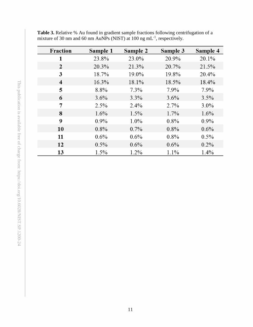
mixture: Since the separation is carried out under low centrifugal force settings, much of the Au distributed within the centrifugation column post-centrifugation should be found within the top few gradient layers (previous experiments in this lab: sample layers, 42.9 % ± 1.5 %; NaCl layers, 37.3 % ± 1.7 %; and 20 % sucrose layers, 15.7 % ± 0.9 %). A small concentration of Au, may be found in the bottom two layers (previous experiments in this lab: the 40 % and 50 % sucrose layers; an average concentration of, 2.3 % ± 0.2 % and 1.8 ± 0.2 %, respectively). With an n of 4, the repeatability of the separation in our lab, data displayed in Table 2 and Table 3, was evaluated by calculating the average concentration of Au found not only in each individual layer, but also as an average sum of Au found in each individual gradient sample. This approach was successfully employed in two published studies for the separation of mixtures of AuNPs at varying sizes, [17],[27] as well as at low and high concentration levels.
19.2.2. Sucrose density separation of the nanosuspension/C. elegans mixture: C. elegans have a specific gravity of approximately 1.08 g cm-3 and can be recovered by flotation in a solution with a specific gravity of 1.15 g cm-3.[28] Sucrose is an ideal solution for this particular density gradient centrifugation protocol, as it is a

8
This publication is available free of charge from: https://doi.org/10.6028/N
IST.SP.1200-24
chemical that is commonly employed for discreet isolation and fractionation of viable worms from other components such as residual bacteria, fungi, debris, etc.3 As mentioned previously, upon introducing the nanosuspension/C. elegans to the top of the gradient column, the C. elegans settles on top of the interface of the NaCl and 20 % sucrose layer (the densities of sucrose concentrations are shown in Table 1). By density, the nematodes migrate easily through the nanosuspension/C. elegans mixture layer and NaCl layer, without the assistance of centrifugation. As displayed in Table 4 and Table 5, for the nanosuspension/C. elegans mixture, a majority of the Au (mean ± SD) distributed within the centrifugation column post-centrifugation will be found within the top few gradient layers (previous experiments in this lab: the sample layer, 45.2 % ± 1.5 %; NaCl layer, 38.9 % ± 2.1 %; and the 20 % sucrose layer, 13.7 % ± 2.0 %). Some Au, but not a significant particle concentration, will be found in the bottom two layers (previous experiments in this lab: the 40 % and 50 % sucrose layers; with Au concentrations (average ± SD) of 1.4 % ± 0.4 % and 0.9 ± 0.2 %, respectively). With an n of 4, the repeatability of the separation can be evaluated by calculating the average concentration of Au found not only in each individual layer, but also as an average sum of Au found in each individual sample.
As can be seen in Table 5, the reproducibility of the separation in our prior experiments was evident, as the average sum of Au found in the nematode migration layers (the three fraction layers denoted as Fraction 8, 9, and 10) was 3.1 %, 3.0 %, 2.3 %, and 2.5 % for sample 1 through sample 4, respectively. This relates to an average AuNP-removal efficiency of 97.3 %.[17]

9
This publication is available free of charge from: https://doi.org/10.6028/N
IST.SP.1200-24
Table 1. Density of sucrose solutions used for the sucrose gradient centrifugation. Sucrose % by weight Density (g cm-3)a
20 1.08140 1.17150 1.230
aValues provided from the Cell Biology Laboratory Manual.[29]

10
This publication is available free of charge from: https://doi.org/10.6028/N
IST.SP.1200-24
Table 2. Total Au, in ng, found in gradient sample fractions following centrifugation of a mixture of 30 nm and 60 nm AuNPs (NIST) at 100 ng mL-1, respectively. Note that 2 mL of the mixture of AuNPs was added to the gradient, accounting for ≈400 ng of total Au.
Recovery percentage represents the calculated % recovery of Au mass applied to the density gradient column prior to centrifugation.
11
This publication is available free of charge from: https://doi.org/10.6028/N
IST.SP.1200-24
Table 3. Relative % Au found in gradient sample fractions following centrifugation of a mixture of 30 nm and 60 nm AuNPs (NIST) at 100 ng mL-1, respectively.

12
This publication is available free of charge from: https://doi.org/10.6028/N
IST.SP.1200-24
Table 4. Total Au, in ng, found in gradient sample fractions following a 0 h nematode exposure to a mixture of 30 nm and 60 nm AuNPs (NIST) at 100 ng mL-1, respectively. Note that 2 mL of the mixture of nematode/AuNP suspension was added to the gradient, accounting for ≈200 ng of total Au.
Recovery percentage represents the calculated % recovery of Au mass applied to the density gradient column prior to centrifugation. *Indicates the location of nematodes within the gradient after centrifugation.

13
This publication is available free of charge from: https://doi.org/10.6028/N
IST.SP.1200-24
Table 5. Relative % Au found in gradient sample fractions following a 0 h nematode exposure to a mixture of 30 nm and 60 nm AuNPs (NIST) at 100 ng mL-1, respectively.
*Indicates the location of nematodes within the gradient after centrifugation.

14
This publication is available free of charge from: https://doi.org/10.6028/N
IST.SP.1200-24
Figure 1. Representative schematics of (A) mixed-stage nematodes, (B) the nanosuspension containing 30 nm and 60 nm AuNPs at 100 ng mL-1, respectively, and (C) the nematode/nanosuspension mixture. The construction of the sucrose density gradient column within a centrifuge tube from top to bottom: 2 mL of the nanosuspension (D) or 2 mL of the nematode/nanosuspension mixture (E), 2 mL of 100 µmol L-1 NaCl (light green) + sucrose [4 mL of 20 % (mass density; purple), 3 mL of 40 % (mass density; orange), and 2 mL of 50 % (mass density; red)].
50 % sucrose
40 % sucrose
20 % sucrose
0.1 mol L-1 NaCl
Nanosuspension
Nematodes +
Nanosuspension
Nematodes
Nanoparticles
D E
2 mL
5 mL
9 mL
11 mL
2 mL
5 mL
9 mL
11 mL
+
A CB

15
This publication is available free of charge from: https://doi.org/10.6028/N
IST.SP.1200-24
Figure 2. Post-centrifugation distribution of Au across the sucrose density gradient centrifugation column of a mixture of 30 nm and 60 nm AuNPs at 100 ng mL-1, respectively. Each colored-line represents individual samples (n = 4). The color designations for the x-axis can denote the color designations within the gradient schematic in Figure 1D. Note: no C. elegans were added in this experiment.

16
This publication is available free of charge from: https://doi.org/10.6028/N
IST.SP.1200-24
Figure 3. Post-centrifugation distribution of Au across the sucrose density gradient centrifugation column following a 0 h nematode exposure to a mixture of 30 nm and 60 nm AuNPs at 400 ng mL-1, respectively. Each colored-line represents individual samples (n = 4). The color designations for the x-axis can denote the color designations within the gradient schematic in Figure 1E.

17
This publication is available free of charge from: https://doi.org/10.6028/N
IST.SP.1200-24
20. Acknowledgements We would like to thank Debra Kaiser for her direction and vision in the coordination of the implementation of the National Nanotechnology Initiative Nano-EHS research strategy at the National Institute of Standards and Technology from which the idea for the development of this series of protocols originated. The authors would also like to thank Andre Striegel (Chemical Sciences Division, NIST), and Danielle Gorka (Materials Measurement Science Division, NIST) for their thorough reviews of the protocol.
21. References [1] CoT/NSET/NEHI (2014) Progress Review on the Coordinated Implementation of the National
Nanotechnology Initiative 2011 Environmental, Health, and Safety Research Strategy. wwwnanogov/node/1157 Visited January 6, 2017.
[2] Hunt PR (2017) The C. elegans Model in Toxicity Testing. J Appl Toxicol 37:50-39. https://doi.org/10.1002/jat.3357
[3] Stiernagle T (C. elegans: A Practical Approach. http://wwwwormbookorg/chapters/www_strainmaintain/strainmaintainpdf:51-67. Accessed September 14, 2015.
[4] Hulme SE , Whitesides GM (2011) Chemistry and the Worm: Caenorhabditis elegans as a Platform for Integrating Chemical and Biological Research. Angew Chem Int Ed Engl 50(21):4774-4807. https://doi.org/10.1002/anie.201005461
[5] Chauhan VM, Orsi G, Brown A, Pritchard DI, Aylott JW (2013) Mapping the Pharyngeal and Intestinal pH of Caenorhabditis elegans and Real-time Luminal pH Oscillations Using Extended Dynamic Range pH-sensitive Nanosensors. ACS Nano 7(6):5577-5587. https://doi.org/10.1021/nn401856u
[6] Hunt PR, Marquis BJ, Tyner KM, Conklin S, Olejnik N, Nelson BC, Sprando RL (2013) Nanosilver Suppresses Growth and Induces Oxidative Damage to DNA in Caenorhabditis elegans. J Appl Toxicol 33(10):1131-1142. https://doi.org/10.1002/jat.2872
[7] Kim SW, Kwak JI, An YJ (2013) Multigenerational Study of Gold Nanoparticles in Caenorhabditis elegans: Transgenerational Effect of Maternal Exposure. Environ Sci Technol 47(10):5393-5399. https://doi.org/10.1021/es304511z
[8] Kim SW, Nam SH, An YJ (2012) Interaction of Silver Nanoparticles with Biological Surfaces of Caenorhabditis elegans. Ecotox Environ Safe 77:64-70. https://doi.org/10.1016/j.ecoenv.2011.10.023
[9] Ma H, Bertsch PM, Glenn TC, Kabengi NJ, Williams PL (2009) Toxicity of Manufactured Zinc Oxide Nanoparticles in the Nematode Caenorhabditis elegans. Environ Toxicol Chem 28(6):1324-1330. https://doi.org/10.1897/08-262.1
[10] Maurer LL, Yang X, Schindler AJ, Taggart RK, Jiang C, Hsu-Kim H, Sherwood DR, Meyer JN (2016) Intracellular Trafficking Pathways in Silver Nanoparticle Uptake and Toxicity in Caenorhabditis elegans. Nanotoxicology 10(7):831-835. https://doi.org/10.3109/17435390.2015.1110759
[11] Meyer JN, Lord CA, Yang XY, Turner EA, Badireddy AR, Marinakos SM, Chilkoti A, Wiesner MR, Auffan M (2010) Intracellular Uptake and Associated Toxicity of Silver Nanoparticles in Caenorhabditis elegans. Aquat Toxicol 100(2):140-150. https://doi.org/10.1016/j.aquatox.2010.07.016
[12] Mohan N, Chen CS, Hsieh HH, Wu YC, Chang HC (2010) In vivo Imaging and Toxicity Assessments of Fluorescent Nanodiamonds in Caenorhabditis elegans. Nano Lett 10(9):3692-3699. https://doi.org/10.1021/nl1021909

18
This publication is available free of charge from: https://doi.org/10.6028/N
IST.SP.1200-24
[13] Wang H, Wick RL, Xing B (2009) Toxicity of Nanoparticulate and Bulk ZnO, Al2O3 and TiO2 to the Nematode Caenorhabditis elegans. Environ Pollut 157(4):1171-1177. https://doi.org/10.1016/j.envpol.2008.11.004
[14] Yang X, Jiang C, Hsu-Kim H, Badireddy AR, Dykstra M, Wiesner M, Hinton DE, Meyer JN (2014) Silver Nanoparticle Behavior, Uptake, and Toxicity in Caenorhabditis elegans: Effects of Natural Organic Matter. Environ Sci Technol 48(6):3486-3495. https://doi.org/10.1021/es404444n
[15] Gao Y, Liu NQ, Chen CY, Luo YF, Li YF, Zhang ZY, Zhao YL, Zhao BL, Iida A, Chai ZF (2008) Mapping technique for Biodistribution of Elements in a Model Organism, Caenorhabditis elegans, after Exposure to Copper Nanoparticles with Microbeam Synchrotron Radiation X-ray Fluorescence. J Anal Atom Spectrom 23(8):1121-1124. https://doi.org/10.1039/b802338g
[16] Arnold MC, Badireddy AR, Wiesner MR, Di Giulio RT, Meyer JN (2013) Cerium Oxide Nanoparticles are More Toxic than Equimolar Bulk Cerium Oxide in Caenorhabditis elegans. Arch Environ Contam Toxicol 65(2):224-233. https://doi.org/10.1007/s00244-013-9905-5
[17] Johnson ME, Hanna SK, Montoro Bustos AR, Sims CM, Elliott LCC, Lingayat A, Johnston AC, Nikoobakht B, Elliott JT, Holbrook RD, Scott KCK, Murphy KE, Petersen EJ, Yu LL, Nelson BC (2017) Separation, Sizing, and Quantitation of Engineered Nanoparticles in an Organism Model Using Inductively Coupled Plasma Mass Spectrometry and Image Analysis. ACS Nano 11(1):526-540. https://doi.org/10.1021/acsnano.6b06582
[18] Xiong B, Cheng J, Qiao Y, Zhou R, He Y, Yeung ES (2011) Separation of Nanorods by Density Gradient Centrifugation. J Chromatogr A 1218(25):3823-3829. https://doi.org/10.1016/j.chroma.2011.04.038
[19] Yanagi K, Iitsuka T, Fujii S, Kataura H (2008) Separations of Metallic and Semiconducting Carbon Nanotubes by Using Sucrose as a Gradient Medium. The Journal of Physical Chemistry C 112(48):18889-18894. https://doi.org/10.1021/jp806822x
[20] Zhang Y, Shi Y, Liou Y-H, Sawvel AM, Sun X, Cai Y, Holden PA, Stucky GD (2010) High performance separation of aerosol sprayed mesoporous TiO2 sub-microspheres from aggregates via density gradient centrifugation. Journal of Materials Chemistry 20(20):4162-4167. https://doi.org/10.1039/B926183D
[21] Johnson ME, Montoro Bustos AR, Winchester MR (2016) Practical utilization of spICP-MS to study sucrose density gradient centrifugation for the separation of nanoparticles. Analytical and Bioanalytical Chemistry 408(27):7629-7640. https://doi.org/10.1007/s00216-016-9844-x
[22] Alberts B, Johnson A, Lewis J (2002) Molecular Biology of the Cell. 4th edition. "Fractionation of Cells.". Available from: https://wwwncbinlmnihgov/book/NBK26936/ Accessed January 11, 2017.
[23] Reina A, Subramaniam AB, Laromaine A, Samuel ADT, Whitesides GM (2013) Shifts in the Distribution of Mass Densities Is a Signature of Caloric Restriction in Caenorhabditis elegans. PLoS One 8(7):9. https://doi.org/10.1371/journal.pone.0069651
[24] Lewis JA , Fleming JT (1995) Methods in Cell Biolgy: Caenorhabditis elegans: Modern Biological Analysis of an Organism. 48:4-29.
[25] Hall DH , Altun ZF (2008) C. elegans Altlas. [26] International A (2012) ASTM E2456-06: Standard Terminology Relating to Nanotechnology. [27] Johnson ME, Montoro Bustos AR, Winchester MR (2016) Practical Utilization of spICP-MS
to Study Sucrose Density Gradient Centrifugation for the Separation of Nanoparticles. Anal Bioanal Chem 408:7629-7640. https://doi.org/10.1007/s00216-016-9844-x
[28] Kuhn-Institut HJ (2013) PM 7/119 (1) Nematode extraction. EPPO Bulletin 43(3):471-495. https://doi.org/10.1111/epp.12077
[29] Heidcamp WH (Table 3.2 Density and refractive indexes of sucrose. Cell Biology Laboratory Manual http://homepages.gac.edu/~cellab/chpts/chpt3/table3-2.html. Accessed January 26, 2016.