SUBTITLE: Expression and Reconstitution of Biologically · PDF fileACETYLCHOLINESTERASE IN E....
38
AD-A261 641 AD_________ CONTRACT NO: DAMD17-90-C-0107 TITLE: PRODUCTION OF ENZYMATICALLY ACTIVE HUMAN ACETYLCHOLINESTERASE IN E. COLI SUBTITLE: Expression and Reconstitution of Biologically Active Human Acetyicholinesterase from E. Coli PRINCIPAL INVESTIGATOR: Marian Gorecki, Ph.D. CONTRACTING ORGANIZATION: Bio-Technology General Corporation 1250 Broadway New York, New York 10001 DTIC REPORT DATE: December 20, 1992 - TIC -ETE TYPE OF REPORT: Midterm Report MAR03 PREPARED FOR: U.S. ARMY MEDICAL RESEARCH AND DEVELOPMENT COMMAND Fort Detrick, Frederick, Maryland 21702-5012 DISTRIBUTION STATEMENT: Approved for public release; distribution unlimited The findings in this report are not to be construed as an official Department of the Army position unless so designated by other authorized documents. -- : ~93-04437 * 83 2 103
Transcript of SUBTITLE: Expression and Reconstitution of Biologically · PDF fileACETYLCHOLINESTERASE IN E....

AD-A261 641
AD_________
CONTRACT NO: DAMD17-90-C-0107
TITLE: PRODUCTION OF ENZYMATICALLY ACTIVE HUMANACETYLCHOLINESTERASE IN E. COLI
SUBTITLE: Expression and Reconstitution of BiologicallyActive Human Acetyicholinesterase from E. Coli
PRINCIPAL INVESTIGATOR: Marian Gorecki, Ph.D.
CONTRACTING ORGANIZATION: Bio-Technology General Corporation1250 BroadwayNew York, New York 10001
DTICREPORT DATE: December 20, 1992 - TIC
-ETE
TYPE OF REPORT: Midterm Report MAR03
PREPARED FOR: U.S. ARMY MEDICAL RESEARCH AND DEVELOPMENT COMMANDFort Detrick, Frederick, Maryland 21702-5012
DISTRIBUTION STATEMENT: Approved for public release;distribution unlimited
The findings in this report are not to be construed as anofficial Department of the Army position unless so designated byother authorized documents.
--: ~93-04437
* 83 2 103

j form Approved
REPORT DOCUMENTATION PAGE OMB No 0704-0188P•uoi,c -eoo,^j towaen for m.s ,cllect-on ýf ntormarlon .% .it•,at-d• toQ ierejqe ' hour oer ýý.wose ncuding Ir'e t~rme ,or .•e.,. nstr.,,•t(cns ox•:•.•*,stinq "qaia wurr -,s
g athe'er" q nd martaining ythe jata needea. ind Como,exr~~s .h.rq woe ot~fl0 nf-tc,,on Sona :omrmensrot-garoorq ms bur-io ot.'-e r' r.th, je 3St In.caeC!onfl 21 ,trmatifl, mluargI' sugqt-s,tons 'of roduul'q !h.% ourcie. , %4$,hnqtofn -1eadqaJriers "eries. Directo~rae 1or nl'!f- ,l on '~' ~n,3 pe "s. '215 etleraonDajs' , .. S..te 1204 .1i0-nj1 n, i - 2202-4302 inr 10 tho Otf, 'e )t Manaqe ret nAnd 9u ad et ' oelwcr e•. Re ctn P r.te 0704-0 !88). ,,sr',nql .n DC 20S03
1. AGENCY USE ONLY (Leave blank) 2. REPORT DATE T. REPORT TYPE AND DATES COVERED
11 April 1992 Midterm Report (9/1/90 - 3/1/92)4. TITLE AND SUBTITLE S. FUNDING NUMBERS
PRODUCTION OF ENZYMATICALLY ACTIVE HUMAN Contract No.ACETYLCHOLINESTERASE IN E. COLI DAMD17-90-C-0107
6. AUTHOR(S) 627 87AMeir Fischer, Ph.D. 3M162787A875.AA.361Marian Gorecki, Ph.D. WUDA335586
7. PERFORMING ORGANIZATION NAME(S) AND ADDRESS(ES) 8. PERFORMING ORGANIZATIONREPORT NUMBER
Bio-Technology General Corporation1250 BroadwayNew York, New York 10001
"9. SPONSORING/ MONITORING AGENCY NAME(S) AND ADDRESS(ES) 10. SPONSORING ' MONITORING
U.S. Army Medical Research & Development Command AGENCY REPORT NUMBER
Fort DetrickFrederick, Maryland 21702-5012
11. SUPPLEMENTARY NOTES
Subtitle: Expression and Reconstitution of Biologically ActiveHuman Acetylcholinesterase from E. Coli
"12a. DISTRIBUTION. AVAILABILITY STATEMENT 12b. DISTRIBUTION CODE
Approved for public release; distributionunlimited
13. ABSTRACT (Maximum 200 words)
Human AChE was cloned and expressed in E. coll under the regulation of theinductible APL and the constitutive deo promoters. A partially purifiedinactive recomoinant protein was recovered from inclusion bodies. After solubiliza-tion, folding and oxidation, a protein with enzymatic properties of AChE wasobtained. Substitution of the C-terminal cysteine residue by serine enhanced therecovery of enzymatically active ACHE. The reconstituted enzyme Vasindistinguishable from native AChE isolated from erythrocytes in terms ofsubstrate specificity and inhibitor selectivity.
14. SUBJECT TERMS 15. NUMBER OF PAGES
RAV; Human Tissues; Acetylcholinesterases; Tissue __._PRICECODE
Cultures; Pseudocholinesterases; Recombinant DNA; 16. PRCE CODEGene Expression; Foreign
17. SECURITY CLASSIFICATION 18. SECURITY CLASSIFICATION 19. SECURITY CLASSIFICATION 20. LIMITATION OF ABSTRACT
OF REPORT OF THIS PAGE OF ABSTRACT
Unclassified Unclassified Unclassified UnlimitpNSN 7540-01-280-5500 Standard Form 298 (Rev 2-89)
ecr0bed bV ANSI StO 139-'S295-i0O2

FOREWORD
Opinions, interpretations, conclusions and recommendations arethose of the author and are not necessarily endorsed by the USArmy.
Where copyrighted material is quoted, permission has beenobtained to use such material.
Where material from documents designated for limited>' distribution is quoted, permission has been obtained to use the
material.
"L ____ Citations of commercial organizations and trade names inthis report do not constitute an official Department of Armyendorsement cr approval of the products or services of theseorganizations.
7 *.' In conducting research using animals, the investigator(s)adhered to the wGuide for the Care and Use of LaboratoryAnimals," prepared by the Committee on Care and Use of LaboratoryAnimals of the Institute of Laboratory Resources, NationalResearch Council (NIH Publication No. 86-23, Revised 1985).
I For the protection of human subjects, the investigator(s)adhered to policies of applicable Federal Law 45 CFR 46.
S/ In conducting research utilizing recombinant DNA technology ,th-einvestigator(s) adhered to current guidelines promulgated bythe National Institutes of Health.
ý\ In the conduct of research utilizing recombinant DNA, theinvestigator(s) adhered to the NIH Guidelines for Research
1Involving Recombinant DNA Molecules.
\/1 In the conduct of research involving hazardous organisms,St-th-einvestigator(s) adhered to the CDC-NIH Guide for Biosafety in
Microbiological and Biomedical Laboratories.

MIDTERM REPORT
1. Contract No.: DAMD17-90-C-0107 2. Report Date: (Revised)
Dec. 20, 1992
3. Reporting period from: September 90 to: March 92.
4. PI: M. Gorecki 5. Telephone No.: 972-8-381223.
6. Institution: BioTechnology General.
7. Project Title: Production of enzymatically active human AChE in
E. Coli
8. Current staff, with percent effort of each on project:
M. Fischer, Ph.D. - 100%
A. Ittach, Res. Asso. - 100%
I. Leifer, Res. Asso. - 100%
9. Contract expenditures to date:
Personnel $ 145,486 Supplies $ 30,615
Travel - Over Head + Fee $164,692
Equipmet - Total $ 340,793
10. Comments on administrative and logistical matters: none
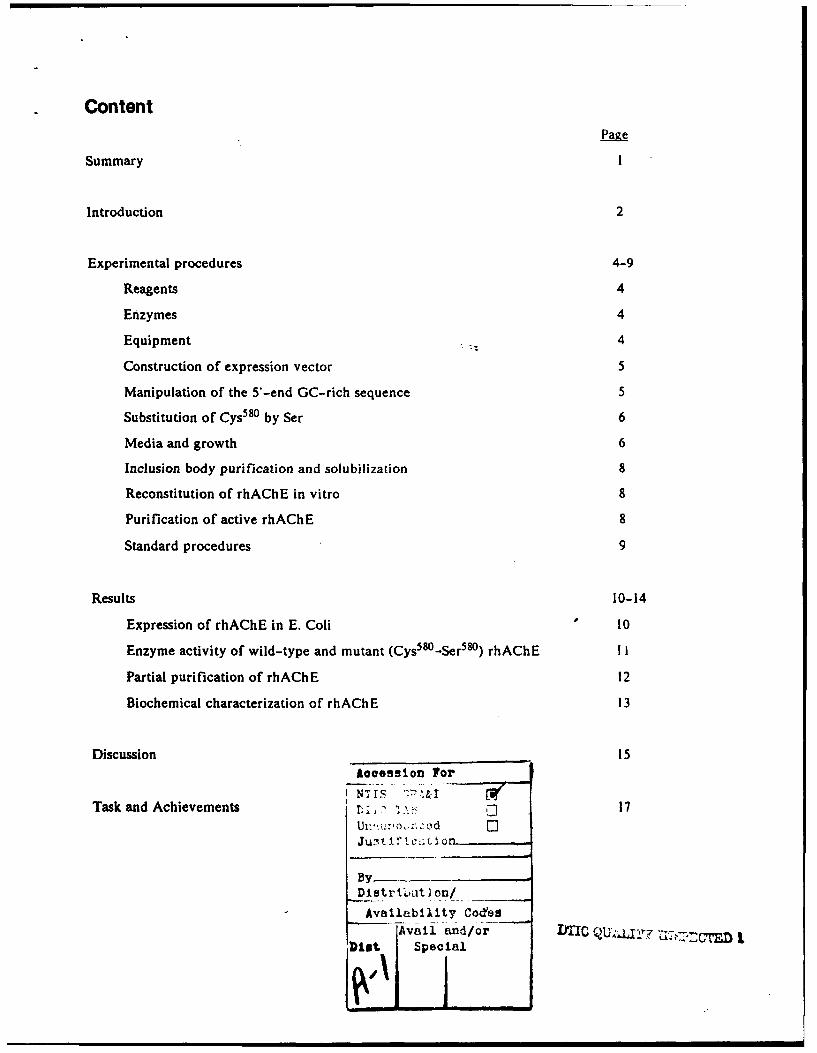
ContentPage
Summary I
Introduction 2
Experimental procedures 4-9
Reagents 4
Enzymes 4
Equipment 4
Construction of expression vector 5
Manipulation of the 5-end GC-rich sequence 5
Substitution of Cys 580 by Ser 6
Media and growth 6
Inclusion body purification and solubilization 8
Reconstitution of rhAChE in vitro 8
Purification of active rhAChE 8
Standard procedures 9
Results 10-14
Expression of rhAChE in E. Coli 10
Enzyme activity of wild-type and mutant (Cys 5 O-Ser 5") rhAChE I I
Partial purification of rhAChE 12
Biochemical characterization of rhAChE 13
Discussion Is£ooession For
Task and Achievements r: •, - 17Ult=, .": o ,oi:,d 0]Just 10 1czlti 1_
ByDIstr hiut I on/
Availability Cod'esAvail and/or DCQU/L
D18% Special

Figures 18-29
Figure 1 18
Figure 2 19
Figure 3 20
Figure 3A 21
Figure 3B 22
Figure 3C 23
Figure 4 24
Figure 5 25
Figure 5A - 26
Figure 6 27
Figure 7 28
Figure 8 29
References 30

SUMMARY
Authentic human acetyicholinesterase (hAChE) was expressed in Escherichia coli under regulation
of the constitutive deo promoter or the thermoinducible )'PL promoter. To facilitate the expression
in the prokaryotic system, the recombinant human AChE (rhAChE) cDNA was modified at the N-
terminus by site-directed mutagenesis in order to replace some of the GC-rich regions by AT. These
modifications did not alter the amino acid sequence but resulted in ample production of the protein.
rhAChE accumulated in the cells and reached a level of 10% of total bacterial proteins. A partially
purified inactive recombinant protein was recovered from inclusion bodies. Active rhAChE was
obtained after solubilization, folding and oxidation, although the recovery of the active enzyme was
low. A 20-40-fold increase in rhAChE enzyme activity was achieved by replacing Cys5'8 by Ser.
Substrate specificity and inhibitor selectivity of the recombinant mutant enzyme were
indistinguishable from native AChE isolated from human erythrocytes.
Preliminary results of this research were presented at the Proceedings of the 1991 Medical Defense
Bioscience Review, 7-8 August 1991. Edgewood, Maryland. A paper summarizing these findings
entitled "Expression and Reconstitution of Biologically active Human Acetylcholinesterase from E.
Coli" by M. Fischer, A. Ittah, I. Liefer and M. Gorecki was accepted for publication in Cellular and
Molecular Neurobiology.

INTRODUCTION
Acetyicholinesterase (AChE, EC 3.1.1.7) is present in the neuromuscular junctions and brain
cholinergic synapses and plays a pivotal role in neurotransmission by degrading acetylcholine,
resulting in termination of muscle or nerve stimulation. The AChE is sensitive to inhibition by a
number of tertiary amines and organophosphates, which in vivo can cause severe impairment and
death. For example, AChE is the primary target of military "nerve gases" and agricultural
insecticides. The therapeutic potential of exogenous AChE has provoked considerable interest.
Acetylcholinesterase and butyrylcholinesterase (BuChE) have'been shown, in several animal studies,
to protect mice against lethal dosages of soman or 7-(methyl-ethoxyphosphonyloxy-l-
methylquinolinium iodide (MEPQ) by intraperitoneal administration of the enzyme prior to exposure
to the toxic agent (Raveh et al. 1989, Ashani et al. 1991). However, more extensive evaluation of the
clinical benefit of AChE has been hindered by limited availability.
Recently, a cDNA prepared from adult basal ganglia cells encoding hAChE catalytic subunit was
isolated and cloned (Soreq et al. 1990). The deduced amino acid sequence of the mature enzyme is
583 residues in length and contains three putative glycosylation sites. Synthetic mRNA, generated
from the cDNA in vitro, was translated in microinjected oocytes into catalytically active enzyme. The
enzyme produced in oocytes exhibits biochemical properties similar to the mature enzyme as
manifested by substrate inhibition and sensitivity to the specific AChE inhibitor BW284C51
(Augustinsson 1963).
The same cDNA was used to transiently express hAChE in embryonic kidney cell line 293. The
recombinant enzyme was enzymatically active and exhibited biochemical properties similar to the
native enzyme (Valen et al. 1991a). The recombinant enzyme secreted into the medium contained
three molecular forms, monomers, dimers and tetrameres in equimolar ratio.
2

In this midterm report, we describe the construction of expression plasmids that support high-level
production of rhAChE in E. Coli and the in vitro reconstitution of the inactive protein from inclusion
bodies into enzymatically active AChE. The in vitro reconstituted enzyme is a monomer and possesses
biochemical properties which are indistinguishable from native AChE in terms of substrate specificity
and inhibitor selectivity.
3

EXPERIMENTAL PROCEDURES
Reagen
Acetyithiocholine, HEPES, S,5-Dithiobisnitrobenzoic acid (DTNB),
tetraisopropylpyrophosphoreamide (iso-OMPA), 1 .5-bis(4--allyldi methylammoni umphenyl) pentan-
3-one dibromide (BW284C5 1), Tris, urea, glutathione-oxidized (GSSG), polyethylenegiycol (PEG),
L-arginine, tetramethylammoniumchloride (TMAC), ethylenediami netetracetic acid (EDTA),
acrylamide, sodium dodecylsulfate (SDS). Coomassie Blue,. bityryithiocholine, ampicillin (Amp),
tetracycline (Tet) were purchased from Sigma Chemical Co. St-Louise Ml, U.S.A.
Guanidinethiocyanate (GTC) was from Fluka Chemical AG, Switzerland, DEAE-Sepharose and
cyanogenbromide activated Sepharose 4B from Pharmacia (Sweeden). I -methyl-9fNB-(e-
aminocaproyl)-13-aminopropylamino] acridinium bromide (MAC) was obtained from the NVeizmann
Institute, Rehovot, Israel.
Enzymes: Restriction endonucleases, T4-ligase, polynucleotide kinase, E. Coli DNA polymerase
(Kienow fragment) were purchased from New England Bio-Labs Inc. (M'VA, U.S.A.). Erythrocyte
derived hAChE and lysozyme were from Sigma Co, (U.S.A.)
Eguiivment Enzyme activities, protein and turbidimetric determinations were done using PU 8700
UV/visible spectrophotometer (Phillips). Fraction collector and monitoring units were from
Pharmacia, Sweden. Densitometric analysis was made with Model 620 Vidio densitometer Bia-Rad
Laboratories Inc.
4

Construction of exnression vectof: A 4Kb DNA fragment harboring the hAChE was isolated from
plasmid GEM-7 (Soreq et al. 1990, harboring the DNA sequences shown in Figure 8) kindly provided
by Prof. H. Soreq (The Hebrew University of Jerusalem). The 2200bp AChE sequence that resides
on the distal part of the 4Kb segment and is flanked by EcoRI-Xhol sites was isolated and ligated into
pBR322 cleaved with EcoRI-Sall restriction endonucleases. The resulting plasmid containing the
AChE was cleaved with Ndel and Nael and the larger of the two fragments generated by the cleavage
was isolated from the agarose gel. A synthetic oligonucleotide containing complementary restriction
sites (Figure 1) was ligated to the purified fragment. The synthetic oligonucleotide contained the
initiation codon ATG and the sequence of AChE starting from base number 253 to base 298 (Soreq
et al. 1990) which corresponds to the beginning of the mature protein after processing of the
sequences encoding leader peptide upstream of base number 253. The resulting plasmid pAIF-2
(Figure IA) was cleaved with Aat1l (at base number 2128), made blunt end by DNA polymerase
(KIenow fragment) and then cleaved with Ndel. The 1875bp fragment generated by these restriction
enzymes was purified from agarose gel and ligated into appropriate expression vectors (not shown).
In these constructs we have placed the hAChE sequence under control of the constitutive dec P
promoters (Fischer et al. 1990) or the thermoinducible XPL promoter (Hartman et al. 1986). The
corresponding expression plasmids containing the authentic GC-rich stretches in the 5 -end of the
hAChE-DNA sequence were designated pAIF-4 and pAIF-l I. respectively (Figure I A).
Manivulation of the 5*-end GC-rich sequence: The GC-rich sequence at the S -end of the hAChE
was substituted with A or T in degenerated codons such that the amino acid sequence was not altered
(Figure IB). A synthetic oligonucleotide flanked by Ndel-Ncol containing 24 base substitution has
ligated to pAIF-4 and pAIF-11. The resulting expression plasmids. driven by the thermoinducible
and constitutive promoters, were designated pAIF-34 and pAIF-5 I, respectively. In plasmid pAIF-S I
the AmpR gene was replaced with the TetR gene to yield pAIF-52 (Figure 2). Replacement of AmpR
by TetR gene in the expression vector pAIF-52 stabilized the plasmid and expression of rhAChE was
maintained at the same level for more than 40 generations.
5
Substitution of Cys580 by Ser: Three intrasubunit disulfide bonds can be deduced from the DNA
sequence (Soreq et al. 1990) and from the three-dimensional structure of the Torpedo AChE (Sussman
et al. 1991). The C-terminus cysteine residue of Torpedo AChE participates in the intersubunit
disulfide bridge between two subunits (MacPhee-Quigley et al. 1986). Since the C-terminus cysteine
residue of human and Torpedo AChE are positioned three amino acids upstream of the last amino acid
we predicted that Cys 580 (the C-terminus) of hAChE is involved in generating a disulfide bridge
b,,Aween two subunits and not required for maintaining the protein integi iry. Thus, this cysteine was
replaced with serine by cleavage of plasmid pAIF-52 with Sacd and Xbal and insertion of a synthetic
oligonucleotide flanked by complementary sites that contained the serine codon TCA instead of the
cysteine codon TGC in the authentic sequence. The resulting plasmid was designated pMFL-52Ser
(Figure 2). Bacterial hosts and plasmid constracts are presented in Table 1.
Media and growth: Cultures were grown in LB supplemented with N19 salts and 0.1% glucose,
100l/ml ampicillin or 12pg/ml tetracycline depending on the culture used. Clones S0930pMFL-52Ser
and S0930pAIF-52 were grown at 300C for 16-18h, harvested by centrifugation for 10 min. at 10,000
RPM in Sorval refrigerated centrifuge, washed with 50mM Tris-HCI, pH 8.0. and inclusion bodies
were prepared as described below. A4255 pAIF-34 (the thermoinducible expression system) was
grown to OD 660 of 0.8 at 300C. To induce expression of rhAChE, the temperature was elevated to
420 and grown for an additional 2h. The culture was harvested by centrifugation in Sorval refrigerated
centrifuge for 10 min at 10,000 RPM. Cell lysates containing about 6004g of protein in 1001 were
prepared by NaOH-SDS lysis solution (Hartman et al. 1986), boiled for 10 min and Il I was applied
per slot of SDS-PAGE. Following electrophoresis at room temperature the gel was stained with
Coomassie Blue R.
6

Table 1:
Plasmid constructs
Plasmid Resistance Promoter upstrem to Ribosomal AChE seqeunceMarker AChE binding site modifications
pAIF-2 Amp none none none
pAIF-4 Amp XPL (thermoinducible) XcII none
pAIF- I1 Amp deoP (constitutive) deo none
pAIF-34 Amp XPL X .cII 24 G and C to Aand T (Figure 1)
pAIF-51 Amp deoP deo 24 G and C to Aand T
pAIF-52 Tet deoP deo 24 G and C to Aand T
pMFL-52Ser Tet deoP deo As pAIF-52 andcys5'°-Ser
pMIF-35Ser* Amp XPL .cll As pAIF-34 andcys580-Ser
E. Coli Hosts
Strains Genotype
A4255 .cI85 7, AHBamH, bio
S0930 &deo, deoR-7, cimA, Alac, udp, upp, ton, thi. (Fischer et al. 1990)
The marked constract was not analayzed.
7

Inclusion body purification and solubilization: To isolate inclusion bodies, lOg of packed cells were
suspended in 100ml of 50mM Tris-HCI, pH 8.0, 10mM EDTA and l0•g/ml of lysozyme. The
suspension was incubated for 2h at room temperature, sonicated intermittently for 5 min to disrupt
cells and centrifuged for 15 min at 15,000 rpm in Sorval refrigerated centrifuge. The pellet was
resuspended in 100ml of distilled water and stirred for 30 min. After centrifugation, the pellet was
suspended in 10mM Tris-HCI, pH 8.0, containing 4M urea and stirred at room temperature for lh.
The inclusion bodies were collected by centrifugation for 30 min at 15,000 rpm, resuspended in 100ml
distilled water and stirred at 40C for 16-18h. Inclusion bodies were then collected by centrifugation
and resuspended in i0ml of 10mM Tris-HCI, pH 8.0. Solid gtiAnidinethiocyanate (GTC) was added
to a final concentration of 5.5M in 25ml. After solubilization of inclusion bodies, the solution was
brought to pH 8.6 with IM NaOH and was allowed to stir for 16-18h at room temperature. The GTC
solubilized inclusion bodies were centrifuged at 15,000 rpm for 30 min at room temperature to
remove undissolved matter and then were diluted 1:10 into 10mM Tris-HCI, pH 8.6, containing 8M
urea.
Reconstitution of rhAChE in vitro: The 1:10 diluted rhAChE in 8M urea was diluted into refolding
solution to yield a final concentration of 30-100 ýlg/ml protein. The refolding solution contained 0.5M
L-arginine, pH 10.0, IM tetramethylammonium chloride, 0.3mM GSSG and 0.3% polyethyleneglycol
4000 at 4"C. After the addition of denatured rhAChE, the solution was incubated for 24-72h at 4-
8"C, dialyzed against 5mM L-arginine, pH 10.0, containing 1mM EDTA, for 16-18h at room
temperature. Acetylcholinesterase activity was determined according to Ellman et al. (1961) at 412nm
using acetylthiocholine as substrate. The assay mixture contained 0.1M HEPES, pH 8.0, instead of
phosphate buffer. We have introduced this modification because no spontaneous hydrolysis of
acetylthiocholine is observed in HEPES.
Purification of active rhAChE: The active rhAChE was subjected to Q-Sepharose column
chromatography. The column was equilibrated with 20mM HEPES, pH 8.0, and the active rhAChE
8

was eluted with NaC! gradient. Fractions containing active AChE were pooled and precipitated with
ammonium sulfate at 45% saturation, dialyzed against 20mM HEPES, pH 8.0, and applied to MAC-
Sepharose 4B prepared according to Dudai et al. (1972). Active rhAChE was eluted from the affinity
matrix with 20mM HEPES, pH 8.0, containing 2M NaCI.
Standard procedures: DNA sequencing was performed according to the dideoxynucleotide
incorporation by the method of Sanger et al. (1977). Restriction endonuclease cleavage and ligation
were done according to the manufacturer's recommendations. SDS-PAGE was according to Laemmli
(1970). Protein determination was according to the procedure described by Bradford (1976). AChE
activity gels were performed as described by Karnovsky and Roots (1964). Amino acid sequence was
determined with Protein Microsequencer (Applied Biosystem 475A) based on the Edman degradation
procedure (Edman 1950; Tarr 1977).
9

RESULTS
Expression of rhAChE in E. Coil: Preliminary attempts to express hAChE gene by two different
expression vectors, pAIF-4 and pAIF-1 1, under control of the XPL or the deoP promoters
respectively, were unsuccessful. The GC-rich sequences at the 5'-end of the gene might have
generated secondary structures in the mRNA that are poorly accessible to ribosomes. Therefore, the
high GC content at the 5-end of the sequence, was replaced by A or T at "wobble" positions which
do not change the authentic amino acid sequence, as shown in Figure 1. Expression vectors
harboring the deoP and the )'PL promoters and the modified se`quence were introduced into E. Coli
S0930 and A4255, respectively. Total cell lysates prepared in SDS-NaOH were analyzed on SDS-
PAGE. Figure 3 shows that an intense protein band corresponding to a molecular weight of 62kD
accumulates in the induced culture of A4255 pAIF-34 (lane 2). This protein band is not seen in
uninduced control culture (Figure 3, lane 1). Western blot analysis, presented in Figure 3A, revealed
that the 62kD protein band immunoreacted with antibodies prepared against erythrocyte-derived
authentic AChE and thus confirmed that the newly produced 62kD protein band represents a genuine
AChE polypeptide. Densitometric analysis of lane 2 (Figure 3) aimed to determine expression level
show that the 62kD protein band, peak number 9, represent about 10% of the total cell protein. A
similar expression level of rhAChE protein was obtained in clone S0930pMFL-52Ser (Figure 3B). In
view of the simpler growth conditions for expression of rhAChE clones harboring the deo promoters
driven system we elected to use it routinely.
To determine the localization of expressed ACHE, the A4255 pAIF-34 and S0930pMFL-52Ser
cultures were sonicated until 99% of the cells were disrupted and the soluble fraction was separated
from insoluble matter by centrifugation. The nonsoluble fraction was resuspended in 1% SDS-NaOH
and 5pg of protein of each fraction was analyzed on SDS-PAGE. The electrophoretic pattern
revealed that rhAChE was localized in inclusion bodies in both cultures (Figure 3, lane 4 and Figure
3B lane 3) and did not possess enzymatic activity. The 62kD protein band, observed on SDS-PAGE,
10
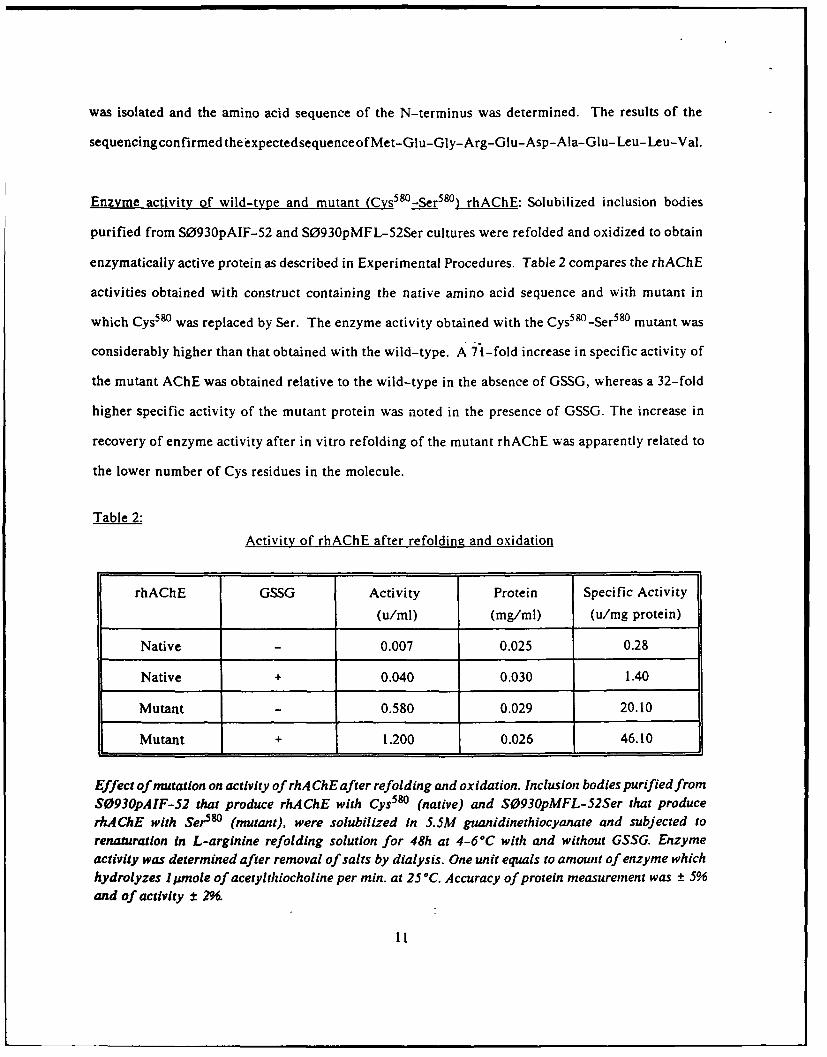
was isolated and the amino acid sequence of the N-terminus was determined. The results of the
sequencingconfirmed theexpectedsequenceof Met-Glu-Gly-Arg-Glu-Asp-Ala-Glu- Leu- Leu-Val.
Enzyme activity of wild-type and mutant (Cys5 8 0 -Ser 58 0 ) rhAChE: Solubilized inclusion bodies
purified from S0930pAIF-52 and S0930pMFL-52Ser cultures were refolded and oxidized to obtain
enzymatically active protein as described in Experimental Procedures. Table 2 compares the rhAChE
activities obtained with construct containing the native amino acid sequence and with mutant in
which Cys580 was replaced by Ser. The enzyme activity obtained with the Cys58 0 -Ser 580 mutant was
considerably higher than that obtained with the wild-type. A 71-fold increase in specific activity of
the mutant AChE was obtained relative to the wild-type in the absence of GSSG, whereas a 32-fold
higher specific activity of the mutant protein was noted in the presence of GSSG. The increase in
recovery of enzyme activity after in vitro refolding of the mutant rhAChE was apparently related to
the lower number of Cys residues in the molecule.
Table 2:
Activity of rhAChE after refolding and oxidation
rhAChE GSSG Activity Protein Specific Activity
(u/ml) (mg/ml) (u/mg protein)
Native 0.007 0.025 0.28
Native + 0.040 0.030 1.40
Mutant 0.580 0.029 20.10
Mutant + 1.200 0.026 46.10
Effect of mutation on activity of rhA ChE after refolding and oxidation. Inclusion bodies purified fromS0930pAIF-52 that produce rhAChE with Cys5 80 (native) and S0930pMFL-52Ser that producerhAChE with Ser580 (mutant), were solubilized in 5.5M guanidinethiocyanate and subjected torenaturatlon in L-arginine refolding solution for 48h at 4-60C with and without GSSG. Enzymeactivity was determined after removal of salts by dialysis. One unit equals to amount of enzyme whichhydrolyzes 1 pmole of acetylthiocholine per min. at 25°C. Accuracy of protein measurement was ± 5%and of activity ± 2%.
11

The zymogram of rhAChE on nondinaturing 6% polyacrylamide gel stained for enzymatic activity
according to Karnovsky and Roots (1964) is shown in Figure 3C. The zymogram revealed that the
activity bank generated by the native enzyme migrated slightly faster relative to the mutant harboring
the Cys5 80-Ser 580 substitution.
Partial purification of rhAChE: Since higher AChE activity was obtained with the mutant enzyme,
we have focused on the purification of this analogue. One liter of reconstituted rhAChE containing
1260 units was concentrated and dialyzed using a "Pellicon" dialysis concentrator and a 30kD MW
cutoff membrane. The volume was reduced to 600ml in 20mMIHEPES, pH 8.0, and applied to a Q-
Sepharose column chromatography.
Table 3 summarizes the results of purification. Pooled fractions, containing AChE activity, were
eluted from Q-Sepharose at about 0.275-0.375mM NaCI. Calculations of yield showed that 64% of
the activity was recovered with a specific activity of I 17u/mg. After precipitation with 45%
saturation of ammonium sulfate and dialysis against 20mM HEPES, pH 8.0, for 180 units were applied
to MAC-Sepharose 4-B affinity solumn (Iml bed volume). As shown in Table 3, the affinity
chromatography step improved purity by 19-fold, with an overall recovery of 84%. SDS-PAGE
analysis of purified rhAChE revealed a single protein band on Coomassie stained gel (Figure 4), and
indicates that the rhAChE was purified to homogeneity.
12

Table 3:
Purification of rhAChE
Volume Activity Protein Specific Activity
(ml) (units) (mg) (u/mg protein)
Q-Sepharose 600 1260 30 42
load
Q-Sepharose 23 805 7.8 117
eluate
MAC-Sepharose 1 180 1.5 120
load
MAC-Sepharose 10 151 0.066 2289
eluate
Inclusion bodies derived from S0930pMFL-52Ser were solubilized and subjected to in vitro refolding.Following concentration-dialysis, rhAChE was first subjected to Q-Sepharose ion-exchangechromatography. The pool of active fractions, eluted by NaCl gradient, was processed and subjectedto MAC-Sepharose 4B affinity chromatography. Note that only 180 out of 805 units were loaded ontoMAC-Sepharose affinity column due to the limiting capacity of the small bed volume. After repetitionof the affinity step several times to process the entire batch of 805 units, active fractions were pooled,concentrated by ammonium sulphate precipitation (45% saturation), resuspended in Imn of 20mMHEPES, pH 8.0. 2.5mM EDTA and dialyzed against 4 liters of the same buffer. A total of 0.290mgprotein containing 661 units of active rhAChE was recovered (52.5% yield by activity).
Biochemical characterization of rhAChE: Km values for acetylthiocholine and butyrylthiocholine for
the Sert mutant rhAChE were calculated by the method of Lineweaver and Burk (Figure 5). The
apparent Km obtained with acetylthiocholine was 0.1 158±0.0067mM (three independent
determinations), while with butyryithiocholine, the Km was 12.5±0.7mM that is more than 100-fold
higher. The Km of the crude recombinant ACHE, derived from clone S0930pAIF-52 harboring the
native amino acid Cys5 , was 0.125mM for acetylthiocholine (Figure 5A) and indicates that the
13

substitution of Cys580 to Ser did not alter the catalytic properties.
To define further the properties of the mutant rhAChE, its inhibition by the AChE specific inhibitor
BW284C51 and the BuChE specific inhibitor iso-OMPA were compared (Figure 6). The sigmoid
profiles of inhibition obtained for the in vitro refolded enzyme and the native erythrocyte AChE are
quite similar. 50% inhibition was obtained at lnM of BW284C51 for both enzymes. A 50% inhibition
with iso-OMPA was obtained at 0.9mM which is six orders of magnitude higher relative to the
inhibitory concentration of BW284C5 1, and is comparable with results obtained for native AChE.
The subunit character of the mutant rhAChE was determined by gel permeation chromatography on
Sephacryl 300. The column was equilibrated with 10mM L-arginine, pH 10.0, and 50mM NaCI.
Bovine serum albumin containing the 67kD monomer and the 135kD dimer was used as an internal
control. About 50 units of the highly purified recombinant enzyme was mixed with BSA and applied
to the column. Figure 7 shows that the 135kD BSA dimer eluted at fraction 45 and the 67kD
monomer eluted at fraction 50, while the activity of the active rhAChE peaked in fraction 53. Similar
analysis by HPLC with Superose 12 column consistently resulted in elution of the 67kD BSA monomer
prior to the elution of active rhAChE (data not shown). These results indicate that the in vitro
reconstituted active rhAChE is a monomer.
14
DISCUSSION
The DNA sequence encoding the human AChE catalytic subunit was inserted into E. Coli expression
vectors under control of ;PL or the E. Coli deo promoters. Cultures harboring these vectors failed
to produce the cloned gene product as determined by SDS-PAGE or Western blot analysis. The lack
of rhAChE expression in clones A4255pAIF-4 and S0930pAIF-Il is attributed to the high GC
content of the cloned gene flanking the ribosomal binding site. GC-rich sequences in mRNA in the
vicinity of the ribosomal binding site often generate stem-loop structures which may block translation
(Kozak 1983, Looman et al. 1986). Disruption of the GC-rich equences at the 5-end of the cloned
hAChE indeed resulted in high expression levels of rhAChE in clones A4255pAIF-34 and
S0930pAIF-52, driven by XPL and deo promoters, respectively.
The rhAChE expressed in E. Coli accumulates as an aggregate in inclusion bodies, as observed for a
number of other cloned eukaryotic genes in E. Coli (Mitraki and King 1989), and possesses no AChE
activity. Solubilization and refolding of the 62kD polypeptide into enzymatically active form was
achieved in the presence of L-arginine and GSSG, which were shown effective in refolding of Fab
fragment produced in E. Coli (Buchner and Rudolph 1991). The attainable enzyme activities
reconstituted in vitro and derived from clones S0930pAIF-52 were low (Table 3). We suspected that
the odd number of seven cysteine residues in the hAChE (Soreq et al. 1990) enhanced incorrect
disulfide bond formation during refolding. Indeed, replacement of cysteine 580 by serine resulted
in considerably higher yield of active rhAChE upon refolding (Table 3). The specific activity of
reconstituted enzyme prepared from inclusion bodies produced in S0930pMFL-52Ser ranged between
35 and 56 units/mg protein.
The data obtained from the gel permeation chromatography of highly purified rhAChE (2289 u/mg
protein) reveal that the active enzyme is a monomer. Valen et al. (199 1b) have recently shown that
replacement of Cys 58° with Ala resulted in secretion of predominantly monomeric hAChE from
15

transiently transfected human embryonic kidney cell line 293. The active rhAChE monomer
produced in tissue culture differs from rhAChE from E. Coli in that the latter enzyme is not
glycosylated since E. Coli does not harbor a glycosylating system. Hence glycosylation appears to be
not essential for catalytic activity.
The catalytic properties of the reconstituted rhAChE, as demonstrated by substrate specificity and
selective inhibition by BW284C51 and iso-OMPA, are quite similar to those of native AChE derived
from erythrocytes (Figure 6) and the rhAChE expressed in tissue culture (Valen et al. 199 la, 199 1b).
The E. Coli-derived enzyme and the erythrocyte AChE show-a 50% inhibition at I nM BW284C5 I
while the enzyme produced in the eukaryotic expression system was inhibited at 8nM (Valen et al.
199 la). The 50% inhibitory concentration of iso-OMPA is in the 100-200mM range for the native and
the recombinant AChE and further elucidates the "true" nature of the E. Coli-derived enzyme.
The bacterial expression system and the in vitro reconstitution of rhAChE opens the possibility to
prepare large quantities of the enzyme to be tested as a potential therapeutic and prophylactic agent
against organophosphates.
16

Task and achievements
Contractor tasks have been defined in USAMRDC log No. ;8351002 dated June 22, 1990. Table 4summarizes the achievements by tasks.
Table 4
Tasks Status
I. Preparation of full-length cDNA clone Full length cDNA was provided by Prof. H.Soreq
2. Construction of AChE expression vectors Completed2.1 Termoinducible (XPL driven)2.2 Constitutive (deo promoter driven)
3. Optimization of AChE expression and scale- In progressup fermentation3.1 XPL expression system3.2 Constitutive expression system
4. Development of an AChE purification scheme In progress4.1 Essessment of biological activity Completed4.2 Reconstitution of inactive AChE to Completed
enzymatically active form4.3 Purification process to obtain highly To be completed
purified rhAChE
5. Characterization of recombinant AChE5.1 M.W. determination of rhAChE Completed5.2 Effect of reducing agent To be determent5.3 Western analysis Completed5.4 Enzymatic activity in solution and Completed
nondenaturing gels5.5 Substrate and inhibitor specificity Completed
5.6 Final recombinant AChE product All to be determined in the futurecharacterization5.6.1 Isoelectric focusing5.6.2 Two dimensional5.6.3 Peptide mapping5.6.4 Amino acid analysis5.6.5 Amino acid sequencing5.6.6 U.V. absorption spectroscopy5.6.7 Fluorescence emission
spectroscopy5.6.8 Circular dichroism5.6.9 Extinction coefficient5.6.10 Stability
6. Scale-up purification process and production To be developed in the futureof rhAChE
17

E Neoi hAChE R
ri I pAIF-2,"'N~el T
[ I IpAIF-.INdel T
dleP l I pAIF-IL
Ndel• : '
''A*I*A*T GAA t; \A i(;'r CAA C•3 AIr A (;,A A 1i1 : ("CT
C C C 6 C C G, G,"'"I) I -4(;'IT ACC" (;TT '(" (;T ., (;; CGT CTI" " (;'(",;T
Al-T CC ( - r( ; A.%,% A i" C 'A (;(;l" 'C( ( , (
('T ( 'M ' "' "IT (;(;(" AT(C (C( TI rT GT(;,,% C(.A (rI N.b+
;I;l'(
pAI1F-51
N..I
Fiure 1: Schematic presentation of rhAChE harboring plasmids,
A: From top to bottom: A linear presentation of the coding sequences for AChE andrespective promoters cloned In pBR322. 4Kb; fragment harboring the hA ChE cDNA.pAIF-2; intermediate construct which contains a synthetic linker harboring ATGtranslation initiation codon and the authentic, GC-rich, DNA sequence starting with thetriplet GAG. encoding Glu, the first amino acid of mantre hA ChE protein. pAIF-4 andpAIF- I I are expression vectors driven by the thermoinducible APL and the constitutivedeoP promoter, respectively. The NdeI site (CA TA__) contain translation initiationcodon (underline). T, indicates translation stop codon located 120bp upstream to AatII.
B: pAIF-34 and 5AIF-S1 plasmid with either "APL or deoP promoters and a syntheticoligonucleotide replacing sequeici" 'betwhii "NdeI-NcoI site. The syntheticoligonucleotlde'equence is shown on the. right. 'The G C (with top letters) nucleotidessubstituted by AT. Ristriction cleava'g site E. H, Sp.P, Sa. Xb. B. and X are EcoRl.Hindil, Sspl, PstI, SaIL Xbal, BamHI and XhoL respectively.
18

Sal { Sa l -"
Bainlil XbaI Sail SspI
T C - %CSal o okf Cop
I)iniFL-52Ser
3' •:•
TGC s eTCA
Figure2: Schematic presentation of expression plasmds, p F-52 cntais the native amino acidsequence. pMFL-52S~er is identical to pAIF-52 with the exception that Cys5s° was replacedby Ser. The three-letter codon for cysteine, tGC, is. replaced by the three-letter codon forserine, TCA."
19

1 2 3 4 5
PEAK Pas IIO I o F4ON RtaELAIVE W1101 41 3 i LN W N AR REL. AREA.. 4. 0011 01 L11 OO I , -e OI, 00. 1 0TTL.
1.. 1 ,2.4 7.10 4 .340 7.25 31 4 11 6.30S 1434 2 40.8 55.2 0.42 7. 700 0.0 0.140 7.30
1,.5 5511.3 0 . . 0.M90 1, I.|00 5.24
1 . 126.1 8I .4 0.04 1.090 1.10 2.067 4.844 '. '.S70 0.070 5.31 1 .910 17.9q
0.(1.) 0,:'. ?'.4 7.75 1.-10 2.47 , ' .0 06I7,51.01 )7 1.6 :M,~ .0 .7 0-040 9.94 0,342 0.20SII 11.,5 01l.1 0 1.040IOi 3.10 1.150 4.32
0.4 AI 12 042.I I7,1 7.00 • 1,0 7.040 1..2
I.S11 154.9 105127 .4 0.4 5.0 .01 .50.4 I2 II 1446 10.* 7 •.0 2 1.440 1.15 .032 • ,, ,0
515 i 9~ 112,1 0.96 , .46 5.04 ?, !05 7,6600.2 14 151." Itll' .IT , .Mo 1.15 7,70 1 ,.0
tI 1 4.4 I02.7 ,VI 0 . 1 . 1 2.11 0 103 1.010.3. M's I 101 II VI 4. 125.90 *794 7.400 0.9§ 70032 1.3
. . . .,. I 1 .... .. . . ... .. I. 1".4 O 12 .9 .797 ,034 ., '.111 7.0
S so40 IQ go T 0 100 I eI 120 l30 ".H
IO0.4
14 4011184111 420 V14,, 1041 00047t
Figure 3:Top: Expression of rhAChE in the thermoinducible clone A4255pAIF-34. Samples of total
lysate and cells disrupted by sonication were analyzed 15% SDS-PAGE. Lane 1: totalcell lysate from uninduced culture. 2: total cell lysate from induced culture. 3:soluble fraction of sonicated culture. 4: insoluble fraction of sonicated culture. 5:molecular weight markers; from top to bottom (kD) - 97,66.47,30,20,14.4.
Bottom: Densitometric analysis of lane 2 shown on top. Peak number 9 correspond to the 62kDrhAChEproteidtband. The numeric display on the right of the scan shows the dataand the relative area, as percent, of the entire lane length.
20

-"94
• • 4m67
'• "-43
-30
-20
-14
Fig 3A: Western blot of rhA ChE. Total cell lysate and inclusion bodies isolated from cloneS0930pMFL-52Ser were subjected to SDS-PAGE on a 1596 acrylamide gel, blottedonto nitrocellulose paper and immunoreacted with human erythrocyte anti-AChE.Lane 1: total cell lysate of coritrol (host) S0930; lAie 2:"'ioial cell lysate ofS093OpMFL-S2Ser; lane 3: Inclusion bodies fraction oselaweyfrompMFL-52Ser. Themost intense immunoreactive band correspond to the 62-64kD polypeptide of rhA ChE.Numbers on right are protiln MWmarkers in kD.M ....
21

Fiure-3B: Expression of rhAChE in clone driven by the constitutive deoP promoter. Lane 1:
Total cell lysate of S0930 host containing no plasmid (control). Lane 2: total cell
lysate of S0930pMFL-52Ser. Lane 3: insoluble fraction of sonicated S0930pMFL-
52Ser. Lane 4:pmolecular wight markers, from top to bottom (kD) - 9 7,66,47,30,20
and 14.4.
22

iRkure 3: Zymo ram of rhACh activity on 696polyacrylamide gel. Lane]I: native rhAChE withCys~gfreconstituted from inclusion bodies derived from clone S093OpA IF-52. Lane2: mutant rhA (ThE with Cys580 -Ser5m substitution reconstituted from inclusion bodiesderived from clone S093OpMFL-52Ser.
23

Eigu¢•A: Affinity purified rhAChE derived from clone S0930pMFL-52Ser. Lane 1: MW markerproteins 94. 67, 43. 30. 20 and l4kD from top to bottom, respectively; lane 2: pautern ofproteins that did not bind to the affinity column MAC-Sepharose 4B; lane 3: the 62-64kDprotein of active rhMAhE eluted from the affinity column. . ...
24

4-
t 5.8013- 0
0.01 0LI1 10 V
MM 5.3-1/V
"I/V2-
4.8
1/v
4.3
kin" 0.1136mMi kmn" 12.5mM
-10 -5 0 5 10 15 20 25 -0.2 0 0.2 0.4 0.6 0.8 1 1.2vS [Acetylthlocho.ine mM-i 1/S IButyrylthiocholine mM- 1]
EzM : Kinetic parameters ofpurified rhA ChE reconstituted in vitro: double-reciprocal Lineweaverand Burk plot and substrate Inhibition (inset). Kmn was determined from Lineweaver-Burkplot. Constant volume of highly purified rhAChE derived from clone S0930pMFL-52Serwas added to the assay mix containing the Indicated concentrations of acetylthiocholine orbutyrylthiocholine. Activity was monitored in *a PU8700 ipectrophotometer.
25

25
20-
15
10
5
Km = 0.125mM
S-5 0 5 10 15 20 25 30 35 40
1/s
Fiture 5A: Km determination of rhAChE derived from clone S0930pAIF-51 which harbors thenative amino acid sequence of hAChE catalytic subunit. Crude non-purified activeenzyme obtained after in vitro refolding, was used for this determination.
26

Inhibition of AChE
A. B..100 120
8W284C51 iso-OMPAA
100 -
0
o A0S..
0
0 60Go-
>%40 A
4040_ 4
UJ 2020O
0 "• rAChE -4- rAChE/%BG*AChE R@C-AChE
0 II.........I....... .... ..... .. ...., .. 0 11 I.H , ,JI.
0.01 0.1 1 10 100 0.001 0.01 0.1 1 10
[inhibitor concentrationi nM (Inhibitor concentrationl mM
Fjigu : Effect of inhibitors on rhAChE. Highly purified rhAChE (o) derived from cloneS093OpMFL-52Ser and erythrocyte AChE (A) obtained from Sigma Co. (St-Louis) wereassayed in the presence of B%284C51 (panel A) or iso-OMPA (panel B) at indicatedconcentrations.
27

0.2 2
0.15 1.5
o E
0 0.1--
0.05 0.5<
"0 0.0 10 20 30 40 50 60 70 80
Fraction
-_u.e: Molecular weight determination of active rhA ChE. Purified rhA ChE obtained from cloneS0930pMFL-52Ser was mixed with bovine serum albumin (BSA) and subjected to sizeexclusion chromatography on Sephacryl-300. Absorbance was monitored at 280nm, andAChE activity was determined in each fraction. The solid line shows the elution pattern ofBSA containing a minor peak corresponding to a M.W of 135D of dimers and the largerpeak corresponds to BSA monomer of 67kD. The dotted lines show the elution profile ofrhA ChE as determined by activity. A total of 8mg of BSA containing 50 units of rhA ChEin 18ml was applied to a column of 720mm x 26mm (Pharmacia).
28

ICC2ZT:ZCA!CCTTTGCCAACCTGCCCCACCTCCTGCAGCCAGCGATAACCCT5 1 TCGG~CC0ACAGTGCCCTALATCTCCTCCCTCCTGGCTTCTCGACCGACCCTTCACCCTTTC
121 CCTTC7TCTCCCAGCAGACGCCGCCTGCCCTGCAGC~kTr.AGrC.CCCCGCAGTGTCTG
L H T P S L A S P L L L L L L W4 L L G G24 1 C.ArCAGGGCCGGGAGGA G ax.GCTGCETFACGGTGCGTGGGGGCCGG
G V G Ak GK R C to A E L LV T V ft G G R 47301 CTGCSGGCCT~iCCCTGAAGACCCCCGCGGGCCCTGTCTCTCTTTCCTGGrCATCCCC
Lf R I RbL KT P G G P V S A F L G I P 67361 TTTGCGGAr.CCACCCATGCGACCCCGVCCC???CtGCCACtGGAGCCCAAGCCAGCCTGG
F A E P P M G P Rt R r L P P E P Kt Q P 14 17421 TCAGGCOG-?GAGACGCTACAACCTTCCAGAGTGTC GCTACCAAATGTGGCA1CCCTA
S G V V 0 A T T r o 3 V MC r 0 Y V V T L 107481 TACCCAG4-TTTGAGGGCACCGAGATGGGALACCC CCGTGAGCT12AGCGAGGAC GC
Y P G Fr KG TE9N WNH P M ft E!LSE D (C 127541I CTGTACCCAACCTGTCGACACCAACCCCCC.GCCTACCATCCCCCACCCCTGTCCTr'.TC
L Y L H V N4 T P Y P ft P T S P T P V L V 147601 TCGATCTATrGGGGTGGCCCTACAGTrGGCGCC-,CCTCCTTGGACGTGTAcG.ATGGCCGC
W4 r Y C C G F Y S G A S S L 0 V Y 0 G ft_ 167661 TCVCGGACAGGCCGAGAGG.ACTCG~rCTGGTGVCCATGAACTACCG(GCVGGGAGCCTTT
F L V 0 A K ft T V L V S H Y R f V G A F 187721 GCTcTCCCGCCCTGCCGGGGArCCCGAr.AGGCCCC=GCCAATGTGGGTCTCCTGGATCAG
G F L A L P G S Rt Z A P G N V G L L D0 -' 07781 ACGCOC2GCCCTAGTCGCTGCACGACAACGTCGCAGCCTCGGGGGTGACCCCACATCA
R L A L 0 V V Q Z N V A A F G G 0 P T S 22794 1 aT5ACCGCTT7GGCGA r.CGCCCGGACCCGCCTCGGTGGCCATGCACCTGCTGTCCCCC
7 L F G K ?I) A C A A S V q M H L L S P 4 741)1 C:AGCCC2CC1CCTGTTCCACAGGGCCCCTGCTCCA.AGCCCGTGCCCCZALATCCACCCTCG
P S ft ; L F H ft A V L 0 3 C A P N G P W4 267961 C:A.;TGCATr.GGAGACGCCCGCCCCAC.CCCCACGCAGCTCGCCCACCTTG1CGrCC
A T 7 G G E A ft R Ft A T Q L A H L V G 2871021 -G-C-ýCCAGCGCGCACCTGOTGGAMZgAgCAGAGCTCGGTAGCC CCCTTCC;GACACGA
081 P C G T G G LN D ) T EL V A CO) L ft T ft 307101LCACC2CA.CTCCVCGTGAACCACGAATGGCACGTGCTGCCTCAAGAAACCGVCTVCCGG
P A C L VMREV H 1K NV L P QtES V F ft 32 711 41 TCTCCVTC0IGCCCGTGGTAGATGGAGACTTCCTCAGTGACACCCr-AGAGGCCCTCTCC
F S F V P V V 0 C 0 T L 5 D T P K A L 1 3471201 AACCCýGC3AGACTTCCA~CGCCTGCAGGTGCTCGGGGGTGVGCGTGAGATGAGGGCTCC
N A G 0 F H G L Q V L V.G V V K 0 K G S 3671261 TAVTTCVGC??TACGGCGrCCCCAGGCTTCAGC.AAAGACAAgreAg1CTCTCATCAGCCGG
Y F L V Y G AP G F S R D(JM E L IS ft 3671321 CCCGAGTrCCTGGCCGGCCTGCCCGTCGCGCTTCCCCAGGTAAGTGACCTGGCACCCGAG
A K T L A G V ft V G V P 0 V S D L A A K 4071361 CCTGVCCTCCTGCATTACACAGACTGGCTGCATCCCCACGACCCGCCACGCCTCAGGGAG
A V V L H Y T 0 W4 L H P K 0 P A Rt L ft K 4271 441 GCCCAC3CCATGTGGTGGGCGACCACAATGTCGTG GCCGCCACVCTC
A L S 0 V V G D0 NH V V (2) P V A 0 L A G 4471501 CSACTGGCGCCCAGrGGGCCCGGGTCTACr.CCTACGTCTTTGA.ACACCGTGCrTCCACG
ft L A A 9 G A ft V Y A Y V F K H Rt A S T 4671551 CTCVCCTGCCCCCVGTCCACGCCCVCCCCCACGGCVACCACATCCAGVVCATCVVCGGG
L S W4 P L N4 H G V P H C Y E 1 K F I F G 4871521 ATCCCCVCTCCCCCTCTCGAAACTACACGGCAGACC.AGAAAACVTCCCCCAGCCACTG
I P L 0 P S ftN A4 K KI F A QftL 5071881 ATGCGATACIGGGCCAACTTTGCCCGCACAGGGGATCCCAATGArCCCCCGAGACCCCAAG
N Rt Y W A N r A It fG0- P N K P ft D P K 5271741 GCCCCACAATGGCCCCCGTACACGCCGGCGGCCCAGCAGTACGVACTCTGCACCTGCCC
A P 0 W4 P P Y T A G A Q 0 Y V S L 0 L Rt 5471601 CCC.CTCGrAGGVGCCGGCGGGGCGCGrCGCCCCAGGCC~XCCVCGACCV~~
F L K V ft ft G L ft A 0 A %~)A F N4 N ft F L 5671661 CCCA.AATTrCTCAGCGCCACCGACACGCTCGACGAG4MCGGAGCGCCAGTGGAAGGCCCAG
P ft L L S A T F 7 0 tU A K R Q WI K A K 5671921 TTCCACCGCCGGAGCTCCTA'rATGGTGrCACTCGAAGAACCAGTTCr.AcCACTACAGCAArG
r N at N S 3 1 H V HN 1 K N! H F T s K 6071961 CGA CCC ?CA"CACCGCCGCGGCCCT CTCC~CTCCGCCCGG
Q 0 t' (Ck S U142041 ~ ~ T CCCVAC'GAATC ArVTCAGGO-CTG~rCTCATVACACACAC.AGcCCACJLA
7101r- C??CCFCCCCACCAGCC CCGCCCCCCTCCVCGCAGcTCcT2161 ?CACrGVACCCCTCCCCCCGTGCCTVCGCCCTCTGCCTCCATAAAVAATGpTACrcA~2221 C A &1S~ 2253
How"t.: Nucleotide and amino acid sequence of hACIE as published by Soreq et al (1990). Thesingle letter code was used to denote Individual amino acids. See the cited reference forexplanation of boxed, underlined sequences and arrows.
29

REFERENCES
Ashani, Y., Shapira, S., Levy, D., Wolfe, A.D., Doctor, B.P., and Raveh, L. (1991).
Buryrylcholinesterase and acetylcholinesterase prophylaxis against Soman poisoning in mice.
Biochem. Pharmacol. 41:37-41.
Augustinsson, K.B. (1963). In Handb. Exp. Pharmak. (G.B. Koelle, Ed), Springer-Varlag, Berlin,
Suppl IS, pp. 89-128.
Bradford, M.M. (1976). A rapid and sensitive method for quantitation of microgram quantities of
protein utilizing the principle of protein-dye binding. Anal. Biochem. 72:248-254.
Buchner, J., and Rudolph, R. (0991). Renaturation, purification and characterization of recombinant
Fab-fragments produced in Escherichia coli. Biotechnology 9:157-162.
Dudai, Y., Silman, I., Shinitzky, M., and Blumberg, S. (1972). Purification by affinity
chromatography of the molecular forms of acetylcholinesterase present in fresh electric-organ tissue
of electric eel. Proc. Natl. Acad. Sci. USA. 69:2400-2403.
Edman, P., (1950). Method for determination of the amino acid sequence in peptides. Acta Chemica
Scand. 4:283-293.
Ellman, G.L., Courtney, D.C., Andres, Jr. V., and Featherstone, R.M. (1961). A new and rapid
colorimetric determination of acetyicholi nesterase activity. Biochem. Pharmacol. 7:88-95.
Fischer, M., Fytlovitch, S., Amit, B., Wortzel, A., and Beck, Y. (1990). A constitutive expression
vector system driven by the deo PIP2 promoters of Escherichia coli. Appl. Microbiol. Biotechnol.
33:424-428.
Hartman, Y.R., Geller, T., Yavin, Z., Bartfeld, D., Kanner, D., Aviv, H., and Gorecki, M. (1986).
High level expression of enzymatically active human Cu/Zn superoxide dismutase in Escherichia coli.
Proc. Natl. Acad. Sci. USA. 83:7142-7146.
Karnovsky, M.J., and Roots, L. (1964). A direct-coloring thiocholine method for cholinesterases.
J. Histochem. 12:219-221.
30

Kozak, M. (1983). Comparison of initiation of protein synthesis in procaryotes, eucaryotes andorganels. Microbiol. Rev. 47:1-45.
Laemmli, U.K. (1970). Cleavage of structural proteins during the assembly of the head of thebacteriophage T 4 . Nature 227:680-685.
Looman, A.C., Bodlaender, J., deGruyter, M., Vogelaar, A., and van Knippenberg, P.H. (1986).Secondarystructure as primary determinant of the efficiency of ribosomal binding sites in Escherichiacoli. Nucl. Acid Res. 14:5481-5497.
MacPhee-Quigley, K., Vedrick, T.S., Taylor, P. and Taylor, S.S. (1986). Profile of the disulfide bondsin acetylcholinesterase. J. Biol. Chem. 261:13565-13570. -
Mitraki, A., and King, J. (1989). Protein folding intermediates and inclusion body formation.Biotechnology 7:690-697.
Raveh, L., Ashani, Y., Levy, D., DeLa Hoz, D., Wolfe, A.D., and Doctor B.P. (1989).Acetylcholinesterase prophylaxis against organophosphate poisoning. Biochem. Pharmacol. 38:529-
534.
Sanger, F., Nicklen, S., and Coulson, A.R. (1977). DNA sequencing with chain terminating inhibitors.Proc. Natl. Acad. Sci. USA. 74:5463-5467.
Soreq, H., Ben-Aziz, R., Prody, C.A., Seidman, S., Gnatt, A., Neville, L., Lieman-Hurwitz, J., Lev-Lehman, E., Ginzberg, D., Lapidot-Lifson, Y., and Zakut, H. (1990). Molecular cloning andconstruction of the coding region for human acetylcholinesterase reveals a G+C rich attenuatingstructure. Proc. Natl. Acad. Sci. USA. 87:9688-9692.
Sussman, J.L., Harel, M., Frolow, F., Oefner, C., Goldman, A., Toker, L., and Silman, 1. (1991).Atomic structure of acetylcholinesterase from Torpedo Californica: A prototypic acetylcholinebinding protein. Science 253:872-879.
Tarr, G.E. (1977) Imprjoved manual sequencing method in Methods of Enzymology. Vol 47, p 335.Hirs C.W. and Timasheff G.N. eds. Academic Press N.Y.
31

a
Valen, B., Kronman, C., Grosfeld, H., Leitner, M., Gozes, Y., Flashner, Y., Sery, T., Cohen, S., Ben-
Aziz, R., Seidman, S., Shafferman, A., and Soreq, H. (1991a). Recombinant human
acetylcholinesterase is secreted from transiently transfected 293 cells as soluble globular enzyme. Cell,
Mol. Neurobiol. 11:143-156.
Valen, B., Grosfeld, H., Kronman, C., Leitner, M., Gozes, Y., Lazar, A., Flashner, Y., Marcus, D.,
Cohen, S., and Shafferman, A. (199 lb). The effect of elimination of intersubunit disulfide bonds in
the activity, assembly and secretion of recombinant human acetyicholinesterase. J. Biol. Chem.
266:23977-23984.
32