Substrate-mediatedStabilizationofaTetramericDrugTarget ... ·...
9
Substrate-mediated Stabilization of a Tetrameric Drug Target Reveals Achilles Heel in Anthrax * □ S Received for publication, June 25, 2009, and in revised form, November 13, 2009 Published, JBC Papers in Press, November 30, 2009, DOI 10.1074/jbc.M109.038166 Jarrod E. Voss ‡ , Stephen W. Scally ‡ , Nicole L. Taylor ‡ , Sarah C. Atkinson ‡ , Michael D. W. Griffin ‡ , Craig A. Hutton § , Michael W. Parker ‡¶ , Malcolm R. Alderton , Juliet A. Gerrard**, Renwick C. J. Dobson ‡1 , Con Dogovski ‡ , and Matthew A. Perugini ‡2 From the ‡ Department of Biochemistry and Molecular Biology and the § School of Chemistry, Bio21 Molecular Science and Biotechnology Institute, The University of Melbourne, Parkville, Victoria 3010, Australia, the ¶ St. Vincents Institute of Medical Research, 9 Princes Street, Fitzroy, Victoria 3065, Australia, the Human Protection and Performance Division, Defence Science and Technology Organisation, Fishermans Bend, Victoria 3207, Australia, and the **School of Biological Sciences, University of Canterbury, Private Bag 4800, Christchurch 8020, New Zealand Bacillus anthracis is a Gram-positive spore-forming bacte- rium that causes anthrax. With the increased threat of anthrax in biowarfare, there is an urgent need to characterize new anti- microbial targets from B. anthracis. One such target is dihydro- dipicolinate synthase (DHDPS), which catalyzes the committed step in the pathway yielding meso-diaminopimelate and lysine. In this study, we employed CD spectroscopy to demonstrate that the thermostability of DHDPS from B. anthracis (Ba-DHDPS) is significantly enhanced in the presence of the substrate, pyru- vate. Analytical ultracentrifugation studies show that the tet- ramer-dimer dissociation constant of the enzyme is 3-fold tighter in the presence of pyruvate compared with the apo form. To examine the significance of this substrate-mediated stabili- zation phenomenon, a dimeric mutant of Ba-DHDPS (L170E/ G191E) was generated and shown to have markedly reduced activity compared with the wild-type tetramer. This demon- strates that the substrate, pyruvate, stabilizes the active form of the enzyme. We next determined the high resolution (2.15 A ˚ ) crystal structure of Ba-DHDPS in complex with pyruvate (3HIJ) and compared this to the apo structure (1XL9). Structural anal- yses show that there is a significant (91 A ˚ 2 ) increase in buried surface area at the tetramerization interface of the pyruvate- bound structure. This study describes a new mechanism for sta- bilization of the active oligomeric form of an antibiotic target from B. anthracis and reveals an “Achilles heel” that can be exploited in structure-based drug design. The etiology of anthrax was first described by Robert Koch in 1876 (1). His work was the first to demonstrate unequivocally that Bacillus anthracis is the causative agent of the disease. Given the tolerance of its spores to extreme physical conditions and their ease of dissemination, the use of B. anthracis in bio- terrorism and biological warfare is a threat to developed coun- tries worldwide. For example, in 2001 a number of letters con- taining B. anthracis endospores were distributed via the U.S. postal system, which resulted in 11 cases of respiratory anthrax. Since then, there has been heightened interest in the develop- ment of anti-anthrax agents (2), especially considering the lim- ited range of current effective therapeutics and the ability of B. anthracis to develop antibiotic resistance (3). One potential therapeutic target is dihydrodipicolinate synthase (DHDPS), 3 which catalyzes the first committed step in the lysine biosyn- thetic pathway (Fig. 1A). The biosynthetic pathway leading to lysine and its immediate precursor, meso-diaminopimelate (meso-DAP), is known as the DAP pathway (4) (Fig. 1A). The first committed reaction in the pathway is the condensation of pyruvate and (S)-aspartate semi-aldehyde (ASA) to form hydroxytetrahydrodipicolinic acid (5), which is catalyzed by DHDPS (Fig. 1, A and B). The DAP pathway in bacteria leads to the de novo synthesis of lysine, essential for protein synthesis, and meso-DAP, which is a vital constituent of the peptidoglycan layer in the bacterial cell wall (6) (Fig. 1A). Thus, targeting the DAP pathway through disrupt- ing the first committed step, DHDPS, will yield a novel class of therapeutic agents to treat anthrax. This notion is supported by recent work (7) that identified dapA, the gene encoding DHDPS from B. subtilis, as one of only 271 genes essential for cell viability from a total of 4,100 genes encoded by its genome. Additionally, mammals do not synthesize lysine and therefore do not possess the DHDPS enzyme. This suggests that specific inhibitors of DHDPS would have selective anti- bacterial activity with low toxicity in a mammalian host. The structure of DHDPS in the absence of substrate has been determined for a number of species of bacteria, including that of B. anthracis (8) (Fig. 1B), Thermotoga maritima (9), Thermo- anaerobacter tengcongensis (10), Escherichia coli (11), Myco- bacterium tuberculosis (12), Corynebacterium glutamicum (13), and Staphylococcus aureus (14, 15). The enzyme usually * This work was supported in part by the Defense Threat Reduction Agency (Project ID AB07CBT004) and by an Australian Research Council Future Fellowship (to M. A. P.) and a Federation Fellowship (to M. W. P.). □ S The on-line version of this article (available at http://www.jbc.org) contains supplemental Figs. 1– 4. The atomic coordinates and structure factors (code 3HIJ) have been deposited in the Protein Data Bank, Research Collaboratory for Structural Bioinformatics, Rutgers University, New Brunswick, NJ (http://www.rcsb.org/). 1 Supported by a University of Melbourne C. R. Roper Research Fellowship. 2 To whom correspondence should be addressed: Dept. of Biochemistry and Molecular Biology, Bio21 Molecular Science and Biotechnology Inst., The University of Melbourne, 30 Flemington Rd., Parkville, Victoria 3010, Aus- tralia. Tel.: 61-3-8344-2355; Fax: 61-3-9348-1421; E-mail: perugini@unimelb. edu.au. 3 The abbreviations used are: DHDPS, dihydrodipicolinate synthase; DAP, dia- minopimelate; ASA, (S)-aspartate semi-aldehyde; Ba, B. anthracis. THE JOURNAL OF BIOLOGICAL CHEMISTRY VOL. 285, NO. 8, pp. 5188 –5195, February 19, 2010 © 2010 by The American Society for Biochemistry and Molecular Biology, Inc. Printed in the U.S.A. 5188 JOURNAL OF BIOLOGICAL CHEMISTRY VOLUME 285 • NUMBER 8 • FEBRUARY 19, 2010 by guest on March 30, 2020 http://www.jbc.org/ Downloaded from
Transcript of Substrate-mediatedStabilizationofaTetramericDrugTarget ... ·...
Substrate-mediated Stabilization of a Tetrameric Drug TargetReveals Achilles Heel in Anthrax*□S
Received for publication, June 25, 2009, and in revised form, November 13, 2009 Published, JBC Papers in Press, November 30, 2009, DOI 10.1074/jbc.M109.038166
Jarrod E. Voss‡, Stephen W. Scally‡, Nicole L. Taylor‡, Sarah C. Atkinson‡, Michael D. W. Griffin‡, Craig A. Hutton§,Michael W. Parker‡¶, Malcolm R. Alderton�, Juliet A. Gerrard**, Renwick C. J. Dobson‡1, Con Dogovski‡,and Matthew A. Perugini‡2
From the ‡Department of Biochemistry and Molecular Biology and the §School of Chemistry, Bio21 Molecular Science andBiotechnology Institute, The University of Melbourne, Parkville, Victoria 3010, Australia, the ¶St. Vincents Institute of MedicalResearch, 9 Princes Street, Fitzroy, Victoria 3065, Australia, the �Human Protection and Performance Division, Defence Science andTechnology Organisation, Fishermans Bend, Victoria 3207, Australia, and the **School of Biological Sciences, University ofCanterbury, Private Bag 4800, Christchurch 8020, New Zealand
Bacillus anthracis is a Gram-positive spore-forming bacte-rium that causes anthrax. With the increased threat of anthraxin biowarfare, there is an urgent need to characterize new anti-microbial targets from B. anthracis. One such target is dihydro-dipicolinate synthase (DHDPS), which catalyzes the committedstep in the pathway yieldingmeso-diaminopimelate and lysine.In this study,we employedCDspectroscopy todemonstrate thatthe thermostability ofDHDPS fromB. anthracis (Ba-DHDPS) issignificantly enhanced in the presence of the substrate, pyru-vate. Analytical ultracentrifugation studies show that the tet-ramer-dimer dissociation constant of the enzyme is 3-foldtighter in the presence of pyruvate compared with the apo form.To examine the significance of this substrate-mediated stabili-zation phenomenon, a dimeric mutant of Ba-DHDPS (L170E/G191E) was generated and shown to have markedly reducedactivity compared with the wild-type tetramer. This demon-strates that the substrate, pyruvate, stabilizes the active form ofthe enzyme. We next determined the high resolution (2.15 A)crystal structure ofBa-DHDPS in complex with pyruvate (3HIJ)and compared this to the apo structure (1XL9). Structural anal-yses show that there is a significant (91 A2) increase in buriedsurface area at the tetramerization interface of the pyruvate-bound structure. This study describes a newmechanism for sta-bilization of the active oligomeric form of an antibiotic targetfrom B. anthracis and reveals an “Achilles heel” that can beexploited in structure-based drug design.
The etiology of anthraxwas first described by Robert Koch in1876 (1). His work was the first to demonstrate unequivocally
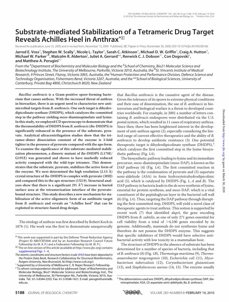
that Bacillus anthracis is the causative agent of the disease.Given the tolerance of its spores to extreme physical conditionsand their ease of dissemination, the use of B. anthracis in bio-terrorism and biological warfare is a threat to developed coun-tries worldwide. For example, in 2001 a number of letters con-taining B. anthracis endospores were distributed via the U.S.postal system, which resulted in 11 cases of respiratory anthrax.Since then, there has been heightened interest in the develop-ment of anti-anthrax agents (2), especially considering the lim-ited range of current effective therapeutics and the ability of B.anthracis to develop antibiotic resistance (3). One potentialtherapeutic target is dihydrodipicolinate synthase (DHDPS),3which catalyzes the first committed step in the lysine biosyn-thetic pathway (Fig. 1A).The biosynthetic pathway leading to lysine and its immediate
precursor,meso-diaminopimelate (meso-DAP), is known as theDAP pathway (4) (Fig. 1A). The first committed reaction inthe pathway is the condensation of pyruvate and (S)-aspartatesemi-aldehyde (ASA) to form hydroxytetrahydrodipicolinicacid (5), which is catalyzed by DHDPS (Fig. 1, A and B). TheDAPpathway in bacteria leads to thede novo synthesis of lysine,essential for protein synthesis, and meso-DAP, which is a vitalconstituent of the peptidoglycan layer in the bacterial cell wall(6) (Fig. 1A). Thus, targeting theDAPpathway through disrupt-ing the first committed step, DHDPS, will yield a novel class oftherapeutic agents to treat anthrax. This notion is supported byrecent work (7) that identified dapA, the gene encodingDHDPS from B. subtilis, as one of only 271 genes essential forcell viability from a total of �4,100 genes encoded by itsgenome. Additionally, mammals do not synthesize lysine andtherefore do not possess the DHDPS enzyme. This suggeststhat specific inhibitors of DHDPS would have selective anti-bacterial activity with low toxicity in a mammalian host.The structure of DHDPS in the absence of substrate has been
determined for a number of species of bacteria, including thatofB. anthracis (8) (Fig. 1B),Thermotogamaritima (9),Thermo-anaerobacter tengcongensis (10), Escherichia coli (11), Myco-bacterium tuberculosis (12), Corynebacterium glutamicum(13), and Staphylococcus aureus (14, 15). The enzyme usually
* This work was supported in part by the Defense Threat Reduction Agency(Project ID AB07CBT004) and by an Australian Research Council FutureFellowship (to M. A. P.) and a Federation Fellowship (to M. W. P.).
□S The on-line version of this article (available at http://www.jbc.org) containssupplemental Figs. 1– 4.
The atomic coordinates and structure factors (code 3HIJ) have been deposited inthe Protein Data Bank, Research Collaboratory for Structural Bioinformatics,Rutgers University, New Brunswick, NJ (http://www.rcsb.org/).
1 Supported by a University of Melbourne C. R. Roper Research Fellowship.2 To whom correspondence should be addressed: Dept. of Biochemistry and
Molecular Biology, Bio21 Molecular Science and Biotechnology Inst., TheUniversity of Melbourne, 30 Flemington Rd., Parkville, Victoria 3010, Aus-tralia. Tel.: 61-3-8344-2355; Fax: 61-3-9348-1421; E-mail: [email protected].
3 The abbreviations used are: DHDPS, dihydrodipicolinate synthase; DAP, dia-minopimelate; ASA, (S)-aspartate semi-aldehyde; Ba, B. anthracis.
THE JOURNAL OF BIOLOGICAL CHEMISTRY VOL. 285, NO. 8, pp. 5188 –5195, February 19, 2010© 2010 by The American Society for Biochemistry and Molecular Biology, Inc. Printed in the U.S.A.
5188 JOURNAL OF BIOLOGICAL CHEMISTRY VOLUME 285 • NUMBER 8 • FEBRUARY 19, 2010
by guest on March 30, 2020
http://ww
w.jbc.org/
Dow
nloaded from
assembles as a tetrameric protein (16), best described as a dimerof tight dimers (Fig. 1B). However, a native active dimer hasrecently been described (14), which makes the quaternarystructure of this enzyme of particular interest. DHDPS fromplants (17, 18) and someGram-negative bacterial species (19) isfeedback-inhibited by (S)-lysine, acting as an allosteric modu-lator through partial inhibition of catalytic activity. However,allosteric regulation by lysine at biologically relevant concen-trations does not occur in DHDPS from Gram-positive species(14, 20), including B. anthracis (21).Recent work in our laboratory has shown that the purifica-
tion of recombinant DHDPS from B. anthracis (Ba-DHDPS) inthe presence of its substrate pyruvate increases the yield andspecific activity of the final product (22). Therefore, the aim ofthis study was to thoroughly characterize the effect of pyruvateon the solution properties and structure of Ba-DHDPS. Here,we report solution and structural studies that unravel themechanism for substrate-mediated stabilization of the activequaternary structure of Ba-DHDPS.
EXPERIMENTAL PROCEDURES
Cloning, Expression, and Purification of Ba-DHDPS—ThedapA gene encoding DHDPS from B. anthracis (Sterne strain)
was amplified by PCR and cloned into the pET11a expressionvector as described elsewhere (22). Briefly, recombinant pro-tein was produced in the host strain E. coli BL21-DE3 as fol-lows. Cells harboring vector were cultured at 37 °C in Luriabroth containing 100 �g ml�1 ampicillin to an A600 of 0.6.Expression of recombinant Ba-DHDPS was induced by addi-tion of isopropyl 1-thio-�-D-galactopyranoside to a final con-centration of 1.0 mM. Cells were harvested 3 h post-inductionand resuspended in 20 mM Tris-HCl, pH 8.0, before lysis bysonication. Ba-DHDPS was subsequently isolated by anion-ex-change and hydrophobic interaction liquid chromatography asdescribed elsewhere (22).Generation of Dimeric Ba-DHDPS (L170E/G191E)—The
dimeric form of Ba-DHDPS was generated using theQuikChange II XL site-directed mutagenesis kit (Stratagene).Themutagenic oligonucleotide primer sets, 5�-GATGCAGGC-GGCGATGTGGAAACAATGACAGAAATCATTG-3� and 5�-CAATGATTTCTGTCATTGTTTCCACATCGCCGCCTGC-ATC-3�, and 5�-GTATACAGCGGTGATGACGAATTAACG-CTACCAGCTATGG-3� and 5�-CCATAGCTGGTAGCGTTA-ATTCGTCATCACCGCTGTATAC-3�, were designed tointroduce amino acid substitutions L170E and G191E, respec-tively. Mutagenesis was performed according to the manufactur-er’s instructions. Briefly, 125 ng of primer and 10 ng double-stranded DNA template utilized per reaction. Second strandsynthesiswas achieved through 18 cycles of amplification. Follow-ing a 1-h DpnI digestion, plasmid was transformed into E. coliXL10-GoldUltracompetentcells.The introductionofpointmuta-tions was verified by DNA sequencing.DHDPS-DHDPR Coupled Enzyme Kinetic Assay—Enzyme
kinetic analyses of Ba-DHDPS and Ba-DHDPS (L170E/G191E)were performed using the DHDPS-DHDPR coupled assay asdescribed in a previous study (23). Assays were routinely per-formed in duplicate at a constant temperature of 30 °C withreaction mixtures allowed to equilibrate in a temperature-con-trolled Cary 4000 UV-visible spectrophotometer for 12 minbefore initiating the reaction with 60 nM DHDPS. Prior to theexperiment, pyruvate and ASA concentrations were routinelyquantified by the addition of limiting amounts of substrate bymeasuring the consumption of NADPH at 340 nm in the Cary4000 UV-visible spectrophotometer. Initial velocity data werebest fitted to a Ping Pong Model using ENZFITTER.4 Rate ver-sus enzyme concentration assays were also conducted with Ba-DHDPS concentrations ranging from 2.5 to 120 nM.CD Spectroscopy—CD spectra were recorded using an AVIV
410-SF CD spectrometer. Wavelength scans were performedbetween 190 and 250 nm in 20mMTris, 150mMNaCl with 0.15mg ml�1Ba-DHDPS and Ba-DHDPS (L170E/G191E) in 1-mmquartz cuvettes. Data were analyzed using the CONTINLLalgorithm from the CDPro software package (25) and the SP29protein data base. For thermal denaturation scans, ellipticity at222 nm was monitored between 20 and 90 °C in 1 °C steps.Analytical Ultracentrifugation—Absorbance-based sedi-
mentation velocity and equilibrium experiments were per-formed in a Beckman model XL-I analytical ultracentrifuge
4 R. J. Leatherbarrow (1987) ENZFITTER, Imperial College of Science and Tech-nology, London.
FIGURE 1. Ba-DHDPS. A, DHDPS catalyzes the condensation of pyruvate andASA into hydroxytetrahydrodipicolinic acid, which is the first committed stepin the lysine biosynthesis pathway in bacteria. B, crystal structure of apo-Ba-DHDPS showing the position of one active site and the self-association inter-faces. Two monomers come together at the tight dimer interface to form thedimeric unit, and then the two dimeric units dock at the weak dimer interfaceto form a tetramer.
Pyruvate-bound B. anthracis DHDPS
FEBRUARY 19, 2010 • VOLUME 285 • NUMBER 8 JOURNAL OF BIOLOGICAL CHEMISTRY 5189
by guest on March 30, 2020
http://ww
w.jbc.org/
Dow
nloaded from
using a 4-hole An-60 Ti or an 8-hole An-50 Ti rotor. Double-sector quartz cells were loadedwith 380�l of sample and 400�lof reference (20 mM Tris, 150 mMNaCl, pH 8.0) for sedimenta-tion velocity, or a 100-�l sample and a 120-�l reference forsedimentation equilibrium. Experiments were conducted at4 °C using a rotor speed of 40,000 rpm (sedimentation velocity)or 12,000 and 18,000 rpm (sedimentation equilibrium) withabsorbance measured at 227 nm (1.6 �M enzyme) or 235 nm(4.8 �M enzyme). Solvent density, solvent viscosity, and esti-mates of the partial specific volume of Ba-DHDPS and Ba-DH-DPS (L170E/G191E) were calculated using SEDNTERP (26).Initial scans were carried out at 3,000 rpm to determine opti-mum wavelength and radial positions for the experiments.Samples monitored in the presence of 0.6 mM pyruvate con-tained pyruvate in both the reference and sample channels.Sedimentation velocity data were fitted to a single discrete spe-cies or a continuous sedimentation coefficient [c(s)]model (27–29) using the program SEDFIT. Additionally, van Holde-Weis-chet analysis (30) was performed using the ULTRASCANsoftware package (31), whereas sedimentation equilibriumdatawere fitted to various self-associating equilibriummodels usingthe program SEDPHAT (32).Crystallization of Ba-DHDPS and X-ray Diffraction Data
Collection—Ba-DHDPS was crystallized according to methodsdescribed previously (22) using sitting- and hanging-drop,vapor diffusion. The crystals used for diffraction analysis weresoaked with 20mM pyruvate overnight to facilitate ligand bind-ing. For x-ray data collection, crystals were transferred to res-ervoir solution containing 20% (v/v) glycerol with 20 mM pyru-vate and directly flash frozen in liquid nitrogen. Intensity datawere collected at the Australian synchrotron using the MX1beamline as described before (22).Phasing and Model Refinement—Diffraction data sets were
processed and scaled using the package MOSFLM (33) andSCALA (34). Initial phase estimates were solved by molecularreplacement using PHASERwith the ligand-unbound structure(PDB ID: 1XKY) as the searchmodel. Structural refinementwasperformed using REFMAC5 (35) with iterative model buildingusing WINCOOT (36). Water, glycerol, sodium ion, and thepyruvate-bound lysine atoms were added at later stages using
WINCOOT. A round of simulatedannealing was performed with astarting temperature of 5000 K toassign the Rfree set of reflectionsfrom the apo structure (PDB ID:1XL9) that shared the same crystalproperties with the newly solvedpyruvate-bound structure (PDB ID:3HIJ). The final model was checkedwith PROCHECK (37).
RESULTS
Effect of Pyruvate on the Second-ary Structure Stability of Ba-DHDPS—Werecently reported thatthe raw enzyme activity and yield ofrecombinant Ba-DHDPS are signif-icantly increased when the enzyme
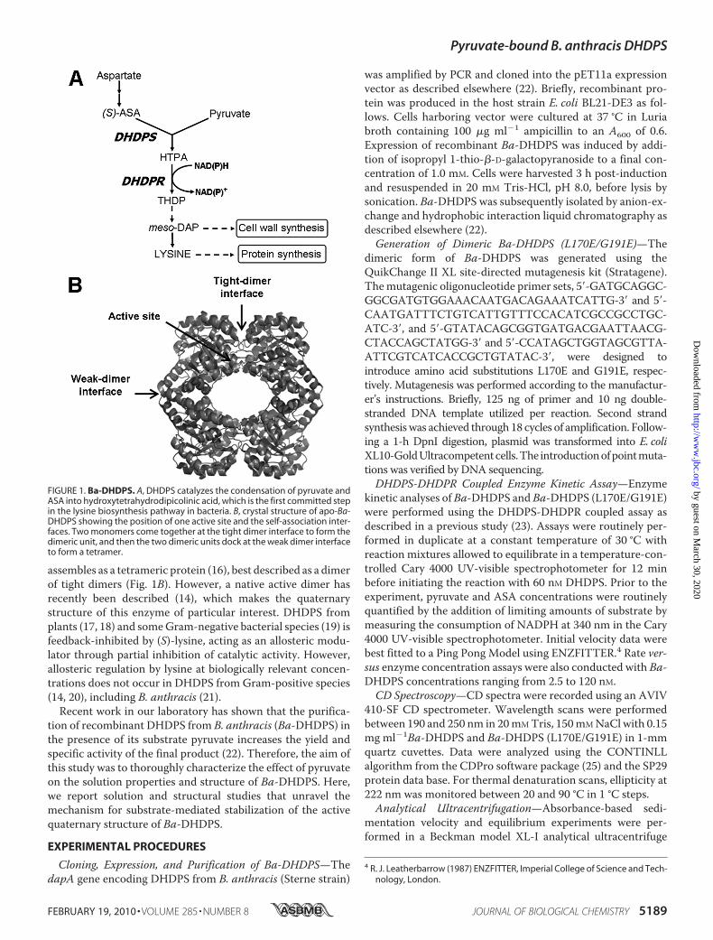
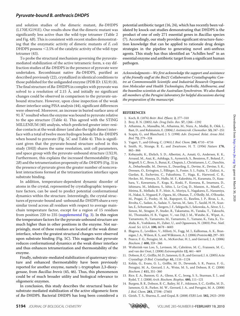
is purified in the presence of its substrate, pyruvate (22). Toexamine the effect of pyruvate on the stability of Ba-DHDPS inaqueous solution, thermal denaturation experiments moni-tored by CD spectroscopy were conducted in the presence andabsence of the substrates pyruvate orASAover the temperaturerange of 20–90 °C. Initially, wavelength scans were performedat 20 °C to monitor global secondary structure in the presenceof substrates, which revealed no change in response to eitherpyruvate or ASA (Fig. 2A). Ba-DHDPS appears to follow athree-state mechanism for thermal unfolding in the absence ofpyruvate and in the presence of ASA, with a potential interme-diate persisting at temperatures of �50–60 °C (Fig. 2B). How-ever, in the presence of 2.0 mM pyruvate, the thermal denatur-ation of Ba-DHDPS is delayed with respect to temperature andunfolding appears to occur via a two-state mechanism withoutthe propagation of an intermediate, indicating that pyruvatestabilizes the folded state of the enzyme. Accordingly, the effectof pyruvate on the quaternary and tertiary structure of Ba-DH-DPS was sought.Effect of Pyruvate on the Quaternary Structure of Ba-DHDPS—
To characterize the quaternary structure of Ba-DHDPS in solu-tion in the absence and presence of pyruvate, absorbance-de-tected sedimentation velocity and equilibrium analyses wereconducted in the analytical ultracentrifuge. The absorbanceversus radial position profiles of Ba-DHDPS (1.6 �M) duringsedimentation velocity in the absence of pyruvate are shown inFig. 3A, whereas Fig. 3B shows the equivalent data sets in thepresence of pyruvate. Two predominant boundaries wereobserved for Ba-DHDPS in the absence of substrate (Fig. 3A).By contrast, the radial absorbance profiles showed a single pre-dominant boundary in the presence of pyruvate (Fig. 3B). Thesedata were analyzed initially using the enhanced van Holde-Weischet method (30), which is a model-independentapproach to analyzing sedimentation velocity data. The result-ing integral distribution for Ba-DHDPS in the absence of pyru-vate (Fig. 3C, white circles) suggests it exists in a reversible self-association with sedimentation coefficients ranging from 2 Sthrough to 6 S. This is consistent with previous reports ofDHDPS from E. coli, which was determined to exist in equilib-rium between a dimer and tetramer (38). In the presence of
FIGURE 2. Secondary structure and thermostability measured by CD spectroscopy. A, wavelength scan ofBa-DHDPS alone (F) and in the presence of 2.0 mM pyruvate (ƒ) or 0.5 mM ASA (E). Ellipticity (mdeg) has beennormalized to mean residue ellipticity (deg cm2 dmol�1 residue�1). B, thermal denaturation of Ba-DHDPSalone (F) and in the presence of its substrates, 2.0 mM pyruvate (ƒ), and 0.5 mM ASA (E).
Pyruvate-bound B. anthracis DHDPS
5190 JOURNAL OF BIOLOGICAL CHEMISTRY VOLUME 285 • NUMBER 8 • FEBRUARY 19, 2010
by guest on March 30, 2020
http://ww
w.jbc.org/
Dow
nloaded from
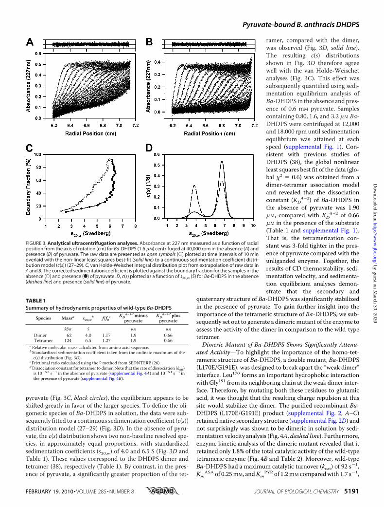
pyruvate (Fig. 3C, black circles), the equilibrium appears to beshifted greatly in favor of the larger species. To define the oli-gomeric species of Ba-DHDPS in solution, the data were sub-sequently fitted to a continuous sedimentation coefficient (c(s))distribution model (27–29) (Fig. 3D). In the absence of pyru-vate, the c(s) distribution shows two non-baseline resolved spe-cies, in approximately equal proportions, with standardizedsedimentation coefficients (s20,w) of 4.0 and 6.5 S (Fig. 3D andTable 1). These values correspond to the DHDPS dimer andtetramer (38), respectively (Table 1). By contrast, in the pres-ence of pyruvate, a significantly greater proportion of the tet-
ramer, compared with the dimer,was observed (Fig. 3D, solid line).The resulting c(s) distributionsshown in Fig. 3D therefore agreewell with the van Holde-Weischetanalyses (Fig. 3C). This effect wassubsequently quantified using sedi-mentation equilibrium analysis ofBa-DHDPS in the absence and pres-ence of 0.6 mM pyruvate. Samplescontaining 0.80, 1.6, and 3.2 �M Ba-DHDPS were centrifuged at 12,000and 18,000 rpm until sedimentationequilibrium was attained at eachspeed (supplemental Fig. 1). Con-sistent with previous studies ofDHDPS (38), the global nonlinearleast squares best fit of the data (glo-bal �2 � 0.6) was obtained from adimer-tetramer association modeland revealed that the dissociationconstant (KD
4–2) of Ba-DHDPS inthe absence of pyruvate was 1.90�M, compared with KD
4–2 of 0.66�M in the presence of the substrate(Table 1 and supplemental Fig. 1).That is, the tetramerization con-stant was 3-fold tighter in the pres-ence of pyruvate compared with theunliganded enzyme. Together, theresults of CD thermostability, sedi-mentation velocity, and sedimenta-tion equilibrium analyses demon-strate that the secondary and
quaternary structure of Ba-DHDPS was significantly stabilizedin the presence of pyruvate. To gain further insight into theimportance of the tetrameric structure of Ba-DHDPS, we sub-sequently set out to generate a dimericmutant of the enzyme toassess the activity of the dimer in comparison to the wild-typetetramer.Dimeric Mutant of Ba-DHDPS Shows Significantly Attenu-
ated Activity—To highlight the importance of the homo-tet-rameric structure of Ba-DHDPS, a double mutant, Ba-DHDPS(L170E/G191E), was designed to break apart the “weak dimer”interface. Leu170 forms an important hydrophobic interactionwith Gly191 from its neighboring chain at the weak dimer inter-face. Therefore, by mutating both these residues to glutamicacid, it was thought that the resulting charge repulsion at thissite would stabilize the dimer. The purified recombinant Ba-DHDPS (L170E/G191E) product (supplemental Fig. 2, A–C)retained native secondary structure (supplemental Fig. 2D) andnot surprisingly was shown to be dimeric in solution by sedi-mentation velocity analysis (Fig. 4A, dashed line). Furthermore,enzyme kinetic analysis of the dimeric mutant revealed that itretained only 1.8% of the total catalytic activity of the wild-typetetrameric enzyme (Fig. 4B and Table 2). Moreover, wild-typeBa-DHDPS had a maximum catalytic turnover (kcat) of 92 s�1,Km
ASA of 0.25mM, andKmPYR of 1.2mM comparedwith 1.7 s�1,
FIGURE 3. Analytical ultracentrifugation analyses. Absorbance at 227 nm measured as a function of radialposition from the axis of rotation (cm) for Ba-DHDPS (1.6 �M) centrifuged at 40,000 rpm in the absence (A) andpresence (B) of pyruvate. The raw data are presented as open symbols (E) plotted at time intervals of 10 minoverlaid with the non-linear least squares best-fit (solid line) to a continuous sedimentation coefficient distri-bution model (c(s)) (27–29). C, van Holde-Weischet integral distribution plot from extrapolation of raw data inA and B. The corrected sedimentation coefficient is plotted against the boundary fraction for the samples in theabsence (E) and presence (F) of pyruvate. D, c(s) plotted as a function of s20,w (S) for Ba-DHDPS in the absence(dashed line) and presence (solid line) of pyruvate.
TABLE 1Summary of hydrodynamic properties of wild-type Ba-DHDPS
Species Massa s20,wb f/f0cKD
4–2d minuspyruvate
KD4–2d plus
pyruvate
kDa S �M �M
Dimer 62 4.0 1.17 1.9 0.66Tetramer 124 6.5 1.27 1.9 0.66
aRelative molecular mass calculated from amino acid sequence.bStandardized sedimentation coefficient taken from the ordinate maximum of thec(s) distribution (Fig. 3D).
cFrictional ratio calculated using the v� method from SEDNTERP (26).dDissociation constant for tetramer to dimer. Note that the rate of dissociation (koff)is 10�5.3 s�1 in the absence of pyruvate (supplemental Fig. 4A) and 10�5.1 s�1 inthe presence of pyruvate (supplemental Fig. 4B).
Pyruvate-bound B. anthracis DHDPS
FEBRUARY 19, 2010 • VOLUME 285 • NUMBER 8 JOURNAL OF BIOLOGICAL CHEMISTRY 5191
by guest on March 30, 2020
http://ww
w.jbc.org/
Dow
nloaded from
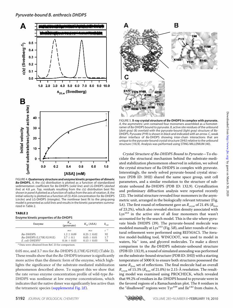
0.05 mM, and 3.7 mM for Ba-DHDPS (L170E/G191E) (Table 2).These results show that theBa-DHDPS tetramer is significantlymore active than the dimeric form of the enzyme, which high-lights the significance of the substrate-mediated stabilizationphenomenon described above. To support this we show thatthe rate versus enzyme concentration profile of wild-type Ba-DHDPS was nonlinear at low enzyme concentrations, whichindicates that the native dimer was significantly less active thanthe tetrameric species (supplemental Fig. 2E).
Crystal Structure of Ba-DHDPS Bound to Pyruvate—To elu-cidate the structural mechanism behind the substrate-medi-ated stabilization phenomenon observed in solution, we solvedthe crystal structure of Ba-DHDPS in complex with pyruvate.Interestingly, the newly solved pyruvate-bound crystal struc-ture (PDB ID: 3HIJ) shared the same space group, unit cellparameters, and a similar resolution to the structure of sub-strate unbound Ba-DHDPS (PDB ID: 1XL9). Crystallizationand preliminary diffraction analysis were reported recently(22). The initial structure revealed fourmonomers in the asym-metric unit, arranged in the biologically relevant tetramer (Fig.5A). The first round of refinement gave an Rcryst of 21.4% (Rfreeof 23.2%), which also revealed election density associated withLys163 in the active site of all four monomers that wasn’taccounted for by the search model. This is the site where pyru-vate binds DHDPS (39). The pyruvate-bound molecule wasmodeledmanually at Lys163 (Fig. 5B), and later rounds of struc-tural refinement were performed using REFMAC5. The itera-tive model-building tool, WINCOOT, was used to model inwaters, Na� ions, and glycerol molecules. To make a directcomparison to the Ba-DHDPS substrate-unbound structure(PDB ID: 1XL9), a round of simulated annealing was performedon the substrate-bound structure (PDB ID: 3HIJ) with a startingtemperature of 5000 K to ensure both structures possessed thesame Rfree set of reflections. The final molecule had an overallRcryst of 15.3% (Rfree of 21.0%) to 2.15-Å resolution. The result-ing model was examined using PROCHECK, which revealedthat 99.2% of residues in Ba-DHDPS bound to pyruvate were inthe favored regions of a Ramachandran plot. The 8 residues inthe “disallowed” regions were Tyr109 and Ile142 from chains A,
FIGURE 4. Quaternary structure and enzyme kinetic properties of dimericBa-DHDPS. A, the c(s) distribution is plotted as a function of standardizedsedimentation coefficient for Ba-DHDPS (solid line) and LG-DHDPS (dashedline) at 4.8 �M. Top, residuals resulting from the c(s) distribution best fitsshown in panel A plotted as a function of radius from the axis of rotation. B, theinitial velocity is plotted as a function of (S)-ASA concentration for Ba-DHDPS(circles) and LG-DHDPS (triangles). The nonlinear best fit to the ping-pongmodel is presented as solid lines and results in the kinetic parameters summa-rized in Table 2.
TABLE 2Enzyme kinetic properties of Ba-DHDPS
Enzyme Km(pyruvate) Km (ASA) kcat
mM mM s�1
Ba-DHDPS 1.2 � 0.09 0.25 � 0.02 92Ba-DHDPS (L170E/G191E) 3.7 � 0.1 0.05 � 0.01 1.7E. coli-DHDPSa 0.16 � 0.03 0.13 � 0.03 78
aData were obtained from Ref. 23 for comparison.
FIGURE 5. X-ray crystal structure of Ba-DHDPS in complex with pyruvate.A, the asymmetric unit contained four monomers assembled as a homotet-ramer of Ba-DHDPS bound to pyruvate. B, active site residues of the unbound(dark gray) (8) overlaid with the pyruvate-bound (light gray) structure of Ba-DHDPS. Pyruvate (PYR) is shown in black and indicated with an arrow. C, weakdimer interface of Ba-DHDPS showing inter-chain interactions that areunique to the pyruvate-bound crystal structure (3HIJ) relative to the unboundstructure (1XL9). Analysis was performed using STING MILLENIUM (40).
Pyruvate-bound B. anthracis DHDPS
5192 JOURNAL OF BIOLOGICAL CHEMISTRY VOLUME 285 • NUMBER 8 • FEBRUARY 19, 2010
by guest on March 30, 2020
http://ww
w.jbc.org/
Dow
nloaded from
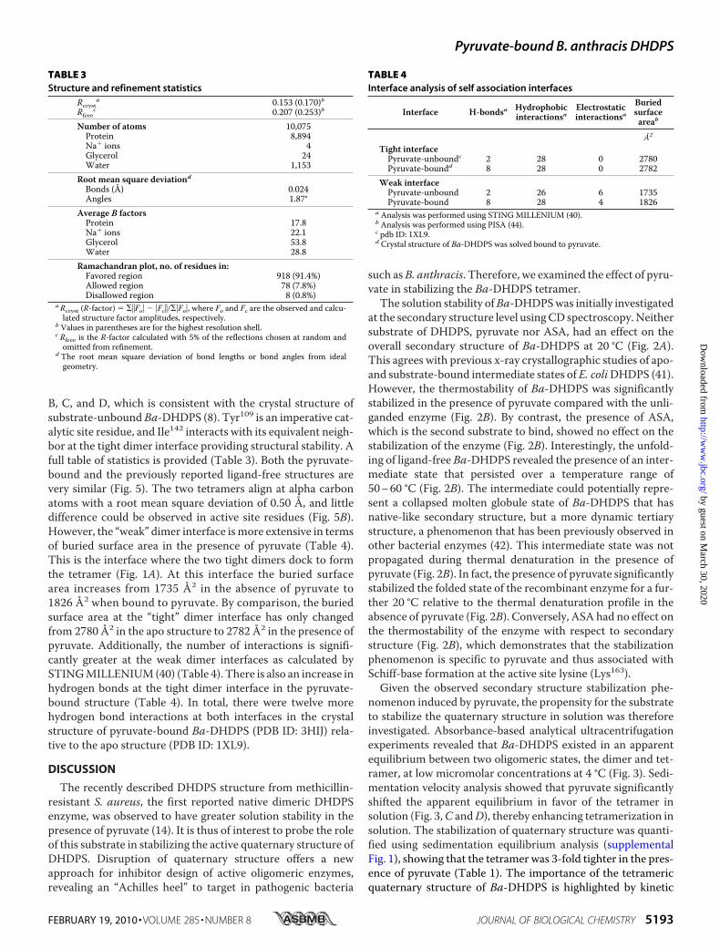
B, C, and D, which is consistent with the crystal structure ofsubstrate-unbound Ba-DHDPS (8). Tyr109 is an imperative cat-alytic site residue, and Ile142 interacts with its equivalent neigh-bor at the tight dimer interface providing structural stability. Afull table of statistics is provided (Table 3). Both the pyruvate-bound and the previously reported ligand-free structures arevery similar (Fig. 5). The two tetramers align at alpha carbonatoms with a root mean square deviation of 0.50 Å, and littledifference could be observed in active site residues (Fig. 5B).However, the “weak” dimer interface ismore extensive in termsof buried surface area in the presence of pyruvate (Table 4).This is the interface where the two tight dimers dock to formthe tetramer (Fig. 1A). At this interface the buried surfacearea increases from 1735 Å2 in the absence of pyruvate to1826 Å2 when bound to pyruvate. By comparison, the buriedsurface area at the “tight” dimer interface has only changedfrom 2780 Å2 in the apo structure to 2782 Å2 in the presence ofpyruvate. Additionally, the number of interactions is signifi-cantly greater at the weak dimer interfaces as calculated bySTINGMILLENIUM(40) (Table 4). There is also an increase inhydrogen bonds at the tight dimer interface in the pyruvate-bound structure (Table 4). In total, there were twelve morehydrogen bond interactions at both interfaces in the crystalstructure of pyruvate-bound Ba-DHDPS (PDB ID: 3HIJ) rela-tive to the apo structure (PDB ID: 1XL9).
DISCUSSION
The recently described DHDPS structure from methicillin-resistant S. aureus, the first reported native dimeric DHDPSenzyme, was observed to have greater solution stability in thepresence of pyruvate (14). It is thus of interest to probe the roleof this substrate in stabilizing the active quaternary structure ofDHDPS. Disruption of quaternary structure offers a newapproach for inhibitor design of active oligomeric enzymes,revealing an “Achilles heel” to target in pathogenic bacteria
such asB. anthracis. Therefore, we examined the effect of pyru-vate in stabilizing the Ba-DHDPS tetramer.The solution stability ofBa-DHDPSwas initially investigated
at the secondary structure level usingCD spectroscopy.Neithersubstrate of DHDPS, pyruvate nor ASA, had an effect on theoverall secondary structure of Ba-DHDPS at 20 °C (Fig. 2A).This agrees with previous x-ray crystallographic studies of apo-and substrate-bound intermediate states of E. coliDHDPS (41).However, the thermostability of Ba-DHDPS was significantlystabilized in the presence of pyruvate compared with the unli-ganded enzyme (Fig. 2B). By contrast, the presence of ASA,which is the second substrate to bind, showed no effect on thestabilization of the enzyme (Fig. 2B). Interestingly, the unfold-ing of ligand-free Ba-DHDPS revealed the presence of an inter-mediate state that persisted over a temperature range of50–60 °C (Fig. 2B). The intermediate could potentially repre-sent a collapsed molten globule state of Ba-DHDPS that hasnative-like secondary structure, but a more dynamic tertiarystructure, a phenomenon that has been previously observed inother bacterial enzymes (42). This intermediate state was notpropagated during thermal denaturation in the presence ofpyruvate (Fig. 2B). In fact, the presence of pyruvate significantlystabilized the folded state of the recombinant enzyme for a fur-ther 20 °C relative to the thermal denaturation profile in theabsence of pyruvate (Fig. 2B). Conversely, ASA had no effect onthe thermostability of the enzyme with respect to secondarystructure (Fig. 2B), which demonstrates that the stabilizationphenomenon is specific to pyruvate and thus associated withSchiff-base formation at the active site lysine (Lys163).
Given the observed secondary structure stabilization phe-nomenon induced by pyruvate, the propensity for the substrateto stabilize the quaternary structure in solution was thereforeinvestigated. Absorbance-based analytical ultracentrifugationexperiments revealed that Ba-DHDPS existed in an apparentequilibrium between two oligomeric states, the dimer and tet-ramer, at low micromolar concentrations at 4 °C (Fig. 3). Sedi-mentation velocity analysis showed that pyruvate significantlyshifted the apparent equilibrium in favor of the tetramer insolution (Fig. 3,C andD), thereby enhancing tetramerization insolution. The stabilization of quaternary structure was quanti-fied using sedimentation equilibrium analysis (supplementalFig. 1), showing that the tetramer was 3-fold tighter in the pres-ence of pyruvate (Table 1). The importance of the tetramericquaternary structure of Ba-DHDPS is highlighted by kinetic
TABLE 3Structure and refinement statistics
Rcrysta 0.153 (0.170)b
Rfreec 0.207 (0.253)b
Number of atoms 10,075Protein 8,894Na� ions 4Glycerol 24Water 1,153
Root mean square deviationdBonds (Å) 0.024Angles 1.87°
Average B factorsProtein 17.8Na� ions 22.1Glycerol 53.8Water 28.8
Ramachandran plot, no. of residues in:Favored region 918 (91.4%)Allowed region 78 (7.8%)Disallowed region 8 (0.8%)
aRcryst (R-factor) � ��Fo� � �Fc�/��Fo�, where Fo and Fc are the observed and calcu-lated structure factor amplitudes, respectively.
b Values in parentheses are for the highest resolution shell.c Rfree is the R-factor calculated with 5% of the reflections chosen at random andomitted from refinement.
d The root mean square deviation of bond lengths or bond angles from idealgeometry.
TABLE 4Interface analysis of self association interfaces
Interface H-bondsa Hydrophobicinteractionsa
Electrostaticinteractionsa
Buriedsurfaceareab
Å2
Tight interfacePyruvate-unboundc 2 28 0 2780Pyruvate-boundd 8 28 0 2782
Weak interfacePyruvate-unbound 2 26 6 1735Pyruvate-bound 8 28 4 1826
a Analysis was performed using STINGMILLENIUM (40).b Analysis was performed using PISA (44).c pdb ID: 1XL9.d Crystal structure of Ba-DHDPS was solved bound to pyruvate.
Pyruvate-bound B. anthracis DHDPS
FEBRUARY 19, 2010 • VOLUME 285 • NUMBER 8 JOURNAL OF BIOLOGICAL CHEMISTRY 5193
by guest on March 30, 2020
http://ww
w.jbc.org/
Dow
nloaded from
and solution studies of the dimeric mutant, Ba-DHDPS(L170E/G191E). Our results show that the dimeric mutant wassignificantly less active than the wild-type tetramer (Table 2and Fig. 4B). This is consistent with recent studies demonstrat-ing that the enzymatic activity of dimeric mutants of E. coliDHDPS possess 2.5% of the catalytic activity of the wild-typetetramer (43).To probe the structural mechanism governing the pyruvate-
mediated stabilization of the active tetrameric form, x-ray dif-fraction studies of Ba-DHDPS in the presence of pyruvate wereundertaken. Recombinant native Ba-DHDPS, purified asdescribed previously (22), crystallized in identical conditions tothose published for the unliganded enzyme (PDB ID: 1XL9) (8).The final structure ofBa-DHDPS in complexwith pyruvatewassolved to a resolution of 2.15 Å, and initially no significantchanges could be observed in comparison to the pyruvate-un-bound structure. However, upon close inspection of the weakdimer interface using PISA analysis (44), significant differenceswere observed. Moreover, an increase in buried surface area of91 Å2 resulted when the enzymewas bound to pyruvate relativeto the apo structure (Table 4). This agreed with the STINGMILLENIUM (40) analysis, which predicted greater inter-resi-due contacts at the weak dimer (and also the tight dimer) inter-face with a total of twelvemore hydrogen bonds forBa-DHDPSwhen bound to pyruvate (Fig. 5C and Table 4). This is signifi-cant given that the pyruvate-bound structure solved in thisstudy (3HIJ) shares the same resolution, unit cell parameters,and space group with the pyruvate-unbound structure (1XL9).Furthermore, this explains the increased thermostability (Fig.2B) and the tetramerization propensity ofBa-DHDPS (Fig. 3) inthe presence of pyruvate given the greater number of noncova-lent interactions formed at the tetramerization interface uponsubstrate binding.In addition, temperature-dependent dynamic disorder of
atoms in the crystal, represented by crystallographic tempera-ture factors, can be used to predict potential conformationaldynamics within the structure. Interestingly, the crystal struc-tures of pyruvate-bound and -unboundBa-DHDPS share a verysimilar trend across all residues with respect to average main-chain temperature factor, except for a region of 15 residuesfrom position 220 to 235 (supplemental Fig. 3). In this regionthe temperature factors for the pyruvate unbound structure aremuch higher than in other positions in the enzyme. Not sur-prisingly, most of these residues are located at the weak dimerinterface, where the greatest structural changes were observedupon substrate binding (Fig. 5C). This suggests that pyruvatereduces conformational dynamics at the weak dimer interfaceand thus enhances tetramerization and thermostability of theenzyme.Finally, substrate-mediated stabilization of quaternary struc-
ture and enhanced thermostability have been previouslyreported for another enzyme, namely L-tryptophan 2,3-dioxy-genase, from Bacillus brevis (45, 46). Thus, this phenomenoncould be of much broader utility and biological relevance tooligomeric enzymes.In conclusion, this study describes the structural basis for
substrate-mediated stabilization of the active oligomeric formof Ba-DHDPS. Bacterial DHDPS has long been considered a
potential antibiotic target (16, 24), which has recently been val-idated by knock-out studies demonstrating that DHDPS is theproduct of one of only 271 essential genes in Bacillus species(7). Accordingly, our study provides significant structure-func-tion knowledge that can be applied to rationale drug designstrategies in the pipeline to generating novel anti-anthraxagents. This study has thus identified an “Achilles heel” in anessential enzyme and antibiotic target from a significant humanpathogen.
Acknowledgments—We first acknowledge the support and assistanceof the friendly staff at the Bio21 Collaborative Crystallographic Cen-tre at Commonwealth Scientific and Industrial Research Organisa-tion Molecular and Health Technologies, Parkville, Melbourne, andthe beamline scientists at the Australian Synchrotron. We also thankall members of the Perugini laboratory for helpful discussions duringthe preparation of the manuscript.
REFERENCES1. Koch, R. (1876) Beitr. Biol. Pflanz. 2, 277–3102. Brey, R. N. (2005) Adv. Drug Deliv. Rev. 57, 1266–12923. Athamna, A., Massalha, M., Athamna, M., Nura, A., Medlej, B., Ofek, I.,
Bast, D., and Rubinstein, E. (2004) J. Antimicrob. Chemother. 53, 247–2514. Scapin, G., and Blanchard, J. S. (1998) Adv. Enzymol. Relat. Areas Mol.
Biol. 72, 279–3245. Yugari, Y., and Gilvarg, C. (1965) J. Biol. Chem. 240, 4710–47166. Smith, H., Strange, R. E., and Zwartouw, H. T. (1956) Nature 178,
865–8667. Kobayashi, K., Ehrlich, S. D., Albertini, A., Amati, G., Andersen, K. K.,
Arnaud, M., Asai, K., Ashikaga, S., Aymerich, S., Bessieres, P., Boland, F.,Brignell, S. C., Bron, S., Bunai, K., Chapuis, J., Christiansen, L. C., Danchin,A., Debarbouille, M., Dervyn, E., Deuerling, E., Devine, K., Devine, S. K.,Dreesen, O., Errington, J., Fillinger, S., Foster, S. J., Fujita, Y., Galizzi, A.,Gardan, R., Eschevins, C., Fukushima, T., Haga, K., Harwood, C. R.,Hecker, M., Hosoya, D., Hullo, M. F., Kakeshita, H., Karamata, D., Kasa-hara, Y., Kawamura, F., Koga, K., Koski, P., Kuwana, R., Imamura, D.,Ishimaru, M., Ishikawa, S., Ishio, I., Le Coq, D., Masson, A., Mauel, C.,Meima, R., Mellado, R. P., Moir, A., Moriya, S., Nagakawa, E., Nanamiya,H., Nakai, S., Nygaard, P., Ogura, M., Ohanan, T., O’Reilly, M., O’Rourke,M., Pragai, Z., Pooley, H. M., Rapoport, G., Rawlins, J. P., Rivas, L. A.,Rivolta, C., Sadaie, A., Sadaie, Y., Sarvas, M., Sato, T., Saxild, H. H., Scan-lan, E., Schumann,W., Seegers, J. F., Sekiguchi, J., Sekowska, A., Seror, S. J.,Simon, M., Stragier, P., Studer, R., Takamatsu, H., Tanaka, T., Takeuchi,M., Thomaides, H. B., Vagner, V., van Dijl, J. M., Watabe, K., Wipat, A.,Yamamoto, H., Yamamoto, M., Yamamoto, Y., Yamane, K., Yata, K., Yo-shida, K., Yoshikawa, H., Zuber, U., and Ogasawara, N. (2003) Proc. Natl.Acad. Sci. U.S.A. 100, 4678–4683
8. Blagova, E., Levdikov, V., Milioti, N., Fogg, M. J., Kalliomaa, A. K., Bran-nigan, J. A.,Wilson, K. S., andWilkinson, A. J. (2006) Proteins 62, 297–301
9. Pearce, F. G., Perugini, M. A., McKerchar, H. J., and Gerrard, J. A. (2006)Biochem. J. 400, 359–366
10. Wolterink-van Loo, S., Levisson, M., Cabrieres, M. C., Franssen, M. C.,and van der Oost, J. (2008) Extremophiles 12, 461–469
11. Dobson, R. C., Griffin,M.D., Jameson,G. B., andGerrard, J. A. (2005)ActaCrystallogr. D Biol. Crystallogr. 61, 1116–1124
12. Kefala, G., Evans, G. L., Griffin, M. D., Devenish, S. R., Pearce, F. G.,Perugini, M. A., Gerrard, J. A., Weiss, M. S., and Dobson, R. C. (2008)Biochem. J. 411, 351–360
13. Rice, E. A., Bannon, G. A., Glenn, K. C., Jeong, S. S., Sturman, E. J., andRydel, T. J. (2008) Arch. Biochem. Biophys. 480, 111–121
14. Burgess, B. R., Dobson, R. C., Bailey, M. F., Atkinson, S. C., Griffin, M. D.,Jameson, G. B., Parker, M. W., Gerrard, J. A., and Perugini, M. A. (2008)J. Biol. Chem. 283, 27598–27603
15. Girish, T. S., Sharma, E., and Gopal, B. (2008) FEBS Lett. 582, 2923–2930
Pyruvate-bound B. anthracis DHDPS
5194 JOURNAL OF BIOLOGICAL CHEMISTRY VOLUME 285 • NUMBER 8 • FEBRUARY 19, 2010
by guest on March 30, 2020
http://ww
w.jbc.org/
Dow
nloaded from
16. Dogovski, C., Atkinson, S. C., Dommaraju, S. R., Hor, L., Dobson, R. C.,Hutton C. A., Gerrard, J. A., and Perugini, M. A. (2009) in Encyclopedia ofLife Support Systems (Doelle, H. W., and Rokem, S., eds) Vol. 11, pp.116–136, Eolss Publishers Co. Ltd., Isle of Man, UK
17. Frisch, D. A., Gengenbach, B. G., Tommey, A. M., Sellner, J. M., Somers,D. A., and Myers, D. E. (1991) Plant Physiol. 96, 444–452
18. Dereppe, C., Bold, G., Ghisalba, O., Ebert, E., and Schar, H. P. (1992) PlantPhysiol. 98, 813–821
19. Laber, B., Gomis-Ruth, F. X., Romao, M. J., and Huber, R. (1992) Biochem.J. 288, 691–695
20. Cremer, J., Treptow, C., Eggeling, L., and Sahm, H. (1988) J. Gen. Micro-biol. 134, 3221–3229
21. Domigan, L. J., Scally, S. W., Fogg, M. J., Hutton, C. A., Perugini, M. A.,Dobson, R. C., Muscroft-Taylor, A. C., Gerrard, J. A., and Devenish, S. R.(2009) Biochim. Biophys. Acta 1794, 1510–1516
22. Voss, J. E., Scally, S. W., Taylor, N. L., Dogovski, C., Alderton, M. R.,Hutton, C. A., Gerrard, J. A., Parker, M. W., Dobson, R. C., and Perugini,M. A. (2009) Acta Crystallogr. Sect. F Struct. Biol. Cryst. Commun. 65,188–191
23. Dobson, R. C., Valegård, K., and Gerrard, J. A. (2004) J. Mol. Biol. 338,329–339
24. Hutton, C. A., Perugini, M. A., and Gerrard, J. A. (2007) Mol. Biosyst. 3,458–465
25. Sreerama, N., and Woody, R. W. (2000) Anal. Biochem. 287, 252–26026. Laue, T. M., Shah, B. D., Ridgeway, T. M., and Pelletier, S. L. (1992) in
Analytical Ultracentrifugation in Biochemistry and Polymer Science (Har-ding, S. E., Rowe, A. J., and Horton, J. C., eds) pp. 90–125, Royal Society ofChemistry, Cambridge, UK
27. Schuck, P. (2000) Biophys. J. 78, 1606–161928. Perugini, M. A., Schuck, P., and Howlett, G. J. (2000) J. Biol. Chem. 275,
36758–3676529. Schuck, P., Perugini, M. A., Gonzales, N. R., Howlett, G. J., and Schubert,
D. (2002) Biophys. J. 82, 1096–111130. Demeler, B., and van Holde, K. E. (2004) Anal. Biochem. 335, 279–28831. Demeler, B. (2005) in Analytical Ultracentrifugation: Techniques and
Methods (Scott, D. J., Harding, S. E., and Rowe, A. J., eds) pp. 210–229,Royal Society of Chemistry, Cambridge, UK
32. Vistica, J., Dam, J., Balbo, A., Yikilmaz, E., Mariuzza, R. A., Rouault, T. A.,and Schuck, P. (2004) Anal. Biochem. 326, 234–256
33. Leslie, A. G. (1991) in Crystallographic Computing 5: From Chemistry toBiology (Moras, D., Podjarny, A. D., and Therry, J. C., eds.) pp. 50–61,Oxford University Press, Oxford, UK
34. Evans, P. R. (2006) Acta Crystallogr. D Biol. Crystallogr. 62, 72–8235. Winn, M. D., Isupov, M. N., and Murshudov, G. N. (2001) Acta Crystal-
logr. D Biol. Crystallogr. 57, 122–13336. Emsley, P., and Cowtan, K. (2004)Acta Crystallogr. D Biol. Crystallogr. 60,
2126–213237. Laskowski, R. A., Moss, D. S., and Thornton, J. M. (1993) J. Mol. Biol. 231,
1049–106738. Perugini, M. A., Griffin, M. D., Smith, B. J., Webb, L. E., Davis, A. J.,
Handman, E., and Gerrard, J. A. (2005) Eur. Biophys. J. 34, 469–47639. Blickling, S., Beisel, H. G., Bozic, D., Knablein, J., Laber, B., and Huber, R.
(1997) J. Mol. Biol. 274, 608–62140. Melo, R. C., Ribeiro, C., Murray, C. S., Veloso, C. J., da Silveira, C. H.,
Neshich, G., Meira, W., Jr., Carceroni, R. L., and Santoro, M. M. (2007)Genet. Mol. Res. 6, 946–963
41. Blickling, S., Renner, C., Laber, B., Pohlenz, H. D., Holak, T. A., andHuber,R. (1997) Biochemistry 36, 24–33
42. Pedone, E., Bartolucci, S., Rossi, M., Pierfederici, F. M., Scire, A., Caccia-mani, T., and Tanfani, F. (2003) Biochem. J. 373, 875–883
43. Griffin, M. D., Dobson, R. C., Pearce, F. G., Antonio, L., Whitten, A. E.,Liew, C. K.,Mackay, J. P., Trewhella, J., Jameson,G. B., Perugini,M.A., andGerrard, J. A. (2008) J. Mol. Biol. 380, 691–703
44. Krissinel, E., and Henrick, K. (2007) J. Mol. Biol. 372, 774–79745. Matsumura, M., Osada, K., and Aiba, S. (1984) Biochim. Biophys. Acta
786, 9–1746. Forouhar, F., Anderson, J. L., Mowat, C. G., Vorobiev, S. M., Hussain, A.,
Abashidze, M., Bruckmann, C., Thackray, S. J., Seetharaman, J., Tucker,T., Xiao, R., Ma, L. C., Zhao, L., Acton, T. B.,Montelione, G. T., Chapman,S. K., and Tong, L. (2007) Proc. Natl. Acad. Sci. U.S.A. 104, 473–478
Pyruvate-bound B. anthracis DHDPS
FEBRUARY 19, 2010 • VOLUME 285 • NUMBER 8 JOURNAL OF BIOLOGICAL CHEMISTRY 5195
by guest on March 30, 2020
http://ww
w.jbc.org/
Dow
nloaded from
Renwick C. J. Dobson, Con Dogovski and Matthew A. PeruginiGriffin, Craig A. Hutton, Michael W. Parker, Malcolm R. Alderton, Juliet A. Gerrard,
Jarrod E. Voss, Stephen W. Scally, Nicole L. Taylor, Sarah C. Atkinson, Michael D. W.Heel in Anthrax
Substrate-mediated Stabilization of a Tetrameric Drug Target Reveals Achilles
doi: 10.1074/jbc.M109.038166 originally published online November 30, 20092010, 285:5188-5195.J. Biol. Chem.
10.1074/jbc.M109.038166Access the most updated version of this article at doi:
Alerts:
When a correction for this article is posted•
When this article is cited•
to choose from all of JBC's e-mail alertsClick here
Supplemental material:
http://www.jbc.org/content/suppl/2009/11/30/M109.038166.DC1
http://www.jbc.org/content/285/8/5188.full.html#ref-list-1
This article cites 43 references, 10 of which can be accessed free at
by guest on March 30, 2020
http://ww
w.jbc.org/
Dow
nloaded from