Subcellular Distribution and Life Cycle of Epstein-Barr ... · Subcellular Distribution and Life...
13
American Journal of Pathology, Vol. 139, No. 1, July 1991 Copyright C) American Association of Pathologists Subcellular Distribution and Life Cycle of Epstein-Barr Virus in Keratinocytes of Oral Hairy Leukoplakia Jorg-Peter Rabanus,* Deborah Greenspan,* Vibeke Petersen,* Ulrike Leser,t Hans Wolf,t and John S. Greenspan* From the Oral AIDS Center, Department of Stomatology,* School of Dentisty, University of California, San Francisco, California; and Max v'on Pettenkofer Institute, t Munich, Federal Republic of Germany The authors investigated the life cycle of Epstein- Barr virus (EBV) in keratinocytes of oral hairy leu- koplakia by combining immunohistochemistry, DNA in situ hybridization, and lectin histochemistry with electron microscopy. Diffuse-staining components of the EBV early antigen complex (EA-D), EBV 150-kd capsid antigen (VCA), EBV membrane antigen (gp350/220), and double-stranded DNA were labeled with monoclonal antibodies. An EBV-DNA probe was used to locate EBV DNA. Wheat-germ agglutinin (WGA) was employed to distinguish Golgi-asso- ciated compartments. The authors found EBV pro- teins and EBVDNA only in keratinocytes with appar- ent viral assembly. In situ hybridization showed EBV DNA in free corelike material and in electron-dense cores of mature nucleocapsids. Monoclonal antibod- ies to nonspecific double-stranded DNA attached to the same structures and to marginated chromatin. Components of EA-D were dispersed throughout the nuclei but accumulated near condensed chromatin and in punched-out' regions of the chromatin. Ep- stein-Barr virus 150-kd capsid antigen was found only in the nuclei, where it appeared preferentially on mature nucleocapsids. As yet unexplained arrays of intranuclear particles that remained unlabeled with all EBV-specific probes reacted intensely with an antiserum against common papillomavirus an- tigen. Gp350/220 was detectable in various cellular membrane compartments and was highly concen- trated on EBV envelopes in peripheral Golgi-associ- ated secretory vesicles. It was less abundant on the extracellular EBV, indicating that viral membrane antigen partly dissociates from the mature virus. Combined lectin-binding histochemistry and elec- tron microscopy demonstrated for the first time that EBV is processed in the Golgi apparatus, which even- tually releases the virus by fusion witb the plasma membrane. These results provide insight into the bi- ologic events that occur during complete EBV repli- cation in vivo. (AmJPathol 1991, 139:185-197) Epstein-Barr virus (EBV) infects more than 90% of the adult population worldwide1 and remains in a fine bal- ance with the immune system for the lifetime of the in- fected host.2 Because the virus replicates at a low rate in healthy persons, the site of continuous and controlled EBV production has long remained obscure. Since the discovery of EBV by Epstein and Barr in 1964,3 assembly of the virus has been observed only in stimulated or superinfected cultures of latently infected B cells. Recent data, however, suggest that epithelial cells may be primary sites of EBV replication.4-8 Human immunodeficiency virus (HIV)-induced de- pression of the immune system can disturb the equilib- rium between EBV and its host.9 This disequilibrium seems to permit the complete replication of EBV in cer- tain oral epithelial sites, which may lead to the develop- ment of hairy leukoplakia, a lesion that most commonly appears on the lateral borders of the tongue of HIV- infected individuals10 1 (Figure 1). Electron-microscopic investigations on hairy leuko- plakia have provided details about ultrastructural pat- terns associated with EBV production in permissive cells in vivo."-19 Immunohistochemistry11,12 and in situ hybridization2 22 have given insight into the distribution of EBV antigens and DNA in hairy leukoplakia. By them- selves, however, these methods are not sufficient to show the assembly and intracellular pathway of EBV as well as the composition of most of the ultramorphologic alter- ations described in EBV-producing cells. In this study, we Supported by NIH-PO1-DE-07946 and Deutsche Forschungsgemein- schaft. Accepted for publication March 7, 1991. Address reprint requests to Jorg-Peter Rabanus, Oral AIDS Center, Department of Stomatology, School of Dentistry, University of California, San Francisco, CA 94143-0512. 185
Transcript of Subcellular Distribution and Life Cycle of Epstein-Barr ... · Subcellular Distribution and Life...

American Journal of Pathology, Vol. 139, No. 1, July 1991Copyright C) American Association of Pathologists
Subcellular Distribution and Life Cycle ofEpstein-Barr Virus in Keratinocytes ofOral Hairy Leukoplakia
Jorg-Peter Rabanus,* Deborah Greenspan,*Vibeke Petersen,* Ulrike Leser,t Hans Wolf,tand John S. Greenspan*From the Oral AIDS Center, Department of Stomatology,*School of Dentisty, University of California, San Francisco,California; and Max v'on Pettenkofer Institute, t Munich,Federal Republic of Germany
The authors investigated the life cycle of Epstein-Barr virus (EBV) in keratinocytes of oral hairy leu-koplakia by combining immunohistochemistry, DNAin situ hybridization, and lectin histochemistry withelectron microscopy. Diffuse-staining components ofthe EBV early antigen complex (EA-D), EBV 150-kdcapsid antigen (VCA), EBV membrane antigen(gp350/220), and double-strandedDNA were labeledwith monoclonal antibodies. An EBV-DNA probe wasused to locate EBV DNA. Wheat-germ agglutinin(WGA) was employed to distinguish Golgi-asso-ciated compartments. The authors found EBV pro-teins andEBVDNA only in keratinocytes with appar-ent viral assembly. In situ hybridization showedEBVDNA in free corelike material and in electron-densecores of mature nucleocapsids. Monoclonal antibod-ies to nonspecific double-stranded DNA attached tothe same structures and to marginated chromatin.Components ofEA-D were dispersed throughout thenuclei but accumulated near condensed chromatinand in punched-out' regions of the chromatin. Ep-stein-Barr virus 150-kd capsid antigen was foundonly in the nuclei, where it appeared preferentiallyon mature nucleocapsids. As yet unexplained arraysof intranuclear particles that remained unlabeledwith all EBV-specific probes reacted intensely withan antiserum against common papillomavirus an-tigen. Gp350/220 was detectable in various cellularmembrane compartments and was highly concen-trated on EBV envelopes in peripheral Golgi-associ-ated secretory vesicles. It was less abundant on theextracellular EBV, indicating that viral membraneantigen partly dissociates from the mature virus.Combined lectin-binding histochemistry and elec-
tron microscopy demonstratedfor the first time thatEBV is processed in the Golgi apparatus, which even-tually releases the virus by fusion witb the plasmamembrane. These results provide insight into the bi-ologic events that occur during complete EBV repli-cation in vivo. (AmJPathol 1991, 139:185-197)
Epstein-Barr virus (EBV) infects more than 90% of theadult population worldwide1 and remains in a fine bal-ance with the immune system for the lifetime of the in-fected host.2 Because the virus replicates at a low rate inhealthy persons, the site of continuous and controlledEBV production has long remained obscure.
Since the discovery of EBV by Epstein and Barr in1964,3 assembly of the virus has been observed only instimulated or superinfected cultures of latently infected Bcells. Recent data, however, suggest that epithelial cellsmay be primary sites of EBV replication.4-8
Human immunodeficiency virus (HIV)-induced de-pression of the immune system can disturb the equilib-rium between EBV and its host.9 This disequilibriumseems to permit the complete replication of EBV in cer-tain oral epithelial sites, which may lead to the develop-ment of hairy leukoplakia, a lesion that most commonlyappears on the lateral borders of the tongue of HIV-infected individuals10 1 (Figure 1).
Electron-microscopic investigations on hairy leuko-plakia have provided details about ultrastructural pat-terns associated with EBV production in permissive cellsin vivo."-19 Immunohistochemistry11,12 and in situhybridization2 22 have given insight into the distributionof EBV antigens and DNA in hairy leukoplakia. By them-selves, however, these methods are not sufficient to showthe assembly and intracellular pathway of EBV as well asthe composition of most of the ultramorphologic alter-ations described in EBV-producing cells. In this study, we
Supported by NIH-PO1-DE-07946 and Deutsche Forschungsgemein-schaft.
Accepted for publication March 7, 1991.Address reprint requests to Jorg-Peter Rabanus, Oral AIDS Center,
Department of Stomatology, School of Dentistry, University of California,San Francisco, CA 94143-0512.
185

186 Rabanus et alAJPJuly 1991, Vol. 139, No. 1
Figure 1. Pronounced oral bai-y leukoplakeia sbowing corrugations at margin of tongue.
investigated the distribution and the pathway of EBVcomponents by combining electron microscopy with im-munohistochemistry, DNA in situ hybridization, and lectinhistochemistry.
Hairy leukoplakia also exhibits histopathologic fea-tures that are similar to those found in human papilloma-virus infection and harbors intranuclear particles that re-semble papillomaviruses. The positive staining of nucleiin epithelial cells of the upper prickle cell layers of hairyleukoplakia with a rabbit serum against common papillo-mavirus antigen10' 11'16 and with probes for human pap-illomavirus DNA23'24 has been controversial.12'19'22 Wetherefore also sought to determine the ultrastructuralbinding sites for the antiserum against papillomavirus.
Materials and MethodsWe obtained punch biopsy specimens of clinically sug-gested hairy leukoplakia from eight HIV-seropositive menaged 25 to 40 years. As negative controls, we used nor-mal-appearing tissue from the vicinity of the lesions andbiopsy specimens of oral gingival warts from two addi-tional HIV-infected patients. All gave informed consent toprovide tissue for the study. The biopsy specimens, takenafter local infiltration with Xylocaine (Astra, Westboro, MA;1 ml, epinephrine 1 :100,000), were cut into blocks mea-suring 1 mm x 1 mm x t mm (where t equals thickness
of epithelium) and immediately fixed in 4% paraformal-dehyde for 2 hours.
Specimens from each case were prepared by twodifferent procedures. The first procedure involved em-bedding tissue blocks in methacrylates; several blocksfrom each case were embedded in Lowicryl K4M (Che-mische Werke, Waldkreiburg, FRG) (hydrophilic) andLowicryl HM20 (hydrophobic). In the second procedure,tissue blocks were infiltrated successively with 10%,20%, 1.2 mol/I (molar), and 2.3 mol/l sucrose in phos-phate-buffered saline (PBS, pH 7.2) and eventuallyshock-frozen in slush nitrogen (-210°C).
Immunoelectron microscopy and DNA in situ hybrid-ization were performed on ultrathin sections of both meth-acrylate-embedded and shock-frozen specimens fromsix patients. In situ hybridization was also carried out onEpon-embedded (Ladd Research Industries, Burlington,VT) sections from two patients.
Immunoelectron microscopy was performed as de-scribed elsewhere.25 Briefly, nonspecific binding siteswere blocked first with 0.1% gelatin and 0.5% bovineserum albumin in PBS, and subsequently with goat non-immune serum. Next the sections were incubated for 1hour with affinity-purified monoclonal antibodies (MAb)specific against different epitopes of galactosidase-fusedEBV proteins that we produced in Escherichia coli. TheMAbs were directed against the following antigens: EBV-
-6-o% C:;

Life Cycle of EBV in HL 187AJPJul' 1991, Vol. 139, No. 1
membrane antigen gp350/220 of reading frameBLLF1 26 EBV-capsid antigen p150 of reading frameBcLF1 ,27 and EBV-early antigen D p138 of reading frameBALF2.28 All MAbs were characterized inasmuch as theyreacted with cloned recombinant products of the respec-tive proteins.2628We also applied the following commercially available
MAbs against EBV antigens: Anti-EBV VCA IgGl (MAb9247/003; DuPont, Billerica, MA) and IgG2a (MAb 9246/003; DuPont), anti-EBV VCA IgG1 k (MAb 817, Chemicon,Temecula, CA), anti-EBV EA-D IgGl (MAb 9240/002; Du-Pont), and anti-EBV MA IgG1 k (MAb 813, Chemicon, ElSegundo, CA). In addition, we applied commerciallyavailable MAb against double-stranded DNA (IgG2a,k;MAb 030/1613, Chemicon), herpes simplex virus typeIgGl (MAb 9251/002; DuPont), and cytomegalovirus(CMV) late nuclear protein IgG3 (MAb 9220/4304; Du-Pont). Finally we concentrated a rabbit antiserum againstbovine ('common') papillomavirus antigen (Dako, SantaBarbara, CA) by the factor 10 in Centricon centrifugal
microconcentrators (Amicon, Danvers, MA) at 4000 rpm
and 40C in a Sorvall RC2-B centrifuge for 2 hours imme-diately before the immunoincubation.
The immunoincubated sections were subsequentlylabeled with affinity-purified goat anti-mouse IgG, goatanti-rabbit IgG, or protein G, conjugated with colloidalgold particles, 1 nm or 5 nm in size (Janssen, Beerse,Belgium). Protein G is a cell wall component of group Gstreptococci and exhibits IgG-binding properties similarto those of protein A, which is a cell wall component of S.aureus.
For indirect lectin-binding histochemistry, we conju-gated ovomucoid (Sigma, Type 111-0) with 10-nm colloi-dal gold particles (Janssen, Beerse, Belgium) as de-scribed previously.29'30 We incubated Lowicryl-K4M-embedded sections with a solution of wheat-germagglutinin (WGA, Sigma) in PBS (100 mg/ml) for 1 hour,and then labeled the lectin-binding sites with the ovomu-coid-gold conjugate, applied for 30 minutes.
In situ DNA hybridization was performed with biotiny-
Figure 2. EBV-producing keratinocivtes inupper third of the epithelium of oral hairpleukoplakia. Note the i)pical ultrastructuralappearances: (1) intranuclear arrays, (2) nu-cleocapsids, (3) extracellular EBV, and (4)marginated chromatin. Bar represents I ,u(X 10,000).

188 Rabanus et alAJP July 1991, Vol. 139, No. I
Figure 3. Condensed and marginated chromatin in nucleus ofEBV-producing keratinocyte after incubation with MAb againstp138 of theearly antigen D complex (EA-D). EA-D has accumulated near marginated chromatin and can be detected in punched-out' regions of thechromatin (long arrows). Inset: Corresponding area after incubation uith MAb against virus-capsid antigen (150 kd). The punched-outregions remain without label, uhereas mature nucleocapsids are distinctly marked (short arrows). l ltrathin sections of shock-frozenspecimens. Bars represent 200 nm (x110,000; inset: x60,000).
lated probes on ultrathin sections of shock-frozen, Low-icryl-embedded, and Epon-embedded specimens. Theprocedure and the biotinylated probes are describedelsewhere.2'21 We used the recombinant plasmid probepBgl 2-U,31 which is specific for the large internal repeatsequence of the EBV genome. Tissue-bound probeswere labeled with streptavidin conjugated with colloidalgold particles, 5 nm in size (Janssen, Beerse, Belgium).
The sections of shock-frozen specimens were stainedwith 2% uranyl acetate. The plastic-embedded sectionswere stained with saturated uranyl acetate and Reynold'slead citrate. All specimens were examined at 80 kV witha Jeol JEM-1200 EX electron microscope.
Results
Of the immunoelectron-microscopic methods applied,the preparation of ultrathin sections of shock-frozen spec-
imens provided the most intense and most specific im-munolabeling. Each antigen showed the same distribu-tion in all cases. Epstein-Barr virus proteins and DNAwere detected only in keratinocytes with apparent viralassembly in the upper prickle-cell layers of the epithelium(Figure 2); they were not found in the control tissue.Monoclonal antibodies specific to different epitopes ofthe same protein showed principally the same label pat-tern, although with varying intensity. The antibodiesagainst herpes simplex virus type and CMV did notreact with either hairy leukoplakia or the control tissue.
The components of the early antigen D complex (EA-D) that were detected with our antibodies were diffuselydistributed throughout the nuclei (Figure 3). Early antigenD was found near condensed chromatin and was usuallyattached to relatively undefined, homogeneous compo-nents of various electron densities. Unlike the other anti-gens examined in this study, EA-D was detected in'punched-out' regions of condensed and marginated
Life Cycle of EBV in HL 189AJPJuly 1991, Vol. 139, No. 1
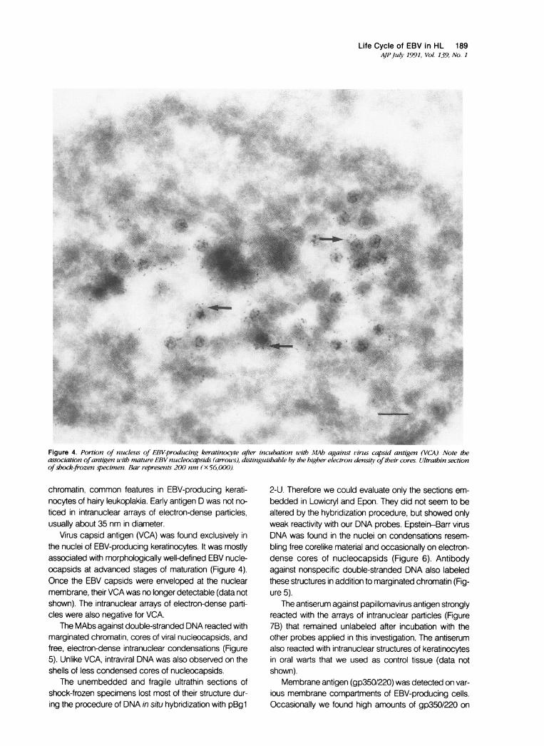
Figure 4. Portion of nucleus of EBV-producing keratinocy'te after incubation with MAb against virus capsid antigen (VCA). Note theassociation ofantigen with mature EBV nzucleocapsids (arrous), distinguisbable by the higher electron densitq, oftheir cores. Ultrathin sectionofshock-frozen specimen. Bar represents 200 nm (x 56,000).
chromatin, common features in EBV-producing kerati-nocytes of hairy leukoplakia. Early antigen D was not no-ticed in intranuclear arrays of electron-dense particles,usually about 35 nm in diameter.
Virus capsid antigen (VCA) was found exclusively inthe nuclei of EBV-producing keratinocytes. It was mostlyassociated with morphologically well-defined EBV nucle-ocapsids at advanced stages of maturation (Figure 4).Once the EBV capsids were enveloped at the nuclearmembrane, their VCA was no longer detectable (data notshown). The intranuclear arrays of electron-dense parti-cles were also negative for VCA.
The MAbs against double-stranded DNA reacted withmarginated chromatin, cores of viral nucleocapsids, andfree, electron-dense intranuclear condensations (Figure5). Unlike VCA, intraviral DNA was also observed on theshells of less condensed cores of nucleocapsids.
The unembedded and fragile ultrathin sections ofshock-frozen specimens lost most of their structure dur-ing the procedure of DNA in situ hybridization with pBgl
2-U. Therefore we could evaluate only the sections em-bedded in Lowicryl and Epon. They did not seem to bealtered by the hybridization procedure, but showed onlyweak reactivity with our DNA probes. Epstein-Barr virusDNA was found in the nuclei on condensations resem-bling free corelike material and occasionally on electron-dense cores of nucleocapsids (Figure 6). Antibodyagainst nonspecific double-stranded DNA also labeledthese structures in addition to marginated chromatin (Fig-ure 5).
The antiserum against papillomavirus antigen stronglyreacted with the arrays of intranuclear particles (Figure7B) that remained unlabeled after incubation with theother probes applied in this investigation. The antiserumalso reacted with intranuclear structures of keratinocytesin oral warts that we used as control tissue (data notshown).
Membrane antigen (gp350/220) was detected on var-ious membrane compartments of EBV-producing cells.Occasionally we found high amounts of gp350/220 on

190 Rabanus et alAJPJuly 1991, Vol. 139, No 1
,^; '.',C:.. ,,,* .' ; . ' .: " :'' ... :' .* >' t.L,'' '* , . 4,; S.s. .... .: Pi bs
¢,.2}erb.S <;#> 2S;;
.X^,t1 ... S.;.>,7Xs
:'o' ;xt'; ..' o-.. .:
:.. [h:*. ....
i.@ _'.;;
* v;:X:
*Up,^;t:,I.
..s::
.gtt.. sS,..v*g..
*5
:.X.i.;
Figure 5. Nucleus of EBV-producing keratinocyte after incubation uitb MAb against double-stranded DNA. 7Te chromatin is intenselylabeled. Note the shell-like gold label on the core surface ofEBV nucleocapsids (arrouws). Ultrathin section of shock-frozen specimen. Barrepresents 100 nm (x 110,000).
the outside of the nuclear membrane, but it seemed to bemore abundant on enveloped EBV capsids that ap-peared in groups in cytoplasmic vacuoles near the cel-lular membrane (Figure 8). Most of the extracellular EBVhad fewer gp350/220-containing membrane projectionson their envelopes than did the cytoplasmic EBV. Gp350/220 could sometimes be detected on the membranes ofthe vacuoles themselves and on the cell membrane. Onrare occasions, small amounts of the antigen were foundin the nuclei near the nuclear membrane.
The peripheral cytoplasmic vesicles that containedgroups of complete EBV particles were heavily and dis-tinctively labeled with WGA (Figure 9A), which distin-guishes them as Golgi-associated secretory vesicles.They released the processed virus by fusing with theplasma membrane (Figure 9B). Envelope and tegumentof peripheral intravesicular and extracellular EBV weremost intensely labeled with WGA.
Discussion
Oral keratinocytes infected with EBV give rise to the onlyknown lesion in which EBV undergoes complete in vivo
replication. Because of its clinical appearance, the lesion,which usually appears at the lateral borders of the tongueof severely immunosuppressed persons, has beennamed oral hairy leukoplakia. Only a few light-micro-scopic investigations have described the distribution ofviral components in hairy leukoplakia.'1,122022 One ad-ditional light-microscopic study described the intracel-lular localization of gp350/220 in the Golgi apparatus.32Those data, however, do not provide direct insight intothe sequence of viral assembly and the intracellular path-way of EBV components. Several investigators describedthe ultrastructural alterations specific for EBV-producingkeratinocytes in hairy leukoplakia.'1 1-19 Yet the composi-tion of most ultrastructural features has remained unclear.In this study, combining immunohistochemistry, lectin-binding histochemistry, and DNA in situ hybridization withelectron microscopy has allowed us to gain direct insightinto the molecular composition of some characteristic ul-trastructural features of hairy leukoplakia and to correlatethem to the distribution and the pathway of EBV constit-uents.
Viral cores and capsids are assembled in the nuclei ofEBV-producing keratinocytes that are found in the upper

Life Cycle of EBV in HL 191AJPJuly 1991, Vol. 139, No. 1
Figure &. Nucleus ofEBV-producing keratinocote afterDNA in situ h¶bridization. EBVDNA is detected onfree corelike material (long arrow)and on electron-dense cores of nucleocapsids (short arrou). Ultrathin sections ofEpon-embedded specimens. For both illustrations the barrepresents 100 nm (x 140,000).
prickle-cell layers of the epithelium. It might be thoughtthat EBV DNA would be replicated in deeper layers andassembly of the translational products would take placein the upper third of the epithelium. We found three EBVantigens early antigen, capsid antigen, and membraneantigen-as well as EBV DNA only in EBV-producingcells of the upper third of the epithelium, providing evi-dence that both replication and assembly are linked toepithelial differentiation.
The plastic-embedded sections prepared for DNA insitu hybridization allowed only very weak labeling, ascompared with the intensity achieved by light-microscop-ic techniques.'22 The weakness was probably due tothe strong cross-linking of already sparse EBV-DNA de-terminants available in ultrathin sections. The poor label-ing obviously does not exclude the occurrence of EBV inlower epithelial layers, although the complete lack of de-tectable EBV DNA in our system suggests a much loweramount in lower cell layers. This indication of an explosiveinitiation of a differentiation-linked EBV replication corre-sponds with previous light-microscopic observa-tions.222 In addition, the labeling was highly specificand appeared mainly on condensations of free core ma-terial and electron-dense cores of nucleocapsids.
The MAb against double-stranded DNA applied onultrathin frozen sections showed the same distribution, inaddition to a strong labeling of marginated chromatin.Although labeling of double-stranded DNA would beweak evidence for EBV DNA at the light-microscopiclevel, the combined observation of the ultrastructurallydefined nucleocapsids and double-stranded DNAstrongly indicates integrated EBV DNA.
Even though components of the early antigen D com-plex usually can be found in both the nuclei and the cy-toplasm of EBV-producing cells, our MAb reacted onlywith intranuclear components, as described previously inRaji cells.33 We found the early antigen p138 rather dif-fusely distributed throughout the nuclei and, unlike theother viral proteins, accumulated along marginated chro-matin and in its punched-out regions. Early antigen in-duces the replication of the EBV genome and the syn-thesis of structural viral proteins.' Hence the accumula-tion of p138 at and in cellular chromatin could indicateintegrated EBV DNA. Epstein-Barr virus DNA has previ-ously been demonstrated to be integrated into the DNAof Namalwa cells35 and may be associated with meta-phase chromosomes.36 It has not yet been shownwhether p138 binds specifically to EBV sequences, how-

192 Rabanus et alAJPJuly 1991, Vol. 139, No. I
Figure 7. Nucleus ofEBV-producing keratinociyte (A) after incubation uith MAb against double-strandedDNA and (B) after incubation withantiserum against common papillomavirus antigen. Note the specific labeling of the arrayled particles u'ith the antiserum against papillo-mavirus antigen, uhereas the clumped chromatin remainsfree of label. Lltrathin sections of shock-frozen specimens. For A and B the barrepresents 100 nm (xIO0,O000).
ever. Hence its peculiar distribution may be attributable tononspecific binding to components of the host cell chro-matin.
Virus capsid antigen was mainly detectable on core-containing nucleocapsids at advanced stages of matu-ration. Only these mature nucleocapsids are translocatedinto the cytoplasm by envelopment at the nuclear mem-brane, as we described recently.37 Envelopment ofherpes simplex virus seems to depend on the completionof DNA packaging and the length of the assembled viralDNA.38 For EBV, we additionally found here that assem-bly of the 150-kd capsid antigen seemed to coincide withthe final stages of DNA packaging. Hence DNA packag-ing seems to be crucial for the assembly of VCA, which inturn seems to be important for the envelopment of thecompleted viral capsids at the nuclear membrane. Thisnotion is supported by the previous observation of empty,thus DNA-free, EBV capsids that accumulated in the nu-clei of superficial keratinocytes.37 Furthermore we foundhere that enveloped capsids were not labeled with theMAb against VCA, indicating that antigenic sites of the150-kd protein were shielded by juxtaposing or perhapseven interacting tegument proteins or the envelope.
Unlike VCA and EA-D, membrane antigen (gp350/
220) was found mostly on extranuclear enveloped EBV,which frequently collected in groups within cytoplasmicvesicles. We were unable to detect gp350/220 on thick-ened and reduplicated nuclear membranes, which occurin herpesvirus-producing cells and are thought to harborvirus-specific proteins.39-40 The fact that the MAb againstgp350/220 most strongly labeled EBV envelopes in pe-ripheral cytoplasmic vesicles suggests that the MAbused are directed toward glycosylated epitopes ofgp350/220. This is not unlikely, because about 50% of thegp350 mass consists of carbohydrates.41 It is also con-sistent with the observations that the glycosylated formsof gp350/220 are stronger immunogens than unglycosy-lated gp350/220 core proteins,42 and that our own MAbsfailed to react with gp350/220 produced in insect cells(data not shown) that glycosylate proteins differently.
Hitherto it has not been clear how EBV is processedand released by its host cells.3-439-40 Our data show, un-ambiguously and for the first time, that EBV follows thesame physiologic pathway as cellular proteins duringtheir final processing and egress. In another study, a 165-kd protein was indirectly shown to be processed andglycosylated to gp350/220 in the Golgi apparatus ofP3HR-1 cells.32 It remained unclear, however, whether

Life Cycle of EBV in HL 193AjP Julv 1991, Vol. 139, No. 1
w.
BFigure 8. Periphery ofEBV-producing keratinocyte after incubation u'ith 4,MAb against membrane antigen (gp350/220). A: Enveloped EBVin the peripheral cytoplasmic vacuole (long arrou) is more intensely labeled than extracellular EBV (short arrows). B: Extracellular EBVis labeled exclusiveely at electron-dense structures projecting out of the viral envelope (long arrow), whereas gp3501220 cannot be detectedon EBV without projections (short arrou). C: K-xtracellular EBV seems to be linked to the plasnia membrane via a gp3501220-positiveprojection of its envelope (arrou'). lltrathin sections ofshock-frozen specimens. For A, B, and C, the bar represents 200 nm (x90,000).
--4
t.
iik; .--...q

194 Rabanus et alAJPJul 1991, Vol 139, Nvo. I
Figure 9. Periphery ofEBV-producing keratinoci'te after incubation with uheat-germ agglutinin (WGA) labeled with ovomucoid-conjugatedgold (10 nm in diameter). WGA binds to N-acetvlglucosamine and stains the Golgi apparatus relativel/v specificallv, but not the endoplasmicreticulum27'36 EBV appears in groups u'ithin peripheral vesicles of the Golgi apparatus (A), uwhich eventually open to the cell surface bymembranefusion to release the processed virus (B). FBV envelope (long arrouws) and tegument (short arrou s) are most intensely labeled.Ultrathin sections ofLowicrml K41-embedded tissue. For A and B, the bar represents 100 nm (X 5-3,000)

Life Cycle of EBV in HL 195AJP July 1991, Vol. 139, No. 1
Figure 10. EBV-producing keratinocyte inoral hairy leukoplakia. (1) Early antigen wasfound in punched-out areas of marginatedchromatin, which also was intensely labeledfor double-stranded DNA. (2) EBVDNA (largeinternal repeat) was detected on condensa-tions resembling free core material. (3) Viruscapsid antigen wasfound on mature nucleo-capsids. Their cores were also labeled withMAbs against dsDNA. (4) After envelopment atthe nuclear membrane, virus capsid antigenwas obscured by the adjacent membrane. (5)Intranuclear arrays of electron-dense parti-cles reacted exclusivelv uith an antiserumagainst papillomavirus antigen. (6) Envel-oped EBV in Golgi-associated vesicles (GAV)reacted with MAb against gp350/220 andwheat-germ agglutinin, u'hicb specifically la-bels the Golgi apparatus and its products by
binding to N-acetylglucosamine. (7) EBV uwasreleased by fusion of GAV and the plasmamembrane. (8) WGA stained extracellularEBV most intensely. ER, endoplasmic reticu-lum; N, nucleus; GA, Golgi apparatus.
the final processing of gp350/220 precursors took placebefore or after their integration into the EBV envelopes. InGolgi-associated secretory vesicles of the keratinocyteswe examined, gp350/220 was preferentially attached toEBV and only occasionally found on the membranes ofthe vesicles themselves, whereas virus-free vacuoles didnot harbor any detached gp350/220. This indicates thatgp350/220 is processed in the Golgi complex of EBV-producing cells after assembly into the viral surface. Ourfinding that tegument and envelope of EBV particles inperipheral Golgi-associated vesicles were heavily la-beled with WGA further supports the idea that EBV getsits final modification in the Golgi apparatus. We also dem-onstrated that completed EBV is released by fusion of theGolgi-associated secretory vesicles with the plasmamembrane. This suggests that egress of EBV dependson physiologic secretory mechanisms of the cell ratherthan on gp350/220 epitopes that regulate trafficking andrelease of EBV.4243
Less gp350/220 was detected on extracellular EBVthat also lacked surface projections; together these ob-servations indicate that membrane antigen is shed fromthe virus rather than blocked by diffusing host antibodies.Antibodies directed toward gp350/220 neutralize infec-tivity of EBV44 and mediate antibody-dependent cytotox-icity of K cells.45 Shed viral surface protein might there-fore bind and deplete protective antibodies againstgp350/220, thus providing a means for EBV to evade thehumoral response of the host.
The selective attachment of the MAb to matured viralstructures, as demonstrated for VCA and gp350/220, in-dicates that the viral antigens are continuously processedafter their putative production at the ribosomes of the en-
doplasmic reticulum. Even though these 'mature' EBVproteins are presumably the most immunogenic, some
MAb may also be specific to less processed determi-nants and attach to different ultrastructural features.
Our results provide, for the first time, direct insight intothe biology of EBV in its naturally permissive cell, the ke-ratinocyte. As summarized in Figure 10, they discloseparts of the intracellular pathway of EBV, its components,and their relation to typical ultrastructural features of hairyleukoplakia. The composition of some common fine-structural appearances, however, remains unclear.We1037 and others19 previously described arrayed het-eromorph and hollow-cored particles in the nuclei of EBV-producing keratinocytes. Similar structures have alsobeen described in cells infected with other herpes-viruseS394649 and have been interpreted as partly ag-
gregated capsids.39 The evidence that these structuresare related to herpesviruses is circumstantial, however,and relies heavily on the observation that structures mor-
phologically indistinguishable from small ringlike compo-nents have been observed in immature nucleocapsids ofherpes simplex virus-1,5i equine herpesvirus-1,51Marek's disease virus, and herpes virus of turkeys.52 Wepreviously described tubular structures in hairy leuko-plakia that were associated with arrays of partially hollow-cored particles.37 Their proximity and compatible calibersuggest that these tubules may consist of linear aggre-
gations of structural components of the granules. Intra-nuclear tubular structures also were found in associationwith intranuclear arrays of other herpesvirus-infectedcells48,49'53'54 and were considered to be a feature ofherpesviruses with oncogenic potential,' which is con-
sistent with the well-known oncogenicity of EBV. In thisstudy, we observed similar tubular fragments that wereinterwoven with the papillomavirus-antigen-positive ar-
rays. None of the other probes we used reacted withthese ultrastructural features.
* *
196 Rabanus et alAJPJuly 1991, Vol. 139, No. I
The heterogenous probes used in combination withimmunoelectron microscopy in this study have given us abetter understanding of the biologic events that occurduring complete EBV replication in the keratinocyte. Be-cause of the lack of ultrastructural alterations and the lowconcentration of viral antigens in latently infected cells,immunoelectron-microscopic analysis may not showmuch beyond that already obtained by light-micro-scopic investigation. The investigation of the ultrastruc-tural distribution of additional late viral proteins and cel-lular antigens, however, may further improve our under-standing of EBV-cell interactions in vivo.
Acknowledgment
The authors thank Evangeline Leash for editing the manuscriptand for helpful discussions.
References
1. Wolf H: Virus-assoziierte Krebserkrankungen des Men-schen. Mnch Med Wochenschr 1986,128:633-638
2. Rickinson AB, Yao QY, Wallace LE: The Epstein-Barr virusas a model of virus-host interactions. Br Med Bull 1985,41:75-79
3. Epstein MA, Barr YM: Cultivation in vitro of human lympho-blasts from Burkitt's malignant lymphoma. Lancet 1964,1:702-703
4. Young LS, Sixbey JW, Clark D, Rickinson AB: Epstein-Barrvirus receptors on human pharyngeal epithelia. Lancet1986, 1:240-242
5. Shapiro IM, Volsky DJ: Infection of normal human epithelialcells by Epstein-Barr virus. Science 1983, 219:1225-1228
6. Becker J, Leser U, Marschall M, Langford A, Jilg W, ReichartP, Gelderblom H, Wolf H: EB-viral expression depends onthe differentiated status of epithelial cells in oral hairy leuko-plakia. In Ablashi DV, ed. Epstein-Barr Virus and Human Dis-ease. Clifton, NJ, Humana Press, 1989
7. Wolf H, Haus M, Wilmes E: Persistence of Epstein-Barr virusin the parotid gland. J Virol 1984, 51:795-798
8. Sixbey JW, Lemon SM, Pagano JS: A second site for Ep-stein-Barr virus shedding: The uterine cervix. Lancet 1986,ii:1 122-1124
9. Birx DL, Redfield RR, Tosato G: Defective regulation of EBVinfection in patients with acquired immunodeficiency syn-drome (AIDS) or AIDS-related disorders. N Engl J Med1986, 314:874-879
10. Greenspan D, Greenspan JS, Conant M, Petersen V, Silver-man S Jr, De Souza Y: Oral "hairy" leucoplakia in male ho-mosexuals: Evidence of association with both papillomavi-rus and a herpes-group virus. Lancet 1984, 13:831-834
11. Greenspan JS, Greenspan D, Lennette ET, Abrams DI, Co-nant MA, Petersen V, Freese K: Replication of Epstein-Barrvirus within the epithelial cells of oral 'hairy' leukoplakia, an
AIDS-associated lesion. N EngI J Med 1985, 313:1564-1571
12. Zhang X, Langford A, Becker J, Rabanus J-P, Pohle HD,Reichart P, Gelderblom H: Ultrastructural and immunohisto-chemical findings in oral hairy leukoplakia. Virchows Arch[A] 1988, 412:533-542
13. Belton CM, Eversole LR: Oral hairy leukoplakia: Ultrastruc-tural features. J Oral Pathol 1986, 15:493-499
14. Kimmig W, Mensing H, Seyfarth G, Janner M, Nasemann T:Orale 'hairy' Leukoplakia-Fruhsymptom bei HTLV-Ill/LAV-lnfektion. Dtsch Med Wochenschr 1986, 111:1394-1397
15. De Maubeuge J, Ledoux M, Feremans W, Zissis G, GoensJ, Andre J, Gourdain JM, Menu R, De Wit S, Cran S,Clumeck N, Achten G: Oral 'hairy' leukoplakia in an AfricanAIDS patient. J Cutan Pathol 1986, 13:235-241
16. Lupton GP, James WD, Redfield RR, Brown C, RodmanOG: Oral hairy leukoplakia. Arch Dermatol 1987, 123:624-628
17. El-Labban N, Rindum J, Nielsen H, Pindborg JJ: Crystallineinclusions in epithelial cells of hairy leukoplakia: A new ultra-structural finding. Scand J Dent Res 1988, 96:353-359
18. Ficarra G, Barone R, Gaglioti D, Milo D, Riccardi R, Romag-noli P, Zorn M: Oral hairy leukoplakia among HIV-positiveintravenous drug abusers: A clinicopathologic and ultra-structural study. Oral Surg Oral Med Oral Pathol 1988,65:421-426
19. Kanas RJ, Abrams AM, Jensen JL, Wuerker RB, HandlersJP: Oral hairy leukoplakia: Ultrastructural observations. OralSurg Oral Med Oral Pathol 1988, 65:333-338
20. De Souza YG, Greenspan D, Felton JR, Hartzog GA, Ham-mer M, Greenspan JS: Localization of Epstein-Barr virusDNA in the epithelial cells of oral hairy leukoplakia by in-situhybridization of tissue sections [Letter]. N Engl J Med 1989,320:1559-1560
21. De Souza YG, Greenspan D, Felton JR, Hartzog GA, Ham-mer M, Greenspan JS: Demonstration of Epstein-Barr virusDNA in the epithelial cells of oral hairy leukoplakia [abstr] JDent Res 1986, 65 (Special Issue):765.
22. Loning T, Heuke P, Reichart P, Becker J: In-situ hybridizationto detect Epstein-Barr virus DNA in oral tissues of HIV-infected patients. Virchows Arch [A] 1987, 412:127-133.
23. Eversole LR, Jacobsen P, Stone CE, Freckleton V: Oralcondyloma planus (hairy leukoplakia) among homosexualmen: A clinicopathological study of thiry-six cases. OralSurg Oral Med Oral Pathol, 1986, 61:249-255
24. Eversole LR, Stone CE, Beckman AM: Detection of EBV andHPV DNA sequences in oral hairy leukoplakia by in-situ hy-bridization. J Med Virol 1988, 26:271-277
25. Gelderblom H, Kocks C, L'age-Stehr J, Reupke H: Compar-ative immunoelectron microscopy with monoclonal antibod-ies on yellow fever virus-infected cells: Pre-embedding la-belling versus immunocryoultramicrotomy. J Virol Methods1985,10:225-239
26. Motz M, Deby G, Wolf H: Truncated versions of the twomajor Epstein-Barr viral glycoproteins (gp250/350) are se-creted by recombinant Chinese hamster ovary cells. Gene1987, 58:149-154

Life Cycle of EBV in HL 197AJPJuly 1991, Vol. 139, No. 1
27. Seibl R, Wolf H: Mapping of Epstein-Barr virus proteins onthe genome by translation of hybrid selected RNA from in-duced P3HR1 cells and induced Raji cells. Virology 1985,141 :1-13
28. Motz M, Fan J, Seibl R, Jilg W, Wolf H: Expression of theEpstein-Barr virus 138-kDa early protein in Escherichia colifor the use as antigen in diagnostic tests. Gene 1986,42:303-312
29. Geoghegan WD, Ackerdam GA: Adsorption of horseradishperoxidase, ovomucoid, and anti-immunoglobulin to colloi-dal gold for the indirect detection of concanavalin A, wheatgerm agglutinin and goat anti-immunoglobulin G on cell sur-faces at the electron microscopical level: A new method,theory and application. J Histochem Cytochem 1977,25:1187-1200
30. Virtanen I, Ekblom P, Laurila P: Subcellular compartmental-ization of saccharide moieties in cultured normal and malig-nant cells. J Cell Biol 1980, 85:429-434
31. Desranges C, Bornkamm GW, Zeng Y: Detection of Epstein-Barr viral DNA internal repeats in the nasopharyngeal mu-cosa of Chinese with IgAlEBV-specific antibodies. Int J Can-cer 1982, 29:87-91
32. Bertoni G, Nguyen QV, Humphreys RE, Sairenji T: Intracel-lular synthesis of Epstein-Barr virus membrane antigengp350/220. Intervirology 1989, 30:61-73
33. Marschall M: Charakterisierung und Kartierung von Epstein-Barr-Virus Proteinen mit Hilfe von Leserahmen-spezifischenAntiseren. Thesis. Munich, Ludwig-Maximilians-UniversitatMunchen, 1987.
34. Miller G. Epstein-Barr virus. In Flelds BN, ed. Virology. NewYork, Raven Press, 1985, pp 563-589
35. Matsuo T, Heller M, Petti L, O'Shiro E, Kieff E: Persistence ofthe entire Epstein-Barr virus genome integrated into humanlymphocyte DNA. Science 1984, 226:1322-1325.
36. Harris A, Young B, Griffin B: Random association of Epstein-Barr virus genomes with host cell metaphase chromosomesin Burkitt's lymphoma-derived cell lines. J Virol 1985,56:328-332
37. Greenspan JS, Rabanus JP, Petersen V, Greenspan D: Finestructure of EBV-infected keratinocytes in oral hairy leuko-plakia. J Oral Pathol Med 1989,18:565-572
38. Vlazny DA, Kwong A, Frenkel N: Site-specific cleavage/packaging of herpes simplex virus DNA and the selectivematuration of nucleocapsids containing full-length DNA.Proc Natl Acad Sci USA 1982, 79:1423-1427
39. Roizman B, Furlong D: The replication of herpesviruses. InFraenkel-Conrat H, Wagner RR, eds. Comprehensive Virol-ogy 3. Reproduction. New York, Plenum Press, 1974, p 346
40. Dargan DJ: The structure and assembly of herpesviruses. In
Harris RH, Horne RW, eds. Electron Microscopy of Proteins,Vol. 5, Viral Structure. London, Academic Press, 1986, p 359
41. Morgan AJ, Smith AR, Barker RN, Epstein MA: A structuralinvestigation of the Epstein-Barr (EB) virus membrane anti-gen glycoprotein, gp350. J Gen Virol 1984, 65:397-404
42. Whang Y, Silberklang M, Morgan A, Munshi S, Lenny AB,Ellis RW, Kieff E: Expression of Epstein-Barr virus gp350/220gene in rodent and primate cells. J Virol 1987, 61:1796-1807
43. Sairenji T, Bertoni G, Medveczky MM, Medveczky PG,Nguyen QV, Humphreys RE: Inhibition of Epstein-Barr virus(EBV) release from P3HR-1 and B95-8 cell lines by mono-clonal antibodies to EBV membrane antigen gp350/220. JVirol 1988, 62:2614-2621
44. Thorley-Lawson DA, Geilinger K: Monoclonal antibodiesagainst the major glycoprotein (gp350/220) of Epstein-Barrvirus neutralize infectivity. Proc Natl Acad Sci USA 1980,77:5307-5311
45. Pearson GR, Orr TW: Antibody-dependent lymphocyte cy-totoxicity against cells expressing Epstein-Barr virus anti-gens. J Natl Cancer Inst 1976, 56:485-488
46. Schaffer PA, Brunschwig JP, McCombs RM, Benyesh-Melnick M: Electron microscopic studies of temperature-sensitive mutants of herpes simplex virus type 1. Virology1974, 62:444 457
47. Cabral GA, Schaffer PA: Electron microscope studies oftemperature-sensitive mutants of herpes simplex virus type2. J Virol 1976, 48:727-737
48. Atkinson MA, Barr S, Timbury MC: The fine structure of cellsinfected with temperature-sensitive mutants of herpes sim-plex virus type 2. J Gen Virol 1978, 40:103-119
49. Stackpole CW: Herpes-type virus of the frog renal adeno-carcinoma: I. Virus development in tumor transplants main-tained at low temperature. J Virol 1969, 4:75-93
50. Nii S: Electron microscopic observations on FL cells infectedwith herpes simplex virus 1. Viral forms. Biken J 1971,14:177-190
51. Perdue ML, Cohen JC, Randall CC, O'Callaghan DJ: Bio-chemical studies of the maturation of Herpesvirus nucleo-capsids species. Virology 1976, 74:194-208
52. Nazerian K, Lee LF, Witter RL, Burmester BR: Ultrastructuralstudies of a herpes virus of turkeys antigenically related toMarek's disease virus. Virology 1971, 43:442-452
53. Cook MK, Sears JF: Preparation of infectious cell-freeherpes-type virus associated with Marek's disease. J Virol1970, 5:258-261
54. Morgan DG, Achong BG, Epstein MA: Unusual intranucleartubular structures associated with the maturation of Herpes-virus saimiri in monkey kidney cell cultures. Br J Cancer1973, 27:434-440