Studying vascularization in fishes using corrosion · PDF fileStudying vascularization in...
8
Studying vascularization in fishes using corrosion casting and microscopy: a Review Mark P.Rogers 1 , Robin L. Sherman 2 and Richard E. Spieler 1 1 Oceanographic Center, Nova Southeastern University, 8000 N. Ocean Dr, Dania Beach, FL 33004, USA 2 Farquahr College of Arts and Sciences, Nova Southeastern University, 3301 College Avenue, Ft Lauderdale, FL 33314, USA The first studies of circulatory anatomy are buried in pre-recorded history. However, the Ebers papyrus, compiled about 1550 BC, already provides a relatively good overview of the circulatory system noting the heart as the center and blood vessels coursing throughout the body. Through the years a variety of substances (i.e., wine, ink, dyes) have been injected into arteries and veins in an effort to trace this circulation. However, it was not until the 1930’s when the use of polymer chemistry came into its own that vascular perfusion with plastics of relatively low viscosity and relatively high resistance to proteolytic chemicals made corrosion casting a method of choice for studying the circulatory system both macro- and microscopically. In essence, corrosion casting consists of injecting a liquid polymer into a blood vessel of an organ or whole animal, allowing it to harden and then corroding away the overlying tissue (typically with a solution of sodium or potassium hydroxide) to produce a 3-D cast of the internal structure of the vessels. Depending on the polymer, these casts can be of the smallest capillaries and with microscopy allow the study of detail to the cellular organelle level. Such studies have produced much of what is currently known about the vertebrate circulatory system anatomy and although micro-CT is beginning to supplant corrosion casting for many studies, corrosion casting remains an active method providing valid results. It is particularly relevant to this volume that corrosion casting is a relatively inexpensive method using readily available, fast-setting material for preparing anatomic molds that are microscopy ready samples. These characteristics provide the opportunity to engage students in small unique research projects. In the process of learning the method of corrosion casting the student is exposed to a host of basic laboratory techniques involving chemistry, animals and animal tissue, anatomy, dissection/surgery, microscopy, etc. Further, because from a comparative standpoint so few species have been examined either as whole specimens or specific organs, the potential for finding something “new” is extremely high. This in turn provides the student a very personal introduction to the excitement of science. We, and our students, have worked extensively with fishes, primarily elasmobranchs. These animals represent the basic blueprint of the vertebrate circulatory system. Here we briefly review the work on corrosion casting of fishes from our laboratories and others. Keywords: corrosion casting; circulatory system; fish 1. Introduction Vascular corrosion casting consists of injecting a liquid polymer into the circulatory system of a whole animal or of an organ, allowing it to harden and then corroding away the overlying tissue (typically with a solution of sodium or potassium hydroxide) to produce a 3-D cast of the internal structure of the vessels. Depending on the polymer these casts can be of the smallest capillaries and, with microscopy, allow the study of detail to the cellular organelle level. Such studies have produced much of what is currently known about the vertebrate circulatory system and although micro-CT is beginning to supplant corrosion casting for many studies, corrosion casting remains an active method providing valid results with more than 250 citations in the first 4 months of 2014 (Google Scholar). The first studies of circulatory anatomy are buried in pre-recorded history. However, the Ebers papyrus, compiled about 1550 BC already provides a relatively good overview of the circulatory system noting the heart as the center and blood vessels coursing throughout the body. Vascular injections may have been initiated as early as the 14th century and by the 1600’s there were multiple anatomists perfusing blood vessels. Through the years a variety of substances (i.e., water, wax, wine, gelatin, ink, dyes) have been injected into arteries and veins in an effort to trace this circulation [1-2]. Although some very elegant preparations were done with India ink, none of these materials were fully successful primarily because of issues with viscosity (infiltrating fine capillaries) and resistance to dissection or maceration of the tissue required to view the vasculature. It was not until the 1930’s, when the use of polymer chemistry came into its own that vascular perfusion with plastics of relatively low viscosity and relatively high resistance to proteolytic/lipolytic chemicals made corrosion casting a method of choice for studying the circulatory system both macro- and microscopically. Much of the early work with acrylic polymers was done by August Schummer [3] with a material he termed PLASTOID and he may be considered the Father of Modern Corrosion Vascular Casting [4]. The microscopic examination of casts was greatly aided by the use of Scanning Electron Microscopy (SEM) in the early 1970’s [5,6]. Microscopy: advances in scientific research and education (A. Méndez-Vilas, Ed.) © FORMATEX 2014 __________________________________________________________________ 95
Transcript of Studying vascularization in fishes using corrosion · PDF fileStudying vascularization in...
Studying vascularization in fishes using corrosion casting and microscopy: a Review
Mark P.Rogers1, Robin L. Sherman2 and Richard E. Spieler1
1 Oceanographic Center, Nova Southeastern University, 8000 N. Ocean Dr, Dania Beach, FL 33004, USA 2 Farquahr College of Arts and Sciences, Nova Southeastern University, 3301 College Avenue, Ft Lauderdale, FL 33314,
USA
The first studies of circulatory anatomy are buried in pre-recorded history. However, the Ebers papyrus, compiled about 1550 BC, already provides a relatively good overview of the circulatory system noting the heart as the center and blood vessels coursing throughout the body. Through the years a variety of substances (i.e., wine, ink, dyes) have been injected into arteries and veins in an effort to trace this circulation. However, it was not until the 1930’s when the use of polymer chemistry came into its own that vascular perfusion with plastics of relatively low viscosity and relatively high resistance to proteolytic chemicals made corrosion casting a method of choice for studying the circulatory system both macro- and microscopically. In essence, corrosion casting consists of injecting a liquid polymer into a blood vessel of an organ or whole animal, allowing it to harden and then corroding away the overlying tissue (typically with a solution of sodium or potassium hydroxide) to produce a 3-D cast of the internal structure of the vessels. Depending on the polymer, these casts can be of the smallest capillaries and with microscopy allow the study of detail to the cellular organelle level. Such studies have produced much of what is currently known about the vertebrate circulatory system anatomy and although micro-CT is beginning to supplant corrosion casting for many studies, corrosion casting remains an active method providing valid results.
It is particularly relevant to this volume that corrosion casting is a relatively inexpensive method using readily available, fast-setting material for preparing anatomic molds that are microscopy ready samples. These characteristics provide the opportunity to engage students in small unique research projects. In the process of learning the method of corrosion casting the student is exposed to a host of basic laboratory techniques involving chemistry, animals and animal tissue, anatomy, dissection/surgery, microscopy, etc. Further, because from a comparative standpoint so few species have been examined either as whole specimens or specific organs, the potential for finding something “new” is extremely high. This in turn provides the student a very personal introduction to the excitement of science.
We, and our students, have worked extensively with fishes, primarily elasmobranchs. These animals represent the basic blueprint of the vertebrate circulatory system. Here we briefly review the work on corrosion casting of fishes from our laboratories and others.
Keywords: corrosion casting; circulatory system; fish
1. Introduction
Vascular corrosion casting consists of injecting a liquid polymer into the circulatory system of a whole animal or of an organ, allowing it to harden and then corroding away the overlying tissue (typically with a solution of sodium or potassium hydroxide) to produce a 3-D cast of the internal structure of the vessels. Depending on the polymer these casts can be of the smallest capillaries and, with microscopy, allow the study of detail to the cellular organelle level. Such studies have produced much of what is currently known about the vertebrate circulatory system and although micro-CT is beginning to supplant corrosion casting for many studies, corrosion casting remains an active method providing valid results with more than 250 citations in the first 4 months of 2014 (Google Scholar). The first studies of circulatory anatomy are buried in pre-recorded history. However, the Ebers papyrus, compiled about 1550 BC already provides a relatively good overview of the circulatory system noting the heart as the center and blood vessels coursing throughout the body. Vascular injections may have been initiated as early as the 14th century and by the 1600’s there were multiple anatomists perfusing blood vessels. Through the years a variety of substances (i.e., water, wax, wine, gelatin, ink, dyes) have been injected into arteries and veins in an effort to trace this circulation [1-2]. Although some very elegant preparations were done with India ink, none of these materials were fully successful primarily because of issues with viscosity (infiltrating fine capillaries) and resistance to dissection or maceration of the tissue required to view the vasculature. It was not until the 1930’s, when the use of polymer chemistry came into its own that vascular perfusion with plastics of relatively low viscosity and relatively high resistance to proteolytic/lipolytic chemicals made corrosion casting a method of choice for studying the circulatory system both macro- and microscopically. Much of the early work with acrylic polymers was done by August Schummer [3] with a material he termed PLASTOID and he may be considered the Father of Modern Corrosion Vascular Casting [4]. The microscopic examination of casts was greatly aided by the use of Scanning Electron Microscopy (SEM) in the early 1970’s [5,6].
Microscopy: advances in scientific research and education (A. Méndez-Vilas, Ed.)
© FORMATEX 2014
__________________________________________________________________
95
A cursory review of the literature indicates possibly 2600+ publications dealing with corrosion casting of vascular systems (Google Scholar). Although there have been castings of respiratory passageways, salt glands, lympatics, bile canniculi, brood-pouch, testicular ducts, plant tissues, etc. [7-10] the vast majority of these publications deal with vascular castings of vertebrate circulatory systems either in whole animal or specific organs. Not surprisingly, most studies deal with mammals but all classes of extant vertebrates from agnatha to elasmobranchs have been examined [7,11-15].
2. Methods
An overview of the casting and SEM methods are provided below. The interested reader is directed to the following references for expanded and more detailed descriptions: [16,17].
2.1 Resins
Schummer’s PLASTOID was too viscous to pass through capillary beds and thus only arterial systems were typically cast [16]. However, many resin types and brands have been marketed for corrosion casting studies since the development and the initial trials with PLASTOID. Early resins had a host of problems. They were often difficult to dilute; had inconsistencies in polymerization times, gave off heat during the curing process, and had differences in viscosity within a batch. In most cases several test subjects had to be sacrificed in order to obtain even partial casts of the target organ and cast failure often was not apparent until after the corrosion process had been completed. As the field of polymeric chemistry advanced, new resins and catalysts were discovered and have since been marketed to the biological and medical communities. One of the major advances in casting technology was the ability to dilute the resin to match the viscosity of natural blood serum while maintaining its ability to fully polymerize and form a stable cured cast. This made it possible to reproduce casts of multiple test specimens and also enabled researchers to make casts of smaller structures, vessels, and even capillaries [18]. In the early 1980’s several researchers set out to define the ideal resin for vascular corrosion casting studies and developed a list of attributes that would fulfill this description. Though none of the resins currently on the market fully meet all of the requirements, the list is ideal for comparing each of them in this review. The accepted criterion are: 1) be nontoxic for the investigator and for the system to be cast, 2) be of sufficiently low viscosity and particle size to pass through the smallest blood vessels (5µm), 3) be physiologically inert in the system to be cast, 4) polymerize within an adjustable period of time (3-15 minutes), 5) not shrink during polymerization, 6) enable dissection and micro-dissection leaving surrounding tissue intact, 7) resist corrosion (maceration) without dramatic damage to cast surface structures, 8) be visible in the dissecting microscope after corrosion (maceration), 9) not change spatial configuration during drying processes, 10) be electron conductive, 11) resist the electron bombardment during scanning electron microscopic (SEM) inspection, 12) replicate all topographical details of cast endothelial luminal surfaces, and 13) indicate the direction of blood flow in the cast system [16]. To date no resin is able to polymerize without shrinkage, conduct electrons, or indicate the direction of blood flow within the system. Many resins have been marketed for use in corrosion casting studies and though none of them meet the criteria of a perfect resin, they have qualities that can benefit anatomical investigations and also have limitations that have to be considered. Rogers [18] did a study examining Batsons #17, Mercox CL-2B, PU4ii, Microfil, and Mercox II. He found the acrylic resins Batsons #17 and Mercox CL-2B had high viscosities that required dilution prior to their use. This can result in inconsistencies among batches including changes in polymerization potential and shrinkage after curing. The polyurethane resin PU4ii had a low viscosity, excellent infiltration potential, and consistently low shrinkage, but is highly toxic to the investigator and target system. However, the high strength and elasticity of casts made with PU4ii are favorable in situations where manipulation of the cast system is necessary [19]. The Microfil resin was able to reproduce large vessels but had limited results filling capillaries. Chemical interactions with the rubber portion of the perfusion syringes could have had an effect on this but nonetheless detracted from its ease of use and ability to reliably produce complete casts. Mercox II had superior surface replication quality and infiltration potential among the acrylate resins making it ideal for general vascular corrosion casting and imaging studies. The resin’s low viscosity and its consistently low, easily calculated, shrinkage further its value in studies involving vascular measurements [20,21]. The resin also has a long shelf life and can be stored for months without noticeable difference in polymerization potential or replication quality. Rogers [18] concluded that Mercox II resin had traits that make it the best currently available resin for general use in vascular corrosion casting studies of fish vasculature. The remainder of this paper concentrates on the use of Mercox II for the formation of vascular casts.
2.2 Precasting Treatment
When using live organisms or tissues a lethal dose of anesthetic is administered to prevent suffering and reduce the likelihood of movement during the initial perfusion process that could compromise the quality of the final cast. An injection site for the perfusates is selected that can be exposed sufficiently to allow clear viewing throughout the perfusion process [22]. A cannula is then inserted into the target vessel and sutured into place to restrict movement and
Microscopy: advances in scientific research and education (A. Méndez-Vilas, Ed.)
© FORMATEX 2014
__________________________________________________________________
96
slippage during perfusion. Placement of the cannula can be aided by a micromanipulator when dealing with small vessels. When dealing with a whole organism a second opening is made downstream from the cannula and the target organ to allow drainage of the excess perfusate. It is recommended that the blood be completely removed from the entire vascular bed before resin is injected so that every detail of the vessel walls including endothelial cell imprints and valve structures can be cast [16,23-25]. Rinsing of blood from the system is done with a saline solution within the physiological range of the animal either with a syringe and hand pressure or with the aid of a perfusion / syringe pump [16,25]. Typically the rinsing solution is mixed with an anticoagulant (heparin). In cases where handling or transport times are required prior to casting (i.e. an organism has recently died and it, or its tissues, have unexpectedly become available), an anticoagulant and a fixative can be administered to the tissue or directly into the bloodstream [16,24].
3. Casting, Resin Preparation and Perfusion
Once the blood has been removed from the system and outflow of perfusate shows no sign of coloration, the resin can be prepared by adding the catalyst to the monomer to initiate polymerization. The mixture is drawn into a second syringe and introduced into the perfusion apparatus taking care to remove any bubbles from the system prior to beginning perfusion of the resin as these can block and or rupture small capillaries. Injection pressure should be the same as during the rinsing process and should continue until polymerization progresses and the resin starts to solidify [25]. At this point the resin is still subject to trauma within the vascular system and it is recommended that the entire setup remain untouched for no less than 30 minutes. The specimen is then placed in a heated water bath for 12-24 hours until polymerization is complete and to temper the solidified resin [16,26]. Following casting, the overlying tissues are macerated, typically in a strong basic solution (i.e. ≈10% KOH or Na OH) or acid. Depending on the amount of overlying tissue this can be time consuming and may require several changes of the macerating liquid. After maceration the casts are washed in distilled water and placed in a 5% formic acid solution for 15-20 minutes then washed again in distilled water. The resulting Mercox casts, cleaned of all tissue, need to be dried and this can be accomplished by air drying or freeze drying. Freeze drying is not required in all cases but prevents surface tension of adhering water deforming or breaking extremely delicate capillary casts.
4. Microscopy
Traditional light microscopy can be used to examine basic cast structures such as supply and drainage vessels and to verify that there was complete filling of the vasculature [2,23]. Manipulation and trimming of cast structures is often required and can be done using a stereo microscope with a large field of view [23]. Several major drawbacks to using light microscopy arise from the fact that most acrylic resins are partially or entirely transparent and examining surface structures of the casted vessels is not possible. They also have a very limited depth of field especially at high magnifications making it difficult to observe or image the three-dimensional angioarchitecture of the casted vasculature [2]. Since its introduction in the 1930‘s the scanning electron microscope (SEM) has become the most widely used tool to examine and image vascular corrosion casts [2,19,27,28]. Several steps are needed to prepare vascular casts for viewing and imaging using the SEM. It is common practice to clean casts in a weak acid solution (typically 5% formic acid) to remove any debris introduced during storage and to remove any residual material left on the cast surface from the corrosion process [22,25]. After specific study areas are identified, most vascular casts are small enough to fit on commercially available aluminum SEM stubs using double sided carbon tape [2, 23,29]. Large casts or ones requiring removal and remounting can be mounted on copper foil sheets that are cut to the size required to receive the cast [23,25,30]. Droplets of colloidal silver and fine copper wires referred to as conductive bridges can be used to attach the cast to the foil sheet and should be applied to areas that will not interfere with the study sites [23,25,30]. Conductive bridges serve as anchor points to hold the cast down and also aid in grounding the cast during SEM imaging thereby preventing charging [30]. As stated previously, none of the currently available resins are able to conduct electrons [23]. Sputter coating is required to prepare the cast surface for interaction with the electron beam so that a sufficient return required for imaging is achieved [23,25,30]. The most widely used materials are Chromium, Gold, Palladium, Palladium and gold, and Carbon and gold of a thickness ranging from 2-20 nm depending on the surface detail required for the study [25,31]. Due to the heat generated in most sputter coating applications it is recommended that for delicate casts or ones requiring thick coatings, several applications lasting no longer than 30 seconds followed by a cooling period be done until the required thickness is achieved [32]. An acceleration voltage of 5-15 kv is recommended and provides good surface detail at most magnifications while minimizing the risk of thermal damage to the cast surface [22,31,32]. There are several common problems encountered when using the SEM to image vascular corrosion casts. Specimen charging is an imaging artifact caused by primary and secondary electrons transferring a negative charge under the cast surface [30,33]. The added electron return received by the microscope causes a reduction in the observed surface detail and results in hot spots on the final image [33]. Charging is common with large casts and ones that lack sufficient
Microscopy: advances in scientific research and education (A. Méndez-Vilas, Ed.)
© FORMATEX 2014
__________________________________________________________________
97
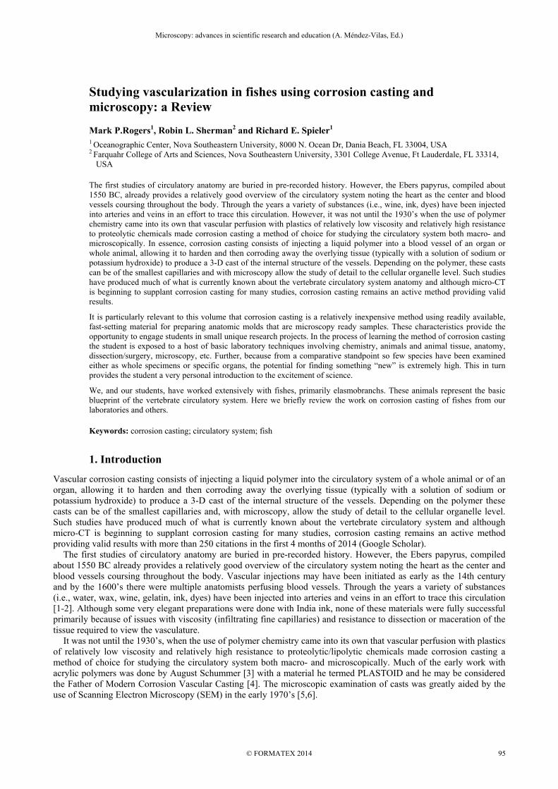
grounding to their stub [30,33]. To reduce the likelihood of specimen charging Lametschwandtner [30] recommends adding conductive bridges to areas of minor interest on the cast surface [25,30] (Fig 1). Another problem that we encountered when using the SEM to image acrylic resin casts was off gassing of the resin within the SEM chamber. On several occasions off gassing resulted in the failure of the microscope filament. To reduce the likelihood of this failure stubs can be kept under vacuum for several hours following the last sputter coating application. Additional sputter coating is also recommended if casts were in storage following their last use.
Fig. 1 Example of fish gills prepared for SEM with conductive bridges attached.
5. Corrosion Casting of Fish Vasculature
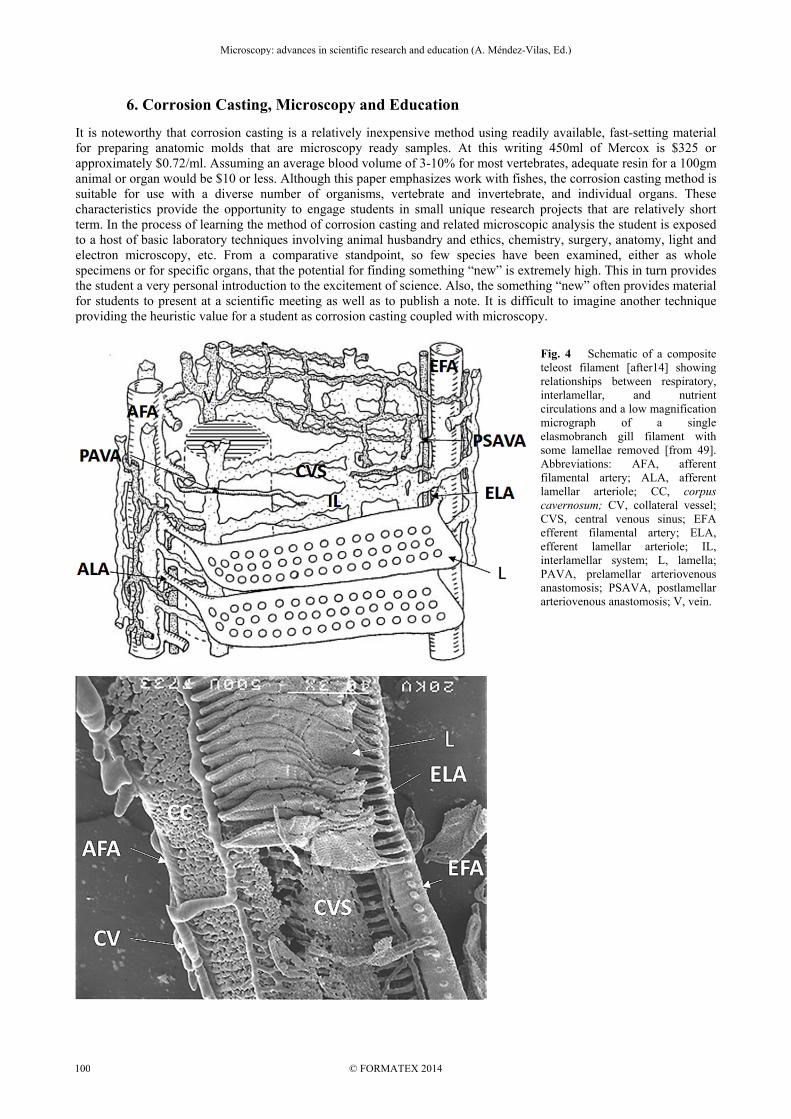
Since the initial corrosion casts of vasculature about 300 studies have examined fishes. This is not surprising as these animals represent the basic blueprint of the vertebrate circulatory system. Circulation in most fishes consists of a single circuit where deoxygenated blood flows from the heart to the gills where it is oxygenated and with a few exceptions (e.g., brain, heart) is then distributed to the rest of the body via the dorsal aorta and secondary arteries and arterioles. Deoxygenated blood returns primarily via the anterior and posterior cardinal veins, the hepatic portal vein, and cutaneous veins which empty into the Ductus Cuvier which, in turn, returns the blood to the sinus venosus of the heart. There are of course substantive exceptions to this plan and gill vascular morphology in air-breathing fishes [34]. The histological structure of the vessels is similar to higher vertebrates [35]. At least 25 species of bony fishes have been studied [14,34] and these studies have concentrated on multiple different organs e.g., brain, eye, heart, swim bladder, kidney (Figure 2)[18,36-39]. However, since early on in the history of corrosion casting and SEM [40] most of the work on fishes has concentrated on the gills and for good reason. “The fish gill is the most physiologically diversified vertebrate organ and its vasculature the most intricate” [14]. Olson [14] has reviewed the gill vasculature of fishes and in the process discussed many of the corrosion cast studies. There are invariable minor morphological differences among species [14,41]. However basically, in bony fishes gill vasculature there are four holobranch gills in a single gill cavity on each side of the animal (Fig. 3) perfused by an arterioarterial pathway and a arteriovenous pathway. The arterioarterial pathway can be considered the primary respiratory pathway which provides oxygenated blood to the trunk and most of the organs of the body. Compared to the arteriovenous system it is relatively simple and un-controversial. Reduced to the basic structures, blood from the heart is carried through the ventral aorta and distributed to the gills through afferent brachial arteries which branch at the level of the gill filament to afferent filamental arteries. The afferent filamental arteries provide the blood to lamella via afferent lamellar arteries. Oxygenation occurs in the lamellae. Blood leaves the lamellae through efferent lamellar arteries then efferent filamental arteries then to the efferent branchial arteries which primarily anastomose with the dorsal aorta. In contrast to the arterioarterial pathway the arteriovenous structures of the gills can be complex and much of the morphology and function of this system(s) is still debated. Olson [14] has broken the arteriovenous pathway into two separate systems: a nutrient system and an interlamellar system; and this appears to be the currently accepted approach [42]. Most of the arterioarterial pathway consists of a series of vessels in a ladder-like structure between the afferent
Microscopy: advances in scientific research and education (A. Méndez-Vilas, Ed.)
© FORMATEX 2014
__________________________________________________________________
98
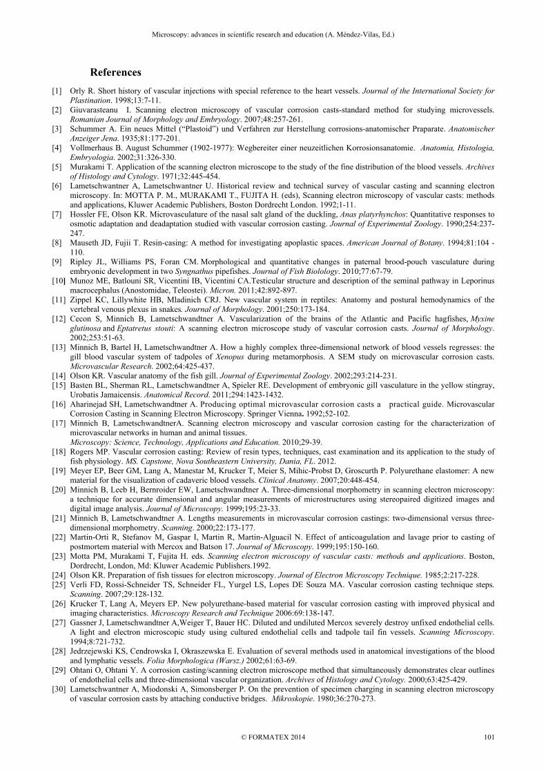
and efferent filamental arteries. The “rung” structures are the interlamellar vessels and the “rails” are the collateral vessels alongside the afferent and efferent filamental arteries. The two systems are apparently linked by small anastomoses. It is primarily a post-lamellar system arising typically from the efferent filamental arteries and the efferent branchial artery, but in some fishes arise from afferent lamellar arteries as well [14]. They drain into the brachial veins at the base of the filament (Fig. 4). The vessels appear to be involved in providing oxygen and nutrients to gill structures but have also been hypothesized to play a role in blood storage, lamellar stability and lymphatic function [14,43]. There has been decidedly less work on elasmobranchs than bony fishes. Nonetheless a number of species and various organs have been examined by corrosion casting e.g., heart, spiral valve, kidney, and rectal gland [37,44-46]. However, like the bony fishes most of the work has been on gill vasculature. At least eight species have been examined [15] and again there are morphological differences among species [47]. Although the general vasculature layout is similar to bony fishes there are distinct differences. The gills are located in pouches, typically five, on either side and the holobranch is split between two pouches (Fig. 3). In the arterioarterial pathway of elasmobranchs there is the inclusion of a corpus cavernosum between the afferent filamental artery and the afferent lammelar artery (Fig. 4) and in the arteriovenous pathway there is an extension of the interlammelar vessels around the septal channel to link with intralammelar vessels of adjacent filaments (not shown). There are a host of questions that remain unanswered regarding the morphology and function of fish gill vascularization and even less is known about other organs. There are an estimated 30,000+ species of fishes (fishbase.org) and they have morphological and physiological adaptations for an extremely wide range of aquatic habitats. Clearly the work with corrosion casting has only scratched the surface of fully understanding the circulatory system in these animals.
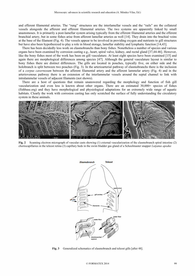
Fig. 2 Scanning electron micrograph of vascular casts showing (1) external vascularizarion of the elasmobranch spiral intestine (2) choriocapillaries in the teleost retina (3) capillary beds in the swim bladder gas gland of a Schoolmaster snapper Lutjanus apodus
Fig. 3 Generalized schematics of elasmobranch and teleost gills [after 48].
Microscopy: advances in scientific research and education (A. Méndez-Vilas, Ed.)
© FORMATEX 2014
__________________________________________________________________
99
6. Corrosion Casting, Microscopy and Education
It is noteworthy that corrosion casting is a relatively inexpensive method using readily available, fast-setting material for preparing anatomic molds that are microscopy ready samples. At this writing 450ml of Mercox is $325 or approximately $0.72/ml. Assuming an average blood volume of 3-10% for most vertebrates, adequate resin for a 100gm animal or organ would be $10 or less. Although this paper emphasizes work with fishes, the corrosion casting method is suitable for use with a diverse number of organisms, vertebrate and invertebrate, and individual organs. These characteristics provide the opportunity to engage students in small unique research projects that are relatively short term. In the process of learning the method of corrosion casting and related microscopic analysis the student is exposed to a host of basic laboratory techniques involving animal husbandry and ethics, chemistry, surgery, anatomy, light and electron microscopy, etc. From a comparative standpoint, so few species have been examined, either as whole specimens or for specific organs, that the potential for finding something “new” is extremely high. This in turn provides the student a very personal introduction to the excitement of science. Also, the something “new” often provides material for students to present at a scientific meeting as well as to publish a note. It is difficult to imagine another technique providing the heuristic value for a student as corrosion casting coupled with microscopy.
Fig. 4 Schematic of a composite teleost filament [after14] showing relationships between respiratory, interlamellar, and nutrient circulations and a low magnification micrograph of a single elasmobranch gill filament with some lamellae removed [from 49]. Abbreviations: AFA, afferent filamental artery; ALA, afferent lamellar arteriole; CC, corpus cavernosum; CV, collateral vessel; CVS, central venous sinus; EFA efferent filamental artery; ELA, efferent lamellar arteriole; IL, interlamellar system; L, lamella; PAVA, prelamellar arteriovenous anastomosis; PSAVA, postlamellar arteriovenous anastomosis; V, vein.
Microscopy: advances in scientific research and education (A. Méndez-Vilas, Ed.)
© FORMATEX 2014
__________________________________________________________________
100
References [1] Orly R. Short history of vascular injections with special reference to the heart vessels. Journal of the International Society for
Plastination. 1998;13:7-11. [2] Giuvarasteanu I. Scanning electron microscopy of vascular corrosion casts-standard method for studying microvessels.
Romanian Journal of Morphology and Embryology. 2007;48:257-261. [3] Schummer A. Ein neues Mittel (“Plastoid”) und Verfahren zur Herstellung corrosions-anatomischer Praparate. Anatomischer
Anzeiger Jena. 1935;81:177-201. [4] Vollmerhaus B. August Schummer (1902-1977): Wegbereiter einer neuzeitlichen Korrosionsanatomie. Anatomia, Histologia,
Embryologia. 2002;31:326-330. [5] Murakami T. Application of the scanning electron microscope to the study of the fine distribution of the blood vessels. Archives
of Histology and Cytology. 1971;32:445-454. [6] Lametschwantner A, Lametschwantner U. Historical review and technical survey of vascular casting and scanning electron
microscopy. In: MOTTA P. M., MURAKAMI T., FUJITA H. (eds), Scanning electron microscopy of vascular casts: methods and applications, Kluwer Academic Publishers, Boston Dordrecht London. 1992;1-11.
[7] Hossler FE, Olson KR. Microvasculature of the nasal salt gland of the duckling, Anas platyrhynchos: Quantitative responses to osmotic adaptation and deadaptation studied with vascular corrosion casting. Journal of Experimental Zoology. 1990;254:237-247.
[8] Mauseth JD, Fujii T. Resin-casing: A method for investigating apoplastic spaces. American Journal of Botany. 1994;81:104 -110.
[9] Ripley JL, Williams PS, Foran CM. Morphological and quantitative changes in paternal brood-pouch vasculature during embryonic development in two Syngnathus pipefishes. Journal of Fish Biolology. 2010;77:67-79.
[10] Munoz ME, Batlouni SR, Vicentini IB, Vicentini CA.Testicular structure and description of the seminal pathway in Leporinus macrocephalus (Anostomidae, Teleostei). Micron. 2011;42:892-897.
[11] Zippel KC, Lillywhite HB, Mladinich CRJ. New vascular system in reptiles: Anatomy and postural hemodynamics of the vertebral venous plexus in snakes. Journal of Morphology. 2001;250:173-184.
[12] Cecon S, Minnich B, Lametschwandtner A. Vascularization of the brains of the Atlantic and Pacific hagfishes, Myxine glutinosa and Eptatretus stouti: A scanning electron microscope study of vascular corrosion casts. Journal of Morphology. 2002;253:51-63.
[13] Minnich B, Bartel H, Lametschwandtner A. How a highly complex three-dimensional network of blood vessels regresses: the gill blood vascular system of tadpoles of Xenopus during metamorphosis. A SEM study on microvascular corrosion casts. Microvascular Research. 2002;64:425-437.
[14] Olson KR. Vascular anatomy of the fish gill. Journal of Experimental Zoology. 2002;293:214-231. [15] Basten BL, Sherman RL, Lametschwandtner A, Spieler RE. Development of embryonic gill vasculature in the yellow stingray,
Urobatis Jamaicensis. Anatomical Record. 2011;294:1423-1432. [16] Aharinejad SH, Lametschwandtner A. Producing optimal microvascular corrosion casts a practical guide. Microvascular
Corrosion Casting in Scanning Electron Microscopy. Springer Vienna. 1992;52-102. [17] Minnich B, LametschwandtnerA. Scanning electron microscopy and vascular corrosion casting for the characterization of
microvascular networks in human and animal tissues. Microscopy: Science, Technology, Applications and Education. 2010;29-39.
[18] Rogers MP. Vascular corrosion casting: Review of resin types, techniques, cast examination and its application to the study of fish physiology. MS. Capstone, Nova Southeastern University, Dania, FL. 2012.
[19] Meyer EP, Beer GM, Lang A, Manestar M, Krucker T, Meier S, Mihic-Probst D, Groscurth P. Polyurethane elastomer: A new material for the visualization of cadaveric blood vessels. Clinical Anatomy. 2007;20:448-454.
[20] Minnich B, Leeb H, Bernroider EW, Lametschwandtner A. Three-dimensional morphometry in scanning electron microscopy: a technique for accurate dimensional and angular measurements of microstructures using stereopaired digitized images and digital image analysis. Journal of Microscopy. 1999;195:23-33.
[21] Minnich B, Lametschwandtner A. Lengths measurements in microvascular corrosion castings: two-dimensional versus three-dimensional morphometry. Scanning. 2000;22:173-177.
[22] Martin-Orti R, Stefanov M, Gaspar I, Martin R, Martin-Alguacil N. Effect of anticoagulation and lavage prior to casting of postmortem material with Mercox and Batson 17. Journal of Microscopy. 1999;195:150-160.
[23] Motta PM, Murakami T, Fujita H. eds. Scanning electron microscopy of vascular casts: methods and applications. Boston, Dordrecht, London, Md: Kluwer Academic Publishers.1992.
[24] Olson KR. Preparation of fish tissues for electron microscopy. Journal of Electron Microscopy Technique. 1985;2:217-228. [25] Verli FD, Rossi-Schneider TS, Schneider FL, Yurgel LS, Lopes DE Souza MA. Vascular corrosion casting technique steps.
Scanning. 2007;29:128-132. [26] Krucker T, Lang A, Meyers EP. New polyurethane-based material for vascular corrosion casting with improved physical and
imaging characteristics. Microscopy Research and Technique 2006:69:138-147. [27] Gassner J, Lametschwandtner A,Weiger T, Bauer HC. Diluted and undiluted Mercox severely destroy unfixed endothelial cells.
A light and electron microscopic study using cultured endothelial cells and tadpole tail fin vessels. Scanning Microscopy. 1994;8:721-732.
[28] Jedrzejewski KS, Cendrowska I, Okraszewska E. Evaluation of several methods used in anatomical investigations of the blood and lymphatic vessels. Folia Morphologica (Warsz.) 2002;61:63-69.
[29] Ohtani O, Ohtani Y. A corrosion casting/scanning electron microscope method that simultaneously demonstrates clear outlines of endothelial cells and three-dimensional vascular organization. Archives of Histology and Cytology. 2000;63:425-429.
[30] Lametschwantner A, Miodonski A, Simonsberger P. On the prevention of specimen charging in scanning electron microscopy of vascular corrosion casts by attaching conductive bridges. Mikroskopie. 1980;36:270-273.
Microscopy: advances in scientific research and education (A. Méndez-Vilas, Ed.)
© FORMATEX 2014
__________________________________________________________________
101
[31] Risco JM, Nopanitaya W. Ocular microcirculation. Scanning electron microscopic study. Investigative Ophthalmology and Visual Science. 1980;19:5-12.
[32] Hodde KC, Nowell JA. SEM of micro-corrosion casts. Scanning electron microscopy. 1980;2:89-106. [33] Joy DC, Joy CS. Dynamic charging in the low voltage SEM. Microscopy and Microanalysis. 1995;3:109-112. [34] Ishimatsu A. Evolution of the cardiorespiratory system in air-breathing fishes. Aqua-BioScience Monographs. 2012;5:1-28. [35] Satchell GH. Physiology and Form of Fish Circulation. Cambridge University Press. 1991. [36] Anderson BG, Anderson WD. Renal vasculature of the trout demonstrated by scanning electron microscopy, compared with
canine glomerular vessels. American Journal of Anatomy. 1976;145:443-457. [37] Tota B, Cimini V, Salvatore G, Zummo G. Comparative study of the arterial and lacunary systems of the ventricular
myocardium of elasmobranch and teleost fishes. American Journal of Anatomy. 1983;167:15-32. [38] Moser B, Lametschwandtner A. Casting the brain vasculature of fishes. European Journal of Cellular Biology. 1991;55:20. [39] Ota D, Lahnsteiner F. Retinal vascularization in the grass goby, Zosterisessor ophiocephalus: a scanning electron-microscopic
study of vascular corrosion casts. Environmental biology of fishes.1996;45:319-324. [40] Gannon BJ, Campbell G, Randall DJ. Scanning electron microscopy of vascular casts for the study of vessel connections in a
complex vascular bed-the trout gill. Proceedings from the Annual Meeting of the Electron Microscopy Society of America. 1973;31:442-443.
[41] Laurent P, Dunel S, Barthe JC. Functional Organization of the Teleost Gill. Acta Zoologica. 1976; 57:189-209. [42] Wegner NC, Sepulveda CA, Bull KB, Graham JB. Gill morphometrics in relation to gas transfer and ram ventilation in high-
energy demand teleosts: Scombrids and billfishes. Journal of Morphology. 2010;271:36-49. [43] Evans DH, Piermarini PM, Choe KP.The multifunctional fish gill: Dominant site of gas exchange, osmoregulation, acid-base
regulation, and excretion of nitrogenous waste. Physiological Reviews. 2005;85: 97-177. [44] Maroni K, Spieler R, Sherman R. A Preliminary Comparison of the Vasculature of the Spiral Valve in the Yellow Stingray,
Urobatis jamaicensis, and the North American Paddlefish, Polyodon spathula. Microscopy and Microanalysis. 2009;15:106-107.
[45] Kent B, Olson KR. Blood flow in the rectal gland of Squalus acanthias. American Journal of Physiology. 1982;243:296-303. [46] Hentschel H. Renal blood vascular system in the elasmobranch, Raja erinacea mitchill, in relation to kidney zones. American
Journal of Anatomy. 1988;183:130-147. [47] Sherman RL, Lametschwandtner A, Spieler RE. Structural variation in gill vasculature among some batoid elasmobranchs
examined using corrosion casting and SEM. Microscopy and Microanalysis. 2005;11:1218-1219. [48] Wilson JM. Laurent P. Fish gill morphology: inside out. Journal of Experimnetal Zoology. 2002;293:192-213. [49] Sherman RL, Spieler RE. Examination of gill vasculature of yellow stingray, Urolophus jamaicensis (Urolophidae), by SEM
observations of resin casts. Italian Journal of Zoology. 1998;65:431-434.
Microscopy: advances in scientific research and education (A. Méndez-Vilas, Ed.)
© FORMATEX 2014
__________________________________________________________________
102