STUDY OF DISEASES OF SUGARCANE - jnkvv.orgjnkvv.org/PDF/09042020210944Diseases_OF_Sugarcane.pdf ·...
46
. Dr. S.P.Mishra Professor Dept. of Plant Pathology College of Agriculture, Rewa STUDY OF DISEASES OF SUGARCANE
Transcript of STUDY OF DISEASES OF SUGARCANE - jnkvv.orgjnkvv.org/PDF/09042020210944Diseases_OF_Sugarcane.pdf ·...
.
Dr. S.P.Mishra Professor
Dept. of Plant Pathology College of Agriculture,
Rewa
STUDY OF DISEASES OF
SUGARCANE


RED ROT OF SUGARCANE

1.The disease RED ROT firstly notic by Went in year 1893.
2.During 1939-1942it caused heavy losses in Bihar and U.P.
3.Butler and his associates in India investigated the disease in great detail during 1914-1918.

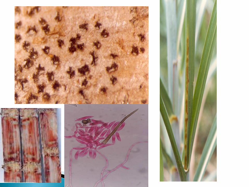
Red spots on the midrib
of the upper leaf surface
develop pale yellow to
white centres, merging
to cover the length of
the leaf. Similar spots
also occur on the leaf
blades.

1.The hyphae are inter and intracellular, thin, septet, hyaline in the beginning turning to dark at maturity.
2.The hyphae produce large number of Chalmydospores in the pith. This can survive in soil for a long time.
3.Thick-walled hyphae, occasionally from dark-green or brown stroma, which develops conidiophores at the epidermis .
4.Long hair like rigid setae,100-200um long develop around in the stroma.
5.This is the developing acervulus.

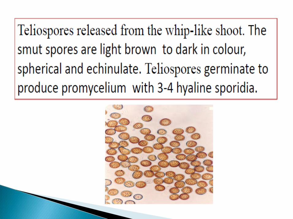
Microscopic view of a single spore mass shows the crescent (the curved
sickle shape of the waxing or waning moon.) moon-shaped spores which
spread to other plants to cause new infections.

6.These acervuli have conidiospores which are small,aseptate,20X8 um.
8.From the vegetative mycelium, conidia produced.
9.Conidia are hyaline, unicellular, falcate or sickle-shaped and some times fusoid (Like macro conidia of Fusarium) measuring 16-48X4-8um with oil globule.


Symptoms
Symptoms 1.first symptom of red rot in the field is
discoloration of the young leaves.
2.The tissues are reddened throughout
the basal portion, especially the vascular
bundles, which are intensely red.
3.In the infected plants the leaves show
symptoms in the form of dark red lesions
in the midrib, which elongate, turning
Blood-red With dark margins and later
on with straw- colored centers.

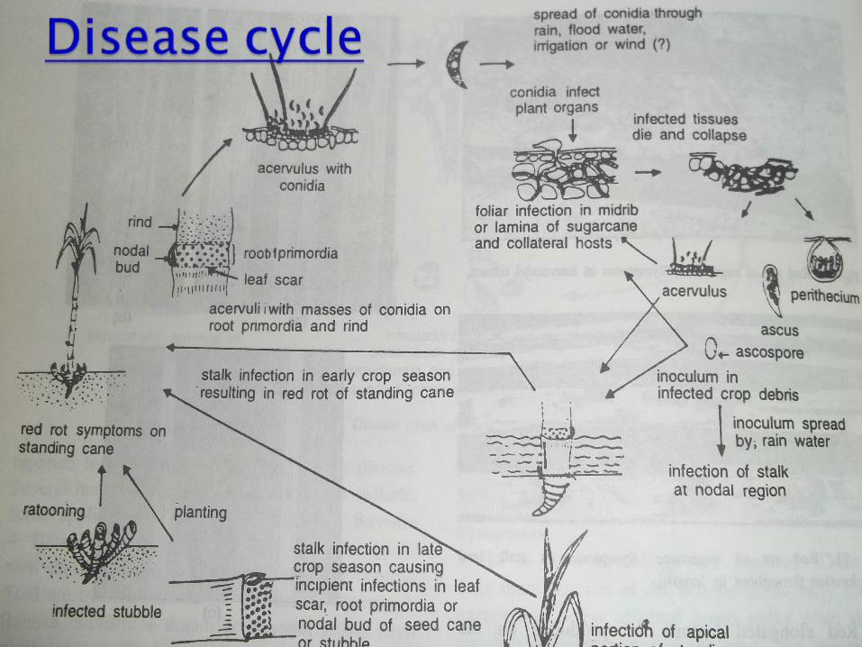
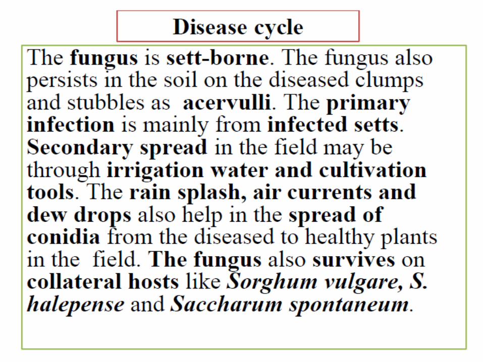
Transmission
Primary transmission through soil
and diseased sets, while the
secondary transmission through
air, rain splash and soil.

The fungus also persists in the soil on diseased clumps and dry leaves left in the field after harvest.
If the conidia settle on the leaves they germinate.
Stem infection takes place through insect bores and root primordial.
The soil-borne fungus also enters the healthy sets through cut-ends, and causes early infection in the shoots.
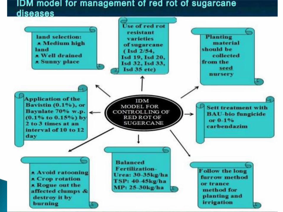

MANAGEMENT
1.The cut-ends, and preferably the entire sett,
should be dipped in a fungicide solution, such as
one per cent Bordeaux mixture, to protect it from
invasion by soil-borne pathogen
. 2.Dipping setts in fungitoxic chemicals like
Bavistan, Benomyl, and Aretan at 0.1 per cent
for 18 min. at 520C gave almost complete
elimination of rot infection.
By the growing of resistant varieties

Co.6806
Co.62198
Co.776
Co.975
Co.8014
Here Co. denotes Coimbatore


Pre disposing (environmental) factors
Mono-culturing of sugarcane
Continuous ratooning
Dry weather during tillering stage
favours the disease

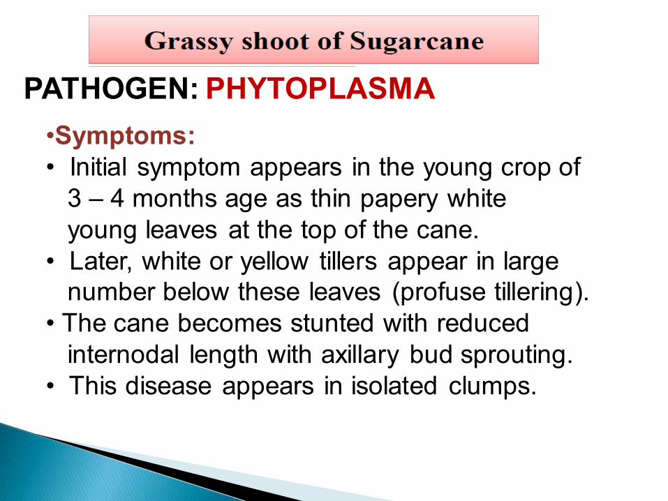
PATHOGEN: PHYTOPLASMA
•Symptoms:
• Initial symptom appears in the young crop of
3 – 4 months age as thin papery white
young leaves at the top of the cane.
• Later, white or yellow tillers appear in large
number below these leaves (profuse tillering).
• The cane becomes stunted with reduced
internodal length with axillary bud sprouting.
• This disease appears in isolated clumps.

Grassy shoot disease


Cultural method:
Growing resistant varieties viz., Co 86249, CoG 93076 and CoC 22
Avoid ratooning if Grassy Shoot Disease incidence is more than 15 % in
the plant crop
If disease symptoms are visible within two weeks after planting, such
plants can be replaced by healthy plants.
Uprooted infected plants need to disposed of by burning them.
Physical method:
Rogue out infected plants in the secondary and commercial seed nursery.
Treat the setts with aerated steam at 50°C for 1 hour to control primary
infection.
Treating them with hot air at 540C for 8 hours and spraying twice a month
with aphidicides.
Chemical method
Spray dimethoate @ 1ml in 1 litre of water to control insect vector
Apply pesticide methyl-demeton @ 2ml/lit of water for controlling aphids.


Causal Organism: Cephalosporium sacchari Class: Deuteromycetes
Order:Moniliales
Family:Moniliacease
1. This is one of the early known diseases of sugarcane in India.
2. It was first reported by Butler and Khan in 1913, from North India.
3. It has been reported to cause severe damage to sugarcane crops in many parts of India. During 1965-1967 it caused severe damage to sugarcane crop in the Deccan plateau

This is an imported disease of
sugarcane and common in Vishakapatnam
and Nizamabad , MP districts and Shrilanka.
The disease occurs singly or in
combination with red rot.
The disease is more is wilt sick soils and in
alkaline soils.
Moisture stress aggravates the disease.

The first symptom of the disease is
visible in the canes of 4-5 months ago.
The canes may wither in groups.
The affected plants are stunted with
yellowing and withering of crown leaves.
The midribs of all leaves in a crown
generally turn yellow, while the leaf
lamina may remain green.
The leaves dry up and stem develop
hollowness in the core or pith.
The pith shows reddish discoloration with
longitudinal red streaks passing from one
internodes to another.
In severe cases, spindle shaped cavities
tapering towards the nodes develop in
each internodes.
The canes emit a disagreeable odour, with
lot of white mycelial threads of the fungus
covering the cavity.
Weight gets reduced due to hollow canes.

WILT DISEASE

•The fungal mycelium is hyaline, septate
and thin walled. The fungal mycelium
are 4-30 x 3-4micron meter.
•The conidiophores are simple, slender or
swollen on which hyaline, single celled,
hyaline, oval to elliptical microconidia
collecting in a slime drop. Macroconidia
are not produced.

High day temperature 30-35dc,
humidity 50-60percent,
low soil moisture, alkaline soils
and excess doses of nitrogenous
fertilizers.
1. The fungal mycelium is abundant in the infected canes.
2. The hyphae are hyaline, thin walled and septate.
3. They produce numerous microconidia on simple or
branched, lateral or terminal hyphae, but NO
macroconidia are produced.
4. This is an important character which is distinct from
that of Fusarium. The conidia are oval to elliptical,and
measure 4-12 x 2-3µm in size. They are mostly
unicellular, but the ones formed later in the advanced
growth of the fungus may be septa.
5. Conidia readily germinate to produce single germ
tubes.


6. When the diseased setts are planted, the eyes may fail to develop or often the shoots arising from the eyes may wilt, due to the infection spreading to the shoots.
7. Root formation in such setts may be very poor. The fungus can also survive in soil as a saprophyte for 2-3 years.
8. Near-neutral and alkaline soils are favoured by the fungus. The perfect stage is not known.

Select the seed material from the
disease free plots.
Avoid the practice of rationing in
diseased fields.
Burn the trash and stubbles in the
field.
Grow coriander or mustered as a
companion crop in the early stages
of crop.

Treat the sets in hot water at 50dc for
2hrs followed by dipping in 0.05 %
Carbendazim for 15 min.
Dip the sets in 40ppm Boron or
Manganese for 10min.
Avoid alkaline soils for growing the
crop
Grow resistant varieties like CO 617
and BP 17.