STUDY MATERIAL FOR B.SC MICROBIOLOGY DAIRY …
52
STUDY MATERIAL FOR B.SC MICROBIOLOGY DAIRY MICROBIOLOGY SEMESTER - V, ACADEMIC YEAR 2020-21 Page 1 of 52 UNIT CONTENT PAGE Nr I MILK PROTEIN 02 II SOURCES OF MICROORGANISMS 07 III DAIRY PRODUCTS 12 IV MILK BORNE BACTERIAL DISEASE 25 V BACTERIOLOGICAL TESTS FOR MILK 41
Transcript of STUDY MATERIAL FOR B.SC MICROBIOLOGY DAIRY …

STUDY MATERIAL FOR B.SC MICROBIOLOGY DAIRY MICROBIOLOGY
SEMESTER - V, ACADEMIC YEAR 2020-21
Page 1 of 52
UNIT CONTENT PAGE Nr
I MILK PROTEIN 02
II SOURCES OF MICROORGANISMS 07
III DAIRY PRODUCTS 12
IV MILK BORNE BACTERIAL DISEASE 25
V BACTERIOLOGICAL TESTS FOR MILK 41

STUDY MATERIAL FOR B.SC MICROBIOLOGY DAIRY MICROBIOLOGY
SEMESTER - V, ACADEMIC YEAR 2020-21
Page 2 of 52
UNIT - I MILK PROTEIN
MILK – Composition of MILK Milk is a nutrient-rich, white liquid food produced by the mammary
glands of mammals. It is the primary source of nutrition for infant mammals (including humans who are breastfed) before they are able to digest other types of food. Early-lactation milk contains colostrums, which carries the mother's antibodies to its young and can reduce the risk of many diseases. It contains many other nutrients including protein and lactose. FOOD AND NUTRITIVE VALUE OF MILK
The major constituents of milk are water, fat, proteins, lactose, ash or minerals matter. The minor constituent are phospholipids, sterols, vitamins, enzymes, pigments etc., Milk is almost an ideal food, with high value. PROTEINS
Milk proteins are complete proteins of high quality ,i.e they contain all essential amino acids in fairly large quantities. MINERALS
All minerals found in milk. Milk is an excellent source of calcium and phosphorus, together with Vitamin D for bone formation. VITAMINS
These are accessory food factors essential for normal growth. Milk is a good source of Vitamin D, thamine, riboflavin etc., FAT
Milk fat plays a significant role in nutritive value, flavour and physical properties of milk and milk products. Milk fat imparts a soft body, smooth, texture and rich taste to dairy products. It increase the incentive of eating good taste. LACTOSE
The principal function of lactose is to supply energy. It establishes the acidic reaction in the intestine and facilities assimilation. PHYSICO – CHEMICAL PROPERTIES OF MILK WATER
Constitutes the medium in which the other milk constituents are either dissolved or suspended. Most of them is free and other is bounded, being firmly bounded by milk proteins, phospholipids., etc., MILK FAT
A bulk of milk fat exists in the form of globules. The surface of fat globule is coated with a absorbed layer of material common known as fat globule.
The membrane contains phospholipids and other proteins in the form of complex, and stabilizes the fat emulsion. The emulsion may be broken by agitation, heating and

STUDY MATERIAL FOR B.SC MICROBIOLOGY DAIRY MICROBIOLOGY
SEMESTER - V, ACADEMIC YEAR 2020-21
Page 3 of 52
freezing. When milk is held undisturbed the fat globules tend to rise to the surface to form creamy layer. Thickest creamy layer have high fat contents. Milk fat is composed of a number of glycerines esters of fatty acids on hydrolysis. MILK PROTEINS
Proteins are among the most complex of organic substances. They are vital for living organisms. The protein in milk consist of casein, B –lacto globulin, a- lacto globulin etc Casein exists only in milk as casein ate phosphate, found in colloidal state. Forms more than 8% in total milk proteins. Precipitate by acid. Rennet, alcohol and heat. MILK LACTOSE
This exists in milk on crystallization it forms crystals.It is one sixth of sucrose. The defect of sandiness in ice cream known as sandilness in ice cream was due to lactose.It composed of two lactose molecule a and b which occur as anhydride or in hydrate.It is fermented into bacteria and yield lactic acid and other organic acids which cause souring. MINERAL MATTER OR ASH:
The mineral matter or salts of milk present in small quantities.The major constituents are potassium, sodium, magnesium, calcium, citrate etc. The trace elements include all other minerals. WHEY PROTEIN
Other milk proteins are present in the whey serum and whey proteins are defined as soluble proteins in the whey after precipitation of caseins at pH 4.6 and at 20°C.Serum proteins include a first protein fraction (80%) consisted of β-lactoglobulin (β-LG), β-lactalbumin (- LA Da), bovine serum albumin (BSA) and immunoglobulin.A second non-protein fraction (20%) is composed of proteose, peptone and nitrogen compounds. VITAMINS AND MINERALS
Milk contains all the vitamins and minerals necessary to sustain growth and development in a young calf during its first months of life.It also provides almost every single nutrient needed by humans — making it one of the most nutritious foods available. The following vitamins and minerals are found in particularly large amounts in milk
➢ Vitamin B12. Foods of animal origin are the only rich sources of this essential vitamin. Milk is very high in B12.
➢ Calcium. Milk is not only one of the best dietary sources of calcium, but the calcium found in milk is also easily absorbed.
➢ Riboflavin. Dairy products are the biggest source of riboflavin — also known as vitamin B2
➢ Phosphorus. Dairy products are a good source of phosphorus, a mineral that plays an essential role in many biological processes.
MILK ENZYMES
Enzymic activity is determined by measuring either the disappearance of the substrate or the production of the end-products.’ Some enzymes are named after the specific substrates they act upon; e.g., lactase catalyzes the hydrolysis of lactose. Certain enzymes are produced in an inactive form and are called zymogens.

STUDY MATERIAL FOR B.SC MICROBIOLOGY DAIRY MICROBIOLOGY
SEMESTER - V, ACADEMIC YEAR 2020-21
Page 4 of 52
LIPASES AND ESTERASES One of the significant aspects of lipases in the dairy industry is that they produce
undesirable rancid flavors in milk and milk products. They may also be essential for development of desirable flavors in certain raw milk cheeses.The presence of lipases and esterases in milk has been known for a long time. Lipase activity has been found in milk of sow, goat, sheep, and humna also. PHOSPHATASES
Phosphatases catalyze the hydrolysis of phosphoric acid esters. A great variety of phosphatases exists in nature--phosphomonoesterases, phosphodiesterases, phosphorylases, pyrophosphatases, nucleotidases, and phytases. Several types of phosphatases reportedly exist in normal milk. LYSOZYME
➢ Lysozyme is an enzyme which lyses certain bacteria by hydrolyzing the fl-linkage between muramic acid and glucosamine of muco polysaccharide of the bacterial cell wall.
➢ It was first isolated from egg-white, a very rich source of this enzyme. It is also widely distributed in many physiological fluids and plant and animal tissues.
➢ It has been established now that lysozyme is inherently present in bovine milk, although until recently its presence in milk as a native ingredient.
➢ Bovine milk contains an average of 13 g of lysozyme per 100 ml. Human milk contains 39 mg/100 ml, or nearly 3,000 times as much as in bovine milk.
PHYSICAL STATE OF MILK
In milk water is present as continuous phase in which other constituents are either dissolved or suspended. Lactose and Portion of mineral salts form Solution. ACIDITY AND PH OF MILK Natural or apparent acidity.Developed acidity or real acidity Titratable acidity = DA = 0.13 to 0.14 % Cow milk 0.14 to 0.15 % Buffalo milk Casein, acid phosphate, citrates, whey proteins, CO2 etc of milk pH of fresh milk 6.4 to 6.6 - Cow milk 6.7 to 6.8 - Buffalo milk DENSITY AND SPECIFIC GRAVITY D = Mass (Weight) / Volume SG is the ratio of density of the substance to density of a standard substance(Water). SG of milk is usually expressed at 600F(15.60C).
➢ Average SG of milk at 600F ranges from 1.028 to 1.030 for cow milk and 1.030 to 1.032 for buffalo milk. For skim milk it ranges from 1.035 – 1.037
➢ Specific gravity of milk is lowered by addition of water and cream and increased by addition of skim milk or removal of fat.
➢ Although buffalo milk contains more fat than cow milk, its specific gravity is higher than the cow milk; this is because buffalo milk contains more SNF with fat which results ultimately results in higher specific gravity. Percentage of TS or SNF in milk is calculated by formula.

STUDY MATERIAL FOR B.SC MICROBIOLOGY DAIRY MICROBIOLOGY
SEMESTER - V, ACADEMIC YEAR 2020-21
Page 5 of 52
COLOR OF MILK
➢ Color of milk is a blend of individual effects produced by Colloidal calcium caseinate/phosphate particles and dispersed/emulsified fat globules, both of which scatter light.
➢ Carotene (to some extent xanthophylls), which imparts a yellowish color. Color ranges from yellowish creamy white (cow milk) to creamy white (buffalo milk).
➢ Intensity of yellow color of cow milk depends on various factors such as breed, feeds, size of fat globules, fat percentage etc.
➢ The greater intake of green feed, results in deeper yellow color of cow milk. Larger fat globules and higher fat percentage also results in increased intensity of yellow color.
➢ Upon heating whiteness increases due to increased reflection of light by coagulate. Skim milk has a bluish and whey a greenish yellow color (due to presence of riboflavin).
FLAVOR OF MILK
Flavor is composed of small (odor) and taste. Flavor of milk is a blend of the sweet taste of lactose and salty taste of minerals.
Phospholipids, fatty acids and fat of milk also contribute to the flavor.Changes in milk flavor may occur due to type of feed, season, stage of lactation, condition of udder, sanitation during milking and subsequent handling during processing. A pronounced flavor of any kind is considered abnormal, source of it may be
1. Bacterial growth 2. Feed 3. Absorbed 4. Chemical composition 5. Processing & handling
6. Chemical changes 7. Addition for foreign material
BOILING POINT Boiling point increases with increase in TS Pure water ® 1000C Milk ® 100.170
FREEZING POINT ➢ Presence of soluble constituents lower or depress freezing point. ➢ For milk ® - 0.525 0C to 0.565 0C ➢ Lactose & minerals affects FP. ➢ Fat & proteins have no effect on FP. Boiling & sterilization increase the value
of FP depression but pasteurization has no effect.
LACTOFERRIN Lactoferrin is an iron-binding protein that is found in the milk, saliva, and other body
fluids of mammals. Purified lactoferrin has been shown in research studies to have some antibacterial activity against Escherichia coli O157:H7, Listeria monocytogenes, and other foodborne pathogens and spoilage organisms.

STUDY MATERIAL FOR B.SC MICROBIOLOGY DAIRY MICROBIOLOGY
SEMESTER - V, ACADEMIC YEAR 2020-21
Page 6 of 52
Purified lactoferrin (>95%) is produced on a commercial scale from skim milk and cheese whey. Although the natural lactoferrin content of milk is low, the availability of large quantities of milk and whey provide a good source of materials for lactoferrin production. The purification technique uses a high heat pasteurization process (194-212°F (90-100°C) for 5 to 10 min) to inactivate bacteria and viruses that may be present in raw milk. Consequently, the pasteurization conditions used for beverage milk do not destroy the activity of lactoferrin.
Purified lactoferrin is used commercially in infant formula, milk, yogurt, and nutritional supplements. The typical concentration of lactoferrin naturally present in beverage milk is 0.1 g/kg. There are currently no reports available in the scientific literature that have evaluated the effectiveness of the natural levels of lactoferrin in milk to prevent against illness from pathogens that may be present in the same milk. LACTOPEROXIDASE
Lactoperoxidase is one of most heat stable enzymes found in milk. Lacto peroxidase has antibacterial activity when it is combined with hydrogen peroxide and thiocyanate. The lacto peroxidase system has been used to reduce spoilage and extend the shelf-life of raw milk in countries where refrigeration may be unavailable (e.g., India ). The lacto peroxidase system has been shown to be effective in reducing the growth of Listeria monocytogenes in raw milk at refrigerator temperatures.
It has been suggested that the presence of lacto peroxidase in raw milk inhibits the disease causing microorganisms (pathogens) present in milk. However, as hydrogen peroxide and thiocyanate must be added to milk in order to activate the system to achieve antibacterial benefits, (since the latter compounds are not naturally present in raw milk), it is unlikely that the lacto peroxidase system contributes significantly to control of pathogens in fresh raw milk.

STUDY MATERIAL FOR B.SC MICROBIOLOGY DAIRY MICROBIOLOGY
SEMESTER - V, ACADEMIC YEAR 2020-21
Page 7 of 52
UNIT - II SOURCES OF MICROORGANISMS
BACTERIA COUNTS IN RAW MILK
The raw milk mostly contains enormous amount of microorganisms. From the time milk is harvested from a cow's udder, milk quality cannot improve. After harvest, there are numerous reasons bacterial levels can be increased. Ideally, bacteria levels within the udder are low and additional bacterial contamination is minimized. SOURCES OF BACTERIA IN RAW MILK
Milk is synthesized by cells within the mammary gland and is sterile when secreted into the alveoli of the udder. Beyond this stage of milk production, bacterial contamination can generally occur from three main sources;
1. within the udder, 2. outside the udder, and 3. from the surface of equipment used for milk handling and storage.
Cow health, environment, milking procedures and equipment sanitation can influence
the level of microbial contamination of raw milk. Equally important is the milk holding temperature and length of time milk is stored before testing and processing that allow bacterial contaminants to multiply. All these factors will influence the total bacteria count (SPC) and the types of bacteria present in raw bulk tank milk. MICROBIAL CONTAMINATION FROM WITHIN THE UDDER
➢ Raw milk as it leaves the udder of healthy cows normally contains very low numbers of microorganisms and generally will contain less than 1000 colony-forming units of total bacteria per milliliter (cfu/ml).
➢ In healthy cows, bacterial colonization within the teat cistern, teat canal, and on healthy teat skin does not significantly contribute total numbers of bacterial neither in bulk milk, nor to the potential increase in bacterial numbers during refrigerated storage.
➢ This natural flora of the cow generally will not influence the SPC or Coliform counts. ➢ While the healthy udder should contain very little to the total bacteria count of bulk
milk, a cow with mastitis has the potential to shed large numbers of microorganisms into her milk.
➢ The influence of mastitis on the total bacteria count of bulk milk depends on type of bacteria, the stage of infection and the percent of the herd infected.
➢ Quarters from infected cows have the potential to shed in excess of 10,000,000 bacterial cfu/ml of milk produced.
➢ Mastitis organisms found to most often influence the total bulk milk bacteria counts are Streptococci (primarily Strepagalactiae and Strepuberis) although other mastitis pathogens have the potential to influence the bulk tank count as well.
➢ While Staph aureus and Strep ag are rarely found outside of the mammary gland, environmental mastitis pathogens (Strep uberis and coliforms) can occur in milk as a result of other contributing factors such as dirty cows, poor equipment cleaning and/or poor cooling.
➢ In general, mastitis organisms will not influence of coliform mastitis, Coli counts may be elevated.

STUDY MATERIAL FOR B.SC MICROBIOLOGY DAIRY MICROBIOLOGY
SEMESTER - V, ACADEMIC YEAR 2020-21
Page 8 of 52
MICROBIAL CONTAMINATION FROM OUTSIDE THE UDDER
➢ The exterior of the cow’s udder and teats can contribute microorganisms that are naturally associated with the skin of the animal as well as microorganisms that are derived from the environment in which the cow is housed and milked.
➢ Of more importance is the contribution of microorganisms from teats soiled with manure, mud, feeds or bedding.
➢ Teats and udders of cows become contaminated while they are lying in stalls or when allowed in dirty lots.
➢ Used organic bedding has been shown to harbor large numbers of microorganisms often exceed 100,000,000 to 10,000,000,000 per gram of bedding.
➢ Organisms associated with bedding materials that contaminate the surface of teats and udders include streptococci, staphylococci, spore-formers, coliforms and other Gram-negative bacteria.
➢ Both thermoduric and psychrotrophic strains of bacteria are commonly found on teat surfaces indicating that contamination on the outside of the udder.
MICROBIAL CONTAMINATION FROM EQUIPMENT CLEANING AND SANITIZING PROCEDURES
➢ The degree of cleanliness of the milking system probably influences the total bulk milk bacteria count as much, if not more, than any other factor.
➢ Milk residue left on equipment contact surfaces supports the growth of a variety of microorganisms. Organisms considered to be natural inhabitants of the teat canal and teat skin are not thought to grow significantly on soiled milk contact surfaces or during refrigerated storage of milk.
➢ This generally holds true for organisms associated with contagious mastitis (Staphaurous and Strepag) though it is possible that certain bacteria associated with environmental mastitis (coliforms) may be able to grow significantly. In general, bacteria from environmental contamination (bedding or manure) are more likely to grow on soiled equipment surfaces. Water used on the farm might also be a source of bacteria, especially psychrotrophs, which could seed soiled equipment.
➢ Cleaning and sanitizing procedures can influence the degree and type of bacterial growth on milk contact surfaces by leaving behind milk residues that support growth, as well as by setting up conditions that might select for specific microbial groups.
➢ Even though equipment surfaces may be considered efficiently cleaned with hot water, more resistant bacteria (thermoduric) may endure in low numbers.
➢ If milk residue is left behind (milk stone) growth of these types of organisms, although slow, may persist. Old cracked rubber parts are also associated with higher levels of thermoduric bacteria.
➢ Significant build-up of these organisms to a point where they influence the total bulk tank count may take several days to weeks though increases would be detected in the LPC.
➢ Less efficient cleaning, using lower temperatures and/or the absence of sanitizers tends to select for the faster growing, less resistant organisms (psychrotrophs), principally Gram-negative rods (coliforms and Pseudomonas) and some Streptococci.
➢ This will result in a high PI and in some case an elevated LPC. Effective use of chlorine or iodine sanitizers has been associated with reduced levels of psychrotrophic bacteria that cause high PI counts.

STUDY MATERIAL FOR B.SC MICROBIOLOGY DAIRY MICROBIOLOGY
SEMESTER - V, ACADEMIC YEAR 2020-21
Page 9 of 52
➢ Psychrotrophic bacteria tend to be present in higher bacteria count milk and are often associated with neglect of proper cleaning or sanitizing procedures and/or poorly cleaned refrigerated bulk tanks.
MILK STORAGE TEMPERATURE AND TIME
Refrigeration of raw milk, while preventing the growth of non-psychrotrophic bacteria, will select for psychrotrophic microorganisms that enter the milk from soiled cows, dirty equipment and the environment.
➢ Minimizing the level of contamination from these sources will help prevent psychrotrophs from growing to significant levels in the bulk tank during the on-farm storage period or at the processing plant.
➢ In general these organisms are not thermoduric and will not survive pasteurization. ➢ The longer raw milk is held before processing (legally up to 5 days), the greater the
chance that psychrotrophs will increase in numbers. Holding milk near the legal limit of 45°F allows much quicker growth than milk held below 40°F.
➢ Although milk produced under ideal conditions may have an initial psychrotrophic population of less than 10% of the total bulk tank count, psychrotrophic bacteria can become the dominant bacteria after 2 to 3 days at 40°F, resulting in a significant influence on PI counts.
➢ Streptococci have historically been associated with poor cooling of milk. These bacteria will increase the acidity of milk. Certain bacteria are also responsible for a "malty defect" that is easily detected by its distinct odor.
➢ Storage temperatures greater than 60°F tend to select for these types of contaminants. The types of bacteria that grow and become significant will depend on the initial contamination of the milk. Once milk leaves the farm, raw milk handling as well as the sub-sample collected by
the milk hauler is beholden to the same sets of rules. If the raw milk or the samples used to run the regulatory tests are maintained at the proper temperature, bacterial counts can be significantly altered. CLASSIFICATION OF MICROBES Psychrophiles Psychrophiles are the organisms that grow at low temperature. ‘Psychros’ means cold and ‘philic’ means loving or prefer to. These psychrophiles grow well at low temperature because the metabolic activates such as protein synthesis, enzyme functioning, transport system work efficiently at low temperature. Whereas psychrophilic microbes have high levels of unsaturated fatty acids in the cell membrane and this fatty acid remain semifluid conditions during cold temperature.
1. The minimum temperature for growth is 0 degree Celsius. 2. The optimum temperature for growth is 15 degree Celsius or it may be lower
than that. 3. The maximum temperature for growth is 20 degree Celsius.
➢ The organism that grows at 0 degree Celsius and the optimum temperature is 20 to
30 degree Celsius and the maximum temperature is 35 degree Celsius are called as Psychrotrophs. These psychotropic bacteria are responsible for spoilage of refrigerated food.

STUDY MATERIAL FOR B.SC MICROBIOLOGY DAIRY MICROBIOLOGY
SEMESTER - V, ACADEMIC YEAR 2020-21
Page 10 of 52
➢ The common habitat of psychrophiles is cold regions like arctic and Antarctic region. ➢ Psychrophiles are unable to grow above 20 degree Celsius because due to increase in
heat the ribosome and other enzymes becomes unstable and which increases membrane disruption resulting in leakage of cell constituents and finally result in lysis of the cell.
➢ Examples of psychrophiles are Chlamydomonasnivalis, Achromobacter, Pseudomonas, Serratia, Candida and Alcaligenes.
Mesophiles Mesophilic bacteria are the bacteria that grow in the middle or we can say the normal temperature range. ’ Meso’ means middle or medium and ‘philic’ means loving or prefer to. Most of the bacteria fall in mesophilic bacteria group.
1. The minimum growth temperature of mesophilic bacteria is 15 to 20 degree Celsius.
2. The Optimum growth temperature range of mesophilic bacteria is 20 to 45 degree Celsius.
3. The maximum growth temperature of mesophilic bacteria is less than 45 degree Celsius.
4. The pathogenic micro-organism is mesophilic in nature as well as the microflora that is present in soil and water also fall in the mesophilic group.
Examplesare Escherichia coli, Staphylococcus aureus, Corynebacterialdiphtheria, Streptococcusaureus, etc Thermophiles Thermophiles are the micro-organism that grows at high temperature that is above 40 to 50 degree Celsius.‘Thermo’ means heat and ‘philic’ means prefer to that means high temperature loving microbes.
1. The minimum growth temperature of thermophilic bacteria is 40 to 42 degree Celsius.
2. The optimum growth temperature of thermophilic bacteria is 65 to 70 degree Celsius.
3. The maximum growth temperature of thermophilic bacteria is 70 degree Celsius. 4. The thermophilic bacteria having optimum growth temperature of 50 to 60 degree
Celsius but can also grow at room temperature is called as facultative thermophilic bacteria.
➢ Thermophilic bacteria are commonly found in hot temperature habitats like hot springs, compost piles and volcanic areas.
➢ There are few algae, fungi and bacteria that are thermophilic in nature. ➢ Some thermophilic bacteria can also be isolated from milk and soil samples. ➢ The thermophilic bacteria are able to survive at high temperature because their
metabolic activity works well at high temperature as well as the enzymes, protein and cellular components are much resistant at high temperature. The saturated fatty acids present in the cell membrane have the high melting point so remain unaffected or stable at high temperature.
Examples of thermophilic micro-organism – Bacillus, Clostridium, Sulphurous and Thermococcus.

STUDY MATERIAL FOR B.SC MICROBIOLOGY DAIRY MICROBIOLOGY
SEMESTER - V, ACADEMIC YEAR 2020-21
Page 11 of 52

STUDY MATERIAL FOR B.SC MICROBIOLOGY DAIRY MICROBIOLOGY
SEMESTER - V, ACADEMIC YEAR 2020-21
Page 12 of 52
UNIT - III DAIRY PRODUCTS
DAHI /CURD Introduction
Since time immemorial, surplus milk has been used in India for preparing a wide variety of dairy delicacies. The first of these products developed was dahi (curds), obtained by fermenting milk. Dahi (Sanskrit:Dadhi) is considered the oldest Indian fermented milk product and is equivalent to Western Yogurt.
➢ It is believed that dahi has valuable therapeutic properties and helps curing gastrointestinal disorders.
➢ In the Indian system of medicine dahi has been recommended for curing dyspepsia, dysentery, and other intestinal disorders.
➢ Dahi is prepared and consumed in the household on a day to day basis and a huge portion of dahi production in the country is still confined to the unorganized sector.
➢ Recently several commercial dairies have come up with production of dahi on large scale and are having good market.
Definition of Dahi
PFA rules (2006), defines dahi or curd as : “It is the product obtained from pasteurized or boiled milk by souring, natural or otherwise, by aharmless lactic acid or other bacterial culture. Dahi may contain added cane sugar. Dahi shall have the same minimum per cent of milk fat and milk solids-not-fat as the milk from which it is prepared. Standards for Dahi
According to Bureau of Indian Standards (1978) specifications for fermented milk, dahi should have a pleasing flavour and a clean acid taste, devoid of undesirable flavour,should have firm, solid body and texture and be uniform with negligible whey separation. Methods of Production of Dahi
The traditional method for preparation of dahi invariably involves a small scale, either in consumers’ household or in the sweet makers shop in urban areas. In the household, milk is boiled, cooled to ambient temperature and inoculated with 0.5 – 1 per cent of starter (previous day’s dahi or butter milk) and allowed to set overnight. It is then stored under refrigeration and consumed. In cooler weather the dahi setting vessel is usually wrapped in a woolen cloth to maintain warmth. In shops, the method is more or less the same except that milk is concentrated somewhat before inoculation and the dahi is usually set in a shallow circular earthen pot, which helps in the absorption of any whey that may ooze out. Household method of preparation of dahi:
Restaurants and sweetmeat shops make dahi by the short set method (curd within 4-6 h). They use inoculum at the rate of 2-4% followed by incubation at 42-45°C till setting of the curd. The procedure for commercial production of dahi.

STUDY MATERIAL FOR B.SC MICROBIOLOGY DAIRY MICROBIOLOGY
SEMESTER - V, ACADEMIC YEAR 2020-21
Page 13 of 52
Commercial production of dahi (short set method) Bowl of dahi Starter cultures for Dahi
The inoculum used to initiate fermentation in milk is called starter. The type and characteristics of starter organisms used in the production of fermented milk is an important factor that determines the type and characteristics of the final product.
Traditionally, the previous day dahi or chhash, containing an unknown mixture of lactic acid bacteria was used as starter culture.
Usually the starter bacteria consist of Lactococcus lactissubsp. lactis, cremoris and diacetylactis, Leuconostocs, Lactobacilli sp. and Streptococcusthermophilus. Lactobacilli dominate in sour dahi due to their higher acid resistence, while streptococci dominate in sweet dahi. BUTTER MILK Introduction
Drinking of butter after churning dahi in to country butter is a very common habit in India. This product has most of the fermented milk solids except fat which goes in butter.It also has mixed lactic acid bacteria, especially Lactococci and Leucostocs, which gives it a typical diacetyl flavour.
Manufacturing cultured butter milk on industrial scale involve selection of good quality raw material, standard cultures and optimized process of fermentation, packaging and storage.
True buttermilk is the fluid remaining after cream is churned into butter. If butter is made from sweet cream, its buttermilk has approximately the same composition as skim milk. Cultured butter milk is prepared by souring true butter milk or more commonly, skim milk with a butter starter culture that produces a desirable flavour and aroma. Starter Culture Cultured butter milk is prepared with the help of normal mesophilic lactic acid bacteria. Making of Cultured Butter Milk Selection of Milk
The quality of raw material decides the quality of final product. The raw milk selected for CBM manufacture should normal composition, be free from off flavour and odours and free from inhibitory substances. It should have lower microbial count. Standardization of Milk
It is usual practice to standardize milk for fat and solids-not-fat content looking to legal requirements and also as per consumer demands. Generally skim milk is the starting material and it may be added with approximately 1.7% fat. In certain commercial processes, fat is added as granules in cold fermented butter milk. Sodium chloride at the rate of 0.1 – 0.2% and sodium citrate at the rate of 0.1 – 0.2% may be added for enhancement offlavour. Heat Treatment
This is to ensure destruction of pathogens and make the product safe for human consumption. Heating below 82°C or above 88°C causes a weak body that allows whey separation in cultured butter milk. Cooling

STUDY MATERIAL FOR B.SC MICROBIOLOGY DAIRY MICROBIOLOGY
SEMESTER - V, ACADEMIC YEAR 2020-21
Page 14 of 52
Milk must be cooled down to inoculation temperature as soon as holding period is over. Inoculation in hot milk will destroy the culture. It should be 22-25°C for cultured buttermilk. Inoculation
The milk should be inoculated with appropriate mesophilic starter culture just after cooling. Inoculation rate varies from 0.5 to 2.0%, depending upon the starter activity and time of incubation. At the time of inoculation, starter should have 0.80-0.85% acidity. Over ripe culture are not suitable as the lactic streptococci have passed their peak of acidity. If it is under ripenedi.e. less than 0.8% lactic acid, it will not have sufficient number of aroma bacteria. Incubation
The typical incubation temperature for CBM is 21.6°C. There are several reasons for selecting 21-22°C as incubation temperature. Cooling, Agitation and Dilution
After desired stage of ripening, the curd must be cooled rapidly to avoid over ripening. Mixing may be necessary to hasten cooling, but it should not be done in a manner that incorporates air. Excessive agitation decreases the stability of butter milk and increases whey separation. Gentle agitation is required to break curd and to have efficient and quick cooling. Packaging & Storage
Bottle or cartons packaging material are commonly used. Packaging material should not excessively increase the microbial load in the product. Product will have better shelf-life if stored below 5°C. Characteristics of Good Quality Cultured Butter Milk
A good quality butter milk, after packaging has a pH 4.5 and possess smooth viscous body giving a slow even flow when poured. The flavour should be clean acid with an integrated aromatic diacetyl and free volatile acid background. It exhibits no free whey or whey separation. The keeping quality of good buttermilk at 5°C is approximately 2 weeks
The standards for quality and hygiene apply to butter milk too. In India the standards for contamination prescribe that coli form count should be less than 10 per ml and yeast & mold count should be less than 100 per ml. CHEESE PRODUCTION
Cheese can be made using pasteurized or raw milk. Cheese made from raw milk imparts different flavors and texture characteristics to the finished cheese. For some cheese varieties, raw milk is given a mild heat treatment (below pasteurization) prior to cheese making to destroy some of the spoilage organisms and provide better conditions for the cheese cultures
Cheese can be broadly categorized as acid or rennet cheese, and natural or process cheeses. Acid cheeses are made by adding acid to the milk to cause the proteins to coagulate. Fresh cheeses, such as cream cheese or queso fresco, are made by direct acidification.

STUDY MATERIAL FOR B.SC MICROBIOLOGY DAIRY MICROBIOLOGY
SEMESTER - V, ACADEMIC YEAR 2020-21
Page 15 of 52
Most types of cheese, such as cheddar or Swiss, use rennet (an enzyme) in addition to the starter cultures to coagulate the milk. The term “natural cheese” is an industry term referring to cheese that is made directly from milk. Process cheese is made using natural cheese plus other ingredients that are cooked together to change the textural and/or melting properties and increase shelf life. Ingredients
The main ingredient in cheese is milk. Cheese is made using cow, goat, sheep, water buffalo or a blend of these milks.
The type of coagulant used depends on the type of cheese desired. For acid cheeses, an acid source such as acetic acid (the acid in vinegar) or gluconodelta-lactone (a mild food acid) is used. For rennet cheeses, calf rennet or, more commonly, rennet produced through microbial bioprocessing is used. Calcium chloride is sometimes added to the cheese to improve the coagulation properties of the milk. Bacterial Cultures
Starter cultures are used early in the cheese making process to assist with coagulation by lowering the pH prior to rennet addition. The metabolism of the starter cultures contribute desirable flavour compounds, and help prevent the growth of spoilage organisms and pathogens. Typical starter bacteria include Lactococcuslactis subsp. lactis or cremoris, Streptococcus salivarius subsp. thermophilus, Lactobacillus delbruckii subsp. bulgaricus, and Lactobacillus helveticus.
Yeasts and molds are used in some cheeses to provide the characteristic colors and flavors of some cheese varieties. Torula yeast is used in the smear for the ripening of brick and limberger cheese. Examples of molds include Penicillium camemberti in camembert and brie, and Penicillium roqueforti in blue cheeses. General Manufacturing Procedure
The temperatures, times, and target pH for different steps, the sequence of processing steps, the use of salting or brining, block formation, and aging vary considerably between cheese types. The following flow chart provides a very general outline of cheese making steps. The general processing steps for Cheddar cheese are used for illustration. General Cheese Processing Steps
➢ Standardize Milk ➢ Pasteurize/Heat Treat Milk ➢ Cool Milk ➢ Inoculate with Starter & Non-Starter Bacteria and Ripen ➢ Add Rennet and Form Curd ➢ Cut Curd and Heat ➢ Drain Whey ➢ Texture Curd ➢ Dry Salt or Brine ➢ Form Cheese into Blocks ➢ Store and Age ➢ Package
STUDY MATERIAL FOR B.SC MICROBIOLOGY DAIRY MICROBIOLOGY
SEMESTER - V, ACADEMIC YEAR 2020-21
Page 16 of 52
The times, temperatures, and target pH values used for cheddar cheese will depend on individual formulations and the intended end use of the cheese. These conditions can be adjusted to optimize the properties of Cheddar cheese for shredding, melting, or for cheese that is meant to be aged for several years. Standardize Milk
Milk is often standardized before cheese making to optimize the protein to fat ratio to make a good quality cheese with a high yield. Pasteurize/Heat Treat Milk
Depending on the desired cheese, the milk may be pasteurized or mildly heat-treated to reduce the number of spoilage organisms and improve the environment for the starter cultures to grow. Some varieties of milk are made from raw milk so they are not pasteurized or heat-treated. Raw milk cheeses must be aged for at least 60 days to reduce the possibility of exposure to disease causing microorganisms (pathogens) that may be present in the milk. Cool Milk
Milk is cooled after pasteurization or heat treatment to 90°F (32°C) to bring it to the temperature needed for the starter bacteria to grow. If raw milk is used the milk must be heated to 90°F (32°C).
Inoculate with Starter & Non-Starter Bacteria and Ripen
The starter cultures and any non-starter adjunct bacteria areaddedto the milk and held at 90°F (32°C) for 30 minutes to ripen. The ripening step allows the bacteria to grow and begin fermentation, which lowers the pH and develops the flavour of the cheese. Add Rennet and Form Curd
The rennet is the enzyme that acts on the milk proteins to form the curd. After the rennet is added, the curd is not disturbed for approximately 30 minutes so a firm coagulum forms. Cut Curd and Heat
The curd is allowed to ferment until it reaches pH 6.4. The curd is then cut with cheese knives into small pieces and heated to 100°F (38°C). The heating step helps to separate the whey from the curd. Drain whey
The whey is drained from the vat and the curd forms a mat. Texture curd
The curd mats are cut into sections and piled on top of each other and flipped periodically. This step is called cheddaring. Cheddaring helps to expel more whey, allows the fermentation to continue until a pH of 5.1 to 5.5 is reached, and allows the mats to "knit" together and form a tighter matted structure. The curd mats are then milled (cut) into smaller pieces. Dry Salt or Brine
For cheddar cheese, thesmaller, milled curd pieces are put back in the vat and salted by sprinkling dry salt on the curd and mixing in the salt. In some cheese varieties, such as

STUDY MATERIAL FOR B.SC MICROBIOLOGY DAIRY MICROBIOLOGY
SEMESTER - V, ACADEMIC YEAR 2020-21
Page 17 of 52
mozzarella, the curd is formed into loaves and then the loaves are placed in a brine (salt water solution). Form Cheese into Blocks
The salted curd pieces are placed in cheese hoops and pressed into blocks to form the cheese. Store and Age
The cheese is stored in coolers until the desired age is reached. Depending on the variety, cheese can be aged from several months to several years. Package Cheese may be cut and packaged into blocks or it may be waxed.
YOGURT
Yogurt is a fermented milk product that contains the characteristic bacterial cultures Lactobacillus bulgaricus and Streptococcus thermophilus. All yogurt must contain at least 8.25% solids not fat. Full fat yogurt must contain not less than 3.25% milk fat, lowfat yogurt not more than 2% milk fat, and non-fat yogurt less than 0.5% milk.
The two styles of yogurt commonly found in the grocery store are set type yogurt and swiss style yogurt. Set type yogurt is when the yogurt is packaged with the fruit on the bottom of the cup and the yogurt on top. Swiss style yogurt is when the fruit is blended into the yogurt prior to packaging. Ingredients
➢ The main ingredient in yogurt is milk. The type of milk used depends on the type of yogurt – whole milk for full fat yogurt, lowfat milk for lowfat yogurt, and skim milk for non-fat yogurt.
➢ Other dairy ingredients are allowed in yogurt to adjust the composition, such as cream to adjust the fat content, and non-fat dry milk to adjust the solids content.
➢ The solids content of yogurt is often adjusted above the 8.25% minimum to provide a better body and texture to the finished yogurt. The CFR contains a list of the permissible dairy ingredients for yogurt.
➢ Stabilizers may also be used in yogurt to improve the body and texture by increasing firmness, preventing separation of the whey (synthesis), and helping to keep the fruit uniformly mixed in the yogurt. Stabilizers used in yogurt are alginates (carageenan), gelatins, gums (locust bean, guar), pectins, and starch.
➢ Sweeteners, flavors and fruit preparations are used in yogurt to provide variety to the consumer. A list of permissible sweeteners for yogurt is found in the CFR.
Bacterial Cultures
The main (starter) cultures in yogurt are Lactobacillus bulgaricus and Streptococcus thermophilus. The function of the starter cultures is to ferment lactose (milk sugar) to produce lactic acid. The increase in lactic acid decreases pH and causes the milk to clot, or form the soft gel that is characteristic of yogurt. The fermentation of lactose also produces the flavor compounds that are characteristic of yogurt. Lactobacillus bulgaricus and Streptococcus thermophilus are the only 2 cultures required by law (CFR) to be present in yogurt.

STUDY MATERIAL FOR B.SC MICROBIOLOGY DAIRY MICROBIOLOGY
SEMESTER - V, ACADEMIC YEAR 2020-21
Page 18 of 52
Probiotic cultures benefit human health by improving lactose digestion, gastrointestinal function, and stimulating the immune system. General Manufacturing Procedure The following flow chart and discussion provide a general outline of the steps required for making yogurt. General Yogurt Processing Steps
➢ Adjust Milk Composition & Blend Ingredients ➢ Pasteurize Milk ➢ Homogenize ➢ Cool Milk ➢ Inoculate with Starter Cultures ➢ Hold ➢ Cool ➢ Add flavours& Fruit ➢ Package
Adjust Milk Composition & Blend Ingredients
Milk composition may be adjusted to achieve the desired fat and solids content. Often dry milk is added to increase the amount of whey protein to provide a desirable texture. Ingredients such as stabilizers are added at this time.
Pasteurize Milk
The milk mixture is pasteurized at 185°F (85°C) for 30 minutes or at 203°F (95°C) for 10 minutes. A high heat treatment is used to denature the whey (serum) proteins. This allows the proteins to form a more stable gel, which prevents separation of the water during storage. The high heat treatment also further reduces the number of spoilage organisms in the milk to provide a better environment for the starter cultures to grow. Yogurt is pasteurized before the starter cultures are added to ensure that the cultures remain active in the yogurt after fermentation to act as probiotics; if the yogurt is pasteurized after fermentation the cultures will be inactivated.
Homogenize The blend is homogenized (2000 to 2500 psi) to mix all ingredients thoroughly and improve yogurt consistency.
Cool Milk The milk is cooled to 108°F (42°C) to bring the yogurt to the ideal growth temperature for the starter culture.
Inoculate with Starter Cultures The starter cultures are mixed into the cooled milk.
Hold The milk is held at 108°F (42°C) until a pH 4.5 is reached. This allows the fermentation to progress to form a soft gel and the characteristic flavour of yogurt. This process can take several hours.

STUDY MATERIAL FOR B.SC MICROBIOLOGY DAIRY MICROBIOLOGY
SEMESTER - V, ACADEMIC YEAR 2020-21
Page 19 of 52
Cool The yogurt is cooled to 7°C to stop the fermentation process.
Add Fruit &Flavours Fruit and flavours are added at different steps depending on the type of yogurt. For set style yogurt the fruit is added in the bottom of the cup and then the inoculated yogurt is poured on top and the yogurt is fermented in the cup. For swiss style yogurt the fruit is blended with the fermented, cooled yogurt prior to packaging.
Package The yogurt is pumped from the fermentation vat and packaged as desired.
ACIDOPHILUS MILK AND BIFIDUS PRODUCTS Acidophilus Products
Acidophilus milk is a sour product that has been allowed to ferment under conditions that favour the growth and development of a large number of Lactobacillus acidophilus organisms.
This acidophilus milk is considered as a probiotic since it aids in the wellbeing of the consumer. Acidophilus milk differs from Indian dahi or curd in body, texture, consistency, flavour, chemical composition and in antibacterial activities.
Sweet acidophilus milk
As natural fermented acidophilus milk was sour and having medicinal type of flavour, it was thought appropriate to sell as non-fermented milk.
This gave birth to sweet acidophilus milk. It is probiotic dairy product based on unfermented milk. It is produced by adding concentrated probiotic bacteria to intensively heat treated and chilled milk.
Heat treatment is necessary to achieve sufficient microbiological stability during storage of the final product.
In some cases it is also prepared by adding concentrated cells of Lb.acidiophilus in chilled pasteurized milk. Acidophilus yoghurts
➢ Yoghurt is a popular product in many parts of the world. Many probiotic products have been developed taking yoghurt as a base.
➢ In some products, acidophilus or other probiotic bacteria are added as a supplement or in other cases one of the yoghurt cultures is replaced by Lb. acidophilus.
➢ The acidophilus yoghurt produced from cow milk is popular in Germany, USA, Scandinavia, Australia, and many other countries.
➢ It is believed that human intestinal strains of acidophilus culture increase the beneficial value of the yoghurt made with them.
➢ The starter culture consists of yoghurt culture (Streptococcus thermophilus and Lactobacillus delbruckii subsp. bulgaricus) and Latobacillus acidophilus.
Acidophilus bifidus yoghurt
➢ This product is very popular in Germany, United States, Japan and several other countries, obtained from cow milk.
➢ The product was first manufactured in Germany in order to improve the nutritive as well as the therapeutic value of yoghurt.

STUDY MATERIAL FOR B.SC MICROBIOLOGY DAIRY MICROBIOLOGY
SEMESTER - V, ACADEMIC YEAR 2020-21
Page 20 of 52
➢ The products involve three groups of bacteria, a. Yoghurt culture (Streptococcusthermophilus and Lactobacillus delbruckii subsp. bulgaricus), b. Strains of Lb. acidophilus and c. Bifido bacterium bifidum or B. longum.
➢ The final product is expected to contain 107 per ml each of Lb. acidophilus and B. bifidum and large numbers of Yoghurt organisms.
Acidophilus yeast milk
➢ The manufacture of the product using the culture Lb. acidophilus and lactose fermenting yeast was mainly for the therapy of the gastrointestinal disorders and tuberculosis.
➢ The viability of the acidophilus bacteria is expected to improve when they are grown together with yeasts. The product has a final acidity of 0.8-1.0% and contains about 0.5% ethanol in addition to carbon dioxide.
KEFIR Introduction
Kefir is a viscous, slightly carbonated dairy beverage that contains small quantities of alcohol and, like yoghurt, is believed to have its origins in the Caucasian mountains of the former USSR. It is also manufactured under a variety of names including kephir, kiaphur, kefir, knapon, kepi and kippi with artisanal production of kefir occurring in countries as widespread as Argentina, Taiwan, Portugal, Turkey and France. Definition
It is a viscous, acidic, and mildly alcoholic milk beverage produced by fermentation of milk with a kefir grain as the starter culture (FAO/WHO 2003).
The Starter culture prepared from kefir grains, Lactobacillus kefir, and species of the genera Leuconostoc,Lactococcus and Acetobacter growing in a strong specific relationship.
Kefir grains constitute both lactose-fermenting yeasts (Kluyveromyces marxianus) and non-lactose-fermenting yeasts (Saccharomyces unisporus, Saccharomyces cerevisiae and Saccharomyces exiguus). Composition
The composition of kefir will be essentially dependant on the type of milk that was used. The major change caused by fermentation measured in term of acid production and alcohol production may also reflect in the composition. Kefir Manufacture
➢ Although commercial kefir is traditionally manufactured from cow’s milk, it has also been made from the milk of ewes, goats and buffalos.
➢ Traditionally, kefir is produced by adding kefir grains (a mass of proteins, polysaccharides, mesophilic, homofermentative and heterofermentative lactic acid streptocci, thermophilic and mesophilic lactobacilli, acetic acid bacteria, and yeast) to a quantity of milk.
➢ Fermentation of the milk by the inoculum proceeds for approximately 24 hours, during which time homofermentative lactic acid streptococci grow rapidly, initially causing a drop in pH.
➢ The presence of yeasts in the mixture, together with fermentation temperature (21-23°C), encourages the growth of aroma producing heterofermentative streptococci. As fermentation proceeds, growth of lactic acid bacteria is favoured over growth of yeasts and acetic acid bacteria.

STUDY MATERIAL FOR B.SC MICROBIOLOGY DAIRY MICROBIOLOGY
SEMESTER - V, ACADEMIC YEAR 2020-21
Page 21 of 52
STARTER CULTURE KEFIR This starter culture mixture has been reported to contain Streptococcus lactis, Lb.
plantarum, Streptococcus cremoris, Lb. casei, Lactococcus lactis subsp Lactis biovar diacetylactis, Leuconostoc cremoris and Saccharomyces florentinus. NUTRITIONAL SIGNIFICANCE OF KEFIR
➢ The composition of kefir depends greatly on the type of milk that was fermented. lactic acid is the organic acid in highest concentrations after fermentation and is derived from approximately 25% of the original lactose in the starter milk.
➢ The amino acids valine, leucine, lysine and serine are formed during fermentation, while the quantities of alanine and aspartic acid increase as compared to raw milk.
➢ Appreciable amounts of pyridoxine, vitamin B12, folic acid and biotin were synthesized during kefir production, depending on the source of kefir grains used, while thiamine and riboflavin levels were reduced. It also contain other exopolysaccharides.
➢ Kefiran contains D-glucose and D-galactose only in a ratio of 1:1. Kefiran dissolves slowly in cold water and quickly in hot water, and forms a viscous solution at 2% concentration.
➢ Many organisms possess enzymes (e.g. proteinases and peptidases) that are able to hydrolyse the protein in a medium.
KOUMISS Introduction
Koumiss (Turkish: kimiz, Mongolian: airag) is a fermented dairy product traditionally made from mare's milk as a result of lactic-acid and alcoholic fermentation. It is a traditional drink of normands-cattle-breeders and remains important to the people of the Central Asian steppes, including the Turks, Bashkirs, Kazakhs, Kyrgyz, Mongols, Yakuts and Uzbeks. It is also known as "MilkChampagne". History
Koumiss is native to parts of southern Russia. This product is also a traditional drink of Central Asia. The name Koumiss was derived from a tribe called Kumanes, who lived along the river Kumane in the Asiatic Steppes.
In earlier days, to accelerate the fermentation of Koumiss, pieces of horse flesh or tendon or some vegetable matter were added to the mare's milk placed into bags made from the skin of lamb presumably to provide microflora needed for fermentation. Types of Koumiss Based on different concentration of lactic acid and alcohol, three types of koumiss can be prepared.
1. Weak koumiss (0.7% lactic acid, 1.0 % alcohol) 2. Ordinary koumiss (1.1% lactic acid, 1.8% alcohol) 3. Strong koumiss (1.8 % lactic acid, 2.5% alcohol)
In addition to acid and alcohol, carbon dioxide is also produced to impart fizziness to the final product. Production of Koumiss
STUDY MATERIAL FOR B.SC MICROBIOLOGY DAIRY MICROBIOLOGY
SEMESTER - V, ACADEMIC YEAR 2020-21
Page 22 of 52
Koumiss is made by fermenting mare's milk. During the fermentation, Lactobacilli bacteria acidify the milk, and yeasts turn it into a carbonated and mildly alcoholic drink.
In western China have it that the skin, partially filled with mares' milk, is hung at the door of each home during the season for making such beverages, and passers-by, who are familiar with the practice, give each such skin a good punch as they walk by, agitating the contents so that they would turn into koumiss. Preparation of Koumiss Starter Cultures
Nowadays purified starters are being used for the production of koumiss. It mainly comprised of L. bulgaricus and Saccharomyces lactis. Various strains of lactic acid bacteria and yeasts have been isolated from commercial koumiss viz., Lactobacillus delbruckei subsp. bulgaricus, Lactobacillus paracasei subsp paracasei, Lactobacillus rhamnosus, Lactobacillus paracasei subsp tolerans, Candida kefir and Kluyveromyces marxianus subsp lactis. It is also possible to find lactic streptococci, coliforms and some spore forming bacilli in koumiss. Nutritive and Therapeutic Advantages of Koumiss
This preparation is used for the treatment of pulmonary tuberculosis. Koumiss possesses diuretic properties and helps in restraining intestinal putrefaction. Koumiss is employed withsuccessin diabetes. Conclusion
Koumiss can be a promising carrier of currently-in-use and future probiotic microorganisms which could provide consumers with health benefits beyond traditional nutrition, thus contributing to sustaining human health and well-being. SOUR CREAM
SOUR CREAM (American English) or soured cream (British English) is a dairy product obtained by fermenting regular cream with certain kinds of lactic acid bacteria. The bacterial culture, which is introduced either deliberately or naturally, sours and thickens the cream. Its name comes from the production of lactic acid by bacterial fermentation, which is called souring. COMPOSITION
Milk is made up of approximately 3.0-3.5% protein. The main proteins in cream are caseins and whey proteins. Of the total fraction of milk proteins, caseins make up 80% while the whey proteins make up 20%There are four main classes of caseins; β-caseins, α(s1)-caseins, α(s2)-casein and κ-caseins. These casein proteins form a multi molecular colloidal particle known as a casein micelle. Processing
The manufacturing of sour cream begins with the standardization of fat content; this step is to ensure that the desired or legal amount of milk fat is present.
➢ During this step in the manufacturing process other dry ingredients are added to the cream; additional grade A whey for example would be added at this time.
➢ Another additive used during this processing step are a series of ingredients known as stabilizers.

STUDY MATERIAL FOR B.SC MICROBIOLOGY DAIRY MICROBIOLOGY
SEMESTER - V, ACADEMIC YEAR 2020-21
Page 23 of 52
➢ The common stabilizers that are added to sour cream are polysaccharides and gelatin, including modified food starch, guar gum, and carrageenans.
➢ The reasoning behind the addition of stabilizers to fermented dairy products is to provide smoothness in the body and texture of the product.
➢ The stabilizers also assist in the gel structure of the product and reduce whey synthesis. The formation of these gel structures, leaves less free water for whey synthesis, thereby extending the shelf life.
➢ Whey synthesisis the loss of moisture by the expulsion of whey. This expulsion of whey can occur during the transportation of containers holding the sour cream, due to the susceptibility to motion and agitation,
➢ The next step in the manufacturing process is the acidification of the cream. Organic acids such as citric acid or sodium citrate are added to the cream prior to homogenization in order to increase the metabolic activity of the starter culture.
➢ Homogenization is a processing method that is utilized to improve the quality of the sour cream in regards to the colour, consistency, creaming stability, and creaminess of the cultured cream. During homogenization larger fat globules within the cream are broken down into smaller sized globules to allow an even suspension within the system.
➢ After homogenization of the cream, the mixture must undergo pasteurization. ➢ Pasteurization is a mild heat treatment of the cream, with the purpose of killing any
harmful bacteria in the cream. The homogenized cream undergoes high temperature short time (HTST) pasteurization method.
➢ In this type of pasteurization the cream is heated to the high temperature of 85 °C for thirty minutes. This processing step allows for a sterile medium for when it is time to introduce the starter bacteria.
➢ After the process of pasteurization, there is a cooling process where the mixture is cooled down to a temperature of 20˚C. The reason that the mixture was cooled down to the temperature of 20˚C is due to the fact that this is an ideal temperature for mesophilic inoculation.
➢ After the homogenized cream has been cooled to 20˚C, it is inoculated with 1-2% active starter culture. The type of starter culture utilized is essential for the production of sour cream.
➢ The starter culture is responsible for initiating the fermentation process by enabling the homogenized cream to reach the pH of 4.5 to 4.8. Lactic acid bacteria (hereto known as LAB) ferment lactose to lactic acid, they are Gram-positive facultative anaerobes.
➢ The strains of LAB that are utilized to allow the fermentation of sour cream production are Lactococcus lactis subsp latic or Lactococcus lactis subsp cremoris they are lactic acid bacteria associated with producing the acid.
➢ The LAB that are known for producing the aromas in sour cream are Lactococcus lactis ssp. lactis biovar diacetyllactis. Together these bacteria produce compounds that will lower the pH of the mixture, and produce flavour compounds such as diacetyl.
➢ After the inoculation of starter culture, the cream is portioned in packages. For 18 hours a fermentation process takes place in which the pH is lowered from 6.5 to 4.6. After fermentation, one more cooling process takes place. After this cooling process the sour cream is packaged into their final containers and sent to the market.

STUDY MATERIAL FOR B.SC MICROBIOLOGY DAIRY MICROBIOLOGY
SEMESTER - V, ACADEMIC YEAR 2020-21
Page 24 of 52
VILLI Villi is a traditional Finnish fermented milk product made
from pastuerized unhomogenized milk with capsule or slime forming starters that have adapted to grow at lower temperature than the ambient temp., viz. 18 - 19C. Starters for villi
➢ Lactic streptococci Lactococcus lactis subsp lactis, L. lactis subsp cremoris ➢ Mould Geotrichum candidum ➢ Because villi is made from unhomogenized milk, the cream rises up to the top in the
cup during fermentation and this mold grows on its, so there is a uniform velvet like layer is seen when the cup is opened for eating.
➢ Geotricum candidum has different metabolic activity and is symbiotic with lactic streptococci.
➢ It assimilates glucose and galactose but not lactose and sucrose, while assimilating sugars in villi, the mold consumes oxygen from air space of air tight cup and produce CO2.
➢ When all O2 is consumed its growth is restricted. So much COis produced by mold that it is partially dissolved in the milk to form carbonic acid and carbonates, and as a consequence of it, slight under pressure is generated in the cup.
➢ The mold also shows lipolytic activity, while growing on the surface layer, the concentration of monoglycerides and free fatty acids increases and gives typical flavour to the product.

STUDY MATERIAL FOR B.SC MICROBIOLOGY DAIRY MICROBIOLOGY
SEMESTER - V, ACADEMIC YEAR 2020-21
Page 25 of 52
UNIT - IV MILK BORNE DISEASES
Introduction The spoilage and pathogenic micro-organisms causing illnesses could come from the
animal, handler, environment, water, equipment, air, and raw materials and due to poor sanitation practices.
Milk is, an efficient carrier for a variety of disease producing microbial agents. With mass collection and distribution of milk in industrial countries, the potential of milk for disease transmission became a widespread problem.
The disease control, however, can be maintained only by constant supervision of the health of dairy animal and by adequate controls at all points from the time the milk leaves the udder until it reaches the consumer.
While the problems of ensuring a safe milk supply are of different orders of magnitude in economically advanced and in developing countries, yet there are essential similarities. In both cases, where a highly mechanized system with extensive distribution services from a centralized milk plant is employed, the slightest relaxation of attention at any crucial links in the milk chain from the farm to consumer invites problems.
A second factor that is common to advanced and developing countries, is the disease causing microbes. Such microbial agents can be conveniently classified as:
1. Communicable disease causing microbes - viruses, rickettsiae, bacteria, protozoa, and other parasites-and/ or their toxins;
2. Specific and non - specific sensitizing agents; and 3. Toxic chemicals - pesticides, preservatives, drugs, radionuclides, and other
substances. Milk as Vehicle of Microbes
Milk, by virtue of possessing all sorts of nutritional factors, can serve as an excellent media for microbes, especially including pathogens.
Bacteria have the ability to utilize various milk constituents to grow and multiply. While growing at the expenses of milk constituents these microbes release certain metabolites like lactic and other organic acids, gases, enzymes, flavouring compounds, pigments, toxins etc. in the system which may be useful and/or harmful, and thus, effects the quality of milk.
Generally, these metabolites lead to different spoilage conditions in milk products and Food safety experts say that pasteurization saves lives. Pasteurization has reduced food-borne illnesses from milk to one-fourth in comparison to before the technique was widely adopted in early 1900s to about 1 percent now.

STUDY MATERIAL FOR B.SC MICROBIOLOGY DAIRY MICROBIOLOGY
SEMESTER - V, ACADEMIC YEAR 2020-21
Page 26 of 52
Milk serves as a potential vehicle for transmission of diseases under certain circumstances. Pathogens grow and multiply to produce certain toxic metabolites and make itself an extremely vulnerable commodity from public health point of view.
The microbiological health hazards arising from the consumption of contaminated milk has grown in recent past and has resulted in intensification of food hygiene programme world over.
Milk spoilage is manifested by a reduction in aroma, flavour, texture and nutritional value of foods. In extreme cases the dairy products become totally unpalatable. In addition, some microbes are known to release toxins that may cause damage to health of consumers. Different Sources of Pathogens
A variety of pathogens may gain access to milk from a number of sources and cause different types of food borne illness. Milk and its products may carry microbes as such or their toxic metabolites called toxins to the consumers. Animals
The health of dairy animals is a very important parameter because a number of diseases including brucellosis, Q-fever, salmonellosis, staphylococcal and streptococcal infections and foot and mouth disease virus may be transmitted to man through milk. The microbes causing these diseases may be transmitted to milk either directly from the udder or indirectly through the infected body discharges that may drop, splash or be blown in to milk. Handlers
The diseased persons may transmit diseases like typhoid fever, scarlet fever, diphtheria, septic sore throat, infantile diarrhoea by contaminated hands or by coughing, sneezing and talking during milking or subsequent handling of milk at farm level. Environment
Dairy farm environment may also introduce pathogens in to milk products at different stages of production and processing.
Some common air borne pathogens are like Group A Streptococci, Corynebacterium diptheriae, Mycobacteriumtuberculosis, Coxiella burnetii and some viruses of respiratory origin. Contaminated water, fodder and unclean vessels and containers used for handling milk and other unhygienic conditions at farm and plant may significantly contribute to pathogens and spoilage causing micro-organisms in milk. Different Terms Used in Milk Borne Infections Outbreak
According to Communicable Disease Centre, an outbreak of food-borne disease is defined as an incident in which two or more persons experience a similar illness usually gastro-intestinal after ingestion of a common food. Etiology
Etiology of a food-borne disease is the confirmation or identification of the causative agent of the disease through laboratory diagnosis.
STUDY MATERIAL FOR B.SC MICROBIOLOGY DAIRY MICROBIOLOGY
SEMESTER - V, ACADEMIC YEAR 2020-21
Page 27 of 52
Epidemiology Epidemiology of a food-borne disease is a systematic approach to locate the cause
and mode of transmission of the disease, so that corrective measures can be applied. Investigation of Milk Borne Diseases
There exists a systematic monitoring and surveillance system to investigate the causes of food-borne illness in developed countries but, there is lack of adequate investigating system in developing countries and as a result, no follow-up action is taken to avoid reoccurrence. Generally gastro intestinal disorders are perhaps the greatest single cause of morbidity in developing countries. For investigation of an outbreak the following steps are to be followed: A detailed description of gastro-intestinal cases should be made.
➢ A record of food eaten and a common source of infection should be identified, if large number of individuals is involved.
➢ History of previous illness of personnel handling milk should be traced. ➢ Evidence of enteric disorders, scratches, wounds, sores, pyogenic infections or other
evidence of sepsis should be looked for and swabs should be taken. ➢ Sanitary facilities and practices used in plant should be recorded. ➢ A detailed veterinary record of animals should be obtained with particular attention
to recent cases of mastitis. ➢ Pooled milk samples from one or several animals should be taken aseptically,
immediately cooled and held cool until delivered for examination. ➢ After identification of the suspected animals carrying the causative microorganism,
the individual samples should be obtained. CALF DIPHTHERIA Diphtheria
➢ Diphtheria is caused by only toxigenic strains of Corynebacterium diphtheriae. Rarely, a diphtheria-like illness is caused by a toxigenic strain of C. ulcerans or C. pseudotuberculosis. C. diphtheriae has three biotypes: gravis, intermedius, and mitis.
➢ The gravis biotype is associated with the most severe disease, but any strain may be toxigenic.
➢ All clinical isolates of C. diphtheriae should be tested for toxigenicity. ➢ Nontoxigenic strains can cause sore throat and other invasive infections, and are
associated with endocarditis. Illness
Classic diphtheria is an upper-respiratory tract infections characterized by sore throat, low-grade fever, and an adherent pseudomembrane of the tonsil(s), pharynx, and/or nose. The disease can involve almost any mucous membrane. For clinical purposes, diphtheria can be classified according to the site of the infection: Anterior nasal diphtheria
Anterior nasal diphtheria usually presents with mucopurulent discharge from nose that may be bloody and a white pseudomembrane on nasal septum. Pharyngeal and tonsillar diphtheria

STUDY MATERIAL FOR B.SC MICROBIOLOGY DAIRY MICROBIOLOGY
SEMESTER - V, ACADEMIC YEAR 2020-21
Page 28 of 52
➢ Pharyngeal and tonsillar diphtheria, the most common type of infection, initially presents with malaise, sore throat, anorexia, and low-grade fever.
➢ Within a few days, a bluish-white pseudomembrane forms on one or both tonsils that can extend to the tonsillar pillars, uvula, soft palate, pharynx and nasopharynx.
➢ Over time, the pseudomembrane evolves, assuming a dirty gray color with areas of green or black necrosis surrounded by a minimal amount of erythema.
➢ Attempts to remove the pseudomembrane cause bleeding. With severe disease patients can develop edema of the anterior neck.
➢ If a significant amount of toxin is absorbed into the blood stream, patients may develop pallor, rapid pulse, coma and death. The differential diagnosis of diphtheria includes streptococcal pharyngitis, viral pharyngitis, Vincent's angina, infectious mononucleosis, oral syphilis and candidiasis.
Laryngeal diphtheria
If the infection involves larynx, it may occur either as an extension of pharyngeal form, or as laryngeal involvement alone. Patients can present with fever, hoarseness and a barking cough. The pseudomembrane can cause potentially fatal airway obstruction. Cutaneous diphtheria
Cutaneous diphtheria, caused by either toxigenic or nontoxigenic strains of C. diphtheriae, is usually mild, typically consisting of non-distinctive sores or shallow ulcers, and rarely causes toxic complications. The disease may present as a scaling rash or as clearly demarcated ulcers. A chronic skin lesion may harbor C. diphtheria along with other micro-organisms. Skin infections with C. diphtheria are common in tropical climates, and this is likely the reason for high levels of natural immunity among local populations in these regions. Reservoir Infected humans are the reservoir. Modes of transmission
Diphtheria is transmitted from person to person through respiratory droplets or less commonly, through contact with discharge from skin lesions. Historically, raw milk and fomites were known to have served as vehicles. Incubation period The incubation period is usually 2-5 days (range 1-10 days). Communicability
Persons are communicable for up to 4 days after treatment with effective antibiotics has been initiated. Untreated persons generally shed bacteria from the respiratory tract or from skin lesions for 2-4 weeks after infection. A chronic carrier state is rare, but known to exist, and such a carrier may shed micro-organisms for 6 months or more. Prevention and control
➢ Adequate heat treatment of milk. ➢ Infected person should not be allowed to handle milk and milk products. ➢ Unhygienic practices like sneezing and coughing by the dairy persons should be
avoided.

STUDY MATERIAL FOR B.SC MICROBIOLOGY DAIRY MICROBIOLOGY
SEMESTER - V, ACADEMIC YEAR 2020-21
Page 29 of 52
➢ Proper vaccination of individuals against disease is an efficient prophylactic measure.
PASTEURELLOSIS ➢ Pasteurellosis in cattle can assume several forms. Pneumonic (lung) pasteurellosis is
mainly a problem in feedlot cattle. ➢ The bacteria Mannheimia (Pasteurella) haemolytica plays a leading role in the
development of bovine respiratory disease (BRD), a condition also known as shipping fever.
➢ All breeds, ages, and sexes in cattle are susceptable. ➢ Cattle have the smallest internal lung surface area per body weight of all mammals
and lack the ability to compensate should a part of the lung be infected. For this reason, cattle easily die from pneumonia.
➢ Bacteria are easily transferred should an infected animal cough or sneeze, releasing a spray of droplets that other animals take in by breathing.
Disease development:
➢ Pasteurellosis is usually preceded by para-influenza type 3 (PI3), contagious bovine tracheitis (IBR), bovine viral diarrhoea (BVD), bovine herpes virus type 1 or respiratory syncytial virus (BRSV); an inflammation of the respiratory passage that initially causes lung lesions and suppresses immunity.
➢ The bacteria Histophilus somni is also involved in such infections. ➢ Other micro-organisms, such as Mycoplasma bovis, may play a role in lung and joint
infection, especially in younger cattle and calves. Sick animals do not react favourably to antibiotic treatment. Infection with Chlamydia bacteria may also play a role in pneumonia in cattle, although their role is still uncertain.
➢ Bacteria such as Trueperella (Corynebacterium, Arcanobacterium) pyogenes and Pseudomonas usually occur secondarily in lung lesions, causing chronic abscesses in the lungs.
Weaning calves
Pneumonic pasteurellosis peaks in autumn and winter, especially after cold and windy days. This period coincides with the traditional weaning period in beef cattle. Weaning shock combined with transport predisposes especially younger calves, which are not yet adapted to cold, to pneumonia. Feedlot cattle
In this environment cattle may become sick at any time, but mostly within 45 days of being admitted. The first cases may be recorded from the fourth day onwards, with the peak between day 14 and day 21. Up to 10% of feedlot cattle may become sick, with up to 50% in exceptional cases. Dairy calves
The prevalence of the disease in intensively reared dairy calves is between 1% and 3%, with mortality below 1%. Incorrect accommodation, poor ventilation, mixing of weaners of different ages and poor hygiene contribute to an outbreak. Disease signs
Other disease symptoms include fever, depressed appetite, nasal discharge, red nose with sores and crusting, sensitivity to light, swollen eyelids, eye infection, depression, rapid

STUDY MATERIAL FOR B.SC MICROBIOLOGY DAIRY MICROBIOLOGY
SEMESTER - V, ACADEMIC YEAR 2020-21
Page 30 of 52
and laboured breathing, coughing fits, extended head and neck, laterally rotated elbows, and breathing through an open mouth with projecting tongue. Weight loss is dramatic and diarrhoea may occur. Dairy cows suffer reduced milk production and may abort. Prevention and management
➢ Timeous antibiotic and supporting treatment of sick animals; ➢ Good care and feeding of sick animals; ➢ Prevention and elimination of stress factors that predispose animals to pneumonic
pasteurellosis; ➢ Vaccinate calves before dehorning, weaning or dosing them against internal
parasites; ➢ Vaccinate against M. haemolytica and virus diseases like PI3, BVD and IBR; ➢ Feedlots must preferably buy weaners directly from their farm of origin, already
vaccinated against M. haemolytica and other bacterial and virus diseases, dosed against internal parasites and fitted with identifying ear tags. The farm of origin should be free of Trueperella abscesses and BVD infection;
➢ Weaners or feedlot cattle of similar size, type and sex must be put in a kraal. Apart from the initial processing on the day of arrival, weaners should not be handled before 21 days after arrival at the feedlot. Molasses and water must be mixed in with dry ration to avoid dust. Adding silage to the ration will keep it moist;
➢ Control dust by providing sprinklers; ➢ Consult a vet.
Vaccination
The control of respiratory diseases caused by pasteurellosis has become more effective during the past few years due to improved vaccines that have been developed for cattle.
➢ Simple toxoid (inactivated pathogen) vaccines against P.Multocida protect cattle well.
➢ The only vaccine available in South Africa containing P. multocida is the Onderstepoort Biological Products Pasteurella vaccine for cattle (G 0478). It contains types A, D and E, as well as M. haemolytica type A.
➢ The Pasteurella group bacteria secrete a harmful leukotoxin factor, which destroys white blood cells (leukocytes). M. haemolytica type A1 is the only type important for cattle in SA.
➢ These vaccines include Bovitect P (G3002), Bovitect P1 (G3001).
Q FEVER Q Fever is caused by Coxiella burnetti. Raw milk is commonly implicated as a vehicle
for transmission of disease. Coxiella burnetti is more heat resistant than Mycobacterium tuberculosis. It can survive pasteurization, if the specified temperature is not maintained and also freezing temperatures. In view of the considerable heat resistance of this microbe the time-temperature combination used in pasteurization is selected on the basis of heat inactivation of this microorganism. It has been found to be viable for 2 years at 20˚C and resist 0.5 per cent formalin and 1 per cent phenol. It has also been observed to remain viable for 25 days in rennet cheese, 42 days in cottage cheese, whereas in yoghurt it is killed

STUDY MATERIAL FOR B.SC MICROBIOLOGY DAIRY MICROBIOLOGY
SEMESTER - V, ACADEMIC YEAR 2020-21
Page 31 of 52
within 24 hours due to acidity. All these features make Coxilla burnetti a micro-organism of great public health significance for its pathogenic potentials.
➢ Individuals, who have frequent direct contact with animals, including veterinarians, meat workers, and sheep and dairy farmers, are at higher risk. Q fever is rarely fatal.
➢ Most patients get Q fever by coming in contact with animals infected with the Q fever bacteria, their tissues, or fluids.
➢ Transmission may occur through breathing contaminated air or dust from an area with a large concentration of animals.
➢ Tissues from animals giving birth pose a particular risk. People can also become infected indirectly from animals through contaminated materials like wool, straw, and fertilizer.
➢ There is a risk of Q fever from consumption of contaminated raw milk. ➢ Sheep, cattle, goats, cats, dogs, some wild animals like bobcats and rodents, birds,
and ticks carry the bacteria. ➢ Most infected animals do not show signs of illness, but Q fever may sometimes cause
abortion. ➢ Only about one-half of all people infected with C. burnetii show signs of illness. For
patients who become ill, the first symptoms of Q fever resemble flu and may include fever, chills, sweats, headache, and weakness.
➢ Q fever may rarely progress to affect liver, nervous system, or heart valve. Q fever is diagnosed by identifying the bacteria in tissues or through a blood test that detects antibody to the micro-organism.
➢ Patients with mild transient illness usually do not require treatment. Placenta, other birth products, and aborted foetus should be disposed of immediately. Seek veterinary assistance, if animals have reproductive or other health problems.
Sources Mostly human infection is by inhalation of infected dust of the faecal matter. Infected cattle continue to excrete the microorganisms in milk for a long time. Symptoms High fever, headache, weakness, malaise, severe sweating and virus like pneumonia. Prevention and control
➢ Adequate heating of milk and cream ➢ Calving sheds should be away from the milking sheds and dairy ➢ Animals should be properly vaccinated ➢ Survey for determining the prevalence of infection in an area should be carried out
SYMPTOMS
Many people infected with Q fever never show symptoms. If you do have symptoms, you'll probably notice them between three and 30 days after exposure to the bacteria. Signs and symptoms may include:
➢ High fever, up to 105 F (41 C) ➢ Severe headache ➢ Fatigue ➢ Chills

STUDY MATERIAL FOR B.SC MICROBIOLOGY DAIRY MICROBIOLOGY
SEMESTER - V, ACADEMIC YEAR 2020-21
Page 32 of 52
➢ Cough ➢ Nausea ➢ Vomiting ➢ Diarrhoea ➢ Sensitivity to light
Risk factors for chronic Q fever The risk of eventually developing the more deadly form of Q fever is increased in people who have:
➢ Heart valve disease ➢ Blood vessel abnormalities ➢ Weakened immune systems ➢ Impaired kidney function
COMPLICATIONS A Q fever recurrence can affect your heart, liver, lungs and brain, giving rise to serious complications, such as:
➢ Endocarditis. An inflammation of the membrane inside your heart, endocarditis can severely damage your heart valves. Endocarditis is the most deadly of Q fever's complications.
➢ Lung issues. Some people who have Q fever develop pneumonia. This can lead to acute respiratory distress, a medical emergency in which you're not getting enough oxygen.
➢ Pregnancy problems. Chronic Q fever increases the risk of miscarriage, low birth weight, premature birth and stillbirth.
➢ Liver damage. Some people who have Q fever develop hepatitis, an inflammation of the liver that interferes with its function.
➢ Meningitis. Q fever can cause meningitis, an inflammation of the membrane surrounding your brain and spinal cord.
PREVENTION
A Q fever vaccine has been developed in Australia for people who have high-risk occupations, but it's not available in the U.S.Whether you're at high risk of Q fever or not, it's important to use only pasteurized milk and pasteurized milk products. Pasteurization is a process that kills bacteria. TUBERCULOSIS
➢ The causative microorganism is Mycobacterium tuberculosis. German physician Robert Koch (1843-1910) revealed the micro-organism, Mycobacteriumtuberculosis from contaminated raw milk.
➢ Koch also reported that another strain, M. bovis, was responsible for tuberculosis in cows, and that it was species specific and believed that cow strain would not infect humans.
➢ The tuberculosis traceable to raw milk was the result of external contamination or lesions in the udders of cows racked with bovine tuberculosis. The milk buckets, too, were easily contaminated by workers.
➢ There are two types of tuberculosis, pulmonary and non-pulmonary type. Pulmonary is caused by human type of microorganisms that affects mainly respiratory tract. Bovine type bacillus cause non pulmonary tuberculosis.

STUDY MATERIAL FOR B.SC MICROBIOLOGY DAIRY MICROBIOLOGY
SEMESTER - V, ACADEMIC YEAR 2020-21
Page 33 of 52
➢ Tuberculosis of cattle is produced by Mycobacterium bovis. Avian type of the microorganism may cause both types of tuberculosis.
➢ Mycobacteriumtuberculosis Human ➢ Mycobacterium bovis Cattle and human ➢ Mycobacterium avium Birds, swine but rarely human ➢ Human type tuberculosis bacilli may gain access to milk from milkers and other
handlers. It causes human type tuberculosis in cattle. ➢ This cannot be immediately noticed and may give tuberculin negative test but after
2- 3 months, this test will be positive. ➢ Such suspected animals are usually held under observation and rested periodically. If
the reaction disappears, these are restored to their normal status in herd. Such cattle may excrete bacilli in their milk from apparently normal udders.
➢ Milch animals other than cattle are also affected with tuberculosis mainly by bovine type.
➢ Buffaloes and goats are less frequently affected by tuberculosis. Bovine type infection in man appears to be practically non-existent, in spite of a considerable proportion of cows being infected.
➢ It may mainly be attributed to the habit of boiling milk before consumption. Sour milk may kill human and bovine tuberculosis bacilli within 18 - 24 h
➢ Avian type tuberculosis bacilli also cause natural infections in cattle. Human infection with avian type bacilli is quite rare.
Symptoms
Tuberculosis is characterized by the onset of paranchymal pulmonary infiltration recognizable by X-ray examination, pleurisy, followed by advanced stage that is accompanied by cough, fever, and fatigue and weight loss. Incubation period is 4 -6 weeks from infection to demonstrable primary lesion. Prevention and control
➢ Animals should be subjected to tuberculin test. ➢ Animal suffering with tuberculosis should be isolated. ➢ Proper heat treatment of milk. The traditional habit of boiling every lot of milk
before consumption in India is good, in combating the incidence of tuberculosis. ➢ Overcrowding of animals must be avoided and living conditions must be improved ➢ Tuberculosis patients should be prohibited from handling cattle as well as milk. ➢ Proper disinfection should be followed.
Diagnostics
➢ Conventional diagnostic tools (i.e., detection of antibodies or antigens) can be used only in the late stages of the disease.
➢ Consequently, the most widely used first bovine tuberculosis diagnostics are based on the cell-mediated immune response, which is determined by either skin or blood testing (IFN-y test).
➢ Differences exist among bovine tuberculosis tests with respect to the time point and the sensitivity for detection of the disease.
➢ The IFN-y test (i.e., BOVIGAM assay) allows for the earliest detection, followed closely by the skin test. Serology tests for antibody response or antigen detection and pathological examinations can be used in later stages of the disease.

STUDY MATERIAL FOR B.SC MICROBIOLOGY DAIRY MICROBIOLOGY
SEMESTER - V, ACADEMIC YEAR 2020-21
Page 34 of 52
➢ PCR is a reliable diagnostic tool for confirmation of the presence of mycobacteria belonging to the tuberculosis complex. Results using the PCR approach can be returned much faster when compared to bacterial culture testing methods.
➢ While the results of a M. bovis culture can take up to six weeks, results using PCR - from sample preparation to testing – take just three hours. The information is then delivered to the farmer or veterinarian in two to three days.
➢ The choice of tests and their applications is dependent on both the risk of bovine tuberculosis infection in a region and the goal of a bovine tuberculosis program.
➢ Optimal TB programs enable sanitary decisions to be made sooner, increase the speed of a test and cull program and helps minimize the duration of farm closures.
BOVINE MASTITIS Introduction
➢ Milk obtained from animals suffering from the infected udder is termed as mastitis. IDF defines mastitis as an inflammation of udder, almost always of microbial origin.
➢ The mastitic milk has higher microbial count and somatic cell count and has altered composition accompanied by reduced yield.
➢ Mastitis is a parenchymal inflammation of the mammary gland that is caused by microbes that invade the udder, multiply and produce toxins, which are harmful to the mammary gland. It is characterized by physical, chemical and usually bacteriological changes in milk and pathological changes in glandular tissues of the host animal.
➢ Mastitis is one of the most important deadly diseases of milch animals, responsible for heavy economic losses due to reduced milk yield, milk discard after treatment (9%) cost of veterinary services (7%) and premature culling.
➢ Mastitis is a global problem that adversely affects animal health, quantity and economics of milk production and huge financial losses.
➢ Unlike clinical mastitis, in sub clinical mastitis there are neither visual abnormalities in milk like blood clots, flecks etc. nor in mammary gland like swelling, hotness etc.
Different Forms of Mastitis Mastitis can be classified based either on symptoms or on causative micro-organisms. Classification based on symptoms Swollen, hot, red and painful udders
➢ Acute or clinical: Macroscopic changes to udder or milk, readily detectable by milker. ➢ Chronic: Little compositional changes with almost complete absence of pain in
udder. ➢ Sub-acute/ sub-clinical: Most common form, udder and milk appear normal.
Diagnosed by detecting pathogens and somatic cells and change in milk composition Causative Microorganisms
Mastitis is caused as a result of udder infection with one or more of the causative micro-organisms. These microbes enter through the teat tip into the teat duct, where these get colonized due to the presence of left over milk and subsequently, spread throughout the udder causing infection. Microorganisms associated with mastitis
➢ Most common causatives are Staphylococcus aureus and Streptococcus agalactiae. ➢ Coliforms : Escherichia coli , Enterobacter, Klebsiella, Citrobacter.

STUDY MATERIAL FOR B.SC MICROBIOLOGY DAIRY MICROBIOLOGY
SEMESTER - V, ACADEMIC YEAR 2020-21
Page 35 of 52
➢ Other Streptococci: Streptococcus uberis, Streptococcus dysgalactiae, Streptococcusfaecalis and Streptococcuspyogenes.
➢ Other Staphylococci : Staphylococcus epidermidis and Staphylococcus albus. ➢ Corynebacteria : Corynebacterium bovis, Corynebacterium pyogenes. ➢ Ricketsia: Coxiella burnetii. ➢ Yeast: Cryptococcus neoformans, Candida pseudotropicalis. ➢ Molds: Asperigillus spp.
Classification based on causative microorganism
➢ Contagious mastitis: Streptococcus agalactiae (as natural inhabitant of udder) ➢ Common mastitis: Species of Streptococcus, Staphylococcus and Escherichia coli ➢ Summer mastitis: Corynebacterium pyogenes ➢ Environmental mastitis: Streptococcus uberis
Compositional Changes in Mastitic Milk The colonization of mammary glands by mastitis causing microorganisms trigger a series of events that in turn causes major compositional alterations.
➢ Initial increased level of pathogenic bacteria occurs, which is closely followed by considerable increase in somatic cell count.
➢ Subsequently, there is a wide range of related effects like impaired synthetic ability of the secretary tissue causing lower milk yield and altered levels of major and minor milk constituents and increased infiltration of blood constituents i.e. serum proteins into milk.
➢ Overall milk from the infected quarters in different cases of mastitis may have the following altered constituents
Increased constituents
Total whey proteins (i.e. bovine serum albumin, immunoglobulins), sodium, chloride and other ions like Cu, Fe, Zn, various enzymes and certain glycoproteins increased significantly in mastitic milk. The pH of milk also increases. Decreased constituents
Lactose, fat, total casein (i.e. alpha and beta fractions) decrease but gamma fraction increase, some whey proteins (i.e. alpha-lactalbumin and beta globulin), potassium and other minerals like calcium, magnesium and phosphorus decreased. On the whole mastitic milk in general has a lower SNF, fat, casein and lactose and higher serum proteins, chloride ions and pH. Significance of Mastitic Milk
Mastitis in lactating animals affects the yield, quality and public health aspects of milk. With severe clinical mastitis, abnormalities of milk are easily observed and milk is discarded.Such milk normally would not enter the milk chain. But when milk of cows with sub-clinical mastitis, i.e. with no visible changes, is accidentally mixed into bulk milk, it enters food chain and can be dangerous to consumer. Although, pasteurization destroys all human pathogens, there is concern, when raw milk is consumed or when pasteurization is incomplete or faulty.
Milk and other dairy products are frequently infected with S. aureus. Milk of infected animals is the main source of enterotoxigenic S. aureus of animal origin. For example,

STUDY MATERIAL FOR B.SC MICROBIOLOGY DAIRY MICROBIOLOGY
SEMESTER - V, ACADEMIC YEAR 2020-21
Page 36 of 52
certain S. aureus strains produce heat-resistant enterotoxins that cause nausea, vomiting and abdominal cramps, when ingested by humans and are responsible for staphylococcal food poisoning.
Toxins are produced due to improper cooling of milk, during cheese making from raw milk and also due to post-processing contamination.
These toxins cannot be destroyed by heating. The bovine mammary gland can be a significant reservoir of enterotoxigenic strains of S. aureus.
S.agalactiae is an important bovine pathogen, especially as a cause of both clinical and sub-clinical mastitis in dairy animals. Mastitis constitutes a source of economic loss for the dairy industry due to its effects on milk quality. It not only lowers the quality of cheese and other milk products and decreases milk yield Lower product yield This might be due to increased fat losses in whey and reduced starter activity. Poorer product quality:
Rennet clotting time is increased causing decreased curd firmness and a loose final texture of cheese. Also results in lack of adequate flavour development due to retarded starter activity. Treatment:
Prevention and Control of mastitis requires consistency in sanitizing the cow barn facilities, proper milking procedure and segregation of infected animals. Treatment of the disease is carried out by penicillin injection in combination with sulphur drug. Control
Practices such as good nutrition, proper milking hygiene, and the culling of chronically infected cows can help. Ensuring that cows have clean, dry bedding decreases the risk of infection and transmission. Dairy workers should wear rubber gloves while milking, and machines should be cleaned regularly to decrease the incidence of transmission. Prevention
A good milking routine is vital. This usually consists of applying a pre-milking teat dip or spray, such as an iodine spray, and wiping teats dry prior to milking. The milking machine is then applied. After milking, the teats can be cleaned again to remove any growth medium for bacteria. A post milking product such as iodine-propylene glycol dip is used as a disinfectant and a barrier between the open teat and the bacteria in the air. Mastitis can occur after milking because the teat holes close after 15 minutes if the animal sits in a dirty place with feces and urine. FOOT-AND-MOUTH DISEASE (FMD)
Foot-and-mouth disease (FMD) or hoof-and-mouth disease (HMD) is an infectious and sometimes fatal viral disease that affects cloven-hoofed animals, including domestic and wild bovids.

STUDY MATERIAL FOR B.SC MICROBIOLOGY DAIRY MICROBIOLOGY
SEMESTER - V, ACADEMIC YEAR 2020-21
Page 37 of 52
➢ The virus causes a high fever for between two and six days, followed by blisters inside the mouth and on the feet that may rupture and cause lameness.
➢ Foot-and-mouth disease (FMD) has very severe implications for animal farming, since it is highly infectious and can be spread by infected animals comparatively easily through contact with contaminated farming equipment, vehicles, clothing, feed, and by domestic and wild predators.
➢ Its containment demands considerable efforts in vaccination, strict monitoring, trade restrictions, quarantines, and occasionally the culling of animals.
➢ The virus responsible for foot-and-mouth disease is a picornavirus. ➢ The incubation period for foot-and-mouth disease virus has a range between one
and 12 days. ➢ SYMPTOMS ➢ The disease is characterized by high fever that declines rapidly after two or three
days ➢ Blisters inside the mouth that lead to excessive secretion of stringy or foamy saliva
and to drooling, and blisters on the feet that may rupture cause lameness. ➢ Adult animals may suffer weight loss from which they do not recover for several
months, as well as swelling in the testicles of mature males, and cows' milk production can decline significantly.
➢ Though most animals eventually recover from FMD the disease can lead to myocarditis (inflammation of the heart muscle) and death, especially in newborn animals.
➢ Some infected ruminants remain asymptomatic carriers, but they nonetheless carry FMDV and may be able to transmit it to others. Pigs cannot serve as asymptomatic carriers.
Cause Cause of the seven serotypes of this virus, A, C, O, Asia 1, and SAT3 appear to be distinct lineages; SAT 1 and SAT 2 are unresolved clades. Transmission
The FMD virus can be transmitted in a number of ways, including close-contact animal-to-animal spread, long-distance aerosol spread and fomites, or inanimate objects, typically fodder and motor vehicles.
The clothes and skin of animal handlers such as farmers, standing water, and uncooked food scraps and feed supplements containing infected animal products can harbor the virus, as well.
Cows can also catch FMD from the semen of infected bulls.
Control measures include quarantine and destruction of infected livestock, and export bans for meat and other animal products to countries not infected with the disease.
Just as humans may spread the disease by carrying the virus on their clothes and bodies, animals that are not susceptible to the disease may still aid in spreading it. Infecting humans
Humans can be infected with foot-and-mouth disease through contact with infected animals, but this is extremely rare.Some cases were caused by laboratory accidents.

STUDY MATERIAL FOR B.SC MICROBIOLOGY DAIRY MICROBIOLOGY
SEMESTER - V, ACADEMIC YEAR 2020-21
Page 38 of 52
Because the virus that causes FMD is sensitive to stomach acid, it cannot spread to humans via consumption of infected meat, except in the mouth before the meat is swallowed.
Symptoms of FMD in humans include malaise, fever, vomiting, red ulcerative lesions (surface-eroding damaged spots) of the oral tissues, and sometimes vesicular lesions (small blisters) of the skin. According to a newspaper report, FMD killed two children in England in 1884, supposedly due to infected milk.
Another viral disease with similar symptoms, hand, foot and mouth disease, occurs more frequently in humans, especially in young children; the cause, Coxsackie A virus, is different from FMDV. Coxsackie viruses belong to the Enteroviruses within the Picornaviridae. MICROSPORUM Cause
Ringworm is one of the commonest skin diseases in such cattle. Ringworm is a transmissible infectious skin disease caused most often by Trichophyton verrucosum, a spore forming fungi. The spores can remain alive for years in a dry environment. It occurs in all species of mammals including cattle and man. Although unsightly, fungal infections cause little permanent damage or economic loss. Direct contact with infected animals is the most common method of spreading the infection. Symptoms
➢ Grey-white areas of skin with an ash like surface ➢ Usually circular in outline and slightly raised ➢ Size of lesions very variable, can become very extensive ➢ In calves most commonly found around eyes, on ears and on back, in adult cattle
chest and legs more common Treatment
Ringworm will usually heal itself without treatment, however this can take up to nine months. Topical treatment, application of the medication directly onto the lesion, is the usual procedure. Medication cannot penetrate the crusts; the crusts should be removed by scraping or brushing. They should be collected and burned to avoid contaminating the premises. Lesions should be treated at least twice, three to five days apart. Prevention
➢ The environment is a major source of infective fungi. ➢ Effective control of ringworm will only occur if the environment is properly cleaned
and disinfected. ➢ This must be done between each batch of animals. ➢ Vaccination is available in some countries. ➢ Reducing the density of animals and direct contact in addition to increased exposure
to sunlight and being maintained on dry lots help prevent the spread between animals.
ASPERGILLUS Mycotoxins Impact Animal Health and Performance:
STUDY MATERIAL FOR B.SC MICROBIOLOGY DAIRY MICROBIOLOGY
SEMESTER - V, ACADEMIC YEAR 2020-21
Page 39 of 52
Many livestock Introduction producers are aware of the common problems associated with molds and mycotoxins. In general, several species of molds {Aspergillus,Fusarium, and Penicilium) are known to produce common mycotoxins (aflatoxin, zearalenone, trichothecenes, vomitoxin and fumonisin) when growing conditions are adequate (organic nutrient source, temperatures ranging from 23 to 140°F, moisture levels >70%, pH 4-7 and oxygen >0.05%).
Much of this knowledge has come from the on-going investigation of the link between Hemorrhagic Bowel Syndrome (HBS), immunosuppression and the mold, Aspergillus fumigatus (AF). Characters
➢ Aspergillus fumigatus (AF) is a ubiquitous, fast-growing, saprophytic fungus that sporulates abundantly, releasing thousands of airborne conidia from each conidial head.
➢ All humans inhale several hundred AF conidia per day. ➢ Healthy people and animals rarely show adverse effects as the conidia are eliminated
by the actions of the innate immune system. ➢ Immunosuppressed people are much more susceptible to invasive aspergillosis as a
result of failure of the immune system to reduce and eliminate the pathogenic effect of the conidia.
➢ It has been a 14-fold increase in human aspergillosis over the past 15 years as a result of increased use of immunosuppressive therapies associated with cancer, AIDS and organ-transplant therapies.
➢ Aspergillus fumigatus and aspergillosis can affect animals as well. ➢ Placentitis and pneumonia are secondary infections occurring as a result of the
spread of AF through the blood from the primary gastrointestinal lesions. ➢ Aspergillosis can account for up to 20% of bovine abortions. ➢ The pathogenicity of AF is attributed to three primary virulence factors. Aspergillus
fumigatus produces compounds called siderophores which compete with iron-binding proteins to steal iron from normal biological functions in the host to support fungal growth.
➢ The fungus also contains specific types of lipid compounds as cellular constituents which allow fungal organisms to effectively evade the immune system by inhibiting specific pathways responsible for activation ofthe complement and phagocytic defence mechanisms.
➢ Thirdly, AF produces proteases which facilitate the penetration of fungal hyphae from the site of infection (lungs, GI tract) to the surrounding tissues.
Cows Inhale, Ingest Aspergillus fumigatus Conidia
➢ Aspergillus infections start with the inhalation or consumption of AF conidia. These infections become invasive with the activation of the conidia to a hyphal form which initiates active destruction of tissue in the lungs and gastrointestinal tract and movement of the fungus into surrounding tissue.
➢ In healthy humans and animals, the innate immune system eliminates Aspergillus conidia, primarily through the actions of phagocytic cells, macrophages.
➢ Good herd health starts with good nutrition, and good nutrition is required to help maintain a healthy immune system in dairy cows.

STUDY MATERIAL FOR B.SC MICROBIOLOGY DAIRY MICROBIOLOGY
SEMESTER - V, ACADEMIC YEAR 2020-21
Page 40 of 52
➢ When cows are under stress from the strains of production and reproduction, or molds and mycotoxins in feed and pathogens in the environment, their natural immune system goes to work fighting off those challenges. In an ongoing fight, the cow’s immune system itself can begin to weaken.

STUDY MATERIAL FOR B.SC MICROBIOLOGY DAIRY MICROBIOLOGY
SEMESTER - V, ACADEMIC YEAR 2020-21
Page 41 of 52
UNIT - V BACTERIOLOGICAL TESTS FOR MILK
Methylene Blue Reduction Test (MBRT)
The methylene blue reduction test is based on the fact that the color imparted to milk by the addition of a dye like methylene blue will disappear more or less quickly. The removal of the oxygen from milk and the formation of reducing substances during bacterial metabolism cause the colour to disappear. The agents responsible for the oxygen consumption are the bacteria. The test is useful in assessing the bacteriological quality of milk by determination of the time taken for the reduction of methylene blue in milk indicated by its colour change. Principle
Oxidation reduction potential of a substrate may be defined generally as the chemical process in which the substrate either loses or gains electrons. When an element or compound loses electrons the substrate is said to be oxidized, while a substrate that gains electrons becomes reduced.
Milk, as it exists in the udder has a sufficiently low redox potential to reduce the methylene blue immediately. The processes like milking, cooling, dumping etc. raise the oxidation reduction potential of milk to +0.3V, because of the incorporation of atmospheric oxygen. At this particular O-R potential, methylene blue is in oxidized state. When bacterial cells multiply in milk these, consume dissolved oxygen and as more and more oxygen is used and gets depleted, the dye starts acting as electron acceptor instead of oxygen. As the oxidation reduction potential decreases from + 0.06 to 0.01 V, methylene blue gets reduced. One atom of hydrogen is taken up by the double bonded nitrogen of the dye that converts it into colourless state. The greater is the number of microorganisms in milk, the greater is the metabolic activity and the faster is the reduction of methylene blue.
MBRT is a rapid, sensitive and low cost, yet a simple quantification method to evaluate viable count during a growth experiment. It is widely used in dairy industry to determine the microbial load in the milk. This test involves the addition of methylene blue into a milk sample and measuring the time required for decolouration. The disappearance of colour in a short time indicates a high microbial load. The disappearance of colour is due to the removal of oxygen from milk and formation of reducing substances during bacterial metabolism
Conversion of methylene blue to leucomethylene blue Standard solution of methylene blue

STUDY MATERIAL FOR B.SC MICROBIOLOGY DAIRY MICROBIOLOGY
SEMESTER - V, ACADEMIC YEAR 2020-21
Page 42 of 52
One tablet of methylene blue thiocyante or chloride is dissolved in 200 ml of cold sterile glass distilled water by gentle heating to facilitate dissolving and then add another 600 ml distilled water. Procedure
The samples of milk are mixed thoroughly. If the milk is in a bottle/ sachet, it shall be inverted at least 25 times to mix the fat uniformly with the milk. Take 10 ml of milk into a test tube and add 1 ml of standard methylene blue solution. Invert the test tube to mix the milk and methylene blue solution. Place the test tube in a thermostatically maintained water bath at 37.5C and note down the time of incubation. Observe the test tubes after 30 min for decolourization reduction of dye. Grading of milk The quality of raw milk is adjusted by making the following observations Table 16.1 Grading of milk based on MBRT as per BIS standard
MBR Time (hr) Quality of raw milk
5 and above Very good
3 and 4 Good
1 and 2 Fair
1/2 and below Poor
Factors affecting the MBRT
a. Cold milk holds more oxygen than warm milk b. Pouring milk back and forth from one container to another increases the oxygen, and c. During milking time much oxygen may be absorbed. d. The rate of reduction of dye depends on the type of microorganism e. Coli forms appear to be the most rapidly reducing microorganisms, f. Closely followed by Lactococcus lactis spp. lactis, some of the faecal Streptococci,
and certain micrococci. RESAZURIN REDUCTION TEST (RRT)
Resazurin reduction t is another method of dye reduction test and the principle of this test is nearly similar to methylene blue reduction test. In MBRT the time for reduction of the dye is measured, while in RRT, at a fixed period time, specific shade of colour and its intensity is measured. Principle
Unlike methylene blue the resazurin undergoes reduction through a series of colour shades viz., blue, purple, and lavender, pink before completely getting reduced to colourless. Resazurin dye which is blue in colour at the oxidation-reduction potential the colour of dye changes to colourless (dihydroresorufin), which is a reversible reaction. Usually, the degree of reduction of the dye is measured after a fixed time of incubation of milk sample in the presence of dye. Procedure
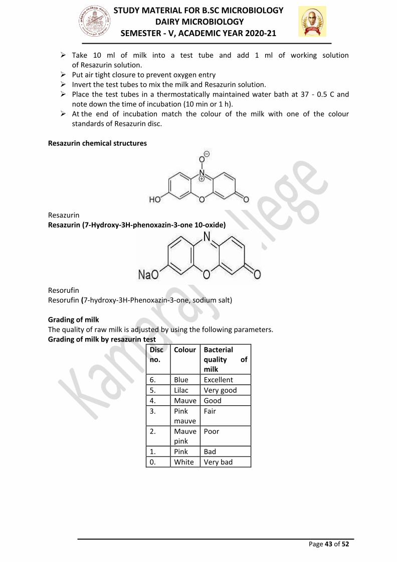
STUDY MATERIAL FOR B.SC MICROBIOLOGY DAIRY MICROBIOLOGY
SEMESTER - V, ACADEMIC YEAR 2020-21
Page 43 of 52
➢ Take 10 ml of milk into a test tube and add 1 ml of working solution of Resazurin solution.
➢ Put air tight closure to prevent oxygen entry ➢ Invert the test tubes to mix the milk and Resazurin solution. ➢ Place the test tubes in a thermostatically maintained water bath at 37 - 0.5 C and
note down the time of incubation (10 min or 1 h). ➢ At the end of incubation match the colour of the milk with one of the colour
standards of Resazurin disc. Resazurin chemical structures
Resazurin Resazurin (7-Hydroxy-3H-phenoxazin-3-one 10-oxide)
Resorufin Resorufin (7-hydroxy-3H-Phenoxazin-3-one, sodium salt) Grading of milk The quality of raw milk is adjusted by using the following parameters. Grading of milk by resazurin test
Disc no.
Colour Bacterial quality of milk
6. Blue Excellent
5. Lilac Very good
4. Mauve Good
3. Pink mauve
Fair
2. Mauve pink
Poor
1. Pink Bad
0. White Very bad

STUDY MATERIAL FOR B.SC MICROBIOLOGY DAIRY MICROBIOLOGY
SEMESTER - V, ACADEMIC YEAR 2020-21
Page 44 of 52
Advantages of dye reduction test ➢ Used for estimating the suitability of milk for liquid consumption. ➢ These tests are cheaper and less time is required.
Disadvantages
➢ Rate of reduction of dye varies considerably and is related to species and the rate at which different micro-organisms grow at a particular temperature.
➢ Inhibitory substances like penicillin and other antibiotics prevent the growth of bacteria and thus increase the reduction time.
PHOSPHATASE TEST
Pasteurisation is an essential process in the production of milk which is safe and free from pathogens.
Alkaline Phosphatase is an enzyme which is naturally present in milk, but is destroyed at a temperature just near to the pasteurization temperature. Alkaline Phosphatase test is used to indicate whether milk has been adequately pasteurised or whether it has been contaminated with raw milk after pasteurisation. This test is based on the principle that the alkaline phosphatase enzyme in raw milk liberates phenol from a disodium para-nitro phenyl phosphate and forms a yellow coloured complex at alkaline pH (Scharer, 1943). The intensity of yellow colour produced is proportional to the activity of the enzyme. The colour intensity is measured by direct comparison with standard colour discs in a Lovibond comparator. The test is not applicable to sour milk and milk preserved with chemical preservatives. Apparatus required
1. Water-Bath -maintained at 37±l⁰C, thermostatically controlled. 2. Comparator - with special discs of standard colour glasses calibrated in µg p-
nitrophenol per ml milk, and 2 x 25 mm cells. 3. Test Tubes - of size 16 x 1.50 mm and rubber stoppers to fit. 4. Pipettes - 1, 5, and 10 ml. 5. Filter Paper - Whatman No. 2 or equivalent. 6. Litmus Paper
Reagants
1. Sodium Carbonate-Bicarbonate Buffer - Dissolve 3.5 g of anhydrous sodium carbonate and 1.5 g of sodium bicarbonate in one litre of distilled water.
2. Buffer Substrate - Dissolve 1.5 g of disodium p-nitrophenyl phosphate in one litre of sodium carbonate-bicarbonate buffer. This solution is stable if stored in a refrigerator at 4°C or less for one month but a colour control test should be carried out on such stored solutions.
Procedure
1. Pipette 5 ml of buffer substrate into a clean, dry test tube followed by 1 ml of the milk to be tested. Stopper the tube, mix by inversion and place in the water-bath
2. At the same time place in the water-bath a control tube containing 5 ml of the buffer substrate and 1 ml of boiled milk of the same kind as that under test that is pasteurized homogenized, low fat.
3. After 2 hours, remove the tubes from the bath, invert each and read the colour developed using the comparator and special disc, the tube containing the boiled

STUDY MATERIAL FOR B.SC MICROBIOLOGY DAIRY MICROBIOLOGY
SEMESTER - V, ACADEMIC YEAR 2020-21
Page 45 of 52
milk control being placed on the left of the stand and the tube containing the sample under test on the right. Record readings which lie between two standard colour discs by adding a plus (+) or minus (-) sign to the figure of the nearest standard.
NOTE - If artificial light is needed when taking these readings, an approved ‘day light’ source of illumination must be used. STANDARD PLATE COUNT Introduction
The standard plate count (SPC) is suitable for estimating bacterial populations in most types of dairy products, and it is a reference method specified in the Grade A Pasteurized Milk Ordinance to be used to examine raw and pasteurized milk. This procedure is also recommended for application in detecting sources of contamination by testing line-samples taken at successive stages in the processing. Principle
The test employs aserial dilution technique for easy quantification of the micro-organisms. The appropriate dilutions of the milk sample are mixed with a sterile nutrient medium that can support the growth of the micro-organisms, when incubated at a suitable temperature. Each bacterial colony that develops on the plate is presumed to have grown from one bacterium or clump of bacteria in the inoculums. The total number of colonies counted on the plates multiplied by the dilution factor to represent the number of viable micro-organisms present in the sample tested. Procedure Sample preparation
➢ Mark each plate with sample number, dilution, and other desired information before making dilutions.
➢ Before opening a sample container, remove from the closure all obvious materials that may contaminate the sample. If desired, wipe the tops of unopened sample containers with a sterile cloth or paper towel saturated with 70% ethyl alcohol.
Dilution of samples
➢ For SPCs, select dilution(s) so that the total number of colonies on a plate is between 30 and 300. For example, where an SPC is expected to reach a number 5000, prepare plates containing 10-2 dilutions.
➢ Use a sterile pipette for initial and subsequent transfers from the same container, if the pipette is not contaminated. If the pipette becomes contaminated before transfers are completed, replace it with another sterile pipette. Do not flame to decontaminate. Use a separate sterile pipette for transfers from each different dilution.
Plating
Melt the required amount of medium quickly in boiling water, in a microwave oven, or by exposing it to flowing steam in a partially closed container, but avoid prolonged exposure to unnecessarily high temperatures during and after melting. Discard melted nutrient agar or tryptone dextrose agar that develops a precipitate. Do not melt more

STUDY MATERIAL FOR B.SC MICROBIOLOGY DAIRY MICROBIOLOGY
SEMESTER - V, ACADEMIC YEAR 2020-21
Page 46 of 52
medium than will be used in 3 hours. Do not re-sterilize the medium.Cool the melted medium. Incubating
Incubate plates at 32 C or 37 C for 48 .3 h for SPC. Plates must reach the temperature of incubation within 2 h. Avoid excessive humidity in incubator to reduce the tendency toward spreader formation, but prevent excessive drying of the medium by controlling ventilation and air circulation. Agar in plates should not lose more than 15% of its weight during 48 h of incubation. Counting of colonies on agar plates
Count the plates after the desired incubation period. Record the dilutions used and number of colonies counted on each plate. If it is impossible to count at once, after the required incubation store the plates at 0 to 4.4C for not more than 24 h. For each lot of samples, record the results of sterility tests on materials used when pouring plates and the incubation temperature used. Table Grading of milk based on standard plate count test (BIS Standards)
Pasteurized milk A standard plate count of lower than 30,000 cfu per ml. of pasteurized milk is indicative of satisfactory quality. Advantages of SPC
➢ Enumeration of only viable microbes. ➢ Cultural and morphological differentiation based on colony characteristics. ➢ Suitable for determination of quality of milk samples like pasteurized milk and high
grade raw milk with low bacterial number. ➢ Useful for pasteurized and for line testing at various stages of processing.
Disadvantages of SPC
➢ Gives only a rough estimate of microbial counts hence, not very accurate. ➢ Time consuming, laborious and cumbersome. ➢ Requires huge quantities of reagents, chemicals and glassware. ➢ Not a rapid method; at least 24 hours are required to get result. ➢ Not suitable for growth of all the species of bacteria present in milk. ➢ Temperature of incubation may not be optimum for the growth of all types of
bacteria.

STUDY MATERIAL FOR B.SC MICROBIOLOGY DAIRY MICROBIOLOGY
SEMESTER - V, ACADEMIC YEAR 2020-21
Page 47 of 52
➢ Amount of sample taken may not be representative. ➢ Pathogens are not detected, because certain microbes like Mycobacterium
tuberculosis cannot grow easily. ➢ Specific information regarding the type of micro flora is not obtained.
DIRECT MICROSCOPIC COUNT Introduction
Direct Microscopic Count (DMC) is a quantitative test and is helpful in assessing the actual number of bacteria present in milk. DMC is used as a platform test to assess the microbiological quality of milk received at the Raw Milk Receiving Dock. The method is useful for rapid estimation of the total bacterial population of a sample of milk and also in giving useful information for tracing the sources of contamination of milk. Method of DMC Principle
It is based on the examination of stained thin film of a measured volume of milk spread over a specified area on a glass slide. The method is useful for rapid estimation of the total bacterial population (including live and dead cells) of a sample of milk.
In this test, milk smear is prepared on one square centimetre area. The smear is stained with a special stain called Newman's stain and examined under microscope. Each microscopic field examined represents a quantitative aliquot of the milk sample. The number of microscopic fields occurring in one square centimetre area of the milk smear will vary as the diameter of the microscopic field varies with different microscope. Microscopic factor
The diameter of the microscopic field is measured with the help of a stage micrometer. Each division on the stage micrometer is equivalent to 0.01 mm. The diameter of the microscopic field varies with the length of draw tube, objective lens and ocular tube. The microscopic factor is calculated as follows:
Preparation of slide
➢ Take 0.01 ml milk with the help of a sterile Breeds pipette and spread it evenly on a grease free slide of 1 cm2 marked area on a Breeds slide.
➢ Dry the smear on a warm surface at 40 - 45C. Do not heat-fix the slide on direct flame.
➢ Rapid drying results in cracked surfaces on the film or peels off during further processing.
➢ Immerse the slide in Newman's stain for to 1 minute. ➢ Newman's stain removes the milk fat, fixes the smear and stains the bacteria in a
single operation. ➢ The tetrachloroethane of the stain helps to dissolve the milk fat globules, ethyl
alcohol fixes the smear and methylene blue stains the smear.
Microscopic examination

STUDY MATERIAL FOR B.SC MICROBIOLOGY DAIRY MICROBIOLOGY
SEMESTER - V, ACADEMIC YEAR 2020-21
Page 48 of 52
Examine under the oil immersion objective and count the number of micro-organisms (individual or clumps of cells) in a number of fields of the film. Thus, a representative number of fields depending on the concentration of bacterial cells in a microscopic field are chosen for counting the bacterial cells as follows. Table 17.1 Number of microscopic fields to be counted in DMC
Grading of milk The quality of raw milk is adjusted using the following details. Grading of milk based on DMC (BIS standards)
Advantages of DMC DMC is widely used to screen incoming raw milk supplies as a platform test to determine whether the milk has an acceptable or legal bacterial load, as per BIS standards.
➢ Easy to perform. ➢ Less time is required to perform the test. ➢ Large number of samples can be screened in a given period of time ➢ Useful in providing the estimated counts, types of bacteria and somatic cells in milk. ➢ DMC can be used as a guide in identifying the types of bacteria present in a milk
sample. Disadvantages of DMC
➢ Not considered as a fool-proof/ legal method. ➢ Strain on the eyes of the operator is too much. ➢ Test is not reliable as both viable and non-viable cells are counted. ➢ Method is not suitable for pasteurized milk. ➢ Results are not reproducible because microbes are unevenly distributed in the
smear. CLOT ON BOILING (C.O.B) TEST

STUDY MATERIAL FOR B.SC MICROBIOLOGY DAIRY MICROBIOLOGY
SEMESTER - V, ACADEMIC YEAR 2020-21
Page 49 of 52
The test is quick and simple. It is one of the old tests for too acid milk (pH<5.8) or abnormal milk (e.g. colostral or mastitis milk). If a milk sample fails in the test, the milk must contain much acid or rennet producing microorganisms or the milk has an abnormal high percentage of proteins like colostral milk. Such milk cannot stand the heat treatment in milk processing and must therefore be rejected. Procedure:
Boil a small amount of milk in a spoon, test tube or other suitable container. If there is clotting, coagulation or precipitation, the milk has failed the test. Heavy contamination in freshly drawn milk cannot be detected, when the acidity is below 0.20-0.26% Lactic acid. Results:
COB test indicates the suitability of milk for pasteurization and other heat treatment processes. Five ml of milk in a test tube is held over a flame and allowed to boil. The formation of flakes or clots indicates that the milk has high developed acidity and is unsuitable for pasteurization or high heat treatments. THE ALCOHOL TEST
The test is quick and simple. It is besed on instability of the proteins when the levels of acid and/or rennet are increased and acted upon by the alcohol. Also increased levels of albumen (colostrum milk) and salt concentrates (mastitis) results in a positive test. Procedure:
The test is done by mixing equal amounts of milk and 68% of ethanol solution in a small bottle or test tube. (68 % Ethanol solution is prepared from 68 ml.96 %( absolute) alcohol and 28 ml distilled water). If the tested milk is of good quality, there will be no coagulation, clotting or precipitation, but it is necessary to look for small lumps. The first clotting due to acid development can first be seen at 0.21-0.23% Lactic acid. For routine testing 2 ml milk is mixed with 2 ml 68% alcohol. THE ALCOHOL-ALIZARIN TEST
The procedure for carrying out the test is the same as for alcohol test but this test is more informative. Alizarin is a colour indicator changing colour according to the acidity. The Alcohol Alizarin solution can be bought readymade or be prepared by adding 0.4 grams alizarin powder to 1 litre of 61% alcohol solution. RESULTS OF THE TEST
Parameter Normal milk Slightly acid Milk Acid milk Alkaline Milk
PH 6.6 – 6.7 6.4 – 6.6 6.3 or lower 6.8 or higher
Colour Red brown Yellowish-brown Yellowish Lilac
Appearance of milk
No coagulation no lumps
No coagulation Coagulation * No coagulation **
Note:
➢ Sour milk looks yellowish with small lumps or completely coagulated. ➢ Alkaline milk looks like lilac and it may be mastitis milk. Clots and flakes too, indicate
mastitis milk.

STUDY MATERIAL FOR B.SC MICROBIOLOGY DAIRY MICROBIOLOGY
SEMESTER - V, ACADEMIC YEAR 2020-21
Page 50 of 52
THE BURRI SLANT PROCEDURE
➢ The Burri slant procedure is not a method which should be considered as a substitute for either the Petri plate or the direct microscopic procedure.
➢ The Burri slant method is of particular value as a means of finding and isolating the long chain streptococci that occur in freshly drawn samples of milk.
➢ The procedure may be employed with excellent results as a means of quickly finding the infected udder or quarter involved in septic sore throat or other epidemics.
➢ The amount of apparatus required is small and inexpensive (largely culture tubes) and within 30 hours streptococcus colonies are easily discernible on the agar slope.
➢ These can be removed for further study. It has a distinct advantage for this work over the Petri plate method because of the limited equipment necessary and over the direct microscopic procedure because it is possible to isolate suspicious colonies.
➢ The Burri slant procedure also is of special value for commercial dairies and food processing establishments for use in finding sources of bacteria that may contaminate the milk or food during processing.
➢ The limited amount of equipment again allows its use in the plant and the examination of a large number of samples taken throughout the processing operation.
➢ It is also of particular value for determining the possible cause of food suspected epidemics. In those cases where a large number of sources of offending organisms must be examined, samples can generally be secured during the field investigation and immediately inoculated onto the agar slopes for subsequent incubation and observation.
➢ There are certain difficulties involved in the use of the Burri slant procedure which should be recognized. Slopes for subsequent incubation and observation.
➢ There are certain difficulties involved in the use of the Burri slant procedure which should be recognized.
PLATFORM TESTS
Platform tests or milk reception tests are the commonly used names for the tests carried out by the persons responsible for raw milk collection and/or reception.
The tests in question are rapid quality control tests - organoleptic tests being of crucial importance - whereby the milks of inferior or questionable quality can be screened out before the milk leaves its original container and is mixed with bulk milk during milk collection and/or reception. LACTOMETER TEST
If the milk appears during organoleptic inspections to be too thin and watery and its colour is "blue thin" it is suspected that milk contains added water. Lactometer test serves as a quick method for determination of adulteration of milk by adding water. The lactometer test is based on the fact that the specific gravity of whole milk, skim milk and water differ from each other’s. Alcohol In case there is any reason to suspect that milk is sour, alcohol test is used as platform test for rapid determination of elevated acidity of milk. Anyhow, if the result of alcohol test indicates too high acidity in milk a sample from milk is to be taken to the laboratory for further testing of titratable acidity. FREEZING POINT DETERMINATION

STUDY MATERIAL FOR B.SC MICROBIOLOGY DAIRY MICROBIOLOGY
SEMESTER - V, ACADEMIC YEAR 2020-21
Page 51 of 52
The freezing point of milk is regarded to be the most constant of all measurable properties of milk. A small adulteration of milk with water will cause a detectable elevation of the freezing point of milk from its normal values of -0.54ºC. Since the test is accurate and sensitive to added water in milk, it is used to detect whether milk is of normal composition and adulterated. Other platform tests are
➢ MBRT ➢ Resazurin ➢ Dye Reduction Test ➢ Direct Microscopic Count (DMC) ➢ Freezing point Fat/SNF ➢ Lactometer
DETECTION OF Staphylococcus aureus IN MILK
Staphylococcus aureus is recognized worldwide as a major pathogen causing subclinical intramammary infection in dairy cows.S.aureus produces coagulase, an extracellular enzyme that binds to protein to form a complex with thrombin-like activity which converts fibrinogen to fibrin. The method more useful in practice for the detection of S. aureus and other coagulase-positive staphylococci in milk from cows with subclinical mastitis. Isolates of Staphylococcus aureus:
From each milk sample, 0.1 ml was plated on 7% sheep blood agar (Oxoid) plate and incubated at 37°C for 48 h. After presumptive identification based on colony morphology and microscopic morphology, biochemical and growth characteristics of the isolates were determined milk sample, 10 ml was added aseptically to 90 ml of 0.1% sterile peptone water. After a 10-fold serial dilution to 10-8, each dilution was spread onto plate containing Baird Parker (B-P) agar (Oxoid) with 20% egg yolk telluride emulsion (Merck) by drop plating technique. Plates were incubated aerobically at 37°C for 24 - 48 h. Typical S. aureus colonies as circular, smooth, convex, moist, 1-3 mm in diameter, black (telluride reaction), and surrounded by an opaque zone with an outer clear zone (lecitinase reaction) were enumerated using a manual method. Microscopic examination
This is helpful in detecting the admixture of mastitic milk with herd milk. Presence of long chains of streptococci is indicative of mastitis due to Streptococcus agalactiae, whereas occurrence of cells in grape like bunches in milk suggests Staphylococcal mastitis. Hotis test
It gives the most accurate information about mastitis infection. It is based on the fact that Streptococcus agalactiae, when growing in milk, produces a characteristic colony like mass of cells adhering to the sides of test tube. By the introduction of an acid indicator (bromocresol purple), the identification of these colonies or 'buttons' is facilitated by their characteristic yellow color. The appearance of yellow colonies of micro-organisms along the sides of tube or in bottom indicates infection with Streptococcus agalactiae. For this, 9.5ml of milk is mixed with 0.5ml of 0.5% aqueous bromocresol purple and incubated at 37’C for 24-48 hrs a test tube.

STUDY MATERIAL FOR B.SC MICROBIOLOGY DAIRY MICROBIOLOGY
SEMESTER - V, ACADEMIC YEAR 2020-21
Page 52 of 52
Interpretation of hotis test
Yellow colonies
Presence of Streptococcus
Flocculent on side of test tube
Presence of especially Streptococcus agalactiae.
Rusty brown color colonies
Presence of Staphylococcus aureus.
Blood agar test Pre incubated (37˚C over night) mastitic milk sample are streaked on blood agar and incubated at 370C for 24hand examined for colony formation and haemolysis. Detection of specific causative organism using blood agar test
Observation Micro-organism
Small colonies, α or β-haemolysis will occur or no haemolysis will occur in some cases
Streptococcus agalactiae
α-haemolysis(small zone around colonies and green discoloration)
Streptococcus dysagalactiae
No reaction Streptococcus uberis
Large colonies than streptococci, β-haemolysis(a wide zone of clearance around colonies)
Staphylococcus aureus
CAMP (Christie, atkin, munch and peterson) test
The CAMP test is quite specific for the detection of mastitis caused by Streptococcus agalactiae. A standard culture of Staphylococcus aureus is streaked vertically down across the center of a blood agar plate. The suspected streptococci plates are cross streaked horizontally at an angle taking care that this streaks do not come into contact with Staphylococcus aureus streak. After incubation at 370C overnight and observe for zones. Clear zone between the streaks of Staphylococcus aureus and milk sample indicates that is positive for mastitis of Streptococcus agalactiae.