STUDIES ON THE FRESHWATER COPEPOD FISH PARASITES OF …
222
*64o&7- STUDIES ON THE FRESHWATER COPEPOD FISH PARASITES OF KERALA THESIS Submitted to THE COCHIN UNIVERSITY OF SCIENCE AND TECHNOLOGY in partial fulfllment of the requirements for the degree of DOCTOR OF PHILOSOPHY BY SHAJU THOMAS, M. Sc. DEPARTMENT OF INDUSTRIAL FISHERIES COCHIN UNIVERSITY OF SCIENCE AND TECHNOLOGY COCHIN ' 682 016 1 988
Transcript of STUDIES ON THE FRESHWATER COPEPOD FISH PARASITES OF …

*64o&7
STUDIES ON THE FRESHWATERCOPEPOD FISH PARASITES OF KERALA
THESISSubmitted to
THE COCHIN UNIVERSITY OF SCIENCE AND TECHNOLOGYin partial fulfllment of the requirements
for the degree ofDOCTOR OF PHILOSOPHY
BY
SHAJU THOMAS, M. Sc.
DEPARTMENT OF INDUSTRIAL FISHERIESCOCHIN UNIVERSITY OF SCIENCE AND TECHNOLOGY
COCHIN ' 682 016
1 988

CERTIFICATE
'Ihis is to certify that this thesis is an authentic record of research
work carried out by Shri Shaju Thomas, M.Sc. under my supervision
and guidance in the Department of Industrial Fisheries, Cochin University
of Science and Technology, in partial fulfilment of the requirements
for the degree of DCIZTOR OF PHILOSOPHY and that no part thereof has
been submitted for any other degree.
CCU-IIN - 16. (M. SHAHUL HAMEED]September , 1988 Supervising Teacher
Dr. M. SHAHUL HAMEEZD
Professor
Department of Industrial FisheriesCochin University of
Science and TechnologyCochin — 682 016.

DECLARATION
I, Shri. Shaju Thomas, do hereby declare that the thesis entitled"STUDIES ON THE FRESHWATER COPEPOD FISH PARASITES OF KERALA" is
a genuine record of research work done by me under the supervision
and guidance of Dr. M. Shahul Hameed, Professor, Department of
Industrial Fisheries. Cochin University of Science and Technology. and
has not been previously formed the ‘basis for the-award of any degree.
diploma, associateship. fellowship or other similar title of any university
or institution.
Cochin — 16 SHAJU THOMASSeptember. 1988

ACKNOW LEDGEMENT
I wish to record my deep sense of gratitude and indebtedness to
Dr. M. Shahul Hameed. Professor, Department of Industrial Fisheries,
Cochin University of Science and Technology for his valuable guidance
and unfailing support given throughout the period of my research work.
I express my sincere thanks to Dr. C.T. Samuel. Professor and Head
of the Department of Industrial Fisheries, for providing the necessary
facilities to carry out the work in the department.
I am thankful to the Cochin University of Science and Technology for
providing me a fellowship during the period of my full-fime research.
My thanks are also due to the Manager, Nirmala College. Muvattupuzha
for deputing me from the College and to the University Grants Commission
for awarding the teacher fellowship for the completion of the study.
I am greatly indebted to Dr. G.A. Boxshall, British Museum (Natural
History), London. U.l<. and Dr. L.S. Roberts, Texas Tech. University.
Texas, U.S.A. for providing a good number of literature in this field.
I am obliged to the authorities of Kerala State Fisheries Department
for giving permission to carry out investigations in the State Fisheries
fish ponds .

My appreciation and thanks goes to Dr. N.K. Sasidharan Pillai. Fish
culture officer, State Fisheries Department and Mrs. Sophy John,
Lecturer, School of Marine Sciences, Cochin University of Science and
Technology, for assisting me in the collection of copepod fish parasites.
I: am greatly indebted to Mr. K. Ashok Kumar. Mr. M. Mukundan. Mr.
I<.C. Bellarmine and Mr. K.V. Pauly, Research Scholars. Department
of Industrial Fisheries, for their wholehearted cooperation and help during
the preparation of this thesis.
I am thankful to all my colleagues, friends and office staff who directly
or indirectly involved themselves with my work.
My thanks are also due to my parents and family members for their
support and encouragement during the course of the work.

CONTENTS
Page No.
PREFACE
CHAPTER I
GENERAL IN'I‘RODUCI'ION 1Classification 6Terminology of cephalic appendages 13Life history 14Host-parasite relationship.pathogenicity and ecology 19Treatment and control 25
CHAPTER II
SYSTEMATICS Introduction 29Description of species 31Ergasilus thammani Sp.nov. 31Ergasilus vembanadi Sp.nov. 36Ergasilus kabati Sp.nov. 41Lamlroglena krishnai Sp.nov. 45Lernaea osphronemi Sp.nov. 49
CHAPTER I11
LIFE HISTORY OF LERNAEA OSPHRONEMI SP.NOV.Introduction 54Materials and methods 54Observations on the life history 57First nauplius 69Second nauplius 71Third nauplius 72First copepodid 73Second copepodid 75

J
Third copepodidFourth copepodid
Fifth copepodidFemale copepodid
Male copepodid
Cyclopoid female
Metamorphosis of the femaleDiscussion
CHAPTER IV
1-I$T— PARASITE RELATIONS}-DP , ECOLCXSY AND TREATMENT
Introduction
Materials and methods
Results and discussion
CHAPTER V
GENERAL OBSERVATIONS AND SUM MARY
General observations
Summary
REFERENCES
77
79
81
81
84
86
87
88
95
96
106
118
124
127

PREFACE
The study of copepods parasitic on fishes was initiated by
Linnaeus (1758) with the description of Lernaea cyprinacea. Since then,
tremendous interest was shown in the study of this particular group
of parasites and many species, new to science have been reported
from different parts of the world. Parasitic copepods exhibit varying
degrees of adaptation to parasitism, which culminates even inendoparasitic forms. Studies on copepods parasitic on fishes reveal
information regarding the origin of parasitism, intricacies of host-parasite
relationship and distribution of host-parasite populations.
At present. the number of copepods parasitic on fishes exceeds
1500 species. Majority of them are parasitic on marine fishes. Reports
regarding freshwater species are less compared to that of marine forms.
Some parasites are capable of infesting even amphibians and aquatic
mammals .
In India. copepods parasitic on fishes have been studied to
a great extent, mainly on systematics. Tripathi (1966). during the
study of parasitic copepods from Indian waters. reported 22 species
from freshwater fishes. 31 from estuarine fishes and 141 species from
marine fishes. His survey was confined mainly to Eastern Indian region
and partly to South India. Hameed (1972) reported 129 species of
parasitic copepods from marine fishes from the coastal waters of Kerala.

Studies on parasitic copepods from freshwater fishes are still
in its infancy. In recent years, there- is a renewed enthusiasm in
the study of freshwater fish parasites due to rapidly increasing
aquaculture practices. The importance of diseases and their control
assumes great significance because of the adverse impact of diseases
on fish production and its economy. Copepods are one of the most
harmful parasites of freshwater fishes. Reports on the damages caused
by copepod parasites from different parts on the world are increasing
alarmingly. But the information on parasitic copepods of freshwater
fishes in India is quite meagre. Knowledge regarding this group of
parasites, their tfiology and pathology from Kerala. is lacking. This
is the main reason why Dr. I-Iameed had entrusted me to undertake
this study.
Copepods parasitic on fishes include three suborders namely
Poecilostomatoizla, Cyclopoida and Siphonostomatoida. The first one
includes five families. the second consists of a single family and the
last one comprises fourteen families [Kabata. 1979). The. suborder
Cyclopoida consists of a single family viz; Lernaeidae, which is
exclusively freshwater. Suborders Poecilostomatoida and Siphonostomatoida
are represented by a few forms parasitic on freshwater fishes.
During the present study, parasitic copepods belonging to the
family Lernaeidae (CyC10p0ith] and Ergasilidae [Poecilostomatoida) were
collected from freshwater fishes in Kerala. They were identified upto
species level and described in detail. In addition to this. the life history

of a new Lernaeid copepod was carried out. Eggs were collected from
adult females and allowed to hatch in controlled conditions. Then thelarvae were released to different host fishes and observed the life
cycle upto the emergence of egg-bearing females. Ecological studies
with special reference to host-parasite relationship , prevalence and
intensity of infection, were conducted for Lernaea osphronemi sp. nov.
Eradication of parasite population from culture ponds is of utmost
importance. S0, prophylaxis and control measures were tried for the
elimination of the new Lernaeid parasite.
The thesis consists of five chapters. The first chapter is a
general introduction which deals with the review of literature on various
aspects of parasitic copepods viz; systematics, life history, host-parasite
relationship, ecology, pathogenicity, prophylaxis and control measures.
Systematics of parasitic copepods of freshwater fishes collected during
the present study forms the second chapter. The third chapter deals
with the life cycle study of the new Lernaeid copepod, Lernaea
osphronemi. The fourth chapter contains host-parasite relationship.
ecology and treatment of ‘the’ new species of Lernaea OnOsphronemus goramy. General observations and a summary of the entire
work constitute the fifth chapter.

CHAPTER - I
GENERAL INTRODUCTION

GENERAL INT'RODUCT'I ON
Copepods- are commonly free-living but some of them are
parasitic. They are parasites or associates on sponges, coelenterates.
polychaete worms. molluscs, echinoderms and aquatic vertebrates.
Copepods are capable of parasitising different parts of the body of
the host. The site preferences of these parasites necessitate adaptation
to a great extent, that leads to varying degrees of diversity in form
and structure. The morphological variations exhibited by the adult
parasitic copepods make it difficult to recognize them as true copepods,
so in the past, some of them were identified as worms. Fish is one
of the major hosts of copepods.
A- survey of literature on parasitic copepods revealed that there
are more marine forms than freshwater species. Though the present
study is confined to freshwater parasitic copepods, it would be
appropriate to mention the pioneers in the study of marine copepod
parasites, because some of'them were experts in both the fields. In
addition to this. some members of the same family or genus parasitize
both marine and freshwater fishes.
The first record of a parasitic copepod Lernaea cyprinacea
by Linnaeus dates back to 1758. His study was succeeded by Muller
(1785) on Caligus. The 19th century workers in the field of parasitic
copepods were Hermann (1804), Risso [1816], Blainville (1822),
Nordmann (1832). Dana (1853). Steenstrup and Lutken (1861). Kroyer

(1863), Heller (1865), Hesse (1873), Richiardi (1883) and Bassett—Smith
(1899). C.B. Wilson (1905) spearheaded the study on both marine
and freshwater parasitic copepods of America. He brought out a long
series of publications for forty years. A 8 T Scott (1913) published
a comprehensive work on British parasitic copepods. Leigh-Sharpe (1925,
1933) has also contributed to the study of parasitic copepods -of British
fishes. Kirtisinghe (1937) concentrated his studies on the copepod
parasites in and around Ceylon and brought out several publications.
It was followed by Heegaard (1943) and Shiino (1952).
Yamaguti (1963) proposed a classification and published a
monograph on "Parasitic Copepoda and Branchiura of Fishes". Bocquet
and Stock (1963) suggested a new approach in the study of parasitic
copepods. Works by Kabata (1958-'87), Ho (1961—'85), Lewis
(1963—'69). Hewitt [1964—'79), Roberts [1963-'70) and Cressey
(1967-'83) are worth mentioning.
The important Indian scienfists in the study of copepod parasites
of marine fishes are, Gnanamuthu (1947—'60), Redkar (1949-'50), Kurian
(1955-'61], Rangnekar and Murthy (1950-'64) ,Tripathi (1952—'69).
Ranganekar (1955—'63). Pillai (1961-'83), Sebastian (1966), Reddiah
(1970). Hameed (1972-'88) and Natarajan and James (1977).
Mar1<evich's (1931—'78) studies on the copepod parasite fauna
of the fishes of the U.S.S.R. brought out a lot of information on this
subject. Hu's (1948) papers on parasitic copepods of China is worth

mentioning. He discussed in detail the taxonomy of the genus Lernaea
and proposed five subspecies. Harding (1950) critically carried out
the systematic study of genus Lernaea and prepared a key for the
identification of this genus.
Fryer (1956-'82) conducted extensive studies on the freshwater
copepod fish parasites of Africa. He described several new species
of parasitic copepods. His approach to the problem was philosophical.
He worked out the zoogeography and phylogeny . of African freshwater
copepod parasites. Causey (1957) reported the parasitic copepods from
Louisiana freshwater fishes, which consist of four genera and twelve
species. Bauer (1962) studied the ecology of parasites of freshwater
fishes. Parasitic crustacea from inland wafer fishes of Israel was
described by Paperna (1964). Sarig (1966) reviewed the diseases and
parasites of fishes in warm—water ponds in the Near East and Africa
and opined that pond—cultured fishes are greatly infected byectoparasites. He suggested precautionary measures to prevent the
hazards of parasitic infection.
Meyer (1966), in his review of the parasites and diseases
of fishes in warm-water ponds in North America pointed out that parasitic
copepods are more harmful than any parasites of cultured fishes.
Hoffman (1967) described i.n detail the parasites of North American
freshwater fishes and provided a key for the identification of parasitic
copepods. Lernaea, Salmincola. Lepeophtheirus, Ergasilus, Lernaeocera
and Achtheres were represented in the key.

The ectoparasitic infection of African freshwater fishes was
studied by Paperna and Thurston (1968). They observed that the
parasific crustaceans in Africa were extremely rich in species and
genera.
Rogers (1969) in a summary of fish disease cases received
over a five year period at South Eastern Co-operative Fish Disease
Laboratory ,reported that 30% of the damage was caused by parasites.
Lernaea was the most damaging of the parasitic crustaceans encountered.
Roberts (1970) carried out an extensive study on the genus
Ergasilus in North America. discussed previous literature in detail and
worked out a key for Ergasilus. A comprehensive account of the
parasites of British freshwater fishes was brought out by Chubb (1970).
He reported the occurrence of eight species of crustaceans fromfreshwater fishes. He concluded that the information on distribution,
life-cycle. seasonal pattern of occurrence. long term cyclical changes
in parasite abundance and other aspects of the biology of the parasites
of freshwater fishes are necessary for a better management of fish
populations. Johnson and Rogers (1973) worked out the distribution
of the genus Ergasilus in several Gulf of Mexico drainage basins and
suggested that some Ergasilus species are" more hostspecific. A check
list of British and Irish freshwater fish parasites with notes on its
distribution was published by Kennedy (1974). It is a valuable
contribution in the study of parasites of freshwater fishes.

Boxshall (1976) created a new genus to include the parasific
copepods coming under the family Lernaeidae. It is interesting to note
that one of the species described under the new genus,Pseudolamproglena, was. from India. Fish parasites of Wisconsin streams
were studied by Amin (1977). Out of the fifteen species of parasites,
copepoda was represented by 1.. cyprinacea. Kabata (1979) published
a monograph on British Parasitic Copepoda . He corrected the errors
and anomalies in the earlier descriptions, critically reviewed the status
of genera and families and proposed a new classification. It was a
milestone in parasitic copepod research.
Thatcher (1981) started studies on parasitic crustaceans of
fishes from Brazilian Amazon and reported several new species. Ergasilid
copepod parasites of Japanese freshwater fishes were studied by Do
(1982). He prepared a valuable key for the identification of eleven
species of Ergasilus reported from Japan. Kabata (1983) created two
new genera of the family Lernaeidae for the description of two new
species. of copepod parasites on freshwater fishes from India. It clearly
indicates that the freshwater copepod parasite fauna in India necessitates
deep and detailed study. Kabata (1985) published a book entitled
‘Parasites and Diseases of Fish Cultured in the Tropics‘. This is a
good guide to those interested in aquaculture practices. Byrnes (1986)
reported the presence of new species of Ergasilids from Australianbream .
Studies on freshwater parasitic copepods from Indian Waters
is fragmentary. Southwell and Prasad (1918) described copepod parasites

from Indian freshwater fishes. The occurrence of a new species of
Ergasilus from Wallago attu was reported by Sundara Raj (1923),
Contributions of Redl<ar et al. (1949), Rangnekar and Murthy (1950-'61)
are quite significant. Karamchandani (1952) described a new species
of Ergasilus and published a key for the identification of seven species
of Ergasilus from India. Gnanamuthu [1951—'56} conducted studies on
Lernaeid parasites infesting freshwater fishes and reported two new
species of Lernaea .
Attention to the study of freshwater fish parasites is revived
due to increased interest in aquaculture activities. Recently, several
scienblsts are engaged in this field of study. Tripathy's (1966. 1969]
work on the parasite fauna of freshwater fishes is noteworthy. Reports
and reviews of Gopalakrishnan (1961. 1964a,b, 1966} gave good account
to the damage of freshwater fishes by copepod parasites. Srinivasachar
and Sundarabai (1974) conducted detailed studies on the crustacean
parasites of freshwater fishes of Mysore and brought out several
interesting observations on copepod ‘parasites. Seenappa et al. (1980)
described a new species of Lernaea parasitic on Catla catla (Ham)
and Labeo rohita (Ham) from Karnataka.
CLASSIFICATION
The morphological variations exhibited by the adult parasitic
copepods led to confusion in identifying the systematic position.
Controversies and disputes regarding the position of species to a parti

cular genus or family are not uncommon in the realm of parasitic
copepods. Within fifty years. three different approaches to the
classification of copepods were suggested. Wilson (1932) divided the
order Copepoda into eight suborders viz; Arguloida, Calanoida,
Harpacticoida . Cyclopoida , Nothodelphoida, Monstrilloida , Caligoida and
Lernaeopoida. This classification was accepted by almost all workers
in the field of copepod research.
Yamaguti (1963) upgraded the status of order Copepoda to that
of a class and Wilson's suborders to orders. He has divided the
subclass Copepoda into six orders viz; Cyclopidea, Caligidea,
Philichthyidea, Andreinidea, Lernaeopodidea and Sarcotacidea.
Kabata (1979) proposed a new classification. He argued that
free living copepods should also be taken into account for the
classification of parasitic copepods. His suggestion is that from the
point of systematics. morphological features can fall into two categories
namely. primary or primitive and secondary or advanced. The
differences between an ectoparasitic copepod and a planktonic copeopd
are, mainly due to the accumulation of specilized features required in
the process of adaptation to their respective modes of life. The primitive
characters are less susceptible to parallelism and convergence. These
include morphological features inherited from the ancestors and
recognisably retained by the later generations. Kabata observed that
the intersegmental articulation and the structure of mouth and mouth
parts remain without much change in the evolutionary process of

8
copepods. These reliable primary characters can be taken as basic
clues to the phylogeny of copepods.
According to the classification (Kabata, 1979), three suborders
under the order Copepoda parasitize fishes. The suborders are
Poecilostomatoida . Cyclopoida and Siphonostomatoida . suborder
Poecilostomatoida is divided into five families viz: Bomolochidae,
Taeniacanthidae, Ergasilidae, Chondracanthidae and Philichthyidae.
Cyclopoida consists of a single family — Lernaeidae. The families under
Siphonostomatoida are Caligidae, Euryphoridae, Trebiidae, Pandaridae.
Cecropidae, Dichelesthiidae, Eudactylinidae, Kroyeriidae. Pseudocynidae,
Hatscheldidae , Lernanthropidae , Pennellidae , Sphyriidae and
Lernaeopodidae. This classification is easy to follow. and also has
advantages over the classifications suggested by Wilson and Yamaguti.
It is accepted by majority of scientist in this field. So for the present
study l<abata's classification is followed.
FAMILY : Ergasilidae and Lernaeidae
During the course of the present investigation parasitic copepods
belonging to the genus Ergasilus Von Nordmann 1832, Lamproglena von
Nordmann 1832 and Lernaea Linnaeus 1758, were collected and studied
in detail. The systematic position of the parasites are also discussed.
Family - Ergasilidae
Members of this family exceeds 100 species, which parasitize
mainly on marine and freshwater teleost fishes. The parasitic Ergasilids

are morphologically least modified by their mode of life, resembles
to free living Cyclopoid. Von Nordmann (1832) first described two
"species of Ergasilus: E. sieboldi and E. gibbus. Wilson (1911) published
a collective account of the family Ergasilidae describing organ system,
ontogeny and ecology from American waters. He suggested three
subfamilies viz; Ergasilinae, Bomolochinae and Taeniacanthinae. Sars
(1918) removed the genus Bomolochus from Ergasilinae and later Wilson
(1932) came to the conclusion that the three subfamilies should be
elevated to full family status. The important genera under Ergasilidae
are, Ergasilus Von Nordmann, 1832; Thersitina Norman, 1905;
Pseudergasilus Yamaguti . 193 6; Paraergasilus Markevich. 1 937 ,
Nipergasilus and Sinergasilus Yin, 1949; Neoetgasilus Yin, 1956 and
Diergasilus Do. 1981.
Genus — Ergasilus von Nordmann, 1832
It forms a major genus under the family Ergasilidae. The
members of this genus exhibit broad range of morphological variations
with respect to segmental boundary between the cephalosome and the
leg bearing segments. The structure of appendages and number of
abdominal segments are also varied. The morphology of the second
antenna is quite variable and constitute one of the best characteristic
features for different species. Adult Ergasilus females are usually
attached to the outer surface or on the gills of the fishes whereas
males remain free swimming throughout their life.

10
Family : Lernaeidae
The members of this family exhibit the ‘highest degree of
adaptation to parasitism. The genera under Lernaeidae belong to two
different groups. Mesoparasitic group undergoes metamorphosis and
their anterior part gets burried in the body of the host while the
posterior part protrudes above the site of penetration. The other group
that does not undergo metamorphosis are ectoparasitic. living mainly
on the gills of their host. The former consists of the genus LernaeaLinnaeus, 1758; Lernaeogiraffa Zimmermann. 1922; Areotrachelus Wilson,
1924; Taurocheros Brain. 1924; Qysphorus Kurtz, 1924; Afrolernaea
Fryer, 1956 and Opistholernaea Yin, 1960. The latter comprises
Lamproglena Nordmann, 1832 and Lamproglenoids Fryer, 1964. Boxshall
(1976) created a new genus Pseudolamproglena and described two new
species. 33. simplex from India and _F_’. annulata from Iraq. Recently,
two more genera were erected by Kabata {I983} for describing two
new species from Indian waters. They are Pillainus volvicollis from
Channa marulius and Indolernaea manohari from Mystus seenghala. At
present . there are 13 genera under Lernaeidae , includingMesolamproglena Kuang, 1980.
Genus — Lamprogna von Nordmann, 1832
This genus, including more than twenty species, all parasitic
on freshwater fishes, is the most primitive group of Lernaeidae. This
genus was included in the family Dichelesthiidae by Fryer (1959) without

1]
referring to the study of Sproston et al. (1950). Because of the
cyclopoid nature of the developmental stages of Lamproglena, Sproston
et al. (1950) removed it from Dichelesthiidae and included in Lernaeidae.
Kabata {1979) also included this genus in Lernaeidae and discussed
in detail the history and systematics.
Genus — Lernaea Linnaeus. 1758
It is the most popular and widely discussed genus under
Lernaeidae. The members of this genus live on the body surface, gill
cavity or mouth of their hosts. Reports and descriptions of the members
of this genus from different parts of the world followed. the
establishment of the type species Lernaea gyprinacea by Linnaeus (1758).
Hu (1948), in his paper "Studies on the Parasitic Copepods
of China", discussed at length the specific and subspecific characters
of L. cyprinacea and proposed five new subspecies. He used the relative
position of the legs as a basis for subspecific identification and also
the differences in first and second antenna. Harding (1950) recognised
twentyeight species of Lernaea, of which fourteen were from the
collections of the British Museum. Nine of them were new species.
In addition, he synonimized seven species and prepared a key for
the twentyeight recognized species. As the characters used to distinguish
between different species of Lernaea were often ill-defined and not
easily visible for identification, Harding suggested ‘the shape of anchor
and its arms‘ the most useful character for taxonomic purposes. The

12
structural differences and systematic position of Lernaeid copepod was
analysed in detail by Fryer (1961a). He did not find any significance
in the relative position of legs for subspecific differentiation. He proved
that morphological variations exhibited by Lernaea depend mainly on
the site of attachment; so, subspecific names are of no value.
Kabata (1979) reviewed the history and systematics of genus
Lernaea and compiled the distribution of Lernaea in different parts
of the world. He opined that seventeen species. the largest number,
occur in Africa, eight species in North America, seven species in
far—eastern Asia; only Europe and India have fewer species. The type
species _I__.. cyprinacea enjoys cosmopolitan distribution.
The morphological variability imposed by the host and site of
attachment makes it difficult to delimit with precision. the boundaries
of the species. This became quite evident after the publication of the
work by Poddubnaya (1973, 1978). On Cyprihus cargio. Poddubnaya
found the "classical" Lernaea gyprihacea which she referred to as
"European". as well as another one. morphologically distinguishable
from the first. which she named as "Asian". The latter is identical
with Leigh-Sharpe's (1925) Lernaea elegans. E. cyprinacea has long
‘T’ shaped dorsal horns whereas in _l:. elegans the shape of the dorsal
horns is 'Y' shaped. The first result of Puddudnaya's work was the
recognition of the validity of _I:. elegans, which was considered
synonymous with _I:. cyprmacea. She then conducted experiments to
check the validity of some species of Lernaea employing different hosts

13
like gyprinus cargio, Ctenopharynggdon idella and Carassius auratus
and suggested that the name Q. cyprinacea must be restricted to the
parasite of the host fish Carassius auratus and all other cyprinids
carry L. elegans .
Fratello and Sabatini (1972) examined the chromosome of Lernaea
collected from Cygrinus carpio, Carassius auratus. Lepomis gibbosus
and Gambusia affinis. 'Ihe chromosomes were identical {2n=16} in all
species. The authors concluded that all these hosts harboured the same
species of Lernaea viz; Lernaea cyqjgnacea. This work appears to
contradict some of Poddubnaya's findings. More work in this direction
is needed for a better understanding of the systematics of genusLernaea.
TERMINOLJIY OF CEPHALIC APPENDAGES
Homology of the cephalic appendages of parasitic copepods still
remains an unsettled problem. Heegarrd (1945). Lang [1946] and Lewis
(1969) studied the homology of the mouth parts of parasitic copepods.
but they failed to arrive at an agreement. Kabata [1979] discussed
in detail. the various aspects of copepod appendages and suggested
that "we shall accept the existence of two pairs of antennae. mandible,
two pairs of maxillae and one pair of maxillipeds in all the copepod
species". This terminology is followed for the present study.

14
LIFE HISTORY
Life history studies are essential for developing methods to
control copepods parasitic on fishes. Several workers contributed in
this field of study. Gurney (1913) worked out the life cycle of Theristina
ggsterostei. Mirzoeva (1972. '73) studied the life history of Sinergasilus
lieni_ and Zamerzlaya (1972) that of Eigasilus sieboldi. Urawa et al.
(1980 a.b) successfully followed the naupliar development of Neoergasilus
japonicus and compared the naupliar development of different species
of Ergasilus. They suggested that there are six nauplii and fivecopepodid stages in the life history of Neoergasilus Eponicus. Male
and female could be first distinguished at the III copepodid stage and
sexual maturation occurs in the adult stage. After copulation, females
enter into a parasitic life.
Sproston (1942) worked out the complete series of developmental
stages of Lernaeocera branchialis. One nauplius. one copepodid and
four chalimus stages were followed by the adult stage. Lewis (1963)
studied the life cycle of gpeophterius dissimulatus, Wilkes (1966) that
of Nectobranchia indivisa. Jones and Mathews (1968) that of Sphygon
lumpi and Izawa (1969) that of Caligus spinosus. Kabata (1972)
described the life cycle of Caligus clemensi and Voth (1972) that of
Epgphtheirus hospitalis. Kabata and Cousens (1973). and Schram (1979)
worked out the life cycle of Salmincola californiensis. and Lernaeenicus
sprattae respectively. Kawatow et al. (1980) studied the life cycle
of Alella macrotrachelus parasitic on cultured black sea bream. It

15
consists of one nauplius, one copepodid, four chalimus and the adult
stage. Perkins (1983) worked out the life history of Cardiodectes
medusaeus. Izawa (1986, 1987} described the development of
Taeniacanthus lagocgihali ’which consists of two naupliar stages. six
copepodid. stages and sexual dimorphism became distinct during the
third copepodid stage. He also worked out the phylogenetic implications
found in the egg and naupliar stages of the Poecilostome cyclopoida.
The life cycle of commonlyknown species of Lernaea cyprinacea
has been studied by several workers; Wilson {1917}, Stolyarov (1935),
Yashouv (1959), Kasahara (1962). Lahav and Sarig (1964). Rogers
(1966), Bauer et al. (1973). Rukyani (1975). and Shields (1978).
The most comprehensive and detailed description of the life history
of _I:. cyprinacea has been given by Gradba (1963). The life cycle
includes three naupliar, five copepodid and cyclopoid stages.
Gnanamuthu (1951 b} in his work on the life cycle of E.chackoensis suggested that the nauplius moulted only once to become
metanauplius followed by the development of six copepodid stages. Fryer
(1968) found that in lake Victoria, the copepodid stages of the race
of L. cyprinacea infect the gills of Bagrus docmac Forskal, but in
the adult stage. they parasitize Tilapia sp. In lakes Edward and George,
_I._.. barnimiana during larval stages were found on Bagrus sps. . but
in the adult stage lived on Tilapia sp. and Haplochromis sp. An
experimental infection on Tilapia sp. proved that there is no need for
an intermediate host for the completion of life cycle of E. barnimiana

15
(Thurston, 1969). Wilson (1917) had opined that members of the genus
Lernaea needs a temporary host for the copepodid stage and a definidve
host for the adult stage.
Kabata [1981] reviewed the reports and descriptions on the
life cycle of parasitic copepods and suggested that most of the life
cycles can be divided into four segments namely naupliar, postnaupliar,
preadult and adult. With the exception of adult, these segments commonly
consist of more than one stage. In order to eliminate the confusion
existing in the terminology in labelling individual stages. the nauplii
have been numbered I-V and the term metanauplius has been dropped.
The postnauplii beginning with the first copepodid end with the last
stage ,earlier to the preadult. Those that remain free swimming and
exhibiting no organogenetic changes foreshadowing parasitism. are
designated by the name copepodid I-V. If they have become attached
and enter the stage of "regressive reconstruction", they are given
the name chalimus. The preadult is that part during which the copepodid
either settles definitively on the host. or attain a definitive level of
organization and reach the adult stage. Raibaut [1985] suggested that
parasitic copepods have. in most cases. one host cycle tholoxenous).
but there are species which use an intermediate host [heteroxenous]
or a facultative host during their life cycle. Shariff and Sommerville
(1986) studied the life cycle of L. polymorpha and _I;. cyprinacea and
observed that the life cycles were similar irrespective of the host.
The two species of parasites could not be (fifferentiated morphologically
at the larval stages. The first copepodid could not develop beyond

17
that stage in the absence of a host. The eggs from both species hatched
into nauplii and required 14-15 days to form young female parasites
at 24.3 to 29.0°C water temperature.
The life cycle of Lernaea bhadraensis was studied by Tamuli
and Shanbhogue (1987). They reported that the first copepodid of this
parasite can undergo successive moulting in the absence of a host
and reach upto fifth copepodid stage within 17-18 days. This finding
is quite interesting, since there are no reports on the survival of
copepodids without host. The fifth stage lived in pond water for about
75 days without host and this also differs from earlier findings.
Nakai (1927) and Nakai and Kokai (1931) studied the development
of Lernaea elegans and reported that at temperature higher than 36.5°C
and lower than 10.1°C, the eggs stopped hatching and the larvae did
not develop beyond the metanauplius stage at the temperature of 14°C.
Kasahara (1962) observed that the newly hatched nauplii of E cflgrinacea
attain the sixth copepodid stage in 18 and 11 days at water temperature
approximating Z2 and 27°C respectively. The life span of the females
is 45 days at 27°C. Rogers (1966) reported that the first copepodid
stage was reached within three days after hatching in the case of
_I:. cyprinacea. At 28°C, the time taken for the completion of life cycle
is 17 days. Bird (1968) studied the life cycle of L. cyprinacea and
noted that copulation took place at the fifth copepodid stage and was
followed by further development, implantation and metamorphosis. The
females which were unable to copulate. could not develop beyond the

18
fifth copepodid stage. Copulation is a trigger. activating development,
but the mechanism is not clear. Thurston (1969) found that in g.
barnimiana sexual dimorphism became distinct during the 5th copepodid
stage and the adult female was visible to naked eye on 19th day at
21—26°C. The first egg sacs was formed on 23rd day and the life span
was 32 days approximately.
According to Sarig (1971), the life cycle of Lernaea cyprinacea
takes 25 days at 20°C, 20 days at 25°C, 17 days at 30°C. and only
14 days at 35°C. He observed that the males and females can be
differentiated during the fourth copepodid stage and copulation occurs
in the cyclopoid stage. after which the males die. Al—Hamed and Hermiz
(1973). showed that the life cycle of L. cmrinacea from egg to mature
adult was completed in 13-14 days at 22—25°C and the first copepodid
stage appeared 3-4days after hatching. Pan et al. (1979) reported
that at 15—20°C, the development of _I:. polymorpha was completed within
14-16 days and at 26-31°C. it took only seven days. The life span
of the adult female is about 20 days at temperatures of 25-37°C.
Lernaea in general has a complex life cycle, characterized
by successive metamorphosis and moulting of the hard inflexible skeleton.
Successive larval stages are characterised by increase in size and
in number of body segments and appendages (Hoffman, 1976]. Reports
on the life cycle of Lernaea are contradictory in certain respcets;
hence species—wise study is essential to establish stfitable methods
for the eradication of the parasites from fishes.

19
HOST — PARASITEI RELATIONSHIP, PATHCISENICITY AND ECOLCBY
The copepod -parasite. when it is free, must find a host and
recognise whether it‘ is suitable for infestation. The parasite has to
select a precise microhabitat on the host before implantation. The
susceptible mechanisms of infestation of the copepod parasites on their
hosts are not known clearly. Many parasites enhance the chances of
finding a new host by synchronizing the reproductive cycle with the
abundance of host population. So far. this phenomenon has not been
observed in the case of copepod parasites.
Knowledge regarding the mechanism of host finding by parasitic
copepods are not clearly known. Fryer (1966). noted "a marked tendency
for a fish which has acquired one parasite to acquire others". Shields
and Tidd (1974) attributed the localization of Lernaea larvae in the
mouth and branchial chambers of tadpoles associated with water currents
occurring in those areas. 'Chemoreception' is suggested as a mechanism
to detect the host, at least in Caligidae, by Kabata (1974, 1981}. He
also opined that the currents caused ‘by the movements or respiration
of the fish is one of the factors directing the copepod to the host.
Boxshall (1974a) observed that all the species of copepods.
with the exception of members of the Lernaeopodidae which inhabit
the gills, were attached with their anterior ends directed towards the
gill arch and the body lying parallel to the primary gill filaments.
Hanek and Fernando (1978 a). found that Eigasflus centrarchidarum
was randomly distributed on the gills of gpomis gibbosus, but it

20
preferred the dorsal and ventral sectors of the anterior halves of
the hemi-branchs in Ambloplites rupestris. The preferred sites of most
of the Lernaea sp. seemed to be the base of dorsal. pectoral and
pelvic fins Shields and Tidd (1974) and Bulow et al. (1979). Amin
et al. [1973] suggested that the site selection of _I:. cyprinacea was
always related to body size of the host and stream conditions. As
Kabata (1981) cited "site selection is undoubtedly determined by a set
of morphological and physiological factors completely unknown at
peresent" .
Host specificity of parasitic copepods are reported by several
workers (Fryer. 1968; Cressey -and Collette, 1970; I-Ianek and Fernando.
1973 b; Kabata. 1979 and Shariff et al. 1986). Lewis et a1. [1969]
suggested that host preference can be indicated by the incidence of
parasitism and the effect of the» host is exerted not only on the sexually
mature copepod but also on all stages in the life cycle. which infest
the host. Although distinct host specificity among copepod occurs. further
work of a statistical nature must be done with these crustaceans before
we formulate significant generalizations about them {Noble and Noble,
1976] .
Lernaea gyprinacea is‘ usually reported as having a wide range
of host susceptibility (Fryer. 1961 b; Demaree, 1967; Hoffman, 1967;
Shields. 1968 and Poddubnaya, 1978). According to Kabata (1979),
over 100 species of fishes have been recorded as hosts of this copepod.
Shariff et al. (1986) reported that fish belonging to the Orders

21
Atheriniformes (Anabantidae and Cyprinodontidae) and Channiformes
(Channidae) are uniformly susceptible to Lernaea. Among Cypriniformes
only some species of Cyprinidae appear to be susceptible to Lernaea.
Only one species of Perciformes, Oreochromis mossambicus (Peters)
became infected by Lernaea cyprinacea.
There is a tendency that larger fishes have higher levels of
paraiste infestation. This was reported by several workers; Dogeil
(1961), Cressey and Collette (1970). Noble and Noble (1976), Kennedy
(1975), Rawson (1977), Bortone et al. (1978), Kabata (1985) and
Conneely and Mc Carthy (1985). Kabata (1981) opined that "the larger
and older fish often carry great numbers of copepods is attributable
mainly to the longer period of Contact and larger attachment surface
available for the parasite". Boxshall (1974 b,c) worked out the
population dynamics of Lepeophtheirus Pectoralis (Muller) in relation
to abundance, age structure and dispersion pattern.
Lower levels of parasitism in the largest size class is also
observed by Noble et al. (1963), Bortone et al. (1978), Amin (1977)
and Eiras (1986). Noble et al. offered a plausible explanation that
older fish may develop some immunity to infestation. Shields and Goode
(1978) and Shariff et al. (1986) also suggested the possible development
of immunity among fishes against the infection of _l:. cyprinacea. Kabata
(1970) reviewed the host-parasite relationship of oopepod parasites
and their host, remarked that the effect of parasite on host could be
classified into local and general. Local effects are those limited to

22
the immediate vicinity of the copepod's attachment and feeding activities.
The general effects are those which manifest themselves at site remote
from the permanent habitat of the adult parasite.
The feeding habit of copepod parasites are also different.
gtgasilus sieboldi feeds on gill epithelium, mucus gland, erythrocytes
and _white blood cells (Einszporn, 1965 a.b). Deeply penetrated copepods
such as Leraneidae feed on tissue debris and blood (Fryer. 1968).
Meyer (1966) observed that the sites of attachment of Lernaea
cyprinacea are usually accompanied by acute haemorrhagic reactions
which frequently become foci for secondary infections by bacteria and
fungi. If the parasites are attached near nerve centres, such as brain
or along the lateral line, the infested fish will frequently swim in a
fight circle or exhibits convulsive movements. Shields and Goode (1978)
described the formation of thickened whorls of hyperplastic epithelium
and fibrous tissue around the parasite Lernaea cyprinacea on Gold
fish. Radhakrishnan and Nair (1981) showed that Lernanthropus gibbosus
and Lernanthropus koenigii were of serious concern to their hosts.
causing irrepairable damage to the gills by way of their mode of
attachment and feecfing activity. Noga (1986) recorded Lernaea cruciate,
as an important initiator of skin lesions in large mouth bass.
Thurston (1965) worked out the pathogenicity of crustacean
parasites and suggested that parasitic infection occur most readily in
crowded condition. Damages to aquaculture industry by I_._ernaea sp.
have been reported by several workers. Nakai (1927), Fryer (1968),

23
Lahav and Sarig (1964), Paperna and Thurston (1968), Sarig (1971),
Kabata (1985) and Sheriff et al. (1986). High mortalibles have occurred
among cultured cat fish, gold fish, baitminnows, carp, trout and other
fishes due to Lernaea infecfion (Post, 1983].
Srinivasachar and Shakuntala (1975) found that Lebistes reticulatus
infested by Lernaea hesaragattensis consumed more oxygen thanuninfected fish. It can be attributed to a ‘stress reaction’ due to
parasitic infestation of the fish. Esch et al. (1975) analysed the
relationship between stress and parasitism, suggested that the impact
of stress upon the dynamics of host and parasite populations were
varied. Such studies regarding copepods and their hosts are wanting.
The ecology of the host has a great significance in the variation
of the rate of infestation by copepod parasites in terms of individual
host. It is not merely the host but also the host's environment. that
forms the environment of the parasite. This is especially true for the
ectoparasites of aquatic animals. For parasite. the "microenvironment"
is the host and the "macroenvironment", host's habitat [Dogie1, 1961].
In the Victoria Nile. where the water was swift. Lernaea barnimiana
occurred in abundance in the mouth, but on the same host in still—water
conditions, most parasites were found at the base of the fins or flanks
(Fryer. 1968). This indicated the influence of external environment
in the selection of site with respect to host's habitat. Margolis et
a1. (1982) worked out definitions of a few terms used by parasitological
ecologists .

24
The effect of temperature on the development of parasitic
copepods has been reported by Grabda (1963), Shields and Tidd (1968),
Pan et al. (1979) and Kabata (1985). According to Shields and Tidd
(1968). in Lernaea "gprinacea, egg production will not occur at
temperatures below 24°C. no naupliar development below 20°C and
copepodid development and penetration to host not below 15—52U°C. They
suggested that the most successful laboratory cultures of Lernaea can
be maintained between 24 and 29°C. Lernaea may overcome the winter
seasons as larval females embedded within the tissue of the host
(Hoffman. 1976] .
Studies by shields and Sperber [1974] revealed that Lernaea
has a limited tolerance’ to increased salinities. Hoffman (1976) ‘reported
that adult Lernaea were not found in waters with salinity greater than
1.8% and larvae proved even more sensitive. In addition, they were
not found in water with pH lower than 7. Seenappa et al. (1985)
observed mortality and reduced hatchability of Lernaea at acidic pH
ranges. Lernaea is considered as a typical freshwater form but its
occurrence was noticed in Philippines on a brackish water fish _qi_ano§
chanos (Kabata. (1985).
Srinivasachar and and Sundarabai (1974) observed that incidence
of Lernaea hesaragattensis. parasitic on Lebistes reticulatus was highest
during July and lowest in January. Viljoen (1985) carried out seasonal
investigation of the genus Lernaea and established that infestation and

25
site preference are related to host and season; body dimensions of
the parasite also varied seasonally.
Timmons and Hemstreet (1980) studied the prevalence rate of
L. cyprinacea on Micropterus salmoides (Lacepedel and reported that
no fish smaller than 25 mm. or longer than 99 mm. were infected.
Uehara et al. (1984) observed that Lernaea infection was different
for the four species of fishes, Carassius auratus L.’ Salmo gairdneni
Richardson, Micropterus salmoides Lacepede and T‘inca tinca L. The
prevalence rate was 8095, 78%, 1295 and 0% respectively.
TREATMENT AND CONTROL
Kabata (1985) defined therapy as treatments intended to restore
normal health to fish that have contracted disease. It is an interplay
of three factors namely the pathogen. the fish and the therapeutic agent
or medication. The choice of medication is based on the nature of
the pathogen. that must be lethal to the pathogen but harmless to the
fish.
Efforts for the control of crustacean parasites of fishes probably
date back to fish culture practices.The first therapeutic measures were
natural organic compounds. They were followed by chemicals and
synthetic compounds (contact insecticides} of ever increasing complexity.
The compounds used for the control of parasitic crustacea fall into
D.D.T. Group, B.H.C. Group and Organophosphate group.

26
The therapy can be divided into three categories: (1) addition
of chemicals to water, {2) addition of chemicals to feed, (3) application
of chen1icals directly to fish. Method of large scale treatment of pond
has been considered best-suited for controlling parasites like Argulus
and Lernaea. Sarig (1971) and Roberts and Shepherd (1974) have
documented the requirements and precautions , while applying chemicals
to pond water .
Eradication of crustacean parasites has been achieved by bath
treatment. It can be divided into three categories: dips, short baths
and long baths. Dips involve immersion for not less than five minutes.
short baths last 5-60 minutes and longer treatments are considered
as long baths. Fairly comprehensive reviews of measures against
Lernaea have been published by Kabata (1970, 1985} and Hoffman and
Meyer (1974).
Various chemicals have been tried to control and eliminate
Lernaea. The easily obtainable and cheap common salt {NaC1) is one
of them. This can be used for fishes which are able to tolerate salt
concentrations required to kill the parasite. It has been reported to
be quite useful against free swimming larval Lernaea. but less effective
against attached females.Putz and Bowen (1964) suggested the use of
0.8 to 1.1% salt solution for three days for the eradication of Lernaea.
Shilo et al. (1960) showed that salt solutions in 25,000 ppm.concentration have an adverse effect on the juveniles of Lernaea, but
not harmful to them beyond the point of recovery.

27
Potassium permanganate [KMnO4 ) has been used to controlLernaea in many parts of the world. applied in a multiplicity of ways.
But, it has not been proved uniformly successful due to several reasons.
The standard method of -treatment using [KMnO has been compiled4 l
by Sarig (1971). Carp infected with Lernaea can be treated in tanks
in which weight of fish to water ratio is not less than 1:2.5. The
-concentration of KMnO4 is slowly built up by gradual additions of the
stock solution until it reaches 25 ppm. in 15 minutes (Kabata, 1970).
After an interval of 20 minutes this procedure is repeated. Bath of
10 ppm. for periods between 50-60 minutes was recommended by some
authors. A concentration of 2 ppm. can be used for indefinite period
{Putz and Bowen, 1964). Sarig [1971] treated infected carps with 20
ppm. of KMnO4 . After 60-120 minutes‘ of exposure. 90-100% of the adult
Lernaea found on fish were killed. He also reported that l<MnO affects4
only the adult Lernaea and does not kill young parasites embedded
in the skin of the fish. Pan et al. (1979) reported that bathing fish
in a solution containing 12.5 pp. of potassium permanganate at water
temperature -24-30°C was effective in killing the adult parasite without
serious injury to host.
Formalin treatment is found to be successful only on the free
swimming larval stages of Lernaea. Putz and Bowen (1964) reported
the use of 30 - 60 minute baths at the concentrafion of 250 ppm.
Baths were repeated every three weeks as long as necessary.Ravindranath et al. (1985) suggested a unique method of treatment
of Lernaeosis using formalin and lime.

28
The use of D.D.T.. B.H.C. and organophosphates were proposed
by Schaperclaus (1954). Shilo et al. (1960), Meyer (1966), Sarig(1971). Gopalakrishnan (1964 b). Lahav et al. (1964), Pan et al.
(1979). Kabata (1985). (1986) and Shariff et al. (1986).
Several p1ant—derived substances were used for the eradicafion
of Lernaea. The oldest known plant remedy is the teaseed cake or
meal used in China for many years. Chen {1933) questioned its
efficiency. though some Chinese farmers still use them. Bundles of
castor plants kept immersed in water are also considered effective
against Lernaea (Kabata, 1985]..
Chinese experts have reported the successful use of another
plant Gelsenium elegans (cited by Kabata, 1985) for the eradication
of Lernaea. Similarly, use of Acanthopanax spinosus (75—1UO Kg/acre)
resulted in complete eradication of Lernaea.
Though, biological control measures have been developed for
the eradication of several pests, it has not been explored for the
elimination of parasitic copepods. Kasahara (1962) observed that a
planktonic copepod, Mesocyclqg. was a main predator of free swimming
larval stages of Lernaea. Recent Indonesian work (Kabata, 1985] on
the susceptibility of various fish species to Lernaea suggests that Tilapia
nilotica is more resistant to infection than other fishes tested. Carp,
along with I. nilotica. reared in a pond were less infected than those
reared alone. Further investigation in this direction is essential tofind out a cheap and harmless method for the eradication of Lernaea.

CHAPTER - II
SYSTEMATICS

29
SYSTEMATI CS
INTRODUCTION
Parasitic copepods of freshwater fishes in India were studied by
Southwell and Prasad (1918) , Gnanamuthu ( 1951 a , 1956] ,
Karamchandani (1952), Tripathi (1966), Srinivasachar and Sundarabai
(1974), Seenappa et al. [1980] and Nandeesha et al. (1984. 1985).
Knowledge regarding the copepod parasites of freshwater fishes in
Kerala is scanty. The aim of the present study is to investigate the
parasitic copepods of freshwater fishes in Kerala.
The specimens described in this chapter were collected by
examining fishes from freshwater fish landing centres and fish markets
in different parts of Kerala. The present collection consists of five
new species which are described in detail. Holotypes and allotypes
will be deposited in the National Museum, Calcutta and paratypes will
be deposited in the Museum of the Department of Industrial Fisheries.
Cochin University of Science and Technology. Cochin.
CLASSIFIED LIST OF SPECIES
Order Copepoda
Suborder Poecilostomatoida
Family Ergasilidae
Genus Ergasilus von Nordmann, 1832.

30
Ergasflus thammani sp. nov.
Ergasilus vembanadi sp. nov.
Ergasilus kabafi sp. nov.
Suborder Cyclopoida
Family Lernaeidae
Genus Lamproglena Von Nordmann, 1832
Lamproglena krishnai sp. nov.
Genus Lernaea Linnaeus, 1758
Lernaea osphronemi sp. nov.
LIST OF HOSTS AND THEIR PARASITES
Channa striatus (Bloch)
Lamproglena krishnai sp. nov.
Mugfl cephalus Linnaeus
Ergasilus thammani sp. nov.
Ergasilus kabati sp. nov.
Osphronemus Loramy Lacepede
Lernaea osphronemi sp. nov.
Puntius sarana (Hamilton-Buchanan)
Ergasilus thammani sp. nov.
Wallago attu (Bloch and Schneider)
Ergasilus vembanadi sp. nov.

31
DESCRIPTION OF SPECIES
Order Copepoda
Suborder Poecilostomatoida
Family Ergasilidae
Genus Ergasilus von Nordmann, 1832
Ergasilus thammani sp. nov.
Figs. 1-12
Material :
Twentytwo females were collected from the gills of Mugil cephalus
Linnaeus) from the freshwater fish landing centres at Thevara, Vail-zom
and Trivandrum and nine females from the gills of Pumius sarana
(Hamilton-Buchanan] at Thammanam fish market.
Female:
Cophalothorax longer than broad, nearly rounded anteriorly and concave
posteriorly with a medio-lateral constriction. Second to fifth leg bearing
segments gradually decreasing in size posteriorly. Fifth segment very
short and narrow. Genital segment broader than long and cup-shaped.
Abdomen three-segmented, each segment somewhat narrower than the
preceding one and the terminal segment with a medial cleft. Uropod
squarish each with four terminal setae, the innermost one highly
elongated. the other three smaller and subequal in length.
First antenna: Five-segmented, segments distinct, tapering terminally
with several setae of varying length. Basal segment stout, longer than

32
broad and bearing the maximum number of setae. Second segment
broader than the third and fourth segments. Fifth segment narrow
compared to all other segments.
Second antenna: Four-segmented, first segment broad. stout and devoid
of any armature. Second segment very long with a sensillum on the
distal half of the medial margin. Third segment long and slender,
slightly curved with a sensillum on the medial margin of the proximal
half. Distal segment a strong stout claw, bearing small spinule on
the disto-lateral margin .
Mandible: Unsegmented. subrectangular with two distal falciform blades;
inner blade extends from a rectangular stalk-like structure and curved
anteriorly. The inner and outer blades provided with rows of short
setae on the ventral margins.
First maxilla: Very small. orbicular, with two setae of unequal length.
Second maxilla: Two-segmented, basal segment broader than long and
stout. Distal segment falciform, long and thickly beset with spiniform
setae dorsally.
Maxilliped: Absent.
First Leg: Sympod two—segmented. Coxa unarmed. basis with a single
plumose seta on the lateral margin to the base of the exopod. Exopod

33
three-segmented. Basal segment broad. longer than the second and
third segments combined, with a short spine distally. Second segment
longer than the third bearing plumose seta on the inner margin.
Terminal segment short with two spines and five plumose setae. Endopod
three—segmented. basal segment longer than the second, bearing one
plumose seta. Second segment slightly shorter than distal with a single
plumose seta. Terminal segment longer than broad with two broad,
spatulate spines unequal in length and four plumose setae. The inner
margin of the basal segment of exopod and the outer margin of the
basal and second segments of endopod with fine hairs.
Second leg: Coxa unarmed. Basis with single plumose seta lateral to
the base of the exopod and a denticular patch in front of the endopod.
Exopod three—segmented, first segment long and stout with a single
spine distally. Second segment slightly longer than third. bearing a
single plumose seta on the inner margin. Third segment short carrying
six plumose setae distally. Endopod also three—segmented. Basal segment
with an inner plumose seta, second segment with two plumose setae
and distal segment with one spine and four plumose setae. The inner
margin of the basal segment of exopod and outer margin of the first
and second segments of endopod bearing fine hairs.
Third leg: Coxa unarmed. Basis bearing a small plumose seta on the
outer margin and a denticular patch on the inner margin. Elxopod
threesegmented. Basal segment long and stout . with an outer spine
distally and a row of fine hairs on the inner margin. Second and third

34
segment subequal in length. Second segment with a single plumose
sets and distal segment carrying six plumose setae. Endopod also
three-segmented. The outer margin of the first and second segments
bearing hairs. The segments are subequal in length. basal segments
with a single plumose seta, second segment carrying two plumose setae
and distal segments with one spine and four plumose setae.
Fourth leg: Coxa unarmed. Basis with a single plumose seta to the
base of the exopod. Exopod two-segmented. First segment long with
a single spine on the distal margin and fine hairs on the inner margin.
Second segment short, carrying five plumose setae distally. Endopod
three-segmented and segments decreasing in length distally. First
segment with a single plumose seta. second segment bearing two plumose
setae and terminal segment carrying one spine and three plumose setae.
First and second segments with fine hairs on the outer margin.
Fifth leg: Two-segmented. Basal segment. very small carrying a plumose
seta. Second segment long, suboval in shape with two plumose setae
distally .
Armature of the Itami as follows
Arabic numerals denote setae: Roman numerals denote spines.
Endopod Ebcopod1 2 3 1 2 3I Leg 0-1 0-1 _II—4 1-0 0-1 11-5II Leg 0-1 0-2 I-4 1-0 0-1 0-6III Leg 0-1 0-2 1-4 1-0 0-1 0-6IV Leg 0-1 0-2 1-3 1-0 0-5 —
36
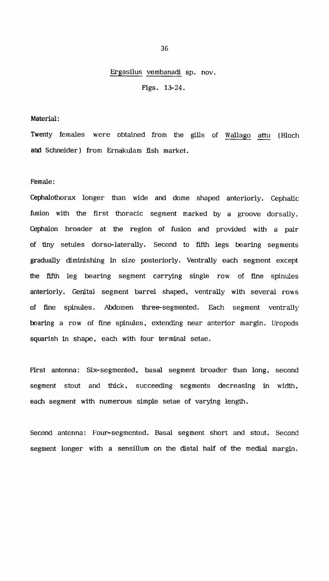
Ergasilus vembanadi sp. nov.
Figs. 13-24.
Material :
Twenty females were obtained from the gills of Wallago attu {Bloch
and Schneider} from Ernakulam fish market.
Female:
Cephalothorax longer than wide and dome shaped anteriorly. Cephalic
fusion with the first thora_cic segment marked by a groove dorsally.
Cephalon broader at the region of fusion and provided with a pair
of tiny setules dorso-laterally. Second to fifth legs bearing segments
gradually diminishing in size posteriorly. Ventrally each segment except
the fifth leg bearing segment carrying single row of fine spinules
anteriorly. Genital segment barrel shaped, ventrally with several rows
of fine spinules. Abdomen three-segmented. Each segment ventrally
bearing a row of fine spinules. extending near anterior margin. Uropods
squarish in shape. each with four terminal setae.
First antenna: Six-segmented, basal segment broader than long, second
segment stout and thick. succeeding segments decreasing in width.
each segment with numerous simple setae of varying length.
Second antenna: Four-segmented. Basal segment short and stout. Second
segment longer with a sensillum on the distal half of the medial margin.

37
Third segment slender and curved. Distal segment is a sharp and strong
claw .
Mandible: Indistinqtly two-segmented. Basal segment massive. from the
base of the distal segment arises an elongated spine. bearing hairs
ventro-laterally; terminal spine beset with fine hairs, posterior to
the spine a large falciform blade fringed with hairs. The mandibular
palp attached to the basal segment with hairs on its inner margin.
First maxilla: Round in shape, armed with two long setae.
Second maxilla: Two-segmented. Basal segment broad and thick and
the terminal segment thickly packed with spiniform setae dorsally.
Maxilliped: Absent .
first Leg: Sympod two—segmented. Coxa devoid of ornamentation. Basis
with a plumose seta on the lateral margin and fine spinules on the
anterior margin. Exopod three-segmented. Basal segment longer than
broad with a spine distally. the spine and distal half of the segment
denticulated. Second segment about half the length of the basal segment
with a single plumose seta on the inner margin and a row of dentlcles
on the inner and outer side of the outer border. Terminal segment
small. bearing two spines with serrated flange and five plumose setae.
The inner margin of the first and second segment with fine hairs.
Endopod three-segmented. Basal segment longer than broad with small
38
denticles and fine hairs on the outer margin and a plumose seta on
the inner margin. Second segment smaller than basal with a row of
line hairs on the outer margin and single plumose seta on the inner
margin. Distal segment equal in size to the second segment, bearing
two subequal spine having denticular flange and four plumose setae.
The outer margin with a row of denticles.
Second leg: Coxa devoid of armature. Basis with a plumose seta on
the lateral margin and fine spinules on the anterior margin. Exopod
three—segmented. Basal segment longer than broad carrying a single
spine on the outer margin and fine hairs on the inner margin. Second
and third segments with very thin serratlon on the outer margin. The
second segment carrying a single seta and fine hairs on the inner
margin. Terminal segment small with six setae. Endopod three—segmented.
First segment stout and long with a single seta. Second and thirdsegment almost equal in size, second segment with two setae and distal
segment carrying a strong spine and four setae. The outer margin
of all the segments with a row of spinules and fine hairs. All setae
are plumose .
Third leg: Sympod two-segmented. Coxa without any armature. Basis
with a small plumose seta lateral to the base of the exopod and
carrying fine spinules on the anterior margin. Exopod three—segmented.
Basal segment longer than the second and third segment combined
together. with a distal spine. Second segment with a single seta on
the inner margin. Distal segment short with a single spine and six

39
setae. The inner margin of the basal and second segment carrying
fine hairs. Endopod three-segmented. Segments decreasing in length
distally. Basal segment with a single seta, second segment bearing
two setae and distal segment with a strong spine and four setae. The
outer margin of all the segments with a row of spinules and fine hairs.
All setae are plumose.
Fourth leg: Coxa unarmed, basis with a lateral plumose seta and fine
spinules on the anterior margin. Exopod two-segmented. Basal segment
long and stout with an outer spine. Distal segment short and carrying
one spine and five setae. Endopod three-segmented, segments subequal
in length. Basal segment with a single seta. second segment with two
setae and terminal segment with one long spine and three setae. The
inner margin of the first exopodal segment and the outer margin of
all the endopodal segments bearing fine hairs. All setae. are plumose.
Fifth leg: Single-segmented. longer than broad and carrying two subequal
plumose setae.
Armature of the rami as follows:
Arabic numerals denote setae: Roman numerals denote spines.
Endopod EXOPUC11 2 3 1 2 3I Leg 0-1 0-1 II-4 1-0 0-1 H-5II Leg 0-1 0-2 1-4 I-0 0-1 0-6I11 Leg 0-1 0-2 1-4 I-O 0-1 1-6IV Leg 0-1 0-2 1-3 1-0 1-5 —

40
Uropod: Squarish with an elognated medial plumose seta and three
subequal setae, the central one elongated and plumose.
Total length: 0.8 mm ---(0.9 mm.
Male: Unknown
Remarks :
Ergasilus vembanadi sp. nov. resembles Ergasilus thammani sp. nov.
in its general body shape. In E. vembanadi. the cephalothroax provided
with a pair of tiny setules dorso-laterally. which is absent in E.
fig The first antenna in the present specimen is six—segmented,
but in E. Egg, it is five-segmented. The second antenna in E._@_Il__lI_lEI_11°_ bearing sensillum on the second and third segments and ‘a
spinule on the claw, whereas in. E. yembanadi, only the second segment
of the second antenna carrying a sensillum. A leaf like maxillary palp
present in the new species is absent in E. thammani. The first leg
endopodal spines in E. thammani are spatulate whereas in E. vembanadi,
_the spines are with serrated flanges’. The ornamentation of thoracic
legs in both these species are also different. In E. vembanadi all the
‘thoracic segments except the fifth leg bearing segment, the genital
segment and the abdominal segment carrying fine spinules ventrally.
whereas spinules are absent in E. thammani. The present species can
be separated from all other species in this genus by the general shape
of the body, presence of a single sensillum on the second segment of

41
the second antenna, structure of the first maxilla, first leg exopodal
spines with serrated flange and the ornamentation on the body and
thoracic legs .
Ergasilus kabati sp. nov.
Figs. 25 - 36.
Material :
Twelve females were collected from the gills of Mugil cephalus Linnaeus
from Murinjapuzha fish landing centre.
Female:
Cephalon broader than long and rounded on either sides anteriorly and
distinct from the posterior part by a dorsal groove. Cephalothorax longer
than broad and laterally with a constriction" in the middle. Second to
fifth thoracic segments gradually decreasing in size, fifth segment
comparatively very small. Genital segment longer than broad and slightly
expanded anteriorly. Abdomen three-segmented. First segment double
the length of the other two, distal segment short. Uropod short, inner
distal corner produced into an elongated seta, laterally a stout spine—like
process and two setae of subequal length.
First antenna: Six—segmented with several setae of varying length. First
segment broader than long, second segment very stout. succeeding

42
segments gradually decreasing in size and terminal segment slightly
longer than broad with apical setae.
$econd antenna: F‘ive—segmented, basal segment squarish, second segment
longer than broad, third segment more than double the length of the
second. fourth segment slender and slightly curved and fifth segment
a curved claw. Second. third and fourth segments enveloped in a thin
sheath, which is wrinkled randomly.
Mandible: Slender, elongated with indistinct segmentation. terminally
bears a large and a small falciform blade with fine hairs. From the
postero-medial part of the mandible arises an elongated palp with short
hairs. A protruberance from the middle of the mandible bears a leai—lil<e
mandibular palp.
First maxilla: Semicircular with two setae of unequal length.
Second maxilla: lndistinctly two—seg_mented, basal segment broad and
subrectangular. The terminal segment anteriorly curved and thickly beset
with spiniform setae on the dorsal surface.
Maxilliped: Absent.
First leg: Sympod two-segmented. coxa unarmed and basis carrying
a single plumose seta on the lateral margin. Exopod three-segmented.
Basal segment longer with an outer spine. Second segment shorter than

43
first and bearing a single plumose seta on the inner margin. Third
segment broader than long with two spines serrated on the outer margin
and five plumose setae. The outer border of the first and second
segments with row of small denticles. Endopod three-segmented. Basal
segment with a single plumose seta, second segment shorter than first
and carrying a single plumose seta. distal segment equal in size to
the second. bearing four plumose setae and two subequal spine
surrounded by serrated membrane.
Second leg: Coxa unarmed, basis with a plumose seta posterior to the
base of the exopod. Exopod three-segmented. First segment longer with
a distally placed spine. Second and third segments almost equal in size.
Second segment bearing a single plumose seta and the distal segment
with six plumose setae terminally. The outer margin of each segment
with a single row of small denticles. Endopod three-segmented. Basal
segment comparatively long with a single plumose seta. Second and third
segments equal in length, second segment carrying a single plumose
seta and distal segment bearing a single spine and four plumose setae.
Third leg: Coxa without oranmentation, basis with a single plumose seta
on the lateral margin. Exopod three-segmented. Basal segment long with
a distal spine. Second segment broad with a single plumose seta. Third
segment short with six plumose setae distally. Endopod three-segmented.
Basal segment long bearing a single plumose seta. Second and third
segment equal in size. Second segment carrying a single plumose seta
and distal segment carrying a spine and four plumose setae.

44
Fourth leg: Basis carrying a plumose seta posterior to the base of
the exopod. Exopod two—segmented. Basal segment slightly longer than
the distal with a single spine. Distal segment short carrying five plumose
setae. Endopod thr§‘e—segmented. Segments subequal in length, basal
segment carrying a single plumose seta, second segment with two plumose
setae and terminal segment bearing one spine and three plumose setae.
Fifth leg: Single segmented, longer than broad with two subequal plumose
setae posteriorly and a small naked seta postero—latera1ly.
Armature of the rami as follows:
Arabic numerals denote setae: Roman numerals denote spines.
Endopod I Exopod1 2 3 1 2 3I Leg 0-1 0-1 11-4 1-0 0-1 11-5II Leg 0-1 0-1 1-4 1-0 0-1 0-6111 Leg 0-1 0-1 1-4 1-0 0-1 0-6"IV Leg 0-1 0-2 1-3 1-0 0-5 —
Upropod: Roughly squarish. bearing an irmer elongated plumose seta,
a stout spineljke structure and two subequal setae in the distolateral
margin .
Total length 0.9 mm - 1 mm.
Male: Unknown.

45
Remarks:
Ergasilus kabati sp. nov. Though having all the characters of the genus
Ergasilus lacks close morphological resemblance with any of "the species
in this genus. The anterior portion of the cephalon broad and rounded
laterally. Cephalothorax longer than broad with a lateral constriction
in the middle. The genital segment differs from all other known species.
This species can easily be identified by the peculiar nature of the second
antenna with sheathing and the prominent spine—1il<e structure on the
uropod.
Suborder CYCLOPOIDA
Family Lernaeidae
Genus Lamproglena von Nordmann, 1832
Lampglena krishnai sp. nov.
Figs. 37 — 46
Material :
Eight females were collected from the gills of Channa striatus (Bloch)
from a freshwater pond at Cochin.
Female:
Body elongate, cylindrical. narrowing towards the hind end. Head
comparatively small. antero—lateral parts expanded and fused with the
thoracic segment, having a pair of fleshy lobe at the anterior end.
First thoracic segment free, broader than long. Second, third and fourth

46
thoracic segments are stout. forming a pear-shaped trunk. Fifth thoracic
segment broader than long. free from the trunk and genital segment.
segment clearly separated from the fifth thoracic segment, also
longer than broad, but gradually tapering posteriorly, having a prominent
lateral constriction near the center. Abdomen clearly three-segmented.
First and second segment cylindrical, distinctly separated and equal
in length. Third segment gradually narrowing posteriorly and having
flhe combined length of the first two segments. Posterior end of the
third segment bifurcated to form the uropod.
First antenna: An elongated structure with indistinct segmentation "having
five naked setae on the middle and four on the tip.
Second antenna: Shorter than the first antenna having two terminal naked
setae .
Maxilla: Basal segment broad and stout having a slender winged claw
pointed towards the tip.
_Maxilliped: Two-segmented, basal segment very stout. Distal segment
short, but slightly longer than broad. Terminally the segment bears
four stout and strong distally curved subequal claws.
First leg: Biramous. basipod stout, with a pectinate ridge near the
postero-ventral margin. Exopod distinctly three—segmented. first segment
with an outer spine distally and an inner naked seta in the middle.

47
Second segment having an inner naked seta. Third segment with two
subterminal stout spines and four elongated naked setae. Endopod
t‘r1ree—segmented. outer margin of the segments with toothed ridges.
First and second segments bearing a single seta and the third segment
with four setae of subequal length on the disto-lateral margin.
Second leg: Biramous, exopod three—segmented, the basal segment of
the exopod’. is comparatively very stout with an outer spine and an inner
naked seta. Second and third segments subequal in length. . Second
segment with an inner median naked seta. Terminal segment with three
‘naked setae and a terminal ‘spine. Ehdopod ‘three-segmented, outer margin
of the endopod with toothed ridges and inner margins armed with setae.
First and second having one each and third having three naked setae.
Third leg: Biramous, exopod three~segmented, basal segment stout, other
two segments comparadvely short. First segment with a spine on the
outer margin and a naked seta on the inner margin. Second segment
with an inner naked seta. Terminal segment with two stout claw—like
spine and four naked setae. Endopod three-segmented. First and second
segments with single naked seta on its inner margin. Third segment
with four naked setae antero-laterally. The outer margin of all the
segments toothed .
Fourth leg: Biramous. exopod three-segmented, basal segment of the
exopod long and stout. armed with an outer spine and an inner naked
seta. Second and third segments subequal in length. Second segment

48
with a naked seta on its inner margin. Terminal segment with two stout
terminal claw-like spine and the inner margin with three naked setae.
Ehdopod three—segmented. outer margin of all segments toothed. Inner
margin of the first and ._ second segments each with a naked seta.
Terminal segment with three naked setae.
Fifth leg: Uniramous. two-segmented; basal segment with two naked setae.
Terminal segment armed with two naked distal setae.
Uropod: Fused with the abdomen, conical in shape. diverging each other
from the postero-median line of the abdomen. having a spine on the
base and distally with three setae of which the central one is longer.
Total length: 3 - 4 mm.
Remarks :
Lamproglena krishnai sp. nov. shows resemblance to E. gihiocephali
Yamaguti (1939) in its general body shape. But the present species
differs clearly in various other details. In g. ophiocephali the genital
‘segment is fused with the thoracic segment and the abdomen is indistinctly
sgemented, whereas in E. krishnai sp. nov. the genital segment is
distinctly separated from the thoracic segment and the abdomen clearly
three segmented. The uropod of the new species completely fused with
the abdomen and conical in shape: but in g. gghiocephali the uropod
is separated from the abdomen and oval in shape. In L. ophiocephali,
in all the thoracic legs both the exopod and endopod are two—segmented,

49
whereas in L. krishnai in all the legs these are distinctlythree-segmented. Postero-ventral margin of the basipod of the first leg
alone is pectinate in the present species. whereas in E. gphiocephali
basipods of all the legs carry pectination. In the present case, the
arrangement of setae and spines on the legs are entirely different from
l._. gihiocephali. _I._.. krishnai sp. nov. differs from all other known
species of the genus by its three-segmented nature of leg rami, distinctly
segmented abdomen and the number of spines and setae present on the
legs.
Genus Lernaea Linnaeus, 1758
Lernaea osphronemi sp. nov.
Figs. 48 - 64.
Material:
Large number of femals were collected from the body surface of
Osphronemus giraniy . Lacepede, from a freshwater fish pond of State
Fisheries Department at Pannivelichira, Kerala, India.
Post-metamorphosis adult female:
Cephalothorax he Ini— spherical . small. anterior to hold fast, bearing
antennae and mouth parts. Holdfast consisting of two pairs of horns
dorso-ventrally placed, ventral branches slightly longer than dorsal.
The branches are simple with blunt tips. Variations in the structure

50
of holdfast is noticed among infrapcpulations. Body subcylindrical,
unsegmented and gradually expanding in breadth posteriorly. Neck
comprising of second to fourth leg—bearing segments and circular in
cross section. At the.., region of the second pair of leg. there is a
node-like swelling, while at the places of third and fourth legs very
slight constrictions are visible laterally. Behind the fifth pair of legs
lies and pregenital prominence which is hemispherical and bilobed.
Abdomen sub-conical with two constrictions ventrally giving the appearance
of three segmented nature and carrying small, single-segmented uropods
dis tally .
First antenna: Uniramous. three—segmented, basal segment is very long
and equal in length of the second and third segments combined, bearing
nine short setae and a long one on the distal part. The second segment
is. short. bearing three small and a long setae. The third segment longer
than the second. carrying ten setae of varying length. seven are apical
in position, out of which two are thicker and longer. than the others.
Second antenna: Uniramous, two-segmented. segments subequal in length.
Basal segment unarmed. Distal segment with two small setae along the
posterior margin, a claw-like spine and four slender setae distally.
Labrum: Small flattened semicircular plate . overlying the mandible and
first maxillae .
Second maxilla: Two-segmented, the basal segment very stout and broad,
distal segment small with two cu.rved strong claws.

51
Maxilliped: Two-segmented. basal segment broad and stout with a small
papilla armed with an apical setule. and slightly projected on the median
margin of_ the distal part. Terminal segment comparatively very short
bearing one short curved and four large subequal strong claws at its
anterior margin .
Thoracic legs: First four pairs of legs are biramous. Ramithree—segmented. Sympod two-segmented. Proximal segment with a small
plumose seta on the inner margin distally. Distal segment with a fine
seta ‘lateral to the base of the exopod. In the first leg a curved
spineelike structure present at the medio-lateral margin of the distal
segment.
Fifth leg: Reduced, one-segmented with four small setae of unequal length
on the distal margin.
Sixth leg: It is represented as a single plumose seta.
Armature of the rami as follows:
Arabic numerals denote setae: Roman numerals denote spines.
Endopod Exopod1 2 3 1 2 3I Leg 0-1 0-1 II-4 I-1 I-1 II~5
11 Leg 0-1 0-2 lI—4 1-1 1-1 IlI—5III Leg 0-1 0-2 11-4 1-1 1-1 111-5IV Leg 0-1 0-2 11-3 I-1 I-1 111-5

52
Uropod: Subcylindrical. each bearing a long straight plumose seta
extended posteriorly. two small setae on either side and another small
seta on the medio-lateral margin.
Total length: 6 - 9 mm.
Remarks:
A total of nine species of Lernaea have been recorded from India and
South—East Asia. They are E. cxprinacea Linnaeus (1758). E. oryzophila
Monod (1932), _g. polymorpha Yu (1938), L. lophiara Harding (1950),
_I:. chackoensis Gnanamuthu (1951) a) k. bengalensis Gnanamuthu (1956),
_l_.. arcuata Soejanto (1965), _I:. hesarangattensis Srinivasachar and
Sundarabai (1974). ll. bhadraensis Seenappa, Manohar and Shetty (1980).
The present new species shows resemblence to ii. lophiara and g.
bengalensis in its general body shape. The ventral arms of the holdfast
slightly shorter than dorsal in L. lophiara, whereas in _I:. osphronemithe dorsal arms are shorter than ventral and also differs in other
characters. 11. osphronemi resembles g. bengalensis in the shape of
the holdfast, but the holdfast arms are non—variab1e in shape in _I:.
bengalensis whereas in the new species. holdfast exhibit variations among
the members of infrapopulation (Margolis, et al. 1982). In the present
species variations such as bifurcations and branching of the tip of the
arms, differences in the length—breadth proportions of the arms were
observed. In addition, it differs from E. bfialensis in the segmentation
and arrangement .of spines and setae of first and second antennae. Fifth
and sixth legs are absent in g. bengalensis whereas it is present in
E. osphronemi. The pregenital prominence in _l;. bengalensis is pressed

53
together to form a ‘heel’ , but in the present species. the pregenital
prominence is bflobed and distinct. Considering the structure of the
holdfast. segmentation and setation of appendages, the present one differs
from all other known species of Lernaea.

CHAPTER - III
LIFE HISTORY OF LERNAEA OSPHRONEMI Sp. nov.

54
LIFE HISTORY OF LERNAEA CBPHRONEMI sp. nov.
INTRODUCTION
Studies on the life cycle of L. cmrinacea was carried out by several
workers such as Wilson (1917), Yashouv (1959), Grabda (1963), Lahav
and Sarig (1964), Bauer et al. (1973), Rukyani (1975), Shields (1978)
Shariff and Sommerville (1986). In India, the life history of _1:.chackoensis was worked out by Gnanamuthu [1951 b). Tamuli and
Shanbhogue [1987] recently published an account on the larval stages
of L. bhadraensis .
'I‘he present study is to establish the life cycle of _I:. 9_sphronemi sp.
nov. and to compare various aspects of its life history with that of
other Lernaeid species. The morphological changes in each stage were
studied in detail to understand the growth of larval stages. The effect
of temperature on larval development is also discussed. The time taken
for the completion of life cycle. the life span of the adult parasite and
the breeding activity of the female parasite were studied. This informafion
is inevitable for the effective control of this parasite in fish culture
"ponds .
MATERIALS AND METHODS
Copepod parasite Lernaea osphronemi with mature egg sacs were collected
from Osphronemus goramy from the fish pond of the Fisheries Department

55
of the Government of Kerala at Pannivelichira in Pathanamthitta district
and brought to the laboratory for the life cycle study. The parasites
were transferred to petricfishes and egg sacs were separated. The egg
sacs collected were kept in petridishes containing dechlorinated tapwater
for hatching. The larvae from the petridishes were released into large
fiber glass aquariam tanks containing Osphronemus goramy of 8-15 cm.
length and were allowed to infest the fish. This host—parassite system
formed the stock population. If an infected fish died, it was replaced
by another fish. Heavily infected fishes were taken from the tanks and
placed separately in glass aquariam of size 75 x 30 x 30 cm. The
matured egg sacs, greenish in colour, were plucked from the parasites.
For convenience five pairs of egg sacs were put together in petridishes
containing 60 ml. of dechlorinated water. The eggs were observed
constantly under a stereo —dissec1ion microscope to study the hatching
and larval development upto I copepodid stage. After 12 hours all
unhatched eggs and remanants of the hatched egg sacs were removed
from the petridishes .
Previous studies on the life cycle of the genus Lernaea indicate. that
I copepodid is the infective stage. To test whether the I copepodid of
g. osphronemi can develop without a host. about hundred larvae were
released into a breaker containing water without host fish and observed
every 24 hours .
‘To study the further sequence of moult and larval stages, two methods
were followed. In the first method. one litre glass beakers [total Nos.

56
36) each with a single host fish of 2-4 centimetres in length were
employed. Fifty numbers of I copepodid were released into each beaker.
At the time of larval release, the beakers were filled with only 250
ml. of water to observe the activities of the larvae in the container
by using a stereo-dissection microscope. Within 20 minutes after the
release. the larvae were found attached on the fins as well as on the
body surface of the hosts. Large number of larvae were found to be
attached on the fins. After this observation, some more water was added
into the beakers for the survival of the fish. The fishes in the beakers
were fed with prepared fish food and kept in the laboratory for further
examinations .
Every twenty-four hours. half the water from the beakers were removed
by siphoning for cleaning and freshwater was added to keep the level.
After 24, 30, 36 and 40 hours, each fish was sacrificed for collecting
larvae. The fishes were dipped in 10% formalin several times. As a
result, a good number of attached larvae got separated from the host
and they were collected with a Pasteur pipette and preserved in 10%
formalin for detailed study. After nine days of the release of I
copepodids. immature females were seen on the fishes in the beakers.
They were collected at every 24 hour duration by killing the fish. to
study the sequence of growth of the parasite. This procedure was
continued till collecting ovigerous females from the fishes.
In the other procedure, about 200 to 300 I copepodid larvae were freed
into two aquariam tanks of 75 x 30 x 30 cm.. each with a single host

57
fish of 18 cm. and 20 cm. in length. A specially designed plankton
net made of bolting silk No. 20 was used for collecting any free-swimming
larvae in the aquariam. The aim of this procedure was to check whether
the larvae can detach-tgfrom the host and swim freely during development.
The net was drawn gently across the fish tank at every 24 hour duration
and the larvae collected were preserved in 10% formalin. Collection
of larvae by using the net was decreased gradually with the advancement
of developmental stages .
Nine days after the release of the I copepodid, immature females were
seen on the fishes in the aquariam. They were kept undisturbed for
noticing the life span of the parasite. Temperature was noted during
the course of the present study.
The larvae. immature females and mature females preserved in 1096
formalin, were mounted on slides and observed under a compound
microscope. The larvae were dissected and temporary slides were
prepared in lactic acid. All the drawings were made by using amirror-type cameralucida .
CBSERVATIONS ON THE LIFE HISTORY
The mature egg sacs separated from the parasites contained an average
of 150 eggs. The eggs were oval in shape and measuring about 0.113
mm. in diameter, the range being 0.099 mm. to 0.127 mm. [the average
obtained by measuring thirty eggs from three different sacs). The eggs

58
were white and opaque in earlier stages but during incubation, they
became transparent and ‘swollen.
The egg sacs in the ..petridishes began hatching within hours. and the
larvae were first released from the postero-lateral part, and the eggs
that hatched last were from the anterior part of the egg sac. Nauplii
have been observed to wriggle within the egg sacs for five to ten minutes
before hatching. It took an average time of 60 to 90 minutes for the
complete hatching of the eggs and hatching percentage was 80 to 90
within this period and the rest usually remained unhatched. When nauplii
escape from the egg sac, they settle on the bottom of the petridish
for a few moments before they start the characteristic short jerky
movements. The hatching time was noted and successive development
was observed carefully. It is found that the life cycle of Lernaea
gphronemi consists of four phases: 1) a free—living naupliar phase
during which the larvae live with the yolk present in the body. 2} a
parasitic post naupliar phase in which the larvae can cling to the body
of their host and free to move over the body. 3) a preadult phase
during which the copepod settles permanently on the host and entered
a period of metamorphosis and 4) the adult phase. The life history
graph (Fig. I}. shows the average length of time taken by each
developmental stage of Lernaea gsphronemi. The temperature fluctuation
was between 27-32°C during the period of this study.
The first nauplius moves in the petridish and settles to the bottom.

59
Between 10 to 14 hours after hatching the nauplius undergoes first
moulting. The second nauplius can easily be recognised by the presence
of two pairs of bristles on the posterior part of the body. After 18
hours the second naiéplius moulted to third, which bears three pairsof bristles and were bigger in size. During the naupliar stages. the
larvae subsist on the stored food material in the body. The third nauplius
underwent moulting within 32 to 36 hours to form the I copepodid. It
was entirely different from the naupliar stages in morphology and move
rapidly in the petridishes.
The I copepodid left in 100 ml. beaker without host started dying from
second day onwards and by fourth day, all copepodids perished. This
clearly indicates that Lernaea osphronemi cannot develop beyond theI copepodid stage in the absence of the host. The length and breadth
of the free-swimming larval stages of p. cyprinacea, E. barnirniana.
E. chackoensis and _I:. osphronemi are shown in Table I.
The I copepodid was followed by the II copepodid and the time taken
for the moulting was between 32 - 38 hours. It was found that the
successive stages also followed this time pattern. It was realised that
the copepodid larvae need not leave the host at the time of moulting,
as the moulted integuments of all copepodid stages were observed to
be attached to the body of the host. All the copepodid larvae were
capable to swim and often release their hold on the host, quickly dart
through the water and take up new position on the host, when the hosts
were disturbed. It was found that during any of these stages. the larvae

50
were capable of surviving four or five days in the absence of a host.
They swim freely in water to find out a new host and if failed. they
would perish .
Sexual dimorphism became pronounced on the V copepodid stage and
during this time. copulation takes place. The males do not survive after
copulation. The inseminated females moulted to cyclopoid and started
-burrowing and penetrate into the flesh of the host. Then they undergo
metamorphosis. the body became elongated, four cephalic horns grew
out and anchored the parasite in the body of the host. The first pair
of egg sac was formed within three or four days of penetration. Relative
increase in body length of different larval stages is represented in
Fig. 2. The increase in length of the cephalothorax is represented in
Fig. 3. Increase in body width of nauplius amnd cephalothoracic width
of copepodids are shown in Fig. 4. Fig. 5 represents body length without
uropod. Fig. 6 depicts the percentage of relative distance between the
thoracic legs in relation to total length to show the diphasic growth
of Lernaea osphronemi.
The first pair of egg sacs was comparatively small. The number of
eggs contained in the egg sacs increased gradually and then decreased
in number towards the end of the breeding period. Fig. 7 shows the
number of eggs collected from a single adult parasite which was detached
from the host- during plucking of the egg sacs on the 16th day. The
total number of egg sacs and eggs produced by the parasite during its
life span could not be observed. It was found that the life span of the
postmetamorphosis female was 20 - 25 days on an undisturbed host
in the aquarium .

domm .. um cmmaamn mmmcmp m.Sum.$n_Eo._.
dmgm ~m..:mEnoHm>mU comm >2 cmxws. mEE E rfiwfimfi mmw.B>m m5 macaw I
§Empow m,.:E®CO.H£QmO umo: 05 co zuoamponwfi may cw om>.Hmmpo .>o: dm Emcounqmo mmmcpmq ho NCOEE 33 A ma
womw mmo 53 mfimemm Ummonapoemumz
Eon 2: B Ema: 2: 5 _DmCoCoc<Amos m5 Ho mam newmoon m5 3 muwizm 9: co uuflmwpmm
m:w>HH mmpm
muse: cw mawe
cow 8m 8m - OOH 8 3 Va omin . . _ . _ _ L
Sm §..§ 3m mom NS Q2 2: 3 cm 8
_ _ _ _ w P _ P _
owm mmm mzmemu
5 H3 QHMEOH mpsuflumum Euoqmaou Euoamaou Bcoamaoo Eooaoaoo Euoamnoo mgfizmz msfiaswz 9:952
83¢: -E8oU>o > 2 H: : H
H: C». H

62
FNH x wow mmfi x mmm mfifi x omm cow x oom ufiuoaoaou H
hm x «ma uwa x omm oofi x com om x omfi msfifiasmz HHH
«m x umfi -- oofi x aha -- msfifiasmz HH
Hm x omfi omfi x omfi vm x mqfi on x ooa mswfiasmz H
couuwe 5 owwm COHOME 5 mmwm couowa 5 omwm cognac 5 mmwman Hmmfi Hwmmfi .mfipmm.>o: dm .:5:Em:mcm: Same .:o~m.5E.. cam >961:
wemcoucawo .m mwmcmoxumno .4 mcwwswcumn .4 mmomcwuawao mm
Asmcoucmmo J. ucm mwmcmoxomno .u_. .w:m.E:c.:5 .n_. .mmom5.H§o hm
MO mmmmum .:w>c.Zw.__ wfiwc.-.:.TSwl®®..H.« MO CHUGOLD UCM QHWCQH OE I H QHDMH.

53
1-30‘
1.20%
ITICOFI rrange11011004 + L
co8_l
Body length (nxnn
as -u$ 5: E
.5004
-3007
.2001
i I-‘(D fl 1‘ TI 1* *1 r i fl *‘1 11 H1 1 111 IV v _Nouplius N. N. Copepodid ——a- Cyclopo1d.Mc1le
Fig. 2. Relation between body length and developmental stages of Lernaea
osphronemi sp. nov.

64
‘’ can2, moL.
030010-250' K i1‘0450-‘ }
Length of Cepholothorox (mm)
0400+
0050+
o.oo ; in 1T1 T 71 Tu 1'v 7/ CydgokfiaoneNauplius ..————+- Copepodid—--e——~——-->
Fig. 3 Relation between cephalothorax length and developmental stagesof Lernaea gphronerni Sp. nov.

65
0-300‘
mean0 -250‘
range
o-200- 1 I I1
01001 I I I I00501
width (mm)
000 *1 j fl jI 7, 17 I E 11 IVNauplius ————-+ COP€P°d'd ' ““*“"‘”"’ Cydopol
T -7d_Mc1lc
{-4
Fig. 4. Relation between body width of nauplius and cephalothoracicwidth of copepodid stages of Lernaea osphronemi sp. nov.

66
1.00
me on1
range
0-900-1
0-800%
J0700
0.600‘
0500*
Length (mm)
0.1400‘
0300‘
0-200‘
01004
0.00 T rrrW1fit*11 11 111 1 11, 1v v .Nouplius Copepodgd : CyCl0pOld.MOl€
Fig. 5. Relation between body length without uropod and developmentalstages of Lernaea osphronemi sp. nov.

Length [%)
Fig. 6.
67
"°‘ MATURE 930
20W r____104
1.0
IMMATURE 930m
204
mi
1.0%
aoj IMMATURE 920
10"
LOTCYCLOPOID
30-1
20
10‘
Head to,l to 11Legntomu:9111l°1VL¢91vaovLegv to tip of the bodyI Leg.
Diphasic growth of Lernaea osphronemi Sp. nov. duringdevelopment.

No. of fertilised eggs/egg gac.
Fig. 7.
68
200i
180:
1eo~
140:
120
100:
80-1
%
50%
E (5 7-41
ffi %2 3 4Serial number of ovulation
osphronemi sp. nov. in each ovulation.Nuber of fertilized eggs laid by a female of Lernaea

69
FIRST NAUPLIUS
Figs. 1 - 4
-The newly hatched ‘nauplius was transparent. light greenish in colour
and elliptical in shape. Major portion of the body was filled with yolk
globules. On the midline of the dorsal side of the body close to the
anterior margin was the ‘X’-shaped red eye. The posterior end of the
body with one pair of bristles.
There were three pairs of appendages. The first antenna uniramous.
with two segments of unequal length. The basal segment possessed a
long plumose seta at its distal end. The terminal segment with two
plumose setae and two very small spines.
Second antenna biramous. Exopod with of four segments, the first segment
largest, second, third and fourth segments gradually tapering distally.
Each segment carrying one plumose seta laterally. The fourth segment
with two slender spines on either side of the seta. Endopod indistinctly
two-segmented, bearing two plumose setae distally and a small spine
medio-laterally.
The mandibles were hiramous, smaller than the second antenna. Exopod
consisted of four segments. the basal segment largest, second. third
and fourth segments diminishing in size distally and each segment with
a long plumose seta. Endopod one—segmented with two unequal plumose
setae distally.

70
The following measurements were made from twenty specimens.
Mean length - 0.120 mm.Range -.5 — 0.110 -0.127 mm.Mean width - 0.081 mm.Range — 0.078 — 0.085 mm.

71
SECOND NAUPLIUS
Figs. 5 - 8
Body size slightly increased than the first nauplius and the shape of
the body was ovate. The yolk globule gradually decreased in quantity
and rudiments of the first maxillae appeared as two spines posterior
to the mandible .
The first antenna two-segmented. similar to the first nauplius, but the
basal segment bearing a small spine posterior to the seta.
The exopod and the endopod of the second antenna resembled that of
first nauplius. but increased in size.
The mandibles were similar to that of the first nauplius.
The posterior end of the body with two—pairs of bristles.
The following measurements were made from twenty specimens:
Mean length - 0.127 mm.Range - 0.113 - 0.142 mm.Mean width - 0.084 mm.Range - 0.078 - 0.085 mm.

72
THIRD NAUPLIUS
Figs. 9 - 12
The body became longer and increased in size. oval in shape and slightly
flattened on the ventral surface. The median eye was distinctly visible
‘and the anterior margin of the cephalothorax was projected slightly.
The amount of yolk globules highly reduced.
The first antenna uniramous and rami two-segmented. The two segments
were almost equal in size. The basal segment with a long plumose seta
and a very small spine. The distal segment bearing two long plumose
setae and six , spines .
The second antenna biramous, exopod similar in structure to that of
the second nauplius. The one—segmented endopod armed at its distal
end with a long plumose seta, a short non-plumose seta and three
spines .
The mandibles were similar to that of second nauplius.
Three pairs of bristles present at the posterior end of the body.
The following measurements were made from twenty specimens:
Mean length - 0.163 mm.Range - 0.142 — 0.184 mm.Mean width — 0.087 mm.Range - 0.078 — 0.099 mm.

'73
FIRST COPEPODID
Figs. 13 - 22
The body was trapsparent and the intestine became conspicuous in itsentire length. Body consisted of cephalothorax. three thoracic segments
and an abdominal segment. The thoracic segments diminishing gradually
in width posteriorly. The abdominal segment rectangular in shape with
a pair of uropod.
The first antenna. second antenna, mandible. maxilla, maxilliped and
first pair of thoracic legs were borne on the cephalothorax. The red
eyes were visible on either side of the mid-line of the antero—dorsal
part of the cephalothorax.
First antenna uniramous and threesegmented. The basal segment unarmed.
second segment longer than broad, with two short setae proximally and
a long seta distally. The terminal segment shorter than the second with
two highly elongated setae on the distal margin and seven setae subequal
in length on the disto-lateral margin.
Second antenna two-segmented, segments subequal ‘in size. Basal segment
unarmed, distal segment with two small setae medio-laterally, a stout
c1aw—]ike spine and three subequal setae apically.
Since the structure of mandibles, maxillae and maxillipeds were similar

74
throughout all the copepodid stages. the description of these appendages
will be included along with lthe fifth copepodid stage.
Two pairs of swimming legs were present. The sympod two—segmented,
coxa unarmed and basis with a small seta lateral to the base of exopod.
Exopod and endopod onesegmented. The exopod of the first leg carrying
four spines and four long plumose setae. The endopod bearing two spines
and five plumose setae. The second leg exopod provided with four spines
and three long plumose setae. The endopod with two’ spines and four
long plumose setae. The outer margin of the endopod with a row offine hairs .
Uropod squarish. each armed with a very long inner plumose seta and
four small setae subequal in length on the distal and lateral margins.
The following measurementes were made from twenty specimens:
Mean length - 0.408 mm.Range - 0.355 - 0.454 mm.Mean length ofcephalothorax — 0'13? mm‘Range — 0.133 - 0.142 mm.Mean width ofcephalothorax - (1095 mm‘Range - 0.078 - 0.113 mm.Mean lengthwithout uropod - 0.251 mm.and setae
Range — 0.227 — 0.284 mm.

'75
SECOND COPEPODID
Figs. 23 - 33
Body consisted of anteriorly rounded cephalothorax with four thoracic
segments and an abdominal segment. The rami of the first two pairs
of legs became two jointed.
First antenna three-segmented. Basal segment with two setae on the
outer margin. Second segment equal to the basal segment with three
setae unequal in length. The distal segment long, bearing two long and
seven unequal setae.
Second antenna similar to that of the first copepodid.
First leg sympod two-segmented. coxa with a small plumose seta on
the inner margin distally, basis with a small seta lateral to the base
of the exopod and a curved spine at the medio-lateral inner margin.
Exopod two—segmented. first segment short, armed with one spine on
the outer margin and second segment with three spines and five plumose
setae. Ehdopod two-segmented. basal segment with a plumose seta and
distal sgement bearing two spines and four plumose setae.
Qoxa of the second leg unarmed and basis with a small seta lateralto the base of the exopod. Both rami two-segmented. Basal segment
of the exopod armed with a spine, distal segment carrying three spines
and four plumose setae. The proidmal segment of the endopod short

76
with a single plumose seta and distal segment long. bearing two spines
and four plumose setae. The outer margin of the endopod having fine
hairs .
Third leg sympod two segmented. coxa unarmed and basis bearing a
small seta posterior to the exopod. Exopod and endopod single-segmented.
Exopod with three spines and three plumose setae. Endopod having two
spines and four plumose setae.
Uropod rectangular in shape, each bearing an inner elongated plumose
seta with two short setae on either side and another seta on theantero-lateral margin.
The following measurements were made from twenty specimens:
Mean length — 0.543 mm.Range - 0.425 - 0.610 mm.Mean length of cephalothorax - 0.159 mm
Range - 0.142 - 0.184 mm.Mean width of cephalothorax - 0.121 mm.
Range - 0.099 — 0.127 mm.Mean length without uropodand same 0.374 mm.Range - 0.284 - 0.440 mm.

77
"man copaponm
Figs. 34 — 45
Cephalothorax elongated with five free thoracic segments and one
abdominal segment. ‘me first three pairs of legs biramous and a new
pair of one-segmented leg. the fourth pair appeared on the third thoracic
segment. The abdomen slighlty enlarged bearing uropod.
First antenna uniramous and three—segmented. Basal segment longer than
broad. with six setae of unequal length. Second segment shorter than
the basal and bearing three small and a long setae. The distal segment
longer than the second with ten setae of varying length. seven apical
in position out of which two were thicker and longer than the others.
Second antenna resembled to that of second copepodid, but increased
in size.
First leg sympod two-segmented. similar to the first leg of second
copepodid. Exopod two-segmented. basal segment equal in length to the
distal segment. bearing a spine on the outer margin and a single plumose
seta on the inner margin. Distal segment with three spines and five
plumose setae. Endopod two segmented, basal segment short with a
single plumose seta and the distal segment large with two spines and
five plumose setae .
The coxa of the second leg carrying a short plumose seta on its inner
margin and basis with a small seta on the outer margin. Exopod

78
two-segmented and segments equal in length. First segment with an inner
plumose seta and an outer spine. Second segment with four spine and
five plumose setae. Endopod two-segmented. basal segment short, bearing
a single plumose seta: and the distal segment with two spines and five
plumose setae. The outer margin of segments with fine hairs.
Third leg biramous. sympod similar to second leg. Exopod two—segmented,
basal segment with a single spine and distal segment bearing three spines
and four plumose setae. Endopod two-segmented, basal segment short
with a single plumose seta and distal segment large with two spines
and four plumose setae. Endopod having fine hairs on the outer margin.
Coxa of the fourth leg unarmed. basis with a small seta to the base
of the exopod. The exopod and endopod one—segmented. Exopod bearing
four spines and three plumose setae and the endopod with two spines
and four plumose setae.
Posterior to the fourth pair of legs the rudiments of the fifth pair of
legs were seen in the form of two fine setae.
Uropod similar in structure to that of the second copepodid.
The following measurements were made from twenty specimens:
Mean length - 0.761 mm.Range - 0.695 - 0.880 mm.Mean length of cephalothorax - 0.216 mm.Range - 0.198 - 0.241 mm.Mean width of cephalothorax - 0.149 mm.Range - 0.142 - 170 mm.Mean length without uropodand setae — 0.534 mm.Range - 0.482 — 0.624 mm.

79
FOURTH CDPEPODID
Figs. 46 - 58
Body consisted of cephalothorax. five thoracic segments and two abdominal
segments with the suropod.
First antenna similar to third copepodid, but the only difference was
in the elongation of the basal segment.
Second antenna resembled that of the previous stage. with the exception
of one more seta on the apical margin of the distal segment.
The first two pairs of thoracic legs were similar to that of the third
copepodid stage .
Sympod of the third leg two—segmented, coxa bearing a short plumose
seta and basis with a small seta lateral to the base of the exopod.
Rami two-segmented. First segment of the exopod with a spine on the
outer margin and a plumose seta on the inner margin. Second segment
long with four spines and five plumose setae. Endopod two-segmented.
basal segment small. bearing a single plumose seta and distal segment
comparatively long with two spines and six plumose setae. Outer margin
of the endopod having fine hairs.
Fourth leg sympod similar to the third leg. Exopod two-segmented, basal
segment with an outer spine and distal segment bearing four spines
and five plumose setae. The basal segment of the endopod carrying

80
a single plumose seta and distal segment with two spines and four
plumose setae. The outer margin of the endopod bearing fine hairs.
Fifth leg single-segmented, with three small plumose setae subequal in
length .
Uropod similar to the third copepodid but slightly larger in size.
The following measurements were made from fifteen specimens:
Mean length — 0.980 mm.Range - 0.852 - 1.079 mm.Mean length of cephalothorax - 0. 248 mm.
Range - 0.227 - 0.269 mm.Mean width of cephalothorax - 0.183 mm.
Range - 0.170 - 0.198 mm.Mean length without uropodand setae — 0.714 mm.Range -0.624 — 0.795 mm.

35
Uropod: Squarish, inner seta very long, plumose and three small
subequal setae laterally, with the medial one plumose.
Total length: 0.8 mm.- 1 mm.
Male: Unknown.
Remarks:
The present new species resembles Egasilus spatulus Cressey (1970)in
the general shape of the body. Exopod and endopod of the first leg
are three-segmented and the endopod with two spatulate spines in both
specimens, but in the present species, the spatulate spines are
comparatively broad. The first antenna of §_. spatulus is six-segmented
whereas in E. £1§_mr_n§_ni_ sp. nov. . it is distinctly five-segmented. Second
antenna devoid of armature in §_. spatulus whereas in E. _EI_lfl1_El_afl'
second antenna bearing sensillum on the second and third segments.
The thoracic appendages exhibit some similarity, but differ in the
ornamentation of basis and rami. In _E_I. spatulus several rows of
spinules present on the genital and abdominal segments, but in the
present species, such structures are absent. f_.. __th_zg_n_1_ag sp. nov.
can be identified from all other known species of the genus by the
broad spatulate endopodal spines of the first leg, five-segmented nature
of first antenna and the arrangement of sensillum on the second antenna.

81
FIFTH CDPEPODJD
when the fourth copepodid larvae passed into the fifth copepodid stage,
sexual dimorphism became distinct. Body consisted of cephalothorax,
five thoracic and three abdominal segments.
The Female Copepodid
Figs. 59 — 71
Body elongated and diminishing in width from the posterior part of the
cephalothorax to the tip of the uropod. The paired spherical eyes situated
upon the median line in the anterior margin of the head.
First antenna similar in structure to that of the fourth copepodid stage
with the exception of an increase in size and the number of setate.
The basal segment bearing ten setae.
Second antenna increased in size compared to the preceding stages and
having one more small seta on the posterior margin of the distal
segment.
Mandible short, one—segmented with an apical claw.
First maxilla very small, one—segmented and wedge—sdaped.
Second maxilla two-segmented, basal segment stout and broad and terminal
segment with two strong curved claws.

82
Maxilliped two-segmented, basal segment broad and stout with a papilla
armed with an apical setule and slightly‘ projected on the median margin
of the distal part. Terminal segment short with one short curved and
four large subequal strong claws at its distal margin.
Thoracic legs: First four pairs of legs biramous. Sympod two-segmented.
proximal segment of the sympod with a small plumose seta on the inner
margin and distal segment with a small seta lateral to the base of the
exopod. Rami three segmented. In the first leg a curved spine-like
structure present at the medio—lateral margin of the distal segment.
The outer margin of the endopods having fine hair-like structures.
Arrangements of spines and setae are given below:
Aratfic numerals denote setae: Roman numerals denote spines
Endopod Exopod1 2 3 1 2 3I Leg 0-1 0-1 II-4 1-1 1-1 11-511 Leg 0-1 0-2 11-4 1-1 1-1 111-5III Leg 0-1 0-2 11-4 1-1 1-1 III-5IV Leg 0-1 0-2 11-3 I-1 I-1 III-5
Fifth leg small, one-segmented with four small plumose setae unequal
in length on the distal margin. Sixth leg not present.
Uropod resembled with that of the fourth copepodid but increased in
size .

83
The following measurements were
Mean 1engI;h.;.
Range
Mean length of cephalothorax
Range
Mean width of cephalothorax
Range
Mean length without uropodand setae
Range
made from fifteen specimens:
.157 mm.
.994 — 1.292 mm.
.260 mm.
.241 — 0.269 mm.
.209 mm.
.198 - 0.213 mm.
.852 mm.
.7120 - 0.951 mm.

84
THE MALE COPEPODID
Figs. 72 .- 85
The body of the male was shorter and broader than that of female.
Paired eyes distincflt. The first free thoracic segment as broad as
cephalothorax. The next three thoracic segments diminish in width
posteriorly. The genital segment large and cup-shaped. Abdomen
three-segmented. gradually decreasing in size and bearing the uropod
posteriorly .
First antenna distinctly four—segmented, longer than that of the female
and heavily armed with setae of varying length along the outer margin.
Second antenna longer than that of female and bearing two more setae
on the distal segment lateral to the claw—1il<e spine.
The mandibles, maxillae and maxillipeds were similar to that of the
female .
Thoracic legs: First four pairs of thoracic legs with two—segmented
sympod and three segmented rami. Coxa with a small plumose seta on
the inner margin distally and basis carrying a small seta lateral to
the base of the exopod. The basis of the first leg armed with a large
double—hool<ed claw on its inner margin. In the same position on the
other three pairs of legs a group of fine hairs present. The outer margin
of the endopod of the four pairs of legs having numerous hair likestructure .

85
Arrangement of spines and setae are given below:
Arabic numerals denote setae; Roman numerals denote spines
Endopod Exopodx:1 2 3 1 2 3I Leg 0-1 0-1 ‘II-4 I-1 I-1 H-511 Leg 0-1 0-2 ~11—4 ::-1 1-1 111-5III Leg 0-1 0-2 11-4 I-1 I-1 III-5IV Leg 0-1 0-2 II-3 1-1 1-1 III-5
Fifth leg two-segmented. the basal segment bearing a small papilla armed
with a small seta. The distal segment broad with six plumose seta of
varying length.
Sixth leg highly reduced bearing three setae. subequal in length.
Uropod similar to that of the female.
The following measurements were made from ten specimens:
Mean length - 0.994 mm.Range - 0.823 - 1.050 mm.Mean length of cephalothorax — 0.236 mm.Range - 0.227 — 0.241 mm.Mean width of cephalothorax — 0.200 mm.Range — 0.184 - 0.213 mm.Mean length without uropodand setae — 0.702 mm.Range — 0.610 — 0.766 mm.

as
CYCLOPOID FEMALE
Figs. 85 - 99
Total length of the body slightly decreased and the cephalothoracic width
increased compared to the fifth copepodid. Body consisted ofcephalothorax, five thoracic and three abdominal segments. The genital
segment larger in size and the uropod similar to that of the fifth stage.
First antenna increased in length, basal segment longest. bearing ten
setae. the disto-median seta elongated. Second. segment half the size
of the first, with four setae. three small and one long. Distal segment
longer than the second with twelve setae of varying length. two terminal
setae stout and highly elongated.
Second antennae, mandibles, maxillae and maxillipeds increased in size
without any structural change.
The thoracic appendages were exactly similar to that of the fifth female
copepodid.
Sixth leg a single plumose seta mounted on a papilla like structure,
just posterior to the fifth leg.
The following measurements were made fromten specimenszzMean length - 1.125 mm.Range - 1.05 - 1.235 mm.Mean length of cephalothorax — 0.259 mm.Range — 0.241 — 0.269 mm.Mean width of cephalothorax — 0.214 mm.Range - 0.213 — 0.220 mmMean length withouturopod and setae - 0.861 mm.Range — 0.766 - 0.951 mm.

87
METAMORPHGSIS or me FEMALE
Figs. 100 — 104
The cyclopoid femaleg penetrated into the host tissue and underwent
metamorphosis to become the adult. The major events observed during
the process of metamorphosis were rapid lengthening of the body andformation of holdfast from the cephalothorax. The thoracic and abdominal
region became narrow and elongated. The segmentation of the body
gradually disappeared.
During the process of metamorphosis, the dorsal horns first erupted
out followed by the ventral horns and in due course. the ventral horns
became longer than the dorsals.
The metamorphosing female attained about 3 mm. in body length within
24 hours and within three to four days. they became 6 — 8 mm. long,
with first pair of egg sacs. Plate.I. shows ovigerous Lernaea osphronemi
on the host Osphronemus goramy.

88
DISCUSSION
The life cycle of Lernaea gphronemi sp. nov. was successfully worked
out in the laboratory. The eggs just before hatching became transparent
and larger in size. ‘Davis (1968), while reviewing the mechanisms of
hatching in aquatic invertebrates pointed out that an outer membrane.
the chorion. splits by pressure from within caused by expansion of
the inner (larval) membrane. The inner membrane expands until it is
much larger than the original egg, and within this blister membrane.
the unhatched nauplius begins to swim actively around and finally comes
out. Davis suggested the change in the osmotic value of the fluid within
the membrane was responsible for the rupturing of the eggs in copepods.
In the present study. it is observed that the nauplii struggled inside
the egg membrane for a few minutes before hatching and certain‘
mechanical pressure is exerted during the process. Lewis (1963) briefly
mentioned hatching in the Caligoid Letfiophtheirus dissimulatus, "as the
movement of the larva within the egg increases. the egg expands and
finally ruptures . . . . . . . The first nauplius by sporadic violent movements
of its appendages, makes its way out of the egg case". Though other
factors -may influence the process of hatching. the present observation
coincides with that of Lewis.
Wilson (1917) suggested the need for an intermediate host to complete
the life cycle of Lernaea. Fryer (1968) observed that the copepodid
stages of g. cyprinacea needed Bagrus docmac as an intermediate host
before they infect Tilaga sp. Thruston [1969] reported that E. barnimiana

89
lived on Tilapia sp. and Haplochromis sp. as an adult. required Bagrus
sp. as an intermediate host for the completion of life cycle. These
observations were from studies in the natural habitat. However. in the
present study, it is fpund that the E. osphronemi sp. nov. can complete
its life cycle on a single host. Lernaea chackoensis completed the life
cycle on one host alone. but it was capable of changing its location
on its host till it metamorphoses into adult (Gnanamuthu, 1951 b}. The
present study clearly indicates that h. osphronemi can complete the
life cycle on a single host and it is able to change its location or find
out another individual of the host species. if the first host dies. Shariff
and Sommerville (1986) opined that Lernaea sp. can complete the life
cycle on a single host ‘species, and the use of an intermediate host
by some forms was flexible and revealed the adaptability of the parasites.
The result of the present study pointed out that the development of
nauplius to adult parasite took an average of 12 - 15 days at 27 - 32°C.
is in accordance with the findings of Shariff and Sommerville (1986).
They noted that in the case of E. polymorpha and _I_._. cyprinacea, the
nauplii required 14 - 15 days to develop into a young female parasite
at. the mean temperature of 27°C. Yashouv (1959) reported that at
26 — 29.5°C_, the development of L. cmrinacea from nauplius to cyclopoid
stages was attained within 12 — 16 days and according to Shields (1978),
this took between 11 — 15 days at 25 - 32°C. The finding of the present
study is slightly different. that at 27 — 32°C, the development of l,_.
osdphronemi from nauplius to cyclopoid stages is accomplished in 9 - 11

90
days. Kasahara (1962) also observed the same rate of growth at 27°C
in the case of E. cxprinacea.
The observations during the present study revealed that the first nauplii
moulted to second nauplji within 10 - 14 hours. second nauplii to third
nauplji within 16 - 20 hours and third nauplii to first copepodid within
32 - 36 hours. So on the third day, the first copepodid appeared. This
finding contradicts with that of Shariff and Sommerville (1986). They
suggested that each nauplius stage lasted for 24 hours before moulting.
Tamuli and Shanbhogue (1987) reported that the nauplii metamorphosed
into. successive stages and reached first copepodid after 65 - 67 hours
at 27.5°C and this agrees with the present study. Thurston (1969)
suggested that at 21 - 26°C, the first nauplii moulted and entered the
second stage 19 hours after hatching and the third stage nauplii developed
42 hours after hatching and first copepodid about 97 hours after hatching.
The variations of these findings with that of the present study may be
due to the difference in temperature at which development takes place.
Thurston [1969] observed that in E. barnimiana, sexual dimoirphism
became distinct during the fifth copepodid stage. Sarig (1971) reported
that males and females of g. cyprinacea can be differentiated during
the fourth copepodid stage and copulation occured in the cyclopoid stage,
after which the males die. In the present study, it is observed that
the males and females of 1.. osphronemi can be differentiated during
the fifth copepodid stage. After copulation, the males die and the females

91
undergo metamorphosis. According to Bird (1968), in the life cycle
of g. cyprinacea. copulation took place at the fifth copepodid stage.
followed by further development. implantation and metamorphosis.
During the present study, it is observed that the first copepodid
appendages viz. . the mandible, maxillae and maxillipeds maintained the
same structure in all the later stages of development except size. The
other cephalic and thoracic appendages became larger and complex in
all the succeeding stages. The males can easily be distinguished during
the fifth copepodid stage from the females by the shape of the body
and differences in cephalic and thoracic appendages.
According to Kasahara (1962), the females, after burrowing into the
flesh of the host, began laying eggs within 4 or 5 days. Egg sacs
appeared on the parasite 4 days after the parasite became visible to
naked eye (Thurston, 1969]. But in the present study, it was observed
that egg sacs were visible 3 - 4 days after the parasites were seen.
Gnanamuthu [1951 b) observed that the first copepodid of g. chackoensis
always lived only upto 3 — 5 days in the absence of a host. Shields
and Tidd (1968), Thurston (1969) and Shariff and Sommerville [1986]
observed the same phenomenon in the first copepodid of Lernaea sp.
It is in accordance with the present observation. Thurston (1969)
reported that the copepodid stages I — IV were living in the gill chamber
of host fish, while V copepodid and cyclopoids on the body surface.
In the present investigation. it is observed that all copepodid stages
and cyclopoid lived on the body surface of the host and not in the gillchamber .

92
Tamuli and Shanbhogue (1987) reported that the first copepodid underwent
successive moulting in the absence of a host to reach the fifth copepodid
stage. This finding is quite interesting, because in the present species
and several of the already described species the first copepodid is
the infective stage and in the absence of a suitable host. it dies within
a few days .
The result of the present study revealed that to complete the life cycle.
i.e. from hatching of the egg to the emergence of the adult with egg
sacs, took 13 - 15 days at 27 — 32°C. According to Lahav and Saring
(1964), Shields (1978) and Shariff and Sommerville (1986), the life
cycle of _I:. cyprinacea took 18 - 25 days at 25 -— 32°C and 14 - 15
days at 27°C: respecfively. Bauer et al. (1973) reported that the life
cycle of _I:. cyprinacea took 25 days at 30°C and Rukyani (1975) opined
that at 28°C. it took 21 — 23 days. Though some of these findings are
contradictory, it is clear that the development rate of Lernaea sp. is
definitely influenced by temperature variation.
Kasahara (1962) observed that the breeding period of the female of
g. cyprinacea lasts for about 3 weeks at 27°C and the number offertilized eggs contained in a pair of egg sacs does not exceed 100
at the beginning for a breeding period. but increases to 400 - 600
in the middle of the breeding period and then decreases towards the
end of the breeding period. A similar phenomenon is observed during
the present study; (Fig. 7), shows that the female produces 8 sets
of eggs before getting separated from the body of the host. Shields

93
(1978) reported that a single female may produce 8 sets of egg sacs
before death or rejection by the host. The capacity of the female to
produce how many egg sacs during its life span could not be estimated
in the present study.
As per Kasahara (1962). the life span of the adult _Ii. cyprinacea was
28 days at 27°C, whereas Pan et al. (1979) suggested 20 days at
26 — 30°C. In the present study, it is observed that the female parasite
lived an average of 20 - 25 days at 27 - 32°C.
Kabata (1981) opined that like other crustaceans. the parasitic copepods
increases in size by a series of moults and on reaching sexual maturity,
they cease to moult. The metamorphosed individual will be entirely
different form the larval forms and this ultimate difference in shape
is due to differential growth of the body. The initial phase of development
associated with feeding and anchoring is restricted to the anterior part
of the body and the later phase associated with reproduction is centred
to the posterior part of the body. Kabata referred this as "diphasic
growth". Fig. 6 shows that in the cyclopoid stage. the cephalothoracic
and abdominal region constitute about 67% of the total length of the body
and the thoracic region comprises only 3395. But in the later stages
of development, this tendency is changed and in the adult female, the
cephalothoracic and abdominal region constitute only 23% of the total
length of the body, whereas the thoracic region forms about 77%. This
shows that the thoracic region of the parasite. which is associated
with reproductive function underwent rapid growth after burrowing into

94
the body of the host. The growth of the various body parts are hence
differential and this observation establishes that the growth of the parasite
is "diphasic" .
Successive larval stages exhibit an increase in length and breadth (Figs.
2.3.485]. However. the female cyclopoid stage in the present species
is slightly shorter than the fifth copepodid. But the cepahlothorax is
broader. Though the dfifference is not very great. it is to be noticed
that upto fifth copepodid stage. the larvae attach to the host almost
permanently. The cyclopoid has to swim freely in search of a new host.
So, this morphometric change might have some advantage for the better
distribufion of the parasite during host finding.

CHAPTER — IV
HOST-PARASITE RELATIONSHIP, ECOLOGY AND TREATMENT

95
1-1%’?-PARASITE RELATIONSHIP. ECOLUSY AND TREATMENT
INTRODUCTION
The study of host-parasite relationship involves physiological and
ecological factors which are of great importance for understanding some
of the basic problems in biology.
Knowledge regarding host finding, site selection and host specificity
are almost lacking in the case of parasitic copepods. Seasonal variafion.
prevalence, intensity of infection and ecological factors affecting parasitic
copepods were studied by Fryer (1968), Shields (1968), Boxshall [1974
a.b), Srinivasachar and Sundarabai (1974), Hoffman (1976), Timmons
and Hemstreet (1980), Kabata (1981).. Uehara et al. (1984) and Viljoen
(1985).
The present study intends to explore the dynamics of host—parasite
relationship and also certain ecological aspects of the host-parasite
system .
Treatment and control measures against Lernaea were compiled by Kabata
(1970, 1985) and Sarig (1971). During the course of the present study.
three chemicals, sodium chloride (NaCl). Potassium Permanganate [KMnO4 ]
) and Formalin were tested to find out the efficiency of these chemicals
against Lernaea. These chemicals were selected, since they are cheap,
easily available and harmless compared to D.D.T. group, B.H.C. group
and organophosphate group. Oreochromis mossambicus (Tilapial was

96
used to find out whether it can act as a ‘Biological Control’ to Lernaea.
MATERIALS AND METHODS
Osphronemus goramy liacepede, was found to be the host of the parasitic
copepod Lernaea osphronemi sp. nov. To test whether this parasite
can infest other fishes. the following experiments were conducted. Five
glass aquarium tanks of 75x30x3U cm. were filled with 50 litre
dechlorinated tap water. Three pairs of Carassius auratus, Oreochromis
mossambicus, Cyprinus Carpio and Osphronemus grainy with an average
length of 7 cm. and Poecilia spherops with 4 cm. length were used
for the experiment. The fishes were observed in the aquariam tanks
for fifteen days to find out any infection. First copepodid larvae of
Lernaea osphronemi sp. nov. "were released into each tanks for infecting
the fishes, which were reared in petridishes. The fishes were fed with
prepared fish food. Faecal matter and remanents of food particles were
removed from the bottom of the fish tanks by siphoning and fresh water
was added to maintain the level of water in the aquariam. The fishes
were left undisturbed for fifteen days and observed every twenty-four
hours .
Fifteen osghronemus grainy were examined on a monthly basis for eight
months from the fish pond at Pannivelichira to find out seasonal variations
of _I:. osghronemi infection (Table 1]. Drag net and cast net were
employed to catch the fishes. The number of fishes infected, number
of parasites on each fish and site of attachment of the parasites were
recorded. Prevalence and mean intensity of parasites were calculated
as suggested by Margolis et al. (1982).

97
To find out the site preference by the parasite, a total of 96 infected
Q. goramy were examined from the fish pond at Pannivelichira (Table
I]. The number and distribution of parasites on the body of the host
fishes were recorded. The total length of all the fishes examined were
noted for establishing any correlation between size of the host and
prevalence and mean intensity of parasite infection (Table 2).
Experiments were conducted to find out whether the commonly used
chemicals against fish diseases, such as KMnO Formalin and NaC14'
are useful for the control of Lernaea osphronemi infection on Q. goramy.
The efficiency of different concentrations of 'l<Mn04 was tested against
_I:. osphronemi in aquarium tanks of size 30x20x2O cm. For the
experiment. Q. goramy with more than three parasites on each individual
was used. Fishes were given bath treatment in 30 ppm. and 20 ppm.
KMn04 solution for 30 minutes twice daily. The time interval betweenthe two treatments were 60 minutes. Aquaria were aerated continuously
throughout the treatment period. The treatment was conducted for seven
days continuously. (Table 3 and 4). Fresh solution of KMnO was4
prepared before starting every treatment.
Nauplii and copepodids were also subjected to 30 ppm. and 20 ppm.
KMn04 treatment. These larvae. hundreds in number. were placed in
109 ml. petridishes and treated for a period of 30 minutes. Treatmentwas given twice a day and the time gap between two treatments was
60 minutes. After each treatment. the larvae were collected from the
petridishes and placed in fresh water for further observation.

98
Formalin [4095 Formaldehyde] was used for Lernaea eradication
experiments. Infected 9. goramy, with at least three parasites. were
subjected to treatment. Formalin solution of 200 ppm. and 400 ppm.
were prepared in 30x20x20 cm. aquarium tanks and the fishes were
given bath treatment for 30 minutes and 15 minutes respectively. Two
treatments were given daily, the time interval was 60 minutes and the
treatment continued for seven days (Table 5 and 6). After each treatment.
fishes were transferred to aerated tap water.
Nauplii and copepodids were subjected to 400 ppm, and 200 ppm. formalin
treatment. These larvae, 50 in each group. were released into 100
ml. petridishes containing formalin solution and treated separately.
Untreated nauplii and copepodid. fifty in each dish were kept as control.
The time of treatment was 5 and 10 minutes respectively.
Two aquaria 75x30x30 cm. . were filled with 50 litre 1% NaC1 solution
and a third one of the same size was filled with freshwater. Fifteen
number of infected Q. goramy of length ranging from 5 - 10 cm. and
each with more than three parasites were selected. They were divided
into three groups of five individuals and released into these tanks. The
fishes were fed with prepared fish food and kept undisturbed.
Sodium chloride at the concentration of 195 was treated against nauplii
and. copepodids. Petridishes of 100 ml. capacity and 50 nauplii and 50
copepodids were used. Similar sets with freshwater acted as control.
Continuous observations were made under the microscope at one hour
interval .

—é;4z2.97—»
99
Oreochromis mossambicus (Tflapia) measuring 5-8 cm. in length were
used to find out whether they have any role in eradicating Lernaea
gsphronemi infection on Q. goramy. Fibre glass tanks of 100 litre
capacity with soil bed were used for the experiment. In one tank, ten
9. goramy measurfitg 5-8 cm. in length were released. In another tank.
ten 9. goramy and ten '1ll:_a;)i_a of about the same size were put together.
‘The fishes were fed with prepared fish food. In each tank. two hundred
first copepodid larvae were released and the system was left undisturbed.
Fishes were collected and examined on 10th, 12th and 15th day after
stocking. The experiment was repeated twice.

100
wmw ow one mwfi «Ha umm mum mm oma
ma 1 H N m m w m ma noumz w3 - N m N m w m 3 Emipom n8 m m m w 3 3 3 3 Emscmn mwofi m ma mm we an «N Na ma pmnsmumo mova a ma ma 5 am uv ma ma uopEm>oz «
omfi «H ow we ma mv av ma ma pmnowoo m9“: w s an 8 mm 3 pm 3 3 Enamamm Nnmm m mm av nv. av av ma ma Hm:mq< H
omflomwcw
mmuwmmuma Efisupmao w zuon mag mwmncwu mwwncfia mmwncwm mmmncfim momma“ umcwewxm
Ho .oz Hausa amm umaoq Ho mouflm Hmusmu oH>_mm Hmpowomm Hmmpoo Ho .oz cmflw Hmpoh awcgz oz
.>o: .Qm wemcounmwo .m.ma«mmpma may mo mmucmpmwmna
mafia udm mcoH~mHpm> Hmcomwmm use mafia op muH:oHHm>Hccmm
um Ucoa cmfim may Eoum UOCHEMXO aampom mnacounmm
H.I mqmtfi

now am DNH
101
«um ma we ow : mmwmm Na Na mm a ombaa ma me on u mmwu «H pa mm a am5 m m om u mfim m ma ma u ofi
ma a HH oa u mmm om mm m u o
vmuumflfloo Umuommcw vmcwewxm A50 cw cymcmfi.
mmuwwwumm mmcwfim no umnesz cmwm Hmuoh macaw sumcma
.HEmcopcamo .m.cofl~ommaH
map o¢_:ow»m~mu cw aampom .m.mo cumcmq
N u mqm<H

102
.mo:mHa mo LQDESC mHmoHv:H mHmm:u:mumQ :H mmp:mHm
H:mHm oH.
u ooH n N em on em m.H mm H H wmH N
~:mHH oH.
H ooH n N om om om w.m mm “ H mm H
ucmeummuu
Q5» m=&.3:3 uflaw. E958: Eu Ea cmmfmn 33:52. can: am: an 33$: mm: B
£mHm m«HmmpwQ no mzmc Hcmeummpu Hw>pmH:H camp Ho mmHHmwpmQ oHumu HnmHm3 .ozHo .oz Ho w mo .oz Ho .oz meek coHump:o woczz mo o: :wmS_ umum3"nmHm mwmum>< HmHuH
..vOC2v— Om wcwms Mnamuom .3 CO wE®CO.H£QmO ....A
no :oHHmoHowpo ma» pea Hcmewumaxm
m I mqm<e

.£m$ mo panes: mumonucw wHmm5:m..._mm 5 mmhsmwm
103
Em: oH.
.: 9% 5 H 8 on on ma mm H H mtom N
Ham: at
I 93 5 N on om om H, mm H H oHH H
32:53:
mH.:mHEmm.HH cmfl nma Em.
8:2 8:3 E253: Eu Ea N 5.838 :.6§cH2. 2&3 mmfimfima Cfiumvs am: Ho
mmnmfl mmflmmpma .32 mzmo HcQ5mm.S Hm>.H®uCH Ema mo .8 .02 ofimu Emflwa .02no .02 go no mo .02 «O .02 m_.:w.H. co3m.H:D ¢OE>2 H.502 umumaunmfi ®mmh®>< Hmwhh.
uI
.vOE>2 Ea om m:Hm: amwlow .m “co Hemcounmmo .4
Ho :oHHmuHuw.Hm 9.: pom Hcmcnumaxm
v ImAm_<.H

.53 Ho .8985: m:.woHHEH flmmficmpmm 5 mmusmfi
.nmH“ oH.
N m.mm u N om om com ¢.m mm N H an N
Hnmfiw oH.
H m.m« b N om om com m.m mm ” H mm H
$9255:
M ..cQEmo.E 2:31 8:3 8:2 ..:QEmm.:. awn Ea N comfioo .8332. am: .55 Efiumvz fix B
wocwfi mfimmpma mo mzwo Emfimmb. Hm>.8:..H pump Ho Eda: mmfimmuma 033 E30: .ozno .02 mo go mo 62 mo .oz mEH.H :oH__m.SD cfimcnom Ho.oz S82 .Hm:.m3EmHm mmm.8>< HMHPH.
.::mE.uom can oom
mfims 32% .0. co u=mcounmm\o .4
Ho :oHumuHum.$ 9: .8“ ..:mEH.$Qxm
m I m._m<H

105
.mm£mC mo LGDEDC mzwuflucfl mwmwcucmpma CH mmiswwm
mnmfia ca.
N m.Nm 5 N ow ma cow o.q mN ” H mm N
.amH% ea.
N v.Hm n N am ma cow m.m mN H H mofi H
33:52.
Hcm::.wm.S L Em“umfiflwx uofifiwx Hcoefimop. zmo pom N cmmaumn flmmuscgz. nmwm gag mpgqumx. :mH% mo
mmcmfi mfiwmpmm No what ~cmE..wm.5 Hw>.$..5 mama mo 3&3 mmfimmpmm oflmp ENE; ozmo .02 no ea No .02 mo 62 mefl. cofimuzo cflwepom No.02 SEE umumauzmwm mmw.5>< date
.cHHmspoH sum cow
wcwms Nmuuaw .0. co Eocoucmmo ..1._.
no cofimufiumpo Qt pom Emefmqxm
m I mqmfi.

105
RESULTS AND DISCUSSION
Of the five sets of fishes maintained for infestation experiment. 9. goramy
and _F_’. spherops were found to be infected. The other fishes were free
from Lernaea infection. This clearly indicates that _L_.. osphronemi is
not having a wide range of hosts. Lernaea gyprinacea is not host specific
and about 100 species of fishes have been recorded as hosts of this
copepod (Kabata, 1979). Carassius auratus and Qyprinus carpio are
the common hosts of Lernaea gyprinacea whereas the infestation
experiments showed that these fishes are not suscepfible to E.
osphronemi. Tilapia sp. is the host of several species of Lernaea like
3. cyprinacea. E. tilagae. _L_. barnimiana and ii. lophiara (Harding.
1950;. Thurston. 1965; Fryer, 1968; and Shariff. et al. 1986). But
the present study establishes that Tilapia is not susceptible to l__.
osphronemi. Two or three parasites were observed on the base of the
pectoral fin and on the body surface of infected E. sgierops. But the
fish died before the parasites produced egg sacs. This indicates that
though the parasite is capable of infesting E. spherops. the fish cannot
withstand infestation. This type of infection is definitely of no value
for the survival of the parasite. Another observation from the field is
also noteworthy. The fish pond owned by the State fisheries department
is for fish seed production. In that pond in addition to Q. goramybreeders of Catla. Rohu, Mrighal and Carps were reared. Examination
of the fishes revealed that they were not infected by Lernaea. The
conclusion of the present study is that l._. osphronemi is host specific
to Q. goramy. The susceptibility of other fishes to this parasite can
be established only after conducting detailed investigation. Further

107
observations from wild fish populations may reveal the host range of
E. osphrone mi .
Knowledge regarding host finding by parasitic copepods is meagre. During
infestation experiments, it was observed that caudal. pectoral and dorsal
fins of the hosts were heavily infected than other parts of the body
by the larvae. The head region was almost free from larvae and adult
parasites. Shields and Tidd [1974] attributed the localization of Lernaea
larvae in the mouth and branchial chambers of tadpoles due to the water
currents occuring in those areas. Kabata (1981) opined that currents
caused by the movements or respiration of the fish are among the factors
directing the copepod to the host. The increased occurrence of larval
forms of g. osphronemi on the fins compared to the body and head
of the host may be due to the movements of water currents produced
by these organs, and it is in accordance with the suggestion of previous
authors ..
After attachment of the larvae. the fishes have been observed dashing
about in the aquarium, rubbing their head and sides of the body against
the wall of the aquarium and even jumping out of water. This may be
one of the reasons why the anterior part of the body was less infected
than the bases of fins and posterior part of the body.
Fryer (1966) reported that a fish which has acquired one parasitecontinues to attract others. He also observed that if two or more
parasites were present. often they were situated in very close proximity
to one another. During the present study, it is observed that _O_. goramy

108
ranging from 0-5 cm. in length, carried only two or three parasites
on their body and the parasites were usually anchored at the base of
the pectoral fin and deeply penetrated into the body.
Bulow et al. (1979) reported that on the body of Black burn "fork fishes
L. cyprinacea incidence was highest on the base of the dorsal fin(27.9%) followed by pectoral fin (19.795), pelvic fin (19.7%), caudal
fin (14.8%). gills (8.2%), body surface (4.995) and operculum (1.595).
According to 'I‘immons and Hemstreet (1980), the most common sites
for attachment of _I_._. cyprinacea on the host fish. Large mouth bass,
were below the dorsal fin (41.6%), behind the pectoral fin (25.0%),
pelvic fin (16.7%), on the side of the body (8.395), the operculum (4.2%)
and on the lower jaw (4.295). In the (present study. the distribution
of L. gphronemi on the host's body was slightly different from the
above observations. The base of the pectoral fin (26.4.95) was the most
preferred site. It was followed by dorsal fin base (25.0%), caudal
peduncle (15.9%), sides of the body (15_.595). pelvic fin base (12.7%).
lower jaw and operculum (4.595). Al-I-Iamed and Hermiz (1973) observed
the maximum number of ll_. cyprinacea behind the pectoral fins (86),
in the buccal cavity (56) and on the base of the caudal fin (46) on
carps. The parasite showed a definite affinity for the body near the
base of fins. Mc_ Neil (1961) studying infections of hatchery rainbow
trout observed that l._. cyprinacea prefers locafions which offered maximum
protection from water currents. Protection from water currents would
be especially important for parasites of fishes living in streams. The
fins also provide protection from the effects of scraping and tissues
near fin bases may be more easily penetrated by the parasite.

.109
During the present investigation. it was observed that older. and larger
fishes have higher levels of prevalence and mean intensity of infection
(Fig. 1.2). Similar observations were made by several "authors such
as Dogiel (1961), Cressey and Collette (1970). Kennedy (1975), Rawson
(1977), and Conneely and Mc Carthy (1985). But. in the present study,
prevalence of infection was more on 0-5 cm. size (group whereas in
5-10 cm., 10-15 cm. and 15-20cm. size groups. the prevalence was
less. Mean intensity of infection was higher in 0-10 cm. length group
than 10-15 cm. and 15-20 cm. length group. Both prevalence and mean
intensity were higher in large size groups. Kabata (1981) suggested
that larger and older fish often carry great number of copepods. It
is (mainly due to the longer period of contact and larger attachment
surface available for the parasite.
Some authors noted lower levels of parasitism in the older and larger
size host (Noble et al. 19639; Bortone et al. 1978). The explanation
is that older fish may develop some immunity against infestation. But
in the present observation, such possibility is ruled out, since older
and larger fishes have highest level of parasitic intensity and prevalence.
The present study shows that prevalence (Fig. 3) of parasite infection
was 100% in August, September and October. Then it decreases slowly;
during February and March. the rate was below 60%. During January,
February and March, smaller fishes were more infected and the site
of attachment of the parasite were mainly at the base of the pectoral
fin. The prevalence rate of parasitic infection of larger fishes decreased

110
during this period and reddish lesions were found on their body. The
mean intensity (Fig. 4) exhibit a different pattern. It was highest in
August, low in September, again increased in October, gradually
decreased and ve.r_;y low during February and March. Bulow et a1. (1979)
reported a similar observation in the case of g. cyprtinacea. The authors
examined 13 species in August and observed that 8 species were infected.
The intensity of infection was highest in August. Srinivasachar and
Sundarabai (1974) observed that incidence of infestation of _l:.hesaragattensis on Lebistes reticulatus was highest during July and
minimum during January. Viljoen (1985) reported that prevalence and
abundance of _1i. barnimiana on Labeo sp. was highest during July. This
variation in prevalence and mean intensity may be related to temperature
and breeding cycle .
Haemorrhagic lesions followed by inflammatory responses were seen
on _Q. goramy infected with _I:.osp_hronemi. Gold fish infested by l._.
cyprinacea shows minute lesions and tumors at the site of penetradon
on the sldn (Tidd and Shields, 1953). Noga (1986) observed many lesions
without Lernaea and suggested that the host may have harboured the
parasite and then rejected it.
The detrimental effects of Lernaea sp. on fish population have been
reported by several workers such as Fryer (1968), Sarig (1971),A1-Hamed and Hermiz (1973), Kabata (1979), Post (1983) and Shariff
et al. (1986). During the present study. mortality of smaller fishes
was observed due to Lernaea infection. The haemorrhagic lesions. caused

111
by Lernaea infection, providing site for infection by bacteria and fungus
was also noticed. The exact influence of Lernaeosis on the growth rate
and weight loss of the host 9. goramy is not studied.
Experiments conducted for the eradication of Lernaea osphronemi, using
different chemicals gave good result. KMnO4 is one of the most commonly
applied chemical against Lernaea and has been used in many parts of
the world. But it hasn't been uniformly successful (Kabata 1985). The
standard method of treatment of Lernaea infected fishes by KMnO4 is
given by Sarig (1971). He suggested that carps survived treatments
with 20 ppm. I<MnO4,if0r upto 120 minutes and 90—100% of the adult
Lernaea found on the fish were killed within 60 to 120 minutes of
exposure. But 9. goramy exhibited great distress in 20 ppm. KMnO4
solution and some of them died after 60 minutes exposure. So a modified
procedure was applied. The present study has established that, 30 minutes
bath in 30 ppm. KMnO4 twice daily at one hour interval for seven days
is the most effective method for the eradication of Lernaea from Q.
goramy [Table 3). KMnO4 of 20 ppm. was also effective. about 90%
of the attached parasites were killed on the 7th day of treatment and
the remaining parasites were found to be degenerated within 2-3 days
after treatment. (Table 4).
This concentration of KMnO4 was not detrimental to the host fish. Lahav
et a1. (1964) reported that the sensitivity of fish to KMnO4 increases
along with the decrease in age and weight, and it was not possible
to use KMnO4 for fish weighing less than 25 gms. But in the present

112
experiment. the fishes treated were less than 25 gms. in weight. They
were unaffected and mortality rate was almost nil. l<MnO4 affect only
the adult Lernaea and does not kill young parasites embedded in the
skin of the fish. -2'I'hese young parasites develop quickly in summer and
appear on the skin of the fish 50-70 hours after treatment .[Sarig, 1971}.
Such observation was lacking in the present investigation. This may
be because of 7 days prolonged treatment of the fish for Lernaea
eradicafion. Spraying KMnO4 directly into pond has not been satisfactory,
because differences in climatic conditions and in the level of organic
content of the ponds unpredictably modify its effects on the fish and
copepod [Kabata , 7 1985] .
The nauplii and copepodid were unaffected by I<MnO4 treatment. They
survived the treatment, moulted in time and infested the untreated fish.
Putz and Bowen (1964) reported the use of 30-60 minutes baths at 250
ppm. formalin, repeated every three weeks, to control Lernaea infection.
In the present study. it was observed that the use of formalin (200
ppm and 400 ppm] against attached Lernaea was not satisfactory [Table
5 and 6]. Only 50% of the parasites were destroyed after 7 days
continuous treatment and fish mortality was also high. So. it is of no
applicability.
‘The larval forms were highly sensitive to formalin treatment. Within
5-10 minutes, all the larvae exposed to 200 ppm. and 400 ppm. were
dead. Kabata (1985) reported that larval stages were killed by formalin
at 166-250 ppm. within one hour.

Prevalence (’/o)
113
I5 10Fish length
Fig. 1. Prevalence of 5.
2'5 50 3'5 4'0(c.m.)
1'5 3'6
osphronerni in relation to length of Q. goramx

114
Kjk)
3512:;
j i O5 34 — I+
~ u.«
Intensity
E5 3I 1
Mean
05 (DL 1
3‘ L
K) I
O T T I I2'0 25 3o 35 40Fish length (cm)
‘I fi—1510wd
Fig. 2. Mean intensity of _g. osphronemi in relation to length of_Q. groamx.

115
Prevalence (‘Ms~ on as ~.a
o oI _L
0 T 1 fit‘ '1‘ fi 1 5 IAUG. SEP. OCT. NOV. DEC. JAN. FEB. MAR.
MONTH.
Fig. 3. Monthly prevalence of _I:._. ogphronemion Osphronemus_ goramy.

116
Mean Intensity.
onI
0 I 1 T W U I fi IAUG. SEP. OCT. NOV. DEC. JAN. FEB. MAR.
MONTH
Fig. 4. Monthly mean intensity of L. 0§ph1"0nE3IIli ongphronernus .

117
Commonsalt ( NaC1). the cheapest and easily available’ chemical used
against Lernaea infection, was found to be very effective. It was
observed that 72 hours bath in 195 NaC1 was capable of -eradicating all
the attached Lernaea from the fish. The fishes were able to tolerate___T___
195 NaC1 solution without any difficulty. Putz and Bowen (1964) suggested
the use of 0.8-1.1 95 salt solution for three days. Though Sarig (1971)
has not recommended salt whereas in the present investigation, it was
found to be useful against both adult and larval forms of L. osphronemi.
Biological control against Lernaea using 9. mossambicus was tried in
the course of the present study as suggested by Kabata (1985). Of the
two sets of experiments conducted, 9. goramy alone was reared in
one tank and both 9. goramy and_ Q. mossambicus in the ratio 1:1 was
reared in the other. It was interesting to note that the individuals of
the tanks containing 9. goramy alone showed greater intensity of infection
(mean No. of parasites on 20 fishes — 56) than the _C_). goramy reared
along with _Q. mossambicus (mean No. of parasites on 20 fishes - 13).
Oreochromis mossambicus were uninfected by the parasite. This clearly
indicates the possibility of predation of larval Lernaea by Q.mossambicus. Detailed investigation in this direction is needed to establish
a solid base for ' the use of _C_l. mossambicus as a biological control
against Lernaea .

CHAPTER - V
GENERAL OBSERVATIONS AND SUMARY

118
GENERAL CBSERVATIONS
The study of copepods parasitic on fishes is of great importance. because
of the adverse impact of these parasites on aquaculture paracites. Earlier
works were mainly on the classification of this group of parasites.
Now. this trend has changed and it is a highly diversified field of study
with many branches of equal importance like life cycle, host-parasite
relationship, ecology. pathology and therapy. Parasitic copepods as a
whole exceed any other group of parasites in number and structural
modification. They are ideal for understanding the impact of parasitism
on an organism. The structural as well as functional modification attained
by these group of parasites range from the mere closeness with their
freeliving relatives, to extreme modification which leads to the loss
of all prominent diagnostic characters.
Copepods parasitic on fishes belong to three suborders; Poecilostomatoida,
Cyclopoida and Siphonostomatoida. The members of these suborders are
widely distributed and found to be parasitising almost all types of fishes.
They are more abundant on marine than freshwater fishes. During the
course of the present study, parasitic copepods belonging to two families
and three genera were obtained from the freshwater fishes of Kerala.
The families are Ergasilidae and Lernaeidae and the genera are Ergasilus,
Lamgroglena and Lernaea .
Ergasilus belongs to the suborder Poecilostomatoida and exhibits primifive
cyclopoid morphology of free-living copepods and are also adapted to
parasifism. The first antennae are sensory in function but the second

119
antennae have become modified into powerful organs for prehension.
Female members of Ergasilus are usually found clinging by their antennae
on the gill filaments of the host fish, for which the terminal segment
of second antennae is typically in the form of a sharp claw. 'I'hirty—twospecies of freshwater fishes were examined during the present study
and only three species of fishes were found to be infected with Ergasilus.
The new species of Ergasilus recorded are E. thammani. §. vembanadi
and E. k_aba_ti_. Roberts (1970) suggested that the second antenna of
Ergasilus is quite variable and consititue one of the best characteristic
features for species identification. During the identification of Ergasilus,
it is found that the structure of second antennae in the three species
are ‘variable. But the mandibles and maxillae maintain a basically
similar structure without much variations in these species.
Family Lernaeidae is more specialised and highly evolved than Ergasilidae.
Genus such as Lernaea exhibit greater modification for parasitism. In
the present study, Lamproglena krishnai sp. nov. and Lernaea osphronemi
sp. nov. are recorded, and established that family Lernaeidae is more
specialised for parasitism. _I:. krishnai is a gill parasite on Channa
striatus. They attach themselves to the gills of the host by means of
hook—lil<e spines on the maxillae and maxillipeds. g. osphronemi penetrates
into the flesh of Q. goramy and gets attached by the anchor—lil<e
expansion of the cephalothoracic region.
The feeding mechanism of parasitic copepods is not adequately known
and the elucidation of this mechanism is a matter of detailed investigation,
particularly in parasite, such as Lernaea, whose head is completely

120
buried in the tissue of the host. One can assume that the mode of feeding
mainly depends on the site of attachment and nature of mouth parts.
The nature of mouth parts indicate that Ergasilus and Lamproglena
probably feed on the gill tissues of the host and Lernaea subsistson tissue or body fluid.
It is difficult to suggest any basic mechanism for the specific host finding
and site selection of parasitic copepods. During the present study. it
is observed that out of the 32 species of fishes examined only five
species were infected. Except E. Mm. all parasites were foundon specific hosts. Definitely, the chances of exposure of these parasites
to other fishes are almost equal. Reasons for this host specificity are
unknown. Chemoreception is considered to be a possible mechanism of
finding a specific host. Several authors suggested the role of tactile
sensation as a possible way to find the hosts. But, this also is a
conjecture, because any report on direct observation in this regard
is lacking and future studies may throw light on this unknown aspect
of copepod parasites .
Site specificity of copepod parasites are observed in this study. Ergasilus
and Lamproglena are gill parasites whereas Lernaea is anchored in
the flesh of the host fish. These parasites are structurally andfunctionally adapted to the specific sites, but how this site specificity
evolved is not known.
Another observation regarding selection of host is that older and larger
hosts have larger number of parasites. Q. ogahronemi sp. nov. infesting

121
Osphronemus goramy is found to be larger in number on older and
larger hosts. But, the medium size fishes are less infected than the
smaller size group. Cressey and Collette (1970) found that specialized
groups of parasites "permanently attached on the host increase in number
with the increase in size of the host. This may be due to the increase
in attachment area with the increasing size of the host.
The life cycle study of Lernaea osphronemi sp. nov. shows that it
consists of four stages. a naupliar stage, post-naupliar stage, preadult
stage and adult stage. Wilson [1911] suggested that the reducfion of
free-living stages reflected the degree of intimacy achieved within the
host—parasite system. In the case of Lernaea it has only three free-living
naupliar stages whereas free-living copepods. ‘have six naupliar stages.
The reduction in free-living naupliar stages may be attributed to an
advancement towards parasitism. Kabata (1981) while reviewing the life
cycle of parasific copepods has not agreed to Wilson's suggestion.
One of the most characteristic features of the life cycle of parasitic
copepods is metamorphosis. Prior to metamorphosis. the copepod develops
by moulting from stage to stage, in time—honoured crustacean fashion.
The onset of metamorphosis is marked by complex physiological and
structural changes as in the case‘ of Lernaea. But. the mechanism behind
the metamorphosis of copepods is sh']l elusive and requires deep and
detailed study .
Kabata (1979) discussed -the phenomenon of metamorphosis from the
point of view of differential growth. The ultimate diflerence in shape
122
of the body is due to the fact that some parts of the copepod's body
grow- faster or more vigorously than others. The periods of accelerated
growth might also differ from one part of the body to another. The
initial morphological gchanges following the attachment to the host. are
mainly directed .towards strengthening of the parasite's prehensile and
feeding abilities. As a result. the early post-attachment growth is more
active in the anterior part of the body associated with this function.
when the parasite is safely established on the host's body. the next
phase follows, that of development of reproductive capacity and consequent
growth‘ of the posterior part of the body. Finally the definitive shape
of the adults body is attained. Hence. the growth consists of two phases
and is referred to as "diphasic growth". The present study on the
development of _I:. gphronemi agrees with the above suggestion.
The effect of temperature on the development of parasitic copepods are
well established. Comparison of the life history of L. osphronemi with
other species of Lernaea indicates the possible influence of temperature
on development.
The life span of E. osphronemi is observed to be 20-25 days and the
first pair of egg sacs contained lesser number of eggs than the
succeeding pairs of egg sacs. This is in accordance with other reports.
Study on the ecology of parasitic copepods is a developing branch. An
eight month study on seasonal variation, prevalence and intensity of
infection of _I:. osphronemi on Q. goramy brought out several interesting

123
observations. It is found that prevalence and intensity of infection are,
to a certain extent. related to host size and also related to particularseasons.
Literature on the treatment and control measures against parasitic
copepods are abundant. Lot of chemicals are found to be effective against
copepod parasites, but some of them are very expensive and capable
of biological magnification and pollute the environment. So. the least
harmful and cheap chemicals were selected for the treatment and control
of 5 o_s1:)hronemi sp. nov. on Osghronemus goramy. The results are
quite encouraging. But, the psfint is that for each host—parasite system,
we have to find out which type of treatment is effective. It will be
easy, if we have a generalised idea of the parasite involved and thechemicals used.

124
SUM MARY
The present study includes systematics of the freshwater copepod
parasites, life history of a new Lernaeid copepod parasite. host-parasite
relationship, ecology’ and treatment.
Copepods parasitic on fishes include three suborders. namelypoecilostomatoida, cyclopoida and siphonostomatoida. Parasitic Copepods
of freshwater fishes for the present study are collected from different
freshwater fish landing centers and fish markets in Kerala. The collected
parasites are identified to species level and described in detail.
It is found that. the five species collected are new to science and they
belong to the genera Ergasilus Von Nordmann. 1832, Lamproglena von
Nordmann, 1832 and Lernaea Linnaeus. 1758. They are named as
Ergasilus thammani, §. vembanadi, E. kabati. Lamproglena krishnai and
Lernaea osphronemi.
The parasites exhibit host and site specificty and they are well adapted
for parasitism. It can be inferred from the structural and functional
modification of the various body parts of the parasites.
The life history of a new lernaeid copepod parasite. L, osphronemi
has been successfully worked out in the laboratory. It has been found
that no intermediate host is required for the completion of the life cycle
of L. osphronemi. The life cycle consists of four phases namely‘;

145
1) free-living naupliar phase.
2} parasitic postnaupliar phase.
3) Pre adult phase and
4) an adult ._phase.
There are nine larval stages in the life cycle of L. ogihronemi. They
are: I Nauplius. I1 Nauplius. D1 Nauplius. I Copepodid, I1 Copepodid,
DI Copepodid, IV Copepodid, V Copepodid ‘and Cyclopoid. Sexual dimorphism
is distinct during V Copepodid stage. During each larval stage the length
and breadth increases except in the cyclopoid female. which is slightly
shorter but broader than the preceding stage. The morphometric
measurements are made for each larval stage and representedgraphic ally .
Based on the data obtained by rearing _l:. osphronemi on Q. goramy
in the laboratory. a life history graph has been made. It represents
the average length of time taken by each developmental stage together
with the relative lengths of time spent by the parasite during development.
The parasite takes an average of 15 days at 27-32°C for the completion
of life cycle. The 'diphasic growth‘ of the Leaneid parasite is observed
during the present study. The life span of the adult parasite is found
to be 20-25 days and the first pair of egg sacs contain less number
of eggs than later stages. Afterwards the number of eggs decreases
during the breeding period.
Lernaea osphronemi sp. nov. is found to be hostspecific and the
preferred sites of attachment are the base of the pectoral fin. dorsal

126
fin, caudal fin. sides of the body, pelvic fin base. lower jaw and
operculum respectively. Monthly prevalence and mean intensity of infection
are varied. The prevalence rate is 100% in August, September and
October and below ._5095 during February and March. Mean intensity of
infection is highest in August and lowest in February and March. Mortality
of smaller Osphronemus Jgoramy is observed due to lernaeosis.
Experimnents conducted for the eradication of _b. osphronemi from its
host. _C_). &I:a_nly using cheap and easily available chemicals are found
to be effective. Treatment with 30 ppm. KMnO4 twice daily for 30 minutes
at one hour interval for 7 days is found to be the efficacious method
for the eradication of Lernaea. Long bath in 1% salt solution is also
effective. Use of 200 ppm and 400 ppm formalin against adult liegnaea
is less satisfactory, whereas the same concentrafion of formalin is highly
useful for eradicating nauplii and copepodids of Lernaea gphronemi.
Studies conducted with Oreochromis mossambicus (tilapial as a biological
control against Lernaea on Q. goramy gave encouraging results, but needs
further detailed investigation for establishing its practice in culture
systems .
A brief discussion on certain aspects of parasitism arising mainly out
of the present study is also included.

REFERENCES

A1-Hamed, M.I. and L. Hermiz
Amin. O.M., .J.S. Balsanoand K.A. Pfalzgarf
Amin O.M.
*Basset—Smith , P. W .
Bauer. 0.N.
127
REFERENCES
1973 Experiments on the control ofthe anchor worm Lernaeacmrinacea . guaculture .2(1) :45-51.
1986 Anchor worm Lernaea gyprinacea
1973
1977
1899
1962
and its control.
Q. Biol. Sci. Res., 1'7[2):131-136
Lernaea cypfinacea Linn. (Copepoda Curstacea ) from RootRiver , Wisconsin , fishes .
A_m. Midl. _I\E1_t_.. 89(2):484—487.
Distribution of fish parasitesfrom two southeast Wisconsinstreams . Wisconsin Academy QSciences. .Arts and letters)65:225-230.
A systematic description ofparasitic copepoda found onfishes , with an enumeration ofthe known species.Proc. Zool. Soc. Lond..
Instt. .
1899 (2): 348-507.
The ecology of parasites of freshwater fish.In:
and the biological basis for theirELL” §ta_ue_. Res.
Lake and River Fisheries,49: 3-215.
parasites of freshwater fish
control . Sci .

*Bauer. O.N.. V.A. Musseliusand V.I. Strelkov
Bird, N.T.
*B1ainviJle . M.H.D. de.
Bocquet, C.. andJ.H. Stock
Bortone, S.A.. Bradley, W.K.and J . L . Oglesby
Boxshall , G.A.
Boxshall . G. A.
128
1973
1968
1882
1963
1978
1974a
1974b
Published
Science
D . C .
of the Interior
Diseases of pond fishes.for the NationalFoundation . Washingtonand the U.S. Dept.by thescientific Transalations,220 PP.
Israel programme forJerusalem .
Effects of mating on subsequentdevelopment of a parasitic copepod_J_. g parasitology,54: 1194-96.Memoire sur les Lernee's(Lernaea, Linn. )-«lo 0 9Some recent trends in workon parasitic copepods.Oceanography and Marine Biology,Annual Review, 1:289—300.
The
of two copepod species and twohost—parasite relationship
fish species. Fish. Biol.,13: 337-350.
The population. dynamics ofLepeophtheirus pectoralis (Muller) :Seasonal variation in abundanceand age structure.Parasitology, 69 : 381-371 .
The
Lepeophtheirus gectoralis (Muller) :population dynamics of
dispersion pattern .Parasitology, 69 : 373-390.

Boxshall . G .A.
Boxshall, G.A.
Bulow, F..]., .].R. Winnjnghamand R.C. Hooper
*Byrnes . T.
Causey , D.
*Chen, T.P.
Chubb, J.C.
129
19740
1976
1979
1986
1957
1933
1970
in
Infections ‘with parasitic copepodain North Sea Marine Fishes.
Q. Mar. Biol. Asso. U.K..54:355-372.
A new genus and two new speciesof copepod parasitic on freshwaterfishes. E- _:fist. §@_1_.. 30[6):209—215.
nat .Mus .
Occurrence of the copepod parasiteLernaea ggprinacea in a streamfish
o_f1tl1e American Fisheries Society,108: 100-102.
population. Transactions
Some ergasilids (copepoda) para.sitic on four species of Australianbeam.
Aust. J.37 (1):81-93.
Mar. Freshwater. Res.,
Parasitic copepoda from Louisianafreshwater fish. _l:m_. Midl. §I_at_.,58(2):378-382.
A study of the methods of preventof fish lice
Lingnan.
and treatmentin pond culture .
12 : 241-244 .
ion
Sci .
The
freshwater fish.parasite fauna of British
In: ‘Aspects of fish parasitology'(Ed. by Taylor A.E. R. and R.Muller ] . Publ . by BlackwellScientific publications . Oxford ,U.K.. 103-118.

Conneely. J.J. andT.K. McCarthy
Cressey. R.F. andB.B. Collette
Cressey, R.F.
*Dana, J.D.
Davis. C.C.
Demaree. R.S. Jr.
130
1985
1970
1983
1853
1968
1967
1982
Ecological factors influencingthe composition of the parasitefauna of European Eel , Anguillaanguilla (L). in Ireland. _J_.Fish. Biol.. 28:207-219.
Copeopds and Needle fishes :a study in host-parasite relationships. Fish. Bull.. 68[3}347-432.
Crustaceans as parasites of otherorganisms.In: "The Biology of Crustacea"Vol. 6 (Ed. by provenzano A.J.Jr.) Publ. by Academic pressN.Y.. U.S.A.. 252-271.
United States exploring expeditionduring the year 1838-42 undercommand of Charles Wilkes,U.S.N., Crustacea. 13:686-1618.
Mechanisms of hatching in aquaticinvertebrate eggs. Oceanographyand Marine Biology; an AnnualReview, 6:325—376.
Ecology and external morphologycyprinacea. _:AI_n.
Midl. $1., 78:416-427.
of Lernaea
Paraergasilus longidigitus Yin 1954 (Copepoda , Poecilostomatoida )
Parasitic on Japanese freshwaterfishes with a key to JapaneseErgasiliclae. Fish. Pathol. Tokyo ,17(2): 139-145.

131
Dogiel, V.A. 1961
*Einszporn. T. 19658
*ELns zporn , T. 1965b
Eiras, J.C. 1986
Esch, G.W., J.W. Gibbons 1975and J.E. Bourque
’-‘Fratello. B. . andM.A. Sabatini
1972
Ecology of the parasites of freshwater fishes.
In: ‘Parasitology of Fishes’(Ed. by V.A. Dogiel, 'G\.K.Petrushevski and Yu. I.Publ.
U.K. .
Polyanski)
by Oliver and Boyd, London,21-47.
Nutrition of Ergasilus sieboldi.Nordmann 1. Histological structurethe §<_:_taParasitologloa Polonica . 13: 71-80 .
of alimentary canal .
Nutrition of Ergasilus sieboldiNordmann ll . The uptake of foodand the food ActaParasitologica Polonica, 13:373-380
material .
infection
(L. ) .
copepod
Some of theof bib. Trisopterus luscusby theLernaeocera lusci (Basset - Smith .1856) Portugueseg_. Fish. Biol.. 28:141-145.
aspects
parasitic
in waters .
An
betweenanalysis of the Relationship
stress and parasitism.fig. Midl. _l\1.£., 93(2):339-352.
Cariologia e Sistematica diLernaea gyLr'inacea L. (Crustacea;
Atti Accdemia Nazionale
Memorie QMatemafiche
e Naturali. (8) 52: 209-213.
Copepoda).
E Lincei,scienze
classeFisiche ,

Fryer . G .
Fryer , G .
Fryer , G .
Fryer, c.
Fryer, G.
Gnanamuthu, C. P.
Gnanamuthu, C. P.
132
1959
1961a
1961b
1966
1968
1951a
1951b
A report on the parasitic Copepodaand Branchiura of the fishesof lake Bangweulu ( NorthernRhodesia}.
Proc. Z001. Soc. Lond..132: 517-550.
Variation and systematic problemsin a group of Lernaeid copepods.Crustaceana. 2(1) :275—285.
The parasitic copepoda andBranchiura of the fishes of lakeVictoria and the Victoria Nile.Proc. Z001. Soc. Lond.,1372 41-60.
Habitat selection and gregariousbehaviour in parasitic crustaceansCrustaceana. 10(2): 198-208.
The parasitic crustacea of Africanfreshwater fishes; their biologyand distribution. g_. Z001. Lond. .156:45—95.
Lernaea chackoensis N. sp.a copepod parasitic on two Madrasfishes. Parasitology,41( 384) :143—147.
Notes on the life history of aparasitic copepod Lernaea chackoensis . Parasitology , 41 ( 3&4] :148-155.

Gnanamuthu. C.P.
Gopalakrishnan, V.
Gopalakrishnan , V.
*Gopala.kr'ishnan, v.
Gopalakrishnan , V.
*Gardba . J .
=~"Gurney , R .
133
1956
1961
1964a
1964b
1966
1963
1963
Lernaea benfglensis sp. nov.a copepod parasi1ic on Channapunctatus.Rec. Ind. Mus., 54:5—8.
Observation on infectious Dropsyof Indian Carps and itsexperimental induction .
J. Sci. _I_n_g. §_e_s. .ji20( 12) : 357-358.
Controlling pests and diseasesof cultured fishes“.
Ind. live stock, 1:57-64.
Recent developments in theprevention and control of parasitesof fishes cultured in Indianwaters.Proc. Zool. Soc. Calcutta,17:915-100.
Diseases and parasites of fishesin warm water ponds in Asiaand Far East. F.A.O. Fish. §§p..44, 5:319-343.
Life cycle and morphogenesisof Lernaea gvprinacea. 3
Polonica ,Acta . Parasitologica .11:159-198. 9
Some notes on the parasiticcopepod Thersitina ggsterosteipagenstecher. _iA11n_. Mag. I_\I_at_.Hist. Ser.. 8[12):415—424.

Hameed. M.S.
Hanek, G. and C.H. Fernando.
Hanek, G. and C.H. Fernando
Harding, J.P.
*Heegaard,
*Heegaard,
*He11er , C .
*Hermann,
P.
P.
J.F.
134
1972
1978a
1978b
1950
1943
1945
1865
1 804
' Studies on Copepod fishparasites‘ Ph.D. Thesis. Universityof Kerala. 287 PP.Spatial distribution of gill parasitesof Lepomis gibbosus andAmbloplites rgpestris, _C__eg. J.Zool., 56. 1235-1240.
The role of season. habitat,host age and sex on gill parasitesof Lepomis gibbosus Can. g_.§3_cl., 56:1247-1250.
On some species of Lernaea(Crustacea . copepoda: parasitesof freshwater fish). B_u1l_. §r_.Mus. nat. Pfist. (Z0o1]..1(1) : 1-27.
Parasitic copepod mainly fromtropical and Antartic seas. Ark.Zool.. 34A[18]:1—37.
Discussion of the mouth appendagesof the copepods. Ark. Zool.,40A[3):1—8.
Crustaceen , Reise derOesterreichischen Fregatte Novaraum die Erde in den J ahren.1857-1859.
Zool. Theil. 2(3):1—Z80.
Memorie Apterologique Strasbourg.144 PP.

*Hesse. C.E.
Hewitt, G.C.
*Ho, J.S.
Hoffman. G.L.
Hoffman. G.L.
Hoffman, G.L. and F.P. Meyer
*Hu-Yung—Tsu
135
1873
1979
1961
1967
1976
1974
1948
Memorie sur dos crustaces raresou nouveaux des cotes de France
22me _P;n_n_1_s.§_gI_*_. 17:1—18.
article .
5 . Z001. .Sci. _n_a_t_.
Copepods of New Zealand's freshwater fishes an enigma._l\1.§.l.Zoo1.. 6(4]:'646.
Parasitic Copepocla . genusLernaea , on Formosan freshwaterfishes . with a speical referenceto Lernaea Qarasiluri Yu. Q.ii. Taiwan Mus., 14:143-158.
Parasites of North American freshPubl.
Press,U.S.A. 486 PP.
water fishes .of CaliforniaCalifornia .
by UniversityBerkely .
fishes .
anchor
freshwaterThe
elegans )
Parasites ofIV. Misceallaneous .
parasite [Lernaea and
related species. Fish. Q13. Leafl.,U.S. Fish. Wildl. Serv. WashingtonD.C., 46: 1-28.
Parasites of freshwater fishes .a review of their control andtreatment. publ. by T.F.H. Publ.Inc. Neptune city, U.S.A., 244PP.
Studies on the parasitic copepodsof China — Part III.19: 86-87.
Sinensia ,

Izawa, K.
Izawa. K.
Izawa, K.
Johnson, S.K. andRogers W .A.
Jones. D.H. andJ.B.L. Matthews
Kabata , Z .
136
1969
1986
1987
1973
1968
1970
Life history of Cahjus glinosusYamaguti. 1939 obtained fromcultured yellow tail . Seriolaguinqueradiata . T. 88 . (Crustacea:Caligoida ] . Rep . Fac . Fish.Perfect. Univ . Mie. 6(3) :127-157 .
On the development of parasiticcopepoda I1] . Taeniacanthusla gocephali pe arse (cyclopoida
lumbi (Kroyer ] .
Taeniacanthidae) . Publ . Seto .Mar. Biol. Lab. 31 (1/2): 37-54.
Studies on the phylogenetic implications of ontogenetic featuresin the poecilostome nauplii
Cyclopoida ) Eufl.Seto,&1_r;. §c_11_. _I:._a2., 32(4/6):157-217.
( copepoda:
Distribution of the genus Ergasilusin Gulf of Mexico drainage basins._}§_u1i. Auburn. Univ. Eric. Res._@1_.. No. 445:1—74.
On the development .of SphgriongCrustaceans ,
supplement I., 177-185.
Crustacea as enemies of fishes.In: "Diseases of fishes" (Ed.by S. Snieszko and H.R. Axelord).Publ. _by T.F.H. Publications,jersey. City, U.S.A.. 171 PP.

Kabata. Z
Kabata, Z.
Katata, Z.
Kabata, Z.
Kabata, Z.
Kabata. Z.
Kabata, Z.and B . Cousens
137
1972 Developmental stages of Caligus
1974
1979
1981
1983
1985
1973
clemensi (Copepoda : Caligidae) .Q. Fish. Res. Board. §_3_a_n.,27:15791-1593.
Two new features in themorphology of Caligidae(Copepoda) . P_I1)_c_. 9: _t_h_e Third
International Congress o_fParasitology . 3: 1 63 5- 163 6 .
Parasitic copepoda of BritishFishes. The Ray Society, London,U.I<. 667 PP.
Copepoda (Crustacea ) parasiticon fishes : problems andperspectives. _A_ri. parasito. ,19 : 1-71 .
Two new genera of the familyLernaeidae [Copepoda , Cyclopoida )parasitic on freshwater fishesof India. Selected Papers QCrustacea. Publ. by Prof. N.Krishna Pillai Farewell Committee,The Aquariam, Trivandrum. Kerala.69-76.
Parasites and Diseases of fishcultured in the tropics. Publ.by Taylor and Francis. Londonand Philadelphia. 295 PP.
Life cycle of Salmincolacaliforniensis [Dana . 1852 )[Copepoda: Lernaeopodidae J .J. Fish. Res. Bd. Can., 30:881-903 .

Karamchandani . S . J .
Kasahara . S.
Kawatow, K.. K. Muroga.K. Izawa and S. Kasahara
Kennedy. C. R.
Kennedy, C.R.
Kirtisinghe , P.
*Kroyer , H.
Kurian , C.V.
138
1952
1962
1980
1974
1975
1937
1863
1955
A new species of Ergasilus fromthe gills of Labeo bata (Hamilton).§_e_g. __I1ld_. Mus., 50:287-293.
Studies on the biology of theparasitic copepod Lernaeagyprihacea Linnaeus and methodfor controlling this parasite inthe fish culture ponds . Contr .Fish . Lab . Fac . ggri . y_n_i.Tokyo” No. 3:103-196.Life cycle of Alella macrotrachelus[Copepoda) parasitic on culturedblack Sea bream.
g_. Fac. 5321; Biol. Sci. Hiroshima_ULi_., 19(2):199-214.
A check list of British and Irishfreshwater fish parasites withnotes on their distribution. A.Fish. Biol.. 6:613—644.
Ecological Animal parasitology.Publ. by Black Well ScientificPublications. Oxford. U.K.
Parasitic copepods of fish fromCeylon . Parasitology. 29 : 435-452 .
Bidrag til Kundskab omSnyltekresene. Naturhi. 'Iidss}<ri.Ser.. 3(2): 75-320.
Parasitic copepods of TravancoreCochin. Bull. Centr. Res. Inst.Univ . Travancore , Trivandrum ,4(11} : 103-106.

139
Kurian. C.V. 1961 Parasitic copepods of fishesfrom Kerala. Bull. Res. Inst.Univ. Kerala . 8: 63-77.
Lahav. M. and S. Sarig 1964 Observation on the biology ofLernaea cygrinacea L. in fishponds in Israel. bamidgeh. 16(3):77-86.
Lahav, M. . S. Sarig and 1964 The eradication of Lernaea inM. Shilo. storage ponds of carps through
destruction of the copepodidalsta ge by dipterx . Ba midgeh ,16 ( 3 ) : 87- 94 .
*Lang, K. 1946 A contribution to the questionof mouth parts of the Copepoda.Ark. Zoo1.. 33A(5) :1—24.
*Leigh-Sharpe , W . H. 1925 Lernaea (Lernaeocera) elegansn. sp. a parasitic copepod ofAnguilla jagonica. Parasitolgy,17: 245-251.
Leigh-Sharpe, W.H. 1933 A list of British fishes with theirparasitic copepoda. Parasitology,25: 109-112.
Lewis. A.G. 1963 Life history of the caligidcopepod. Lepeophtheirusdissimulatus Wilson , 1905(Crustacea : Caligoida). fa§_'1ii__gScience. 17 (2): 192-242.
Lewis, A.G. 1969 A discussion of the maxillaeof the "Caligoida" (Copepoda].Crustaceana, 16 [2):65-77.

Lewis, A.G.. J. Deanand E. Gilfillan
*L.innaeus , C.
Margolis. L.. G.W. Esch.T.C. Holmes, A.M. Kurisand G.A. Schad
*Marl<evich, A. P.
McNeil. R.L. Jr.
14b
1969
1758
1982
1964
1961
ordines ,
Taxonomy and host associationsof some parasitic copepods{ Crustacea }
fishes .
414-437.
from Pelagic TeleostPacific Science. 23{ 4):
Systema naturae . per re gnatria naturae, secundum classes,
genera. species. cumdifferentliis.
Editio decima,Laurenfius
I-Iolmiae . 832 PP.
characteribus .
synonymis , locis .reformata . Salvius :
The use of Ecological termsin parasitology. (Report of anadhoc committee of the AmericanSociety of parasitologists) . .3. parasitologx. 68[1]:131-133.
(On phylogenetic relationshipsof copepoda parasitica) . IurglUkrainskog RespubljkanskogoNauchnogo ObshchestvaParazitologv , [ 3 ,) .1 3- 8 [INRus sian] .
The use of benzene hexachlorideas a copepodicide and someobservations on Lernaean parasitesin trouts rearing units . TheProge. Fish. Cult., 23:127-133.

Meyer. F.P.
*M_irzoeva , L . M .
*Mirzoeva , L . M .
*Mu]ler. O.F.
Nakai. N.
Nakai. N. and E. Kokai
141
1966
1972
1973
1785
1927
1931
A review of the parasites anddiseases of fishes in warm-water
pons in North America. .IEQ. _}§3_p., 44, 5: 290-318.
Life cycle and biology ofSinergasflus Ii_e_ni Yin. 1949{Crustacea. Copepoda, parasitica]Parazitologiya, 6: 252-258 (INRussian].
(Life cycle and morphology ofSinergasilus lieni , Yin , 1949[Copepoda parasitica) . TurdxVsesoyuzno go Nauchno-iss1edovatelskogo Instituta Prudovo gogybnogo Khozyaystva, 22; 143-148[In Russian).
Entomostraca, seu Insecta testaceaque in aques Daniae et Norvegiae
Thiele:
an Copenhagen, 136 PP.reperit . F. W . Leipzig
On the development of a parasiticcopepod. Lernaea elegans Leigh
infesting on Qyprinusgaa_r;3i_o _L_. Imp. Fish. Inst. Tokyo,23 (3):39—59.
Sharpe ,
On the biological study of aparasitic copepod, Lernaea elegans1eigh—Sharpe,- infesting on Japanese
fishes . (Abstract) .L]: Stat. Tokyo.2: 93-128.
freshwater

Nandeesha, M.C.. K.V. Devarajand C.K. Murthy
Nandeesha, M.C.. D. Sgenappa.I<.V. Devaraj and C.K. Murthy
Natarajan P. andP.,_S.B.R. James
Noble, E.R.. R.E. Kingand B.L. Jacobs
Noble. E.R. and G.A. noble
Noga, E._J.
*Nordmann, A. Von
142
1984
1985
1977
1963
1976
1986
1832
Incidence of Crustacean parasiteLernaea bhadraensis on fingerljngs
‘_l._§_b_e_o_ fimbriatus (Bloch).
_(2_u_1:.is_.. 13: 80-82.
of
Incidence of anchor worm Lernaeaon new hosts of fishes.Environ. Eco1., 3(5): 293-295.
A bibliography of parasites andfreshFish.
diseases of marine andwater fishes of India. J.Biol., 10(4): 347-369.
Ecology ofof GillichthysEcology, 44, 295-305.
the gillmirabilis
parasitesCooper ,
The
Animal Parasites.
Publ. byPhiladelphia. U.S.A.,
of
Fourth edition .and
' Parasitology ' Biology
Lea Febiger .527 PP.
The importance of Lernaea cruciata(Le Sueur), in the initiafionof slcin lesions in the largemouth bass , Micropterussalmoides [Lacepede). in theChowan River, North Carolina,U.S.A. Q. lg. 9(4):295—302.
Mikrtographische Beitrage zurNaturgeschichte der WirbellosenThiere. _l_le_ft_ 2, 1-XVIII. 1-150.G. Reimer . Berlin.

*Pan, J.. T. Yang and G. Xu
Paperna. I.
and
J . P. 'I'hrustonPaperna, I.
Perkins, P.S.
Pillai, N.K.
*Poddubnaya . A. V.
143
1979
1964
1968
1983
1965
1973
Studies on the biology of theparasitic copepod Lernaeapolymorpha of silver carp(liypgiithal michthys molitrix)and big head carp (A1"1Sl1°ChthEp_o_t£i_s_] and its control. 5_cia_.hxdroboii. girl .,5: 377-391.
Para sific Crustacea (Copepodaand Brachiura) from Inland WaterFishes of9: Zool.. 13:
Israel.58-68.
Israel. 1.
Report on Ectoparasitic infectionsfish in Africa .1391.. 69 (7-8):
of freshwaterBull. fl. _iy£.1197-1206.
The ]i.fe history of Cardiodectesmedusaeus (Wilson), a copepodparasite of latern fishes [Myctophidae). Q. Crust. Bio1., 3(1):70-87.
Copepod parasitic on Indian marinefishes. fly.on Crustacea, 5:[Variability and
A review. Proc.1556—1680.
specificity ofLernaea parasific on pond fishes).luiflx Vsesoyouznog Nauchno—issledo valskogo InstitutePrudovogo Rflnogo Khozyaystva ,22: 159-173. (In Russian].

*Poddubnaya . A. V.
Post, 6.
Putz, R.E. and J.T. Bowen
Radhakrishnan, S. andN. Balakrishnan Nair
Raibaut . A.
Rangnekar . M. P.
144
1978
1983
1964
1981
1985
1956
(Contribution to the knowledgeof zoogeography of the crustaceangenus Lernaea Linne; 1746];Thurgy Vsesoyuznogo Nauchnoissiedo-vatelskogo InstitutaPrudovogo Ryhnogo Khozygystva,27: 111-124. [In Russian).
Text book of fish health. Publ.by T.F.H. Publications Inc. Ltd.,Neptune City, U.S.A.,256 PP.
New Jersy,
Parasites of fresh water fishes.Anchor
(Lernaea gyprinacea] and_1l-_9_- ESE: W_'1lQ
Serv. Fish. Leaflet. 575. 4 PP.
IV. Miscellaneous . TheWOFIII
related species .
Nature of infestiation of fishes bylernanthropus gbbosus Pi].1aiand ti. Koenig” stp. and Ltuk.(Copepoda : Anthosomatidae ) alongthe south west {Trivandrum )coast of India. 3129. 339i§\_c_a_g1. E. [Anim. Sci.]., 90(1):209-223.
Life cycles of parasitic copepodsand infestation modalities. AnneeBio1., 24(3): 223-274.
Parasitic copepods from marinefishes of Bombay. 1. Univ. Bombay24: 42-65.

Rangnekar. P.(3. andN.N. Murti
Ravindranath. K.. K.GL Raoand M.Y. Kama}
Rawson, M.V. Jr.
Redkar, M.V., P.G. Rangnekarand N.N. Murti
*Reddiah , K
*Richiardi , S.
*Risso . A.
145
1959
1985
1977
1949
1970
1883
1816
Caligus Bassett-Smith.a copepod parasitic on a marinefish at Bombay. Univ. Bornpj.28: 78-81.
cossackii
A. case of Lernaeosis and itscontrol in an air-breathing fishculture system . Curr. _S_c_i. .54(17): 885-885.
Population Biology of parasitesof striped Mullet , M ugilcephalus L. Crustacea. Q. Fish.Biol., 10: 1-10.
Four new species of parasiticcopepods from the marine fishesof Bombay. _.]_. Univ. Bombay.18: 36-50.
Fecundity and its significancein parasitic copepods. g_.parasitolog , 56 Sect. II, Part I, 278.
Descrizione di una specie nuovadi crostacei parassiti. philichkhysdoderleini. Atti. §_o_c. tosc. _S§i_._r}_zg., 3:297-280.
Histoire naturelle des crustacesdes environs de Nice LibrairieGracque- Latine — Allemandet Paris,175 PP.

Roberts.L.S.
*fRoberts,R.J. andC.J. Shepherd
Rogers, W .A.
Rogers , W .A.
*Rukyani , A.
Sarig , S .
Sarig , S .
146
1970
1974
1966
1969
1975
1966
1971
Ergasilus [Copepoda : Cyclopoida)Revision and key to ‘s peciesin North America . Trans . Amer .Microsc. Soc., 89(1): 134-161.
Handbook of Trout and salmondiseases . Publ . by Fishing new sBooks, Farnham. U.K., 172 PP.
The biology and control of theanchor worm, Lernaea gprinacea._Ej.§._C_J. Fisheries Report, 44,5: 393-398.
A summary of Fish Disease casesreceived over a five—year periodat the south-eastern co-operativefish disease laboratory._P£c_1_g. gf_ 23rd Annual conference
gf_ _t_h§ South Eastern Assoc.9_f_ Game and Fish Commissioners:353-358.
Some aspects on the biologyof Lernaea sp. Biotrop Newsletter.14:17.
A review of diseases 6 parasitesof fishes in warm-water pondsin the Near-East and Africa.3.5.9; Fisheries Report , 44 .5: 278-289.
"Diseases of Fishes". Book 3:The prevention and Treatment

*Sars . G. O.
*Schaperc1aus , W .
*Schram , T.A.
Scott, T. and A. Scott
Sebastian, M . J .
Seenappa, D. . L. Manoharand H.P.C. Shetty
147
1918
1954
1979
1913
1966
1980
of diseases of warm water fishesunder sub-tropical conditions ,with special emphasis on intensive
Publ. by T.F.H.Publications. Inc . Ltd. , N.J .U.S.A., 127 PP.
fish farming.
Copepoda Parts X111and XIV. Lichomolgidae (concluded)
Cyclopoida
Oncaeidae .
lidae .
Supplement In: "An
Corycaeidae . Ergasi—Clausiidae , Eunicicolidae
account ofwith
figures6: 176—225 .
the crustacea of Norwayshort descriptions andof all the species".
Fischkrankheiten , 3rdPubl. by Akademie, verlag: Berlin,708 PP.
The life history of the eye-maggotof the
edition .
8 part , Lernaeenic ussparttae (Sowerby] (Copepoda .Lernaeoceridae) . Sarisa . 64 :7279-316.
The BritishV01 . I
Society ,
Parasiticand 11. Publ .London, U.K..
Copepoda
by Ray257 PP.
Observation on a few parasiticcopepod from South India. Bull.Dept. Mar. Biol. Oceano. Univ.Kerala, 2: 19-26.
A new species of Lernaea parasiticon Catla catla (Ham.) and Labeo
Qgric.rohita (Ham.). Mysore Q .14: 574-577.

Seenappa, D. . M.C. Nandeesha,P.W. Basarkar andC. N. Srinivasan
Shariff. M. and C. Sommerville
Shariff. M. . Z. Kabataand C . Sommerville
Shields , R.J.
Shields, R.J.
Shields, R.J. and W.M. Tidd
148
1985
1986
1986
1968
1978
1968
Effect of pH on survival andhatching of anchor worm Lernaeabhadraensis .
3(1):Environ.
107-109 .
and Ecol. .
The life cycles of Lernaea pcgy—morpha and g. gyprinacea. (Ed.by J . L. Maclean . L. B. Dizonand L.V. Hosfllos . ) The firstAsian Fisheries Forum. AsianFisheries Society , Manila ,Philippines: 273-278.
Host susceptibility to Lernaeacyprtinacea L. and its treatmentin a large aquariam system.g_. Fish. p_i_s_.. 9: 393-401.
Experimental infestation of Fundulusheteroclitus (L) ( Pisces ) byLernaea cyprinacea L. Copepoda.Crustaceana, 15 , (1) :111-112.
Procedures for the laboratoryrearing of Lernaea cyprinacea L.{Copepoda) Crustaceana. 35(3):259-264.
Effect of temperature on thedevelopment of larval and transformed females of Lernaeacyprmacea L . ( Lernaeidae ) .Crustaceana flpgld studies onCopepoda: 87-95 .

Shields, R.J. and R.C. Sperber
Shields, R.J. and W.M. 'I‘idd
fshields, R.J. and R.P. Goode
Shiino, S.M.
*Shilo. M.M., S. Sarig andR. Rosenberger
Southwell, T. and B. Prasad
Sproston, N. G.
*Sporst0n, N.G.. W.Y. Yinand Y.T. Hu
149
1974
1974
1978
1952
1950
1918
1942
1950
osmotic Relationships of Lernaeacyprinacea L . (Copepoda ] .Crustaceana, 26 (2): 156-171.
Site selection on hosts bycopepodids of Lernaea gyprinaceaL. (Copepoda) Crustaceana, 27(3):225-230.
Host rejection of Lernaeacyprinacea L . (Copepoda)Crustaceans, 35(3): 301-307.
Copepods parasistic on Japanesefishes. I. On the species ofCaligus and Lepeophtheirus. Esp.Pkg. fig. Pref. vi». _l\4i_e,1:79-113.
Ton Scale treatment of Lernaeainfected carps. bamidgeh, 12(2):37-42.
Indian Fish parasites. Two newspecies of parasitic copepods.Egg. _Inc_1. Mos. , 15:352-355.
The developmental stages ofLernaeocera branchialis (Linn. ) .Q. _I\.'I_ar'_. §5o_1_. gs. ii. 25:441-66.
The genus Lamproglena (Copepodathe
and males ofparasitica) :life
Chinese species from food fishes
discovery ofhistory two

Srinivasachar , H. R. antiA; Sundarabai
Srinivasachar. H.R. andK. Shakuntala
*Steenstrup. J.J.S andC.F. Lutken
Stolyarov , V. P .
150
1974
1975
1861
1936
revealing their relationship withLernaea and of both to cyclopoideaSinensia. news. Ser.. 1: 51-84.
Studies on crustacean parasitesof fresh water fishes of Mysore,Part I. Morphology of a newcopepod parasite Lernaeahes aragattensis sp . nov . onLebistes reticulatus ( Peters) .Proc. _I@1'_e1r1_ flgag. §_c_i__. (B),80:139-146.
Ecophysiology of a host—parasitesystem: Effect of infection of aparasitic copepod Lernaeahesaragttensis on the oxygenconsumption of the fig Lebistesreticulatus (Peters) . ELLE ,s_c1_,_l_ 44(2): 51-52.
Bidrag til kundskab om det aabnehavs synltekrebs og LernaeerSamt om nogle andre nye ellerhidtil kun ufulstaendigt kjendteparasitiske copepoder. £<_. danskevidensk. §e_1§5. §l_<_r_‘. EH 5,5: 341-352.
Observation of life cycle ofLernaea cXp_rinacea and itspathogenic effect on cutaneoustissues of fish. Trudy Leningradobsh. Estestvois otdel Zool. ,65(2): 1-15.

Sundara Raj . B.
Tamuh, K.K. and
S.L. Shanbhogue
*Thatoher , V. E.
Thruston. J .P.
Thruston, J . P.
Tidd, W.M. and R.J . Shields
T.J.W.G. HemstreetTimmons , and
151
1923
1987
1981
1965
1969
1963
1980
A new copepod parasite fromthe gills of Wallago attu.Fish Bull.. 17(2): 45-48.
Madras
Life cycle of Lernaea bhadraensis1980) A.F.S.I.B.(Seenappa et al .
Newsletter. Oct. 1987 - March1988: 4-5.
A crustacean parasite found inthe Brazilian Amazon, Ergasilusbryconis sp. nov. (Copepoda:Cyclopoidea) on the martrineha[Brycon melafugpterus J . gala .Amazon. . 11(3): 43.
The pathogenicity ‘of fishparasitesin Uganda.
Proc. E. fifi. Acad.. 1II:45—51.
The biology of Lernaea barnimianafrom lake
Beg.
git. flf_z;.. LXXX (1-2):15-32.
(Crustacea: Copepoda)George , Uganda . Zool .
Tissue damage inflicted by Lernaea
cxprinacea Linnaeus, A copepodparasitic on Tadpoles. Q. 9:Parasitology, 49(4): 693-696.
Prevalence rate of LernaeaCyprinacea L . (Copepoda :Lernaeidae ) on the young of-theyear largemouth bass, Micropterussalmoides (Lacepede] in West

Tripathi , Y. R.
Tripathi, Y. R.
Uehara, .J.K.. A.T. Scholz,B.Z. Lang and E. Anderson
Urawa, S. , K.S. Kasahara
Muroga and
Urawa, 5.. K. Murogaand S. Kasahara
1 52
1966
1969
1984
1980a
1980b
Point Reservior. Alabama. Georgia.
U.S.A. :1. Fish. 3(6):529-30.
Survey of Fish parasites andMortality of Fish due to parasitesand adverse environmental
India. Bull. Q._1p_t_. 15:912.. 65(5-6): 589-602 .
conditions in
Common diseases of fish in India.
-Indian Fmg., 19(9): 42-45.
Prevalence of the ectoparasiticcopepod lternaea cyprinacea L.on four species of fish in MedicalLake, Spokane county, Washington.
Q. o_f_ Parasitology, 70 (1 ] :1 83- 84 .
Na upliar Development of Neoer-tEEEILQS (C0pep0da=Ergasilidae) . Bull. Jap. Soci.0_f_S_(:_i_. Fish. 46(8): 941-947.
Studies on Neoergasilus japonicus(Copepoda : Ergasilidae) . a parasiteof freshwater fishes. 2. Development in copepodid
gtppl . Biol.stage. i.
Fac . Sci . HiroshimaUniv., 19: 21—38.

Viljoen, B.C.S.
*Voth, D.R.
*Wilkes, S.N.
Wilson, C.B.
Wilson, C.B.
Wilson. C.B.
153
1985
1972
1966
1905
1911
1917
A seasonal investigation of thegenus Lernaea [ Crusta cea:Copepoda) on cyprinid fish inBoskop Dam, Transvaal, "SouthAfrica.
S. Q. _J_. Wildl. Res., 16(1):27-31.
Life history of the Caligid copepodLepeophtherius hospitalis Fraser,1920 [Crustacea, Caligoida) 933.Abst. Inst. 1_3_: §c_i_. Q3” 32:5547-5548.
The life history of Nectobrachiaindivisa Fraser, 1920 (Copepoda:Lernaeopodoida) , a parasiteof the starry flounder. $.93Thesis, Oregon State UniversityU.S.A.
North American parasitic copepodsbelonging to the family Caligidae.Part I. The Caliginae. Pic’.U.S. nat. Mus., 28: 479-672.
North American parasitic copepodsbelonging to the family Ergasflidae.Proc. Iii ga_t_. l\_/IE” 39:363-400.
The economic relation, anatomyand life history of the genusLernaea. Bulletin. U.S. Bureauof Fisheries. 35: 163-198.

154
Wilson, C.B. 1932 The copepods of the Woods Holeregion , Massachusettus Bull .U.S. nat. Mus.. 158:1-635.
Yamaguti, S. 1939 Parasitic copepods from fishesof Japan, Part 5. Caligoida HI._\_@_1_. gig. P_ro_1:. Yoshida, 2:445-483.
Yamaguti, S. 1963 Parasitic copepoda and Branchiuraof fishes. Publ. by IntersciencePublishers, New York. U.S.A.1104 PP.
Yashouv, A. 1959 On the biology of Lernaea infish ponds. Barnidgeh, 11(4):80-89.
*Zmerzlaya. 13.1. 1972 (Egasilus sieboldi Nordmann. 1832,its development , biology andepizootiological significance) .Izvestiya Gosudartvennogo Nauchnoissledovatelskogo Instituta Ozernogo
1 Rechnogo Rxbnogo Khogyaystav,80: 132-177 (In Russian].
J-6 Not referred to in original.

EXPLANATION OF FIGURES
Ergasilus thammani Sp. nov.
Figs. 1 -— 12
1 Female. dorsal View
2 First antenna
3 Second antenna
4 Mandible
5 First Maxilla
6 Second Maxflla


EXPI_ANAT-ION OF FIGURES
Egasilus thammani Sp. nov.
'7 First leg
8 Second leg
9 Third leg
10 Fourth leg

10

EXPLANATION OF FIGURES
Ergasilus tharnmani sp. nov.
11 Fifth leg
12 Uropods
Ergasilus vembanadi sp. nov.
Figs. 13 - 24
13 Female. dorsal View
14 First anemna
15 Second antenna
16 Mandible
17 First maxilla
18 Second maxilla

....\\\\\\§ M

EXPLANATHIQ OF FIGURES
Ergasilus _.x/embanadi sp. nov.
19
20
21
22
23
24
First leg
Second leg
TTfird leg
Fourflmleg
Fifih.1eg
Ventral View of genital segment.abdomen and uropods .

‘ 20__,..--"'-T
\I|""."||I]:“'9. .1.'I ,,~I ..I. . I I ".\ \ " ‘| '; , I I
\ ._ .0.vr,'l 7/7"'. '
,7-,7r;7 r/I7/7
, , //. \
.'l -_. ‘O v9-ta‘ I' '--M.1 _ \u I .5 _‘ A
.___,_
._ \

EXPLANATION OF FIGURES
Er'gas1'lusk.abat'1 ep. nov.
Figs. 25 ~ 36
25 Female. dorsal View
26 First antenna
27 Second antenna
28 Mandible
29 First maxilla
30 Second maxilla
31 First leg
32 Second leg


EXPLANATION OF FIGURES
Ergasilus kabati sp. nov.
33 Third leg
34 Fourth leg
35 Fifth leg
36 Uropods
Lamproglena krishnai Sp. nov.
Figs. 37 - 46
37 Female. dorsal view
38 First antenna
39 Second antenna
40 Maxflla

:2
36
go C L..\. ‘ .$...¢..u.A....g... /
/._. __.
_ , I
; .... ..4 /. /
33
"”%39

EXPLANATION OF FIGURES
Lamproglena krishnai sp. nov.
41
42
43
44
45
46
47
Maxilliped
First leg
Second leg
Third leg
Fourth leg
Fifth leg
Uropods
Lernaea osphronemi sp. nov.
Figs .
48
48-64
Female. dorsal View


EXPLANATION OF FIGURES
Lernaea ogghronenfi
49
50
51
52
53
54
55
56
57
58
$§§%$%%$£$£I%%
variations in the
structure of holdfast .
First antenna
Second antenna
Second maxilla
M axilliped
First leg


EDG1fi¢MflTON CW'fHGURES
Lernaea osphronenu sp. nov.
59 Second leg
60 TTfird leg
61 Fourfllleg
62 Pifih leg
63 Sbdh.1eg
64 Uropods


EXPLANATION OF FIGURES
First Nauplius
Figs. 1 - 4
1 I Nauplius, dorsal View2 First antenna3 Second antenna4 Mandible
Second Nauplius
Figs. 5 - 8
5 I1 Nauplius, dorsal View8 First antenna


EXPLANATION OF ‘ FIGURES
H Naupljus
7 Second antenna
8 Mandible
Third Nauplius
Figs 9 — 12
9 1]] Nauplius. dorsal View
10 First antenna
11 Second antenna
12 Mandible

u t . 1'1’ ‘I II.‘ n’f' ‘
it
al. I I n. I \
pv.\l. . .\\c\.‘.‘¥uI . . \
. . .... _..\\ . .
n \ \ . .I .... ? ... .

EXPLANATION OF A FIGURES
First Copepodid
Figs .
13
14
15
16
17
18
19
20
21
22
13 -22
I Copepodid, dorsal View
First antenna
Second antenna
Mandible
First maxilla
Second maxilla
Maxmiped
First leg
Second leg
Uropod


EXPLANATION OF . FIGURES
Second Copepodid
Figs
23
24
25
26
27
28
29
30
31
23-33
II Copepodid
First antenna
Second antenna
Mandible
First maxilla
Second maxilla
Maudlliped
First leg
Second leg


EXPLANATION T FIGURES
II Copepodid
32 Third leg
33 Uropod
Third Copepodid
Figs. 34 - 45
34 III Copepodid, dorsal View
35 First antenna
36 Second antenna
37 Mandible
38 First maxilla
39 Second maxilla
40 M ax‘ Uiped


EXPLANATION OF FIGURES
I11 Copepodid
41 First leg
42 Second leg
43 Third leg
44 Fourth leg
45 Uropod

4241
.\\\\\N\\\
I . u I a .
\\
m N. . \.\L.\\\\m.V\k_ Kr. .
44
_ T
. . U. I .. \ ... .\ . \\ ..
.....H ‘I .
‘ I I.n In
fo.» .. .. .
..:.\I..
x _ N . \...\.
4m .
.1 .,r4
.../.(...4.,
I-.95‘. c .
c - . a....
It
/ I/.
ll.’
_. m6.0o.o.$éWVso\
o...<.wu._oIu.Vs..... \ x .
310.. .

EXPLANATION OF FIGURES
Fourth Copepodid
Figs. 46 - 58
46 IV Copepodid, dorsal View
47 First antenna
48 Second antenna
49 Mandible
50 First maxilla
51 Second maxilla
52 Maxflliped


EXPLANATION OF FIGURES
IV Copepodid
53 First leg
54 Second leg
55 Third leg
56 Fourth leg

5453

EXPLANATION OF FIGURES
IV Copepodid
57 Fifth leg
58 Uropod
Female Copepodid
Figs. 59 — 71
59 Female Copepodid. dorsal View
60 First antenna
61 Second antenna
62 Mandible
63 First maxilla


EXPLANATION OF FIGURES
Female Copepodid
64
65
66
67
68
Second ma)-jlla
Ma)dJJ_iped
First leg
Second leg
Third leg

6764
EXPLANATION OF FIGURES
Female Copepodid
69 Fourth leg
70 Fifth leg
71 Uropod
Male Copepodid
Figs. 72 - 85
72 Male copepodid, dorsal view
73 First antenna
74 Second antenna
75 Mandible
76 First maxilla

/ /,\-3.‘ \\\\m\ §\\\\\ 73
756 Am
74

EXPLANATION OF FIGURES
Male Copepodid
77
78
79
80
81
Second maxilla
Maxillped
First leg
Second leg
Third leg

/ 7977

EXPLANATION OF FIGURES
Male Copepodid
82 Fourth leg
83 Fifth leg
84 Sixth leg
85 Uropod
Cyclopoid Female
Figs. 86 - 99
86 Cyclopoid female. dorsal view
87 First antenna
88 Second antenna
89 Mandible
90 First maxilla


EXPLANATION OF FIGURES
Cyclopoid female
91
92
93
94
95
Second maxilla
Maxilliped
First leg
Second leg
Third leg


EXPLANATION OF FIGURES
Cyclopoid female
96 Fourth leg
97 Fifth leg
98 Sixth leg
99 Uropod
Metamorphosis of Female
Figs. 100 — 104
100 Immature female,
24 hours after penetration
into the body of the host.

96
m

EXPLANATION OF FIGURES
Metamorphosis of female
101
102
103
104
Immature female .
48 hours after penetration.
Cephalic region of immature
female. 60 hours after penetration.
Cephalic region of immature
female. 72 hours after penetration.
Mature female. 80 hours
after penetration.


Plate No.1. Ovigerous Lernaea osphronemi Sp.nov.infested on Osphronemus goramy.