STUDIES OF THE IMPACT OF MYCOFLORA ASSOCIATED WITH …
226
ii STUDIES OF THE IMPACT OF MYCOFLORA ASSOCIATED WITH ORYZA SATIVA (RICE) IN SOUTH AFRICA By MOHAMMED TUFAZZAL HOSSAIN Submitted in accordance with the requirements for the degree of DOCTOR OF PHILOSOPHY in the subject ENVIRONMENTAL SCIENCE at the UNIVERSITY OF SOUTH AFRICA SUPERVISOR: PROF D. M. MODISE CO-SUPERVISOR: DR I. H. RONG JUNE 2013
Transcript of STUDIES OF THE IMPACT OF MYCOFLORA ASSOCIATED WITH …

ii
STUDIES OF THE IMPACT OF MYCOFLORA ASSOCIATED WITH ORYZA SATIVA (RICE) IN SOUTH AFRICA
By
MOHAMMED TUFAZZAL HOSSAIN
Submitted in accordance with the requirements for the degree of
DOCTOR OF PHILOSOPHY
in the subject
ENVIRONMENTAL SCIENCE
at the
UNIVERSITY OF SOUTH AFRICA
SUPERVISOR: PROF D. M. MODISE
CO-SUPERVISOR: DR I. H. RONG
JUNE 2013

iii
TABLE OF CONTENTS PAGES
DECLARATION OF ORIGINALITY viii
ACKNOWLEDGEMENTS ix
DEDICATION xi
PUBLICATIONS xii
LIST OF SYMBOLS AND ABBREVIATIONS xiii
LIST OF TABLES xv
LIST OF FIGURES xvi
LIST OF APPENDIX xvii
SUMMARY xviii
Chapter 1: General Introduction
1.1 Rice cultivation and consumption 1
1.2 Fungi associated with rice 3
1.3 Mycotoxicosis 4
1.4 Environmental factors in mycotoxin production 5
1.5 The research question and specific aims and objectives 6
References 8
Chapter 2: Literature Review
2.1 Mycotoxins 14
2.1.1 Moniliformin 16
2.1.2 Fumonisins 17
2.2 Mycoflora (fungi) in rice plants and rice seeds 18

iv
2.2.1 Fusarium spp. 19
2.2.1.1 Fusarium anthophilum (A. Braun) Wollenweber 19
2.2.1.2 Fusarium chlamydosporum (Wollenweber & Reinking) 20
2.2.1.3 Fusarium compactum (Wollenweber) 21
2.2.1.4 Fusarium equiseti (Corda) Sacc. Sensu Gordon 22
2.2.1.5 Fusarium fujikuroi Nirenberg 23
2.2.1.6 Fusarium semitectum (Berk. & Rav.) 25
2.2.2 Other fungi 25
2.2.2.1 Alternaria species 26
2.2.2.1.1 Alternaria alternata (Fr.) Keissler 26
2.2.2.1.2 Alternaria longipes (Ellis & Everth) Mason 27
2.2.2.2 Cochliobolus miyabeanus (Ito & Kuribayashi) Drechler ex Dastur 28
2.2.2.3 Nigrospora sphaerica (Sacc.) 28
2.2.2.4 Phoma Species 29
2.2.2.4.1 Phoma eupyrena (Sacc.) 29
2.2.2.4.2 Phoma jolyana Pirozynski & Morgan Jones 30
2.2.2.4.3 Phoma sorghina (Sacc.) Boerema, Doren B. & van Kest 30
2.2.2.5 Pithomyces Species 31
References 32
Chapter 3: The Mycoflora Associated with Diseased Plants and Seeds of Oryza sativa (Rice)
3.1 Abstract 65
3.2 Introduction 65
3.3 Materials and Methods 66

v
3.3.1 Field Surveys 67
3.3.2 Isolation of Fungal Species from Diseased Rice Plants 67
3.3.3 Isolation of Fungal Species from Rice Seeds 67
3.3.4 Identification of Fungal Isolates Based on Morphological Characteristics 68
3.3.5 Identification of Fusarium Isolates on the Basis of Molecular Characteristics 68 3.4 Results 70
3.4.1 Field Surveys 70
3.4.2 Isolation and Identification of Different Species of Fungi 71
3.4.2.1 Isolation and Identification of Fusarium Isolates Based on Morphological 72
Characteristics
3.4.2.2 Identification of Fusarium Isolates based on Molecular
Characteristics 74
3.4.2.3 Isolation and Identification of Other Fungi 75
3.5 Discussion and conclusion 77
References 84
Chapter 4: Pathogenicity of Fusarium anthophilum and Fusarium fujikuroi Associated with Bakanae Disease of Rice
4.1 Abstract 105
4.2 Introduction 105
4.3 Materials and Methods 106
4.3.1 Collection of Rice Seeds and Inoculation Procedure 106
4.3.2 Statistical Analysis 108
4.4 Results 108

vi
4.5 Discussion and conclusion 109
References 112
Chapter 5: Responses of 54 Rice Cultivars and Lines to Bakanae Disease Caused by Fusarium fujikuroi and Fusarium anthophilum
5.1 Abstract 120
5.2 Introduction 120
5.3 Materials and Methods 123
5.3.1 Collection of Rice Seeds and Inoculation Procedure 123
5.3.2 Statistical Analysis 124
5.3.3 Determination of Resistance and Susceptibility 124
5.4 Results 124
5.4.1 Fusarium fujikuroi (MRC 5807) 125
5.4.2 Fusarium anthophilum (MRC 5806) 125
5.5 Discussion and conclusion 126
References 128
Chapter 6: Comparative Effectiveness of Two Different Fungicides in Controlling Bakanae Disease of Rice
6.1 Abstract 148
6.2 Introduction 148
6.3 Materials and Methods 151
6.4 Results 152

vii
6.5 Discussion and conclusion 153
References 156
Chapter 7: Studies on the Production of Mycotoxins by Selected Fusarium Species Isolated from Oryza sativa (Rice)
7.1 Abstract 164
7.2 Introduction 164
7.3 Materials and Methods 168
7.4 Results 170
7.5 Discussion and conclusion 170
References 174
Chapter 8: General Discussion 186
References 196
Chapter 9: Conclusions, Recommendations and 205
Further Areas of Research
References 208

viii
Student number: 4559-679-4
I declare that Studies of the impact of Mycoflora Associated with Oryza sativa (rice) in South
Africa is my own work and that all the sources that I have used or quoted have been
indicated and acknowledged by means of complete references. Ethics clearance for this
work was sought and approved by the Ethics Committee of University of South Africa
(UNISA).
______________________________ ___________________ SIGNATURE DATE (MR MT HOSSAIN)

ix
ACKNOWLEDGEMENTS
I would like to thank my supervisor Professor D.M. Modise PhD, Director: School of
Agriculture and Life Sciences, University of South Africa (UNISA) and my co-supervisor Dr.
I.H. Rong PhD Programme Manager: Plant Microbiology, ARC-Plant Protection Research
Institute, Pretoria, South Africa for their guidance, advice and encouragement that enhanced
the progress of this research, and in preparation of the thesis. I am very grateful for the
prompt and critical review they gave on this thesis. I also extend my gratitude to Dr. W.J.
Jooste D.Sc. (retired Professor) of North West University, Potchefstroom Campus, for
advice, support and encouragement to initiate this study.
I gratefully acknowledge the Department of Agriculture and Rural Development, North West
Provincial Government of South Africa for financial support and for use of laboratory
facilities. I also gratefully acknowledge North-West University, Mafikeng Campus for use of
laboratory facilities.
Many people helped me during this study but I am especially thankful to the following:
Dr. W.F.O Marasas PhD, PROMEC Medical Research Council, Tygerberg, Capetown, Dr.
F.C. Wehner PhD (retired Professor), Department of Microbiology and Plant Pathology,
University of Pretoria, Dr. A.R. Jacobs PhD Manager of the National Collection of Fungi,
Biosystemics, ARC-Plant Protection Research Institute, Pretoria, Dr. G.S. Shephard PhD, Dr
H.F. Vismer PhD and Ms L.van der Westhuizen Medical Research Council, Tygerberg,
Capetown and Mr Christ Labuschagne of Inqaba Biotechnology, Pretoria.
Dr J. van der Mey PhD (retired) of ARC - Grain Crops Institute, Potchefstroom, Mr W. Lue
and Mr G.N. Stander of Lowveld College of Agriculture, Department of Agriculture, Nelspruit,
South Africa and others who made rice seeds of different cultivars and lines available.
Mr J. Habig of ARC-Plant Protection Research Institute, Pretoria, South Africa for support
with statistical analyses of data, Dr. A.K. Saha PhD (retired) Associate Scientist, Statistical

x
Institute, Kolkata, India and Dr. H. Iqbal PhD, North West University, Mafikeng Campus, for
valuable advice.
Professor C.M. Khalique PhD and Professor M.D. Maselesele PhD of Faculty of Agriculture,
Science and Technology, North West University, Mafikeng Campus, Dr. A M Karodia PhD
(retired) Superintendent General of Department of Education, North West Province, and Dr
A. Rahman (retired) Deputy Director General of the Department of Health and Social
Development, Gauteng Province for continued moral support and encouragement.
Highly appreciated are Mr France Mahlangu, Ms Jabulile Mokena (National Department of
Agriculture) and Ms Leslie Adriaanse (UNISA) for continued support and help they provided,
as well as that of other people particularly at the Department of Agriculture and Rural
Development (North West).
Finally I highly appreciate my wife Afroza Hossain, my sister Asma Hossain and my sons
Towhid, Tanzir, Tamzid and Tanvir and my daughters in law Homairah, Hawa Bibi, Umaira
and Roquyya for their continued moral support and encouragement during the years
required for this study.

xi
DEDICATION
I dedicate this thesis to Almighty God who gave me an opportunity to undertake this
research for the doctoral thesis in Environmental Science. I dedicate this thesis in the sacred
memory of my beloved late parents Moulavi A.H. Mohammed Makim Uddin and Shumarun
Nesa and my wife’s late parents Dr. M. Jamshed Uddin and Khodeza Khatun. In the same
vein, this thesis is also dedicated to the late Mr. Nalini Mohan Das, Head Master and late Mr
Matiur Rahman, teacher of Mathematics and Physics, Nandina M.H.K. Pilot High School,
Jamalpur, Bangladesh, who encouraged me to study science, my late brother Mohammed
Ashraf Hossain, Mohammed Mosharraf Hossain, Mohammed Muazzem Hossain and my
late sister Firoza Hossain and Razia Hossain. All of them would be extremely happy to see
this achievement, if they were alive.
I hope this research at my advanced age will encourage and inspire my sons and daughters
in law as well as my grand-children to work hard in life and to learn the importance of
education so as to contribute to the development of national and international communities.

xii
PUBLICATIONS
Articles in preparation
• Hossain, M.T., Jacobs, A., Modise, D.M., Rong, I.H. 2013. Fusarium Species associated with Oryza sativa (rice) in South Africa.
• Hossain, M.T., Rong, I.H. and Modise, D.M. 2013. Studies on the pathogenicity of
Fusarium anthophilum and Fusarium fujikuroi associated with bakanae disease of rice.
• Hossain, M.T., Rong, I.H. and Modise, D.M. 2013. Reaction of some rice cultivars and lines to bakanae disease caused by Fusarium anthophilum and Fusarium fujikuroi.
• Hossain, M.T., Rong, I.H. and Modise, D.M. 2013. Production of mycotoxins by
strains of Fusarium anthophilum and Fusarium fujikuroi isolated from rice plants with bakanae disease.
Presentation at seminar
• Hossain, M.T. and Modise, D.M. 2010. Mycoflora associated with Oryza sativa (rice) in South Africa and their negative impact. A presentation was made at the Post Graduate Research Symposium by the College of Agriculture and Environmental Sciences, at the University of South Africa, July 2010.

xiii
List of Symbols and Abbreviations
α Alpha % Percentage °C Degree Celsius mℓ Millilitre µg Microgram mg Milligram g Gravity g Gram kg kilogram µM Micrometer Lux SI unit of illuminance AAL Toxin produced by fungus Alternaria alternata AFLP Amplified fragment length Polymorphism AIDS Acquired immune deficiency syndrome ANOVA Analysis of Variance BLAST Basic local alignment search tool Ca Calcium CO₂ Carbon dioxide C₃ Crop- Photo synthetically less efficient crop cultivar C₄ Crop Photo synthetically more efficient crop cultivar CI Consistency indices DNA Deoxyribonucleic acid DEAE Sephadex
Used as a support for ion-exchange chromatography
DNTPS Used for DNA amplification EF 1 Elongation factor 1 EF 2 Elongation factor 2 ELEM Leukoencephalomalacia FAO Food and Agricultural Organization of United Nations FB₁ Mycotoxin fumonisin B₁ FB₂ Mycotoxin fumonisin B₂ FB₃ Mycotoxin fumonisin B₃ FIESC Fusarium incarnatum- equiseti Species Complex h Hour HIV Human immunodeficiency virus HPLC High Performance Liquid Chromatography IARC International Agency for Research on Cancer ISTA International Seed Test Association ITS Internal transcribed spacer LSD Least Significant Differences MAFFT Multiple Sequence Alignment Program Mgcℓ₂ Magnesium chloride MLST Multilocus Sequence Typing MPA Mating Population A (Fusarium verticillioides)

xiv
MPC Mating Population C (Fusarium fujikuroi) MPD Mating Population D (Fusarium proliferatum) MRC Medical Research Council, Tygerberg, Cape Town, South Africa NaOCℓ Sodium hypochlorite solution NCBI A Gene Bank NERICA New Rice for Africa ND Not detected O₂ Oxygen OPA Ophthaldihyde PAUP Phylogenetic analysis using parsimony PCR Polymerase chain reaction PDA Potato Dextrose Agar PIC Paired Iron Chromatography PPRI National Collection Of Fungi Plant Protection Research Institute,
Agriculture Research Council, Pretoria, South Africa RAPD Random Amplified Polymorphic DNA RI Retention indices RNA Ribosomal nucleic acid SSA Sub- Saharan Africa TBR Tree bisection reconnection TEF Translation Elongation Factor T-2 MycotoxinTrichothecene 2 USA United States of America USDA Department of Agriculture of United States of America UK United Kingdom Voℓ Volume

xv
LIST OF TABLES Pages
2.1 Mycotoxins in food commodity 58
2.2 Fungi in association with diseases of rice and other plants 59
2.3 Summary on Fusarium fujikuroi 63
2.4 Summary on Alternaria alternata 64
3.1Rice diseases found in the fields of South Africa 96
3.2 Isolation of fungi from diseased rice plant tissues and rice seeds 97
3.3 Identification of Fusarium isolates from diseased rice plants and rice
seeds based on Fusarium MLST, Fusarium ID and NCBI (GenBank)
databases
98
4.1Pathogenicity of Fusarium anthophilum and Fusarium fujikuroi isolates to
seedlings of four rice cultivars/lines
117
5.1Number of infected cultivars/lines seedlings after artificially inoculation
with Fusarium fujikuroi (MRC5807)
132
5.2 Number of infected cultivar/lines seedlings after inoculation with
Fusarium anthophilum (MRC 5806)
140
6.1Efficacy of Benomyl and Thiram as seed treatment fungicides for
controlling bakanae disease caused by Fusarium anthophilum (MRC 5806)
162
6.2 Efficacy of Benomyl and Thiram as seed treatment fungicides for
controlling bakanae disease caused by Fusarium fujikuroi (MRC 5807)
163
7.1 Production of moniliformin and fumonisins by isolates of Fusarium
anthophilum and Fusarium fujikuroi
185

xvi
LIST OF FIGURES Pages
3.1: Phylogenetic tree produced using parsimony of the translation elongation factor
100
4.1 Cluster analyses of four rice cultivars and lines for pathogenicity test was performed to assign treatment combinations similar to each other into groups (Tree Diagram for eight variables)
118
4.2 Cluster analyses of four rice cultivars for pathogenicity test was performed to assign treatments to each other into groups (Tree Diagram for four variables)
119
5.1 Cluster analyses of disease severity of rice cultivars and lines after inoculation with F. fujikuroi (MRC 5807)
137
5.2Cluster analyses of disease severity of rice cultivars and lines after inoculation with F. anthophilum (MRC 5806)
145

xvii
LIST OF APPENDIX Pages
3.1 Fusarium isolates from diseased rice plants and rice seeds in the
molecular study
101
3.2 Fusarium isolates included in the phylogenetic study 102

xviii
SUMMARY
The objective of this research was to investigate the occurrence of mycoflora in rice plants
and rice seeds in South Africa and their negative impact.
A total of six species of Fusarium were isolated from diseased rice plants and rice seeds and
identified as F. anthophilum, F. chlamydosporum, F. compactum, F. equiseti, F. fujikuroi and
F. semitectum. In the translation elongation factor data set, Fusarium equiseti isolates
grouped together within the F. incarnatum - equiseti Species Complex (FIESC). The isolates
from rice clustered together in a single clade with the F. equiseti and F. incarnatum isolates
forming two separate sub-clades.The isolates of F. equiseti present a new phylogenetically
distinct species in FIESC.
In the pathogenicity tests, isolates of both F. anthophilum and F. fujikuroi caused bakanae
disease to rice plants. Fifty four rice cultivars and lines were tested by the standardized test
tube inoculation method for resistance and susceptibility against bakanae isolate of F.
anthophilum and the bakanae isolate of F. fujikuroi. None of the rice cultivars and lines was
found to be resistant to bakanae isolates of Fusarium spp.
The fungicide, benomyl was found to be most effective as a seed treatment for controlling
bakanae disease of rice due to isolates of both F. anthophilum and F. fujikuroi. Thiram was
found to be the least effective fungicide for controlling bakanae disease of rice caused by
isolates of both the Fusarium spp.
Apart from Fusarium species, other fungi that were also isolated from diseased rice plants
and rice seeds were identified as Alternaria alternata, Alternaria longipes, Cochliobolus
miyabeanus, Nigrospora sphaerica, Phoma eupyrena, Phoma jolyana, Phoma sorghina and
Pithomyces sp.

xix
In mycotoxin tests, the isolates of both F. anthophilum and F. fujikuroi produced
moniliformin. None of the isolates of F. anthophilum and F. fujikuroi produced fumonisins.
This research is important as it identifies many fungal species in rice plants and seeds in
South Africa for the first time. Currently, there is very little literature that makes reference to
such findings under South African conditions. In addition, this investigation unravels
previously unknown information on the resistance of rice to bakanese disease. Finally,
information is provided on the effectiveness of commonly used fungicides (benomyl and
thiram) to control rice diseases. This knowledge is crucial information that is useful to plant
pathologists, the farming community and the scientists that are involved in strategies of
fighting or reducing rice diseases so as to help contribute to food security.
Key terms:
Mycoflora; Oryza sativa; Fusarium species; Fungi; Translation Elongation Factor (TEF); Pathogenicity; Disease control; Mycotoxins; Impact

1
CHAPTER 1
GENERAL INTRODUCTION
1.1 Rice cultivation and consumption
Rice (Oryza sativa L.) is the world’s most extensively cultivated cereal crop after
wheat and it constitutes the staple diet of more than 50% of the world’s population in
terms of cultivation and consumption (Makun et al., 2007; FAO, 2012). It is grown in
over one hundred countries on more than 150 million hectares of land and about
90% of the annual harvest is consumed in Asia and about 5-6% of rice produced, is
traded on the world market (Bayer, 1997). While production and consumption are
concentrated in Asia, rice is also grown in specific regions in North and South
America, Africa, Australia and Europe (Grist, 1959; Sarla and Mallikarjuna Swamy,
2005; FAO, 2012). In the year 2011, about 721 million tonnes of paddy rice was
produced in the world. China produced 28% of world production of rice, India 21%,
Indonesia 9% and Bangladesh 7% (FAO, 2012).
China and India are the top two producers of rice in the world. These two countries
consume the majority of rice produced domestically leaving little to be traded
internationally. South Africa depends on imports to supply its domestic rice
requirements (Wailes et al., 2000). In South Africa, per capita rice consumption was
projected to range from 10.7 to 11.7 kg/annum over the period 1998-2010. Total
consumption of rice was projected to grow from 450 000 tonnes in 1998 to 554 000
tonnes by 2010, an annual growth of 1.8%. Wailes et al. (2000) reported that until
1982, South Africa had to import 80-90% of its requirement of rice from United
States of America (USA). Later on, the importance of the USA as a rice supplier to
the country declined substantially. In the marketing years 2010/2011, South Africa
imported 721 000 tonnes of rice and Thailand, with almost 80% of the market share,
was South Africa’s major importing country (USDA, 2012).
The production of rice in South Africa has a history of over 92 years. During 1920,
rice was produced in the KwaZulu - Natal coastal area and also in the lowveld area

2
of Mpumalanga on a small scale for home consumption. Areas in North West and
Northern Cape provinces are also suitable for rice production although most of the
soil is currently under maize production.The Makhathini Flats of North Eastern part of
KwaZulu - Natal is one of the most promising areas for cultivation of rice due to the
warm climate and good rainfall (Dippenaar and Clarke, 1979; Dreyer, 2004). Rice
has been grown intermittently on a commercial scale in South Africa since 1943
(Dippenaar and Clarke, 1979). However, cultivation of rice as a South African crop
has not been particularly successful. Its cultivation was introduced to the former
Bophuthatswana homeland, presently part of the North West, Free State and
Northern Cape provinces of South Africa by Taiwanese rice experts in 1981 and
produced about 253 tonnes of paddy rice during the 1986/87 crop season (Kung,
unpublished). At the same time, cultivation was also introduced in KaNgwane, now
Mpumalanga province, and in the Makhathini Flats in the KwaZulu - Natal province.
Research in rice production and especially the development of new cultivars may
change the prospects for this crop in future in South Africa. The development of
photosynthetically more efficient cultivars with a so called C4 cycle as opposed to
the current C3 cycle, together with new innovations in breeding for nutritional value
may change the future of this crop. In comparison to C3 crops such as rice, C4 crops
have higher yields; reduced water loss and increased nitrogen use efficiency
particularly when grown in hot and dry environments (Sage, 2004; Hibberd et al.,
2008). The African rice species (Oryza glaberrima Steud) has great importance for
cross breeding with the high yielding Oryza sativa (O. sativa) of South East Asia
(Van der Walt, 2004).
Oryza glaberrima (O. glaberrima) has many important traits such as weed
competitiveness, drought tolerance and ability to respond to low input conditions. It is
also resistant to various pests and diseases of rice (Sarla and Mallikarjuna Swamy
2005). Oryza glaberrima has been grown in Africa for more than 3000 years and is
well adapted to the African environment (Van der Walt, 2004).The cultivar “NERICA”
(New Rice for Africa), a cross breed rice cultivar may offer a new dimension to rice
production in South Africa through its drought tolerance and high yield potentials
(Dreyer, 2004; Van der Walt, 2004). It is reported by the African Rice Centre (2009)
3
that some of the NERICA varieties have high yield advantage over O. glaberrima
and O. sativa parents either through superior weed competitiveness, drought
tolerance, and pest or disease resistance or simply through high yield potentials.
Apart from that, the grain quality of some of the NERICAs is often better than that of
their parents. For example, the protein content of some of the NERICAs is 25%
higher than that of the Asian rice in the market (African Rice Centre, 2009). All these
advantages combined can significantly contribute to food security and improved
nutrition in Sub-Saharan Africa.
In the first 4-5 years of rice cultivation in the former Bophuthatswana homeland (now
part of North West province, Free State province and Northern Cape province), no
fungal diseases were reported by farmers. Later farmers complained about diseases
of rice in the fields due to monoculture of the crop. Plant diseases caused by
mycoflora (fungi) were reported to have reduced the quantity and quality of plant
produce (Padmanabhan, 1973; Marin Sanchez and Jimenez Diaz, 1982; Copco and
Karaca, 1983; Desjardins et al., 2000). Agrios (2005) also alluded to the fact that
diseased plants may sometimes contain mycotoxins and render the produce unfit for
consumption.
1.2 Fungi associated with rice
The fungus Cochliobolus miybeanus Ito and Kuribayashi Drechsler ex Dastur is
known to cause the brown spot disease of rice (Padmanabhan, 1973). In 1942, an
epidemic of the disease in Bengal (presently Bangladesh and West Bengal State of
India) had caused yield losses of 40-90% and was largely responsible for the Bengal
famine of 1943 (Padmanabhan, 1973; Webster and Gunnell, 1992; Agrios, 2005).
Two million people died from starvation due to the famine. The fungus Fusarium
fujikuroi Nirenberg has been reported as highly virulent and is the only Fusarium
species involved in causing bakanae disease of rice in Malaysia and Indonesia
(Zainudin et al., 2008; Heng et al., 2011), in Italy (Amatulli et al., 2010) and in
Pakistan (Iqbal et al., 2011). It was reported by Ito and Kimura (1931) that bakanae
disease caused a 20% loss in Hokkaido and losses were also reported to be as high
as 40% in the Kinki-Chugoku Region of Japan (Anonymous 1975). The bakanae

4
disease has been reported as seed-borne disease and has become major limiting
factor in rice production throughout the world (Ghazanfar et al., 2013).Therefore, in
terms of rice production and diseases caused by fungi to rice have negative impact.
Gorter (1977) reported some diseases of rice caused by fungi in South Africa but
there was no information on the toxicity and their negative impact on human and
animal health.
1.3 Mycotoxicosis
Fungal mycoflora not only cause diseases of plants, as well as they can produce
mycotoxins (Desjardins et al., 2000; Fandohan et al., 2003; Murray et al., 2005;
Desjardins, 2006; Magan and Aldred, 2007; Nayaka et al., 2011; Van Rensburg,
2012; Cao et al., 2013) that have been implicated in a variety of illnesses and clinical
syndromes in humans and animals. Mycotoxins are secondary fungal metabolites
that cause diseases known collectively as mycotoxicoses after ingestion, inhalation
or through direct contact with the toxin and mycotoxicosis may manifest as acute or
chronic disease ranging from rapid tumor formation to death (Peraica et al., 1999;
Murray et al., 2005).
Mycotoxicosis is caused or facilitated by one or a combination of genetic and
environmental factors such as the consumption of plant material with sufficient
quantities of toxin to cause disease but also by genetical as well as physiological
susceptibility of the consumer (Marasas and Nelson, 1987).The toxins act as
allergens or irritants and some might inhibit other micro-organisms with penicillin
being a good example (Keller et al., 2005).The occurrence of mycotoxins in food is
often caused by pre-harvest contamination of the material by toxigenic fungi that are
plant pathogens.The mycotoxin moniliformin (sodium or potassium salt of 1-
hydroxycyclo but-1-ene-3, 4-dione) is a highly toxic compound which was first
isolated by Cole et al. (1973) from maize that had been inoculated with Fusarium
moniliforme Sheldon (presently known as F. verticillioides Sacc. Nirenberg).The
mycotoxin caused pathological lesions on heart tissue of experimental animals and
ultimately caused the death of the animal (Burmeister et al., 1980; Allen at al., 1981;
Zhao at al., 1993).The production of high levels of moniliformin (5000 to 7000µg /g)

5
by isolates of F. fujikuroi and (200 to 5000µg/g) by isolates of F. proliferatum
(Matsushima) Nirenberg was obtained from rice (Desjardins et al., 1997).
Fumonisins are mycotoxins with cancer promoting activity produced by F.
moniliforme, was first reported in rats (Gelderblom et al., 1988) and there have been
many publications dealing with the importance of fumonisins in maize in South Africa
(Rheeder et al., 2002). Fumonisin B1 has been associated with a high incidence of
oesophageal cancer in people living in China, Italy and South Africa (Murray et al.,
2005).
1.4 Environmental factors in mycotoxin production
Hussein and Brasel (2001) postulated that the production of mycotoxins depend on
the surrounding intrinsic and extrinsic environments and they vary greatly in their
severity, depending on the organisms infected and their susceptibility, metabolism
and defence mechanisms.The key environmental determinants pre- and post-
harvest are water availability and temperature (Sinha, 1995; Magan et al., 2003;
Magan and Olsen, 2004). Kungu (2004) reported that a few strains of mycoflora can
nevertheless produce mycotoxins at some point during their growth even under sub-
optimal growth conditions or limited nutrients.The conditions that favour production of
one type of mycotoxin may not be favourable for production of another type. For
example, aflatoxin production by Aspergillus is dependent on concentrations of
oxygen, carbon dioxide, zinc and copper, as well as physical location, while
ochratoxin production relates to air exhaustion (Kungu, 2004). Plant stress factors
such as high temperatures, drought, poor fertilization and competition for nutrients
are some of the aspects known to increase the mycotoxin production in the field
(Rodrigues, 2008). Various environmental factors in mycotoxin production were
categorized by D’Mello and MacDonald (1997) as physical factors such as
temperature, relative humidity and insect infestation, chemical factors include the
use of pesticides and or fertilizers and biological factors based on the interactions
between the colonizing toxigenic fungal species and substrates. Marin et al. (1995)
reported on the effect of different water activities (aw) of 0.968, 0.956, 0.944 and
0.925 and temperature of 250C and 300C on colonization and production of
fumonisin B1 (FB1) and B2 (FB2) by F. proliferatum and F. moniliforme on maize

6
grain. Isolates of both F. proliferatum and F. moniliforme grew faster with increasing
water activities and best at 300C. Isolates of both F. moniliforme and F. proliferatum
produced more FB1 than FB2 regardless of water activity or temperature. Very little
FB1 and FB2 were produced at 0.925 aw, with maximum produced at 0.956 and
0.968 aw at both temperatures tested.The growth of Fusarium spp. and production of
fumonisin result from the complex interaction of several factors such as biotic and /or
abiotic. Water stress and temperature are the most relevant environmental factors
that influence fungal growth and mycotoxin production (Charmley et al., 1994). It has
been suggested that a differential regulation of fumonisin biosynthesis in the isolates
of F. verticillioides and F. proliferatum might be related to their different host range
and play an ecological role and additionally, environmental condition leading to water
stress might result increased risk of fumonisin contamination of maize caused by F.
verticillioides (Marin et al., 2010). The fungal infection and fuminisin accumulation
during the development and drying of white maize kernels are related to
environmental factors (Cao et al., 2013).
1.5 The research question and specific aims and objectives
The only information available on the mycoflora associated with rice in South Africa
was given by Gorter (1977). However, he did not provide any recommendation on
control measures.There was also no information on toxigenicity caused by fungi
associated with rice in South Africa. The research question that was addressed in
this research study was to gain an increased understanding of the mycoflora
associated with rice plants and rice seeds in South Africa and their negative impact
on the health of rice plants which has effect on rice production due to diseases
caused by fungi and negative impact on human and animal health due to mycotoxins
produced by fungi. Accurate species identification of plant pathogenic and toxigenic
fungi is very important. Any error can have far reaching consquenecs impacting on
biodiversity assessment, ecological studies and management decisions (Bortolus,
2008). It was important to devise methods to control or minimize their harmful effects
or impacts on this economically important food crop. It was important to know
whether the fungi could produce mycotoxins such as fumonisins and moniliformin.
Production of mycotoxins by fungi has potential harmful effects on the plant health,

7
human health and animal health. It was also important to understand the identity of
mycotoxins produced by the fungi associated with rice, a staple diet in many
countries including South Africa. This knowledge would enable better strategies to
combat the incidences of occurrence of these toxins and to able to ameliorate their
harmful effects. In order to answer these questions a series of experiments were
conducted and important informations were obtained on the mycoflora associated
with rice, a staple diet in many countries including South Africa with available
resources and fund.The specific objectives were:
• isolate and identify the mycoflora (fungi) associated with diseased rice plants
and rice seeds of Oryza sativa (rice) that should be of concern because of
their pathogenicity and toxigenic potential in South Africa.
• test the pathogenicity of some isolates of fungi associated with important
diseases of rice in South Africa.
• screen the rice cultivars and lines for resistance to control important rice
diseases of rice as part of economic and environmental friendly method of
disease control.
• evaluate the effectiveness of seed treatment fungicides to control important
diseases of rice.
• determine the potential for production of mycotoxins such as fumonisins and
moniliformin by selected isolates of specific species of fungi with available
resource and fund.
• assess the risks and potential harmful impact of mycoflora on the health of
rice plants and risks on health of humans and animals due to mycotoxins.

8
References
Africa Rice Center. 2009. Africa Rice. http://www.warda.org/warda/uplandnerica.asp
date accessed: 2010/05/30
Agrios, G.N. 2005. Plant Pathology. Publisher: Elsevier Academy Press, 30
Corporate Drive, Suite 400, Burlington, MA 01803, USA, 525 B Street, 1900,San
Diego, California 92101- 4495, USA, Theobald’s Road, London WC1X8RR, UK,
ISBN 0-12-044565-4. 922p.
Allen, N.K., Burmeister, H.R., Weaver, G.A. and Mirocha, C.J. 1981. Toxicity
ofdietary and intravenously administered moniliformin to broiler chickens. Poultry
Science 60:1415-1417.
Anonymous, 1975. Cooperative studies on control of bakanae, brown spot and
damping off of rice plant. Report of the Kinki-Chugoku National Agricultural
Experimental Station, Japan, 1-77.
Bayer, A.G. Crop Protection Business Group, 1997. Rice blast will lose its threat.
Courier 2nd Issue 12-15.
Bortolus, A. 2008. Error cascades in the biological sciences: the unwanted
consequences of using bad taxonomy in ecology. American Biology 37: 114-118.
Burmeister, H.R., Grove, M.D. and Kwolek, W.F. 1980. Moniliformin and butenolide:
effect on mice of high level, long term oral intake. Applied and Environmental
Microbiology 40:1142-1144.
Cao, A., Santiago, R., Ramos, A.J., Marin, S., Reid, L.M. and Butron, A. 2013.
Environmental factors related to fungal infection and fumonisin accumulation during

9
the development and drying of white maize kernels. International Journal of Food
Microbiolgy 164(1): 15-22.
Charmley, L.L., Rosenber, A., and Trenholm, H.L. 1994. Factors responsible for
economic losses due to Fusarium mycotoxin contamination of grains, food and
feedstuffs. Mycotoxins in Grain Compounds other than Aflatoxin (Miller JD &
Trenholms, eds), pp. 471-486. Eagan Press, St Paul, MN.
Cole, R.J., Kirksey, J.W. Cutler, H.G., Douphic, B.L. and Peckham, J.C. 1973. Toxin
from Fusarium moniliforme: effects on plants and animals. Science (USA) 179: 1324-
1326.
Copcu, M. and Karaca, I. 1983. Investigation of rice diseases caused by fungi, their
distribution, prevalence and incidence, overwintering in the Aegean of Turkey, 1.
Detemination of rice diseases causal agents and distribution, prevalence of
incidence. Journal of Turkish Phytopathology 12: 61-72.
Desjardins, A.E., Platter, R.D. and Nelson, P.E. 1997. Production of fumonisin B1
and moniliformin by Gibberella fujikuroi from rice from various geographic areas.
Applied and Environmental Microbiology 63: 1838-1842.
Desjardins, A.E., Manandhar, H.K., Plattner, R.D., Manandhar, G.G., Poling, S.M.
and Maragos, C.M. 2000. Fusarium species from Nepalese rice and production of
mycotoxins and gibberellic acids by selected species. Applied and Environmental
Microbiology 66 (3): 1020-1025.
Dippenaar, M.C. and Clarke, P.F. 1979. Historical review of rice cultivation in South
Africa. Rice A.1/1979. Farming in South Africa.
Dreyer, J. 2004. Feasibility study on rice production in South Africa. Agricultural
Research Council, Potchefstroom, South Africa.74p.

10
Fandohan, P., Hell, K., Marasas, W.F.O. and Wngfield, M.J. 2003. Infection of maize
by Fusarium spp. and contamination with fumonisin in Africa. African Journal of
Biotechnology 2: 570-579.
FAO, 2012. Rice Market Monitor, Trade and Markets Division, Food and Agriculture
Organization of the United Nations 15 (1): 33p.
Gelderblom, W.C.A., Taskicwiez, K., Marasas, W.F.O., Thiel, P.G, Horak, R.M.,
Vleggaar, R. and Kriek, N.P.J.1988. Fumonisins-novel mycotoxins with cancer
promoting activity produced by Fusarium moniliforme. Applied and Environmental
Microbiology 54: 1806-1811.
Ghazanfar, M.U., Javed, N., Wakil, W. and Iqbal, M. 2013. Screening of some fine
and course rice varieties against bakanae disease. Journal of Agricultural Research
51 (1): 41-49.
Grist, D.H. 1959. Tropical Agricultural Series Rice. Publisher: Longmans, Green and
Co Ltd, London, UK. 472p.
Gorter, G.J.M.A. 1977. Index of plant pathogens and the disease they cause in
cultivated plants in South Africa. South African Department of Agricultural Technical
Services, Scientific Bulletin 392: 1-177.
Hibberd, J.M., Sheehy, J.E. and Langdale, J.A. 2008. Using C4 Photosynthesis to
increase the yield of rice rationale and feasibility. Current Opinion in Plant Biology.
11: 228-231.
Hussein, H.S. and Brasel, J.M. 2001. Toxicity, metabolism and impact of mycotoxins
on humans and animals. Toxicology 167: (2): 101-134.
Ito, S. and Kimura, J. 1931. Studies on the “bakanae” disease of the rice plant.
Report of Hokkaido National Agricultural Experiment Station. 27: 1-95 +5. [J, en].
11
Keller, N.P., Turner, G. and Bennet, J.W. 2005. Fungal secondary metabolism from
biochemistry to genomics. National Review in Microbiology 3 (12): 937-947.
Kungu, J. 2004. Mycotoxins in indoor environment, their health, effect and the molds
theme. www.moldbacteria.com/newsletters/2004-html. Date accessed: 2012/07/17.
Leslie, J.F., Marasas, W.F.O., Shephard, G.S., Sydenham, E.W., Stockentrom, S.
andThiel, P.G. 1996. Duckling toxicity and the production of fumonisin and
moniliformin by isolates in the A and F mating populations of Gibberella fujikuroi
(Fusarium moniliforme). Applied and Environmental Microbiology 62: 1182-1187.
Magan, N., Sanchis, V. and Aldred, D. 2003. Role of fungi in seed deterioration, in
Arora, D Fungal Biotechnology in Agricultural, Food and Environmental Applications,
New York, Marcel Dekker, 311-323.
Makun, H.A., Gbodi, T.A., Akanya, O.H., Ezekiel, A.S., Ogbadu, G.H. 2007. Fungi
and some mycotoxins contaminating rice (Oryza sativa) in Niger State, Nigeria.
African Journal of Biotechnology 6 (2): 99-108.
Marasas, W.F.O., Thiel, P.G., Rabie, C.J., Nelson, P.E. and Tousson, T.A. 1986.
Moniliformin production in Fusarium section Liseola. Mycologia 78 (2): 242-247.
Marasas, W.F.O. and Nelson P.E. 1987. Mycotoxicology: Introduction to the
mycology, plant Pathology, chemistry, toxicology and pathology of naturally occuring
mycotoxicoses in animals and mans. The Pennsylvania State University Press.
University Park and London, ISBN 0-271-00442-8. 102p.
Marin-Sanchez, J.P and Jimenez-Diaz, R.M. 1982. Two new Fusarium species in
Southern Spain. Plant Disease 66 (4): 332-334.
Marin, S., Sanchis, V., Vinas, I., Canela, R. and Magan, N. 1995. Effect of water
activity and temperature on growth and fumonisin B1 and B2 production by Fusarium

12
proliferatum and F. moniliforme on maize grain. Applied Microbiology 21 (5): 298-
301.
Marin, P., Magan, N., V’azquez, C. and Gonz’alez-Ja’en, M.T. 2010. Differential
effect of environmental conditions on the growth and regulation of the fumonisin
biosynthetic gene FUM1 in the maize pathogens and fumonisins producers Fusarium
verticillioides and Fusarium proliferatum. FEMS Mycrobiology Ecology 73: 303-311.
Murray, P.R., Rosenthal, K.S. and Pfaller, M.A. 2005. Medical Microbiology. Elsevier
Mosby, ISBN 0-323-03303-2, Philadelphia, Pennsylvania, USA. 963p.
Nayaka, S.C., Wulff, E.G., Udayashankar, A.C., Nandini, B.P., Niranjana, S.R.,
Mortensen, C.N. and Prakash, H.S. 2011. Prospects of molecular markers in
Fusarium. Applied Microbiology and Biotechnology 90: 1625-1639.
Padmanabhon, S.X. 1973. The Bengal famine. Annual Review of Phytopathology 11:
11-26.
Peraica, M., Radic, B., Lucic, A. and Pavlovic, M. 1999. Toxic effects of mycotoxins
in humans. Bulletin of the World Health Organization 77(9): 754-766.
Rheeder, J.P., Marasas, W.F.O. and Vismer, H.F. 2002. Production of fumonisin
analogs by Fusarium species. Applied and Environmental Microbiology 68 (5): 2101-
2105.
Rodriques, D.I. 2008. Mycotoxins: a simple explanation for a complex topic
(11): http://en,engormix.com/MA-pig-industry/health/articles/mycotoxins-simple-
explanation- complex 1120.htm date accessed: 2008/05/09.
Sage, R.F. 2004. The evolution of C4 photosynthesis. New Phytologist 161: 341-
370.

13
Sarla, N. and MallikarjunaSwamy, B.P. 2005. Oryza glaberrima: A source for
improvement of Oryza sativa. Current Science 89 (6): 955-963.
Sinha, R.N. 1995. The stored grain ecosystem, in Jayas D.S., White N.D.G., Muir
W.E., Stored Grain Ecosystems, New York, Marcell Dekker, 1-32.
USDA (Foreign Agricultural Service), 2012. Grain Report, South Africa-Republic of
Grain and Feed Annual. 17p.
Van Rensburg, B.J. 2012. Modelling the incidence of Fusarium and Aspergillus toxin
producing species in maize and sorghum in South Africa. PhD thesis in Plant
Sciences, University of the Free State, Blomfontein, South Africa. p150.
Van der Walt, W. 2004. African and Asian rice Crossed. Farmers Weekly 27 August
2004.
Wailes, E., Cramer, G., Chavez, E. and Hansen, J. 2000. Arkansas Global rice
Model. International Baseline Projections for 2000-2010. Special Report- Arkansas
Agricultural Experiment Station No. 200: 106p. ISSN 0571-0571-0189.
Webster, R.K.and Gunnell, P.S.eds.1992.Compendium of Rice Diseases. American
Phytopathological Society, St. Paul, M.N. 62p.
Zainudin, N.A.I.M., Razak, A.A. and Salleh, B. 2008. Bakanae disease of rice in
Malaysia and Indonesia: etiology of causal agent based on morphological,
physiological and pathogenicity characteristics. Journal of Plant Protection Research
48 (4): 475-485.
Zhao, D., Feng, Q., Yah, X., Li, C., Pan, Y. and Cui, Q. 1993. Ultrastructural study of
moniliformin induced lesions of myocardium in rats and mice. Biomedical
Environmental Science 6: 37-4

14
CHAPTER 2
LITERATURE REVIEW
2.1 Mycotoxins
Fungal mycoflora cause diseases to plants as well as they could affect negatively
human and animal health with their mycotoxins (Marasas et al., 1984; Hussein and
Brasel, 2001; Zaccardelli et al., 2006; Glenn, 2007; Wulff et al., 2010; Van Rensburg,
2012). Mycotoxins are secondary metabolites of fungi that cause diseases known
collectively as mycotoxicoses. Mycotoxicosis is caused or facilitated by one or a
combination of genetic and environmental factors such as the consumption of plant
material with sufficient quantities of toxins to cause disease but also by genetical as
well as physiological susceptibility of the consumer (Marasas and Nelson, 1987).
Production of mycotoxins by fungi and their impacts on animal and human health
have also been reported by other research scientists (Scott et al., 1987; Desjardins
et al., 2000; Desjardins, 2006; Nayaka et al. 2011). Murray et al. (2005) reported that
mycotoxins are involved in a variety of illnesses and clinical syndromes in humans
and animals. Mycotoxicosis is caused after ingestion, inhalation or through direct
contact with the toxin. Mycotoxicosis is manifested as acute or chronic disease
ranging from rapid tumor formation to death (Murray et al., 2005). Mycotoxins act as
allergens or irritants and some mycotoxins might inhibit other micro-organisms such
as fungi or bacteria, penicillin being a good example (Keller et al., 2005). Mycotoxins
can cause chronic toxicity in humans and animals that eat contaminated foods or
crops depending on the quantities produced and consumed (Barrett, 2000; Lezar
and Barros, 2010). Mycotoxins that adversely affect human and animal health are
found in every variety of food and feed which supports fungal growth including pre
and post harvest crop such as maize, rice, sorghum, wheat, oil seeds and edible
nuts (Marasas et al., 1984; Marasas and Nelson, 1987; Sydenham and Thiel, 1996;
Glenn, 2007; Magan and Aldred, 2007; Reddy et al., 2008; Van Rensburg, 2012;

15
Cao et al., 2013). However, mycotoxin contamination is less reported for rice than
many other cereal crops (Tanaka et al., 2007; Reddy et al., 2008). It has been
reported that there are economic losses due to Fusarium mycotoxins contamination
of grains, foods and feedstuffs (Charmley et al., 1994; Mellor, 2003). It is estimated
that 25% of the world’s food crops are affected by mycotoxins each year. Mycotoxins
have very negative roles in contamination of grains, foods and feedstuffs and
mycotoxin produced by fungi pose a continuous challenge to the safety and quality
of food commodities in South Africa (Lezar and Barros, 2010).
Fungi can produce a wide range of mycotoxins such as aflatoxin, fumonisins,
trichothecenes, ochratoxin,, zearalenone and moniliformin on food raw materials
under environmental conditions which are conducive to growth and the key
environmental determinants pre- and post-harvest are water availability (water
activity, aw) and temperature (Sinha, 1995; Peraica et al., 1999; Magan et al., 2003;
Magan and Olsen, 2004; Glenn, 2007; Morgensen et al., 2010). Production of
mycotoxins by Fusarium species have been reported in wheat, oats, barley, corn and
rice as pre-harvest (Pitt, 1995; Amadi and Adeniyi, 2009). Aflatoxins occur in food
raw materials such as nuts, maize, sorghum and rice and two major Aspergillus
species such A. flavus and A. parasiticus produce aflatoxins (Peraica et al., 1999;
Magan and Olsen, 2004).The aflatoxins are carcinogenic; therefore, contamination of
food materials with the toxin has significant economic impact (Peraica et al., 1999;
Magan and Olsen, 2004). Makun et al. (2007) showed rice samples contaminated
with aflatoxin B1 at cocentrations of between 20-1642µg/kg with a mean value of
200.19µg/kg. Fumonisins are produced by various Fusarium species including F.
anthophilum, F. fujikuroi, F. konzum, F. proliferatum and F. semitectum (Desjardins,
2006; Lezar and Barros, 2010). Aspergillus niger aggregate strains isolated from
harvested maize in Portugal have also been reported to produce FB2 (Soares et al.,
2013). Fumonisin producing Fusarium species can infect a wide variety of crop
plants such as barley, maize, millet, rice, sorghum and wheat; thus, fumonisins could
occur in a wide variety of foods and feeds (AOAC, 1994; Dutton, 1996; Desjardins et
al., 1997; Rheeder et al., 2002; Desjardins, 2006). However, Maize is well known
important component in the food and feed chain and fumonisins have been
commonly found in a wide range of maize-based food in different parts of the world

16
(Doko and Visconti, 1994; Sanchis et al., 1994; Velluti et al., 2001; Magan and
Olsen, 2004). Trichothecenes are mycotoxins (carcinogenic type A and B) produced
mostly by the members of the Fusarium genus and other genera (e.g.Trichoderma,
Trichothecium, Myrothecium and Stachybotrys) can also produce these compounds
(Peraica et al., 1999; Magan and Olsen, 2004). Contamination of cereal grains such
as wheat, maize and rice with trichothecene mycotoxins such as deoxynivalenol
(DON) and nivalenol (NIV) has been a particular problem for the flouring milling and
baking industry and brewing industry (Magan and Olsen, 2004). Ochratoxins are
produced by Aspergillus and Penicillium strains and found on a wide variety of foods
including cereal products, nuts, spices, coffee, raisins and wine as well as on all
kinds of food commodities of animal origin in many countries of the world (Speijers
and Van Egmond, 1993, Peraica et al., 1999; Magan and Olsen, 2004). Ochratoxin A
(OTA) has received particular attention because of its association with cancer-
promoting activity (IARC, 1993; Magan and Olsen, 2004). Zearalenone is produced
by mainly by F. graminearum species complex in wheat, maize, sorghum, rice,
barley and compounded feeds (Peraica et al., 1999; Magan and Olsen, 2004; Makun
et al., 2007). Zearalenone and its derivatives produce estrogenic effects such as
infertility, vulval oedema, vaginal prolapse and mammary hypertrophy in females and
feminization of males- atropy of testes and enlargement of mammary glands in
various animal species (Peraica et al., 1999). Moniliformin was initially isolated as a
culture extract of F. moniliforme; hence the name moniliformin (Burmeister et al.,
1979; Glenn, 2007).There is international legislation for aflatoxin, fumonisins,
trichothecene and ochratoxin. For moniliformin, there is no legislation at present.
Important mycotoxins found in different food commodities and their negative impacts
on human and animal health are shown in Table 2.1(adapted from Smith and
Henderson, 1991; GASGA, 1997; Peraica et al., 1999; Trucksess and Pohland,
2001; Magan and Olsen, 2004; Desjardins, 2006).
2.1.1 Moniliformin
Cole et al. (1973) first revealed the mycotoxin moniliformin (sodium or potassium salt
of 1-hydroxycyclobut-1-ene-3, 4-Dione) a highly toxic compound was first isolated
from maize that was inoculated with Fusarium moniliforme (F. verticillioides).The

17
production of moniliformin by strains of Fusarium species isolated from rice in
Taiwan, Japan and Philippines with bakanae disease that resembled Fusarium
fujikuroi (Nirenberg, 1976) was confirmed by Marasas et al. (1986). Marasas et al.
(1986) also confirmed the production of moniliformin by strains of F. anthophilum (A.
Braun) Wollenweber isolated from soil debris in New Guinea and from wheat in the
USA.The production of moniliformin by various Fusarium species including F.
anthophilum, F. chlamydosporum (Wollenweber and Reinking), F. equiseti (Corda)
Sacc. Sensu Gordon and F. semitectum (Berkeley and Ravenel) has been reported
by Farber et al. (1988).The production of moniliformin has been detected from
various species of Fusarium (Chelkowski et al., 1990), however, moniliformin was
not detected from the isolates of F. chlamydosporum, F. compactum (Wollenweber)
W.L. Gordon and F. equiseti. Marasas et al. (1991) reported the production of
moniliformin from 19 isolates of F. nygamai (Burgess and Trimboli) from millet in
Africa (4300- 18200μg/g) and 15 isolates from rice with bakanae disease (2300-
19300μg/g) caused by uncertain taxa.The production of moniliformin (5000-
7000μg/g) by isolates of F. fujikuroi and (200-5000μg/g) by isolates of F. proliferatum
was reported from rice (Desjardins et al., 1997). These isolates of F. fujikuroi could
not produce fumonisins (Desjardins et al., 1997).
Mycotoxin moniliformin caused death by developing pathological lesions on heart
tissue of experimental animals (Kriek et al., 1977; Burmeister et al., 1980; Allen et
al., 1981). Zhao et al. (1993) confirmed that moniliformin might induce lesions of
myocardium in rats and mice and could cause death of the experimental animals.
However, Desjardins (2006) reported that although moniliformin had efficacy in
experimental animals, the mycotoxin had not been associated with any chronic or
fatal animal disease outbreak.
2.1.2 Fumonisins
Fumonisins are a group of mycotoxins produced by F. verticillioides (F. moniliforme)
strain MRC 826 isolated from maize in the Transkei region of South Africa
(Gelderblom et al., 1988). Gelderblom et al. (1988) first revealed the cancer
promoting activity of the mycotoxin fumonisin in rats, produced by this strain of F.

18
verticillioides. Fumonisin has been reported as hepatoxic to ducklings and rats
(Colvin and Harrison, 1992). Fumonisins B1 and B2 have toxicological significance,
while the others such as B3, B4, A1 and A2 occur in very low concentrations (Peraica
et al., 1999). Rheeder et al. (2002) explained the different aspects of novel,
carcinogenic mycotoxin fumonisins. Fumonisin B1 is responsible for leukoen
cephalomalacia (ELEM) disease in horses (Kellerman et al., 1990; Wilson et al.,
1990).Thiel et al. (1991) and Gelderblom et al. (1992) confirmed that fumonisin could
cause ELEM disease in horses. Harrison et al. (1990) revealed that Fumonisin B1 is
responsible for pulmonary edema in pigs and other studies (Ross et al., 1990;
Gelderblom et al., 1992; Nelson et al., 1994; Sala et al., 1994; Moheshwar et al.,
2009) also showed fumonisin B1 is responsible for pulmonary edema in pigs.
Sydenham et al. (1991) and Thiel et al., (1992) suggested that fumonisins could be
associated with oesophageal cancer in humans. Murray et al. (2005) also reported
that fumonisins were associated with a high incidence of oesophageal cancer in
people living in South Africa, China and Italy. Domijan et al. (2005) reported that
International Agency for Research on Cancer (IARC) evaluated FB1 as a possible
carcinogen (Group 2B) to humans. Recenty, production of fumonisin B1 has been
shown by13 of the 32 strains of Fusarium fujikuroi isolated from rice in the
Philippines (Cruz et al., 2013). Based on phylogenetic studies, these scientists
showed that the strains of F. fujikuroi are distinct from F. proliferatum strains.
However, they could not find any relationship between fumonisin production and
pathogenicity.
2.2 Mycoflora (fungi) in rice plants and rice seeds
Different species of fungi such as Fusarium species and other fungi have been
reported from diseased rice plants and rice seeds and from other crop plants (Table
2.2) and these fungi negatively affect human and animal health (Ou, 1985;
Desjardins et al., 2000; Hussein and Brasel, 2001; Murray et al., 2005; Desjardins et
al., 2006; Makun et al., 2007; Nayaka et al., 2011).Therefore, accurate species
identification of plant pathogenic and toxigenic fungi in association with rice plants
and rice seeds is very important. Any error can have far reaching consequences

19
impacting biodiversity assessment, ecological studies and management decisions
(Bortolus, 2008).
2.2.1 Fusarium spp.
Fusarium species have been reported from different geographical regions of the
world to cause diseases of rice and they produce mycotoxins that negatively affect
human and animal health (Marin-Sanchez and Jimenez –Diaz, 1982; Desjardins et
al., 1997; Desjardins et al., 2000; Desjardins, 2006; Zainudin et al., 2008; Wulff et al.,
2010; Heng et al., 2011 Van Rensburg; 2012; Latiffah et al., 2013).
2.2.1.1 Fusarium anthophilum
Gordon (1956) first reported the isolation of F. anthophilum from copra in Trinidad
without any information on pathogenicity and toxigenicity of the fungus. However,
Gordon (1960) revealed that F. anthophilum has the ability to produce substances
with gibberellin like biological properties and this was confirmed by Marasas et al.
(1984).
Abdel-Hafej et al. (1987) reported the isolation of F. anthophilum from paddy grains
in Egypt but no information on pathogenicity and toxigenicity of the fungus. Nelson et
al. (1992) confirmed the isolation of F. anthophilum from rice grains in Australia and
from pearl millet in Zambia but without any information on pathogenicity and
toxigenicity. Nyvall et al. (1999) also confirmed the isolation of F. anthophilum from
both cultivated rice (Oryza sativa) and wild rice (Zizania palustris) with head blight
symptoms in Minnesota, USA but pathogenicity was demonstrated. Mantecon et al.
(1984) reported the isolation of F. anthophilum from wheat in Argentina and
demonstrated that the fungus could reduce the height of wheat seedlings. This was
the first report on the pathogenicity of the fungus F. anthophilum. Mansuetus et al.
(1997) and Sreenivasa et al. (2008) confirmed the isolation of F. anthophilum from
sorghum in Tanzania and from sorghum in India respectively. However, they could
not provide any information on the pathogenicity of the fungus. Sala et al. (1994)
confirmed the isolation of F. anthophilum from maize in Spain. Montazeri and

20
Mojaradi (2008) isolated F. anthophilum from barnyard grass (Echinochloa crus-galli)
in Guilan, the North of Iran. Several other studies suggest the association of F.
anthophilum with other crop plants and a negative impact on plant health (Sharfun
and Mustaq, 2006; Moghaddam and Taherzadeh, 2007). Mustaq and Sharfun (2006)
found F. anthophilum to be the causal agent of wilting and damping off of sunflower
in Pakistan. Moghaddam and Taherzadeh (2007) confirmed F. anthophilum as the
causal pathogen of crown and root rots of hazelnut in Iran.
Marasas et al. (1984) revealed that F. anthophilum could cause toxicity to rabbit skin and Desjardins (2006) also suggested that the fungus could cause toxicity to rabbit
skin. Summerbell et al. (1988) reported the isolation of F. anthophilum along with F.
solani (Mart.) Sacc. from disseminated cutaneous Fusarium infection in a leumic
patient in Japan but unfortunately they could not establish the degree to which each
of the two Fusarium species contributed to the infection. The production of
beauvericin, fumonisins and moniliformin by F. anthophilum has been reported
(Marasas et al., 1986; Desjardins, 2006).
2.2.1.2 Fusarium chlamydosporum
Nath et al. (1970) first isolated F. chlamydosporum from rice seeds in India.
Desjardins et al. (2000) and Broggi and Molto (2001) isolated F. chlamydosporum
from rice seeds in Nepal and Argentina respectively.They suggested that F.
chlamydosporum is important as a seed-borne pathogen of rice. Marasas et al.
(1987) also confirmed the isolation of F. chlamydosporum from other cereal crop
plants such as maize and sorghum but without any information on pathogenicity of
the fungus. However, Sharfun and Mushtaq (2006) suggested that F.
chlamydosporum is also an important pathogen of other crops than cereals. Sharfun
and Mushtaq (2006) reported F. chlamydosporum as the causal pathogen of collar
rot, wilting and stunting of sunflower in Pakistan. Chaudhury et al. (2009) isolated of
F. chlamydosporum from lentils and suggested that the fungus can cause root rot
and wilting at different growth stages of lentil. Palmero et al. (2009) reported F.
chlamydosporum as the causal pathogen of pre-and post-emergence damping-off on
melon and tomato. These were confirmed by pathogenicity tests.

21
Segal et al. (1998) reported the isolation of F. chlamydosporum from an infected
patient with plastic anemia. Naiker and Odhav (2004) and Desjardins (2006)
confirmed the association of F. chlamydosporum in human infection. Rabie et al.
(1978) and Marasas et al. (1984) isolated F. chlamydosporum from millet linked to
onyalai disease, hemorrhagic syndrome in Namibia. Cultures of F. chlamydosporum
caused toxicity to ducklings and rats in experiments but these cultures of F.
chlamydosporum could not produce the characteristics symptoms of onyalai disease.
Rabie et al. (1978) reported the production of moniliformin, a mycotoxin from F.
chlamydosporum (previously known as F. fusarioides). Dejardins (2006) confirmed
that F. chlamydosporum can produce moniliformin. Savard et al. (1990) revealed the
production of chlamydosporal, a new mycotoxin with relatively low toxicity from F.
chlamydosporum. Solfrizzo et al. (1994) and Desjardins (2006) confirmed the
production of chlamydosporal from F. chlamydosporum.
2.2.1.3 Fusarium compactum
Marasas and Van Rensburg (1986) and Desjardins (2006) isolated F. compactum
from maize in South Africa. Schj (2002) was also able to isolate this species from
maize in Zambia. These studies provided no information on relative pathogenicity
and toxigenicity stratus of this fungus. Van Wyk et al. (1987) reported the isolation of
F. compactum from wheat in South Africa and suggested that the fungus could
cause root and crown rot disease. Rochacic and Hudec (2005) confirmed the
isolation of F. compactum from wheat kernels in the Slovak Republic. Gonzalez and
Trevathan (2000) in the USA showed that F. compactum was responsible for root
and crown rot of wheat grown on the upper coastal plains of the Mississipi. Sarbjeet
et al. (2000) reported that the fungus had caused head-blight of wheat. Onyike and
Nelson (1992) were also able to isolate F. compactum from sorghum grains in
Lesotho and Nigeria. Somorin and Bankole (2010) reported the isolation F.
compactum from rice seeds in Nigeria. This was the first report of F. compactum in
association with rice. However, Wang et al. (2004) suggested that the fungus not
only caused disease to cereal crops but it could also cause wilting disease of
Gossypium in Australia.

22
Marasas and van Rensburg (1986) investigated mycotoxicological aspects on maize
and groundnuts from the endemic areas of Mseleni joint disease in the KwaZulu-
Natal area of South Africa. They found that strains of F. compactum isolated from
maize were toxic to ducklings but they did not produce the characteristics symptoms
of Mseleni disease. Desjardins (2006) suggested that the fungus F. compactum may
not be responsible for Mseleni disease. Cole et al. (1988) confirmed the isolation of
trichothecenes from F. compactum that was suspected in the aetiology of a major
intoxication of sandhill cranes in Western Texas, USA.These scientists further
reported that trichothecenes of F. compactum were the cause of fatal toxicoses and
death of 9500 wild birds in the USA. Nelson et al. (1990) confirmed that
trichothecenes of F. compactum isolated from waste peanuts was the cause of fatal
toxicoses of sandhill cranes. Desjardins (2006) also suggested that trichothecenes of
F. compactum was the cause of this environmental disaster. Lezar and Barros
(2010) also suggested that F. compactum has the ability to produce fumonisins.
2.2.1.4 Fusarium equiseti
Joffe and Palti (1967) isolated F. equiseti from soils and a wide range of host plants
in Israel but no information on pathogenicity and toxigenicity of the fungus was
reported. Marin-Sanchez and Jimenez-Diaz (1982) isolated F. equiseti from mature
rice plants in Spain and showed that fungus can cause discoloration in vascular
tissues of the plants. Singh and Khare (1983) reported F. equiseti as a seed-borne
pathogen of rice in India. Kang and Rattan (1983) isolated F. equiseti from rice
plants with sheath rot symptoms in India. In a pathogenicity test, Kang and Rattan
(1983) found that the fungus really caused sheath rot symptoms on rice plants.
Reckhause and Adamon (1986) also reported that F. equiseti can cause disease on
rice plants. Van Wyk et al. (1987) isolated F. equiseti from wheat plants in South
Africa. Marasas et al. (1987) confirmed the isolation of F. equiseti from maize, wheat,
sorghum and other crop plants such as pasture and from soils in South Africa.
Onyike and Nelson (1992) also isolated F. equiseti from sorghum grains in Lesotho,
Nigeria and Zimbabwe. Sriovasta et al. (2008) suggested that F. equiseti not only
causes disease on cereal crops, it can also cause disease on other crop such as
wilting disease of guava seedligs.

23
Fusarium equiseti was found in association with myelomonocytic leukemia (Wray et
al., 1979; Marasas, et al., 1984). Marasas et al. (1984) reported that F. equiseti from
mouldy rice straw caused Degnala disease of water buffaloes in India. Fusarium
semitectum was also present in the mouldy straw and therefore the roles of these
two Fusarium species in Degnala disease remained unclear. However, Hokonohara
et al. (2003) and Desjardins (2006) suggested that F. equiseti is not responsible for
Degnala disease because it was alleviated by tetracycline treatment of affected
water buffaloes and that suggested that the disease is not primarily a mycotoxicosis.
O’Donnell et al. (2009) reported that F. equiseti is a member of the Fusarium
incarnatum-equiseti species complex (FIESC) and this comprises at least 20
mycoses among 28 reciprocally monophyletic lineages resolved by multilocus
molecular phylogenetics.
Hossein et al. (1991) and Desjardins (2006) reported that F. equiseti has the ability
to produce the mycotoxins trichothecenes, zearalenone and moniliformin.
2.2.1.5 Fusarium fujikuroi
Ito and Kimura (1931) revealed that Gibberella fujikuroi (Sawada) Ito (anamorph:
Fusarium moniliforme) was the causal agent of bakanae disease of rice in Japan.
Thomas (1931) isolated G. fujikuroi from diseased rice plants in India and confirmed
that the fungus could cause bakanae disease of rice plants. Reyes (1939) isolated
G. fujikuroi in the Philippines and confirmed the fungus as the causal organism of
bakanae disease of rice. Gibberella fujikuroi was found to cause bakanae disease in
India and Thailand (Pavgi and Singh, 1964; Kanjanasoon, 1965). Fifty four glutinous
and 78 non-glutinous varieties of rice were examined to find possible resistant
varieties for controlling bakanae disease of rice in Thailand. He found only 2 varieties
moderately resistant and both were non-glutinous. Booth (1971) considered the
causal organism of bakanae disease as Fusarium moniliforme (presently known as
F. verticillioides). However, Nirenberg (1976) differentiated it as a separate species,
Fusarium fujikuroi. Nelson et al. (1983) did not accept F. fujikuroi as a separate
species but included it in F. moniliforme as the “short-chained” type of F.

24
moniliforme. Mia and Zaman (1973) isolated G. fujikuroi from rice plants with
bakanae symptoms in Bangladesh and confirmed the fungus as causal organism of
bakanae disease of rice. Ou (1985) also described G. fujikuroi as the causal
pathogen of bakanae disease of rice. The most common symptom caused by G.
fujikuroi is the elongation of the stems of diseased plants. Bakanae symptoms may
be observed in the seedbeds as well as in the field. Infected seedlings are taller than
normal seedlings and are thin and pale yellow-green in colour. Diseased seedlings
usually die after transplanting although healthy seedlings may be infected in the
fields after transplanting. Bakanae disease has been reported from other rice
producing countries such as Philippine (Lee et al., 1989), Malaysia and Indonesia
(Zainudin et al., 2008) and Italy (Amatulli et al., 2010). Desjardins et al. (1997)
confirmed the isolations of F. verticillioides and F. proliferatum from bakanae infected
rice seedlings from various geographic areas. Desjardins et al. (2000) suggested
that bakanae disease of rice is caused by one or more Fusarium species and is a
complex of disease symptoms including root rot and crown rot, abnormal elongation
of stems, wilting, stunting and formation of adventitious roots at nodes on the lower
portions of stems. Several studies (Yamanaka and Honkura, 1978; Sun and Snyder,
1981; Ou, 1985; Webster and Gunnell, 1992) confirmed that these symptoms are
characteristic of bakanae disease of rice due to G. fujikuroi. Carter et al. (2008)
showed F. fujikuroi as the causal pathogen of bakanae disease of rice (Oryza sativa
L.), water grass (Echinochloa oryzoidis) and barnyard grass (Echinochloa crus-galli)
in California, USA. Amatulli et al. (2010) isolated several Fusarium species
associated with rice plants with bakanae symptoms in Italy. However, in
pathogenicity tests, they found only F. fujikuroi could cause bakanae disease of rice
plants. Wulff et al. (2010) found that F. fujikuroi, F. proliferatum and F. verticillioides
could cause bakanae disease of rice plants. Heng et al. (2011) suggested that both
F. fujikuroi and F. proliferatum have the ability to cause bakanae disease of rice
plants.
Realms et al. (1997) reported that F. fujikuroi with a known concentration of
moniliformin caused sudden death syndrome in chicks and turkey poults. Fusarium
fujikuroi culture material containing moniliformin could cause toxicity to turkey poults
(Bermudez et al., 1997; Bermudez and Ledoux, 1997; Kubena et al., 1997).

25
Desjardins et al. (2000) and Desjardins (2006) confirmed that F. fujikuroi could
produce moniliformin and fusaric acid. Proctor et al. (2004) found that only one of six
strains of F. fujikuroi was able to produce fumonisins. Webster and Gunnell (1992)
and Desjadins (2006) also reported that F. fujkuroi could produce gibberellic acid. A
summary on F. fujikuroi has been shown in Table 2.3.
2.2.1.6 Fusarium semitectum
Fusarium semitectum has been isolated from different crop plants such as mung
beans, soya beans, paddy rice, sorghum grains and different types of beans,
sesame seeds and cassava in Thailand (Pitt et al., 1994), from redfleshed dragon
fruit (Hylocereus polyrhizus) and from several vegetable fruits in Malaysia (Masratul
Hawa et al., 2010; Latiffah et al., 2013) and from rice in Argentina and Paraguay
(Sergio et al., 1997). Broggi and Molto (2001) confirmed that F. semitectum was a
seed borne pathogen of rice in Argentina. Butt et al. (2011) isolated F. semitectum
from stored rice grains in Pakistan and it was a seed borne pathogen. Madbouly et
al. (2012) isolated F. semitectum from rice and maize seeds in Egypt and showed
the fungus as a seed-borne. Little information is available on the pathogenicity of F.
semitectum. However, previously Jain et al. (1979) reported that F. semitectum could
cause stalk rot and ear rot of maize. Francis and Burgess (1975) isolated F.
semitectum from maize with stalk rot symptoms in Eastern Australia.
Marasas et al. (1984) reported an association of strains of F. semitectum with moldy
rice straw with Degnala disease of water buffaloes. However, Hokonohara et al.
(2003) and Desjardins (2006) found that Degnala disease was alleviated by
tetracycline treatment of affected water buffaloes, suggesting that the disease is not
due to mycotoxicosis. Lezar and Barros (2010) demonstrated the ability of F.
semitectum to produce fumonisins.
2.2.2 Other fungi

26
Not only Fusarium species, but other fungi have also been reported from rice plants
and rice seeds and some are plant pathogenic and toxic to animals and humans
(Ou, 1985; Rheeder et al., 2002; Reddy et al., 2008).
2.2.2.1 Alternaria species
Copcu and Karaca (1983) reported Alternaria species as plant pathogenic. Mirocha
et al. (1996) and Rheeder et al. (2002) demonstrated that Alternaria species could
be toxigenic.
2.2.2.1.1 Alternaria alternata (Fr.) Keissler
Siddiqi (1980) isolated A. alternata from rice and suggested that it caused glume
spoting in Malawi. Nesterov (1981) found that A. alternata was a pathogen causing
root rot of wheat in Kazakhstan. Copcu and Karaca (1983) also isolated A. alternata
from rice plants in Turkey and demonostrated it as the causal pathogen of minute
leaf spot of rice. Koroleva et al. (1984) isolated A. alternata from rice in the Ukraine.
Ou (1985) suggested that A. alternata could cause leaf spot of rice. In the same
year, Saini (1985) reported that A. alternata causes discoloration of rice grain in
India. Lee et al. (1986) revealed A. alternata as the causal pathogen of dirty panicle
disease of rice in Philippines. Broggi and Molto (2001) confirmed A. alternata as a
seed borne pathogen of rice in Argentina. Butt et al. (2011) reported A. alternata as a
seed borne pathogen of rice in Pakistan. Pose et al. (2009) suggested that A.
alternata is responsible fungus for black mould of ripe tomato fruits in Argentina.
Taba et al. (2009) suggested A. alternata as pathogen of leaf spot of basil in Japan.
Thomidis et al. (2009) also confirmed A. alternata as a pathogen of fruit rot of
peaches in Greece.
Rabie and Thiel (1985) reported the isolation of strains of A. alternata from sorghum
malt which was found to be toxic to ducklings. Gruber-Schley and Thalmann (1988)
suspected animal illness due to A. alternata toxin in animal feeds. Ahn et al. (2009)
reported that A. alternata induced rhinosinusities. Alqurashi (2009) confirmed the

27
isolation of A. alternata in eastern Saudi Arabia and suggested that the fungus has a
role in causing ophthalmic infection.
Production of mycotoxins such as alternariol, alternariol methyl ether, altenuene and
and a derivative of tetramic acid and tenuazonic acid by A. alternata has been
reported (Meronuck et al., 1972; Pero et al., 1973; Harvan and Pero, 1976; Wei and
Swartz, 1985; Gruber-Schley and Thalmann, 1988). Production of these mycotoxins
by Alternaria species has been shown on wheat (Magan and Lucey, 1985), tomato
(Harwig et al., 1979), sorghum (Sauer et al., 1978; Magan and Baxter, 1994), pecans
(Schroeder and Cole, 1977) and on cotton (Young et al., 1980). A culture of A.
alternata on corn flower has been found to be carcinogenic in rats and culture
extracts were mutagenic in various microbial and cell systems (Dong et al., 1987;
Zhen et al., 1991). It has been reported that A. alternata might be one of the
etiological factors for human oesophageal cancer in Linxian, China (Dong et al.,
1987; Trucksess and Pohland, 2001). Chen et al. (1992) revealed that A. alternata
can produce mycotoxin fumonisin B1 and AAL-toxin in culture. Abbas and Riley
(1996) and Mirocha et al. (1996) confirmed the production of fumonisins FB2, FB3
and AAL- toxin by A. alternata. Rheeder et al. (2002) alluded to the fact that A.
alternata does not belong to the genus Fusarium, but has the ability to produce
fumonisins (FB1, FB2 and FB3). Frisvad et al. (2007) also found that fungus
Aspergillus niger that does not belong to the genus Fusarium could also produce
fumonisin B2. Later, Noonim et al. (2009) showed production of fumonisin B2 by A.
niger on coffee. Morgensen et al., (2010) detected fumonisin B2 and B4 by A. niger
on grapes and raisins. Recently Soares et al. (2013) confirmed the production of
fumonisin B2 by A. niger aggregate strains isolated from harvested maize in three
Portuguese regions. A summary on A. alternata has been shown in Table 2.4.
2.2.2.1.2 Alternaria longipes (Ellis & Everth) Mason
Giri and Murugesan (1996) reported that A. longipes causes small, chlorotic, water
soaked lesions on groundnut leaves in India. The fungus has been reported to cause
leaf blight of Medicago sativa in India (Maiti et al., 2007). Vintal et al. (2002) isolated
A. longipes from carrots in Israel and confirmed the fungus as the causal pathogen

28
of leaf blight of carrot. Yiyong et al. (2009) confirmed A. longipes as the causal
pathogen of brown spot of tobacco in China. Long et al. (2009) also confirmed A.
longipes as the causal pathogen of leaf spot of smilax in China. Pose et al. (2004)
reported the production of several toxins such as alternariol, alternariol monomethyl
ether and tenuazonic acid. However, the fungus has not been reported to cause any
human or animal disease.
2.2.2.2 Cochliobolus miyabeanus
Padmanabhan (1973) confirmed C. miyabeanus as the causal pathogen of brown
spot disease of rice and largely responsible for the Bengal (presently Bangladesh
and West Bengal of India) famine of 1943. In 1942, the fungus caused yield losses of
40 to 90% in this region. Two million people died from starvation due to famine
(Webster and Gunnell, 1992; Agrios, 2005). Reddy and Khare (1978) and Singh and
Khare (1983) isolated the fungus from rice seeds in India. Zainun and Nik (1977) and
Mujim et al. (1983) also isolated the fungus from rice seeds in Malaysia and
Indonesia respectively. Kim et al. (1981) reported the isolation of C. miyabeanus
from rice seeds in Vietnam. In Africa, Gorter (1977) isolated C. miyabeanus from rice
plants with brown spot disease in South Africa. Imolehin (1983) also confirmed the
isolation of the fungus from rice seeds in Nigeria.
Ishlah and Gendeh (2005) reported that C. miyabeanus has a role in the
pathogenesis of allergic diseases of rhinitis patients in the Klang Valley in Malaysia.
2.2.2.3 Nigrospora sphaerica (Sacc.)
Nigrospora sphaerica has been reported to cause disease on turf grasses in China
(Liu and Pu, 2004), foliar dieback on carpet grass (Axonopus compresses) in
Argentina (Palmucci and Arcidiacono, 2003), leaf spot on Nyctanthese arbortistis
(Panday and Roy, 2008), leaf spot, twig and shoot blight on blueberry (Wright et al.,
2008), leaf spot on Glycyrrhiza glabra (Verma and Gupta, 2008) and on Aloe
barbadensis Mill (Sharma and Samota, 2007) in India. Luan et al. (2004) confirmed
the isolation of N. sphaerica from wheat seeds with black embryo disease in China.

29
Isolation of N. sphaerica from various plants in Argentina has also been reported
(Piontell et al., 2005) but without any information on the pathogenicity of the fungus.
Wright et al. (2008) showed that N. sphaerica causes leaf spot, twig and shoot blight
on blueberry in Argentina. Lucca et al. (2008) reported the isolation of N. sphaerica
from diseased grape berries and stems but no report on the pathogenicity of the
fungus.
Nigrospora sphaerica has been reported to produce a compound (mycotoxin) called
nigrosporolide which inhibited growth in etiolated wheat coloeptiles (Harwooda et al.,
1995). However, N. sphaerica has not been reported to cause any disease to
humans or animals.
2.2.2.4 Phoma species
Phoma species are widely distributed in warm regions of the world (tropical,
subtropical and continental climate) and they have been reported from plants, soil,
human beings, animal and air (Dorenbosch, 1970; Boerema et al., 1971; Boerema et
al., 1973b; Ou, 1985). Some species of Phoma such as P. sorghina secrete
phytotoxins and anthraquinone pigments as secondary metabolites which have
potential for biological control of weeds. Rai (2009) reported that secondary
metabolites secreted by some species of Phoma are antitumor, antimicrobial and
anti-HIV. Equisetin and phomasetin obtained from species of Phoma are useful
against AIDS (Rai, 2009). Ou (1985) also reported the isolation of Phoma species
from discolored rice grains.
2.2.2.4.1 Phoma eupyrena (Sacc.)
Malcolmson (1958) first found P. eupyrena as a secondary harmless fungus on
potato tubers. Dorenbosch (1970) isolated P. eupyrena from potato tubers and from
underground parts of all kinds of other plants but was not pathogenic. The fungus
has not been reported from any country of the world as rice mycoflora. The fungus
has not been reported to cause disease in humans and animals and has not been
reported to produce any toxins.

30
2.2.2.4.2 Phoma jolyana Pirozynski and Morgan Jones
Boerema et al. (1971) reported the isolation of P. jolyana from all kinds of plants
including rice. Boerema et al. (1973b) confirmed that the fungus acts as a secondary
invader of dead or weakend plant material. Phoma jolyana has not been reported to
cause any disease in humans or animals. The fungus has not been reported to
produce any toxins.
2.2.2.4.3 Phoma sorghina (Sacc.) Boerema, Dorenb.and van Kest
Chantarasnit (1971) and Zainun and Parbery (1974) reported the isolation of P.
sorghina from various crop plants and showed that the fungus caused considerable
reduction in germination and post-emergence death of seedlings of various crops.
Boerema et al. (1973a) confirmed P. sorghina as a pathogen of maize, millet, rice,
sorghum, sugarcane and wheat. Gorter (1977) reported P. sorghina as the causal
agent of glume blight of rice in South Africa. Marley and Ajay (1999) also showed P.
sorghina to be the causal pathogen of sorghum in West and Central Africa. Amaral
et al. (2004) found P. sorghina caused leaf spot of maize in Brazil. Ram et al. (2005)
confirmed P. sorghina as a pathogen of sheath rot of rice in Haryana, India. Raj et al.
(2007) found P. sorghina to be a seed borne pathogen of paddy, sorghum, sunflower
and cowpea seeds in Karnataka State, India. Velez-Rodriques and Rivera-Vargas
(2007) confirmed that P. sorghina is pathogenic to young roots and bulbs of onions.
Boerema et al. (1973b) reported that P. sorghina produces a metabolite which is
acutely toxic to rats and chickens and other mammals and birds. The following
anthraquinones have been reported to be produced by P. sorghina (Borges and
Pupo, 2006):1,7-dihydrox-3-methyl-9,10-anthraquinone,1,6-dihydroxy-3-methyl-9,10-
anthraquinone and 1-hydroxy-3-methyl-9, 10-anthraquinone, one new anthraquinone
(1,7- dihydroxy-3-hydroxy methyl-9, 10 - anthraquinone) and two new hexahydro
anthraquinone derivatives, dendryols E and F, have been isolated from culture of the
endophytic fungus P. sorghina and the fungus was found in association with
Tothonia diversifolia ( Asteraceae).

31
2.2.2.5 Pithomyces species
Liu and Zhang (2007) reported the isolation of Pithomyces species from soil in the
warm temperate zone of Eastern China. Manoch et al. (2007) isolated Pithomyces
species from various substrates such as soil, water, plants, food stuffs and paper in
Thailand. Jeamjitt et al. (2006) isolated Pithomyces species from dung in Thailand.
Green et al. (2006) reported Pithomyces species from North New South Wales,
Australia. Hilda et al. (2003) reported P. chartarum from rice seeds in Cuba. Toth et
al. (2007) confirmed that fungus P. chartarum causes leaf damage to wheat in
Europe.
Brewer et al. (1988) reported the production of sporidesmin and sporidesmolides by
P. chartarum. Aas and Ulvund (1989) found that P. chartarum caused hepatic
damage when ingested by grazing lambs in Norway and that the fungus could
produce the toxin sporidesmin. Waghorn et al. (2002) reported that P. chartarum
causes facial eczema in cattle and sheep. Dodd and Stewart (2003) confirmed these
findings. Hum (2005) reported that P. chartarum caused chronic hepatotoxicity in a
2-year old, male Eastern grey kangaroo in Australia and killed the animal with
exposure to the mycotoxin sporidesmin.

32
References
Aas, O. and Ulvund, M.J. 1989. Do microfungi help to induce the phytotoxic disease
alveld in Norway? Veterinary Record 124(21): 563.
Abbas, H.K. and Riley, R.T. 1996. The presence and phytotoxicity of fumonisins and
AAL-toxin Alternaria alternata. Toxicon 34 (1): 133-136.
Abdel-Hafez, S.I.I., El-Kady, I.A., Mazen, M.B. and El-Maghraby, O.M.O. 1987.
Mycoflora and trichothecene toxins of paddy grains from Egypt. Mycopathologia
100:103-112.
Agrios, G.N. 2005. Plant Pathology, Elsevier Academic Press, 30 Corporate Drive,
Suite 400, Burlington, MA 01803, USA, 525 B Street, 1900, San Diego, California
92101- 4495, USA, 84 Theobald’s Road, London WC1X 8RR, UK, ISBN 0-12-
044565-4. 922p.
Ahn, B.N., Park, Y.H. and Shin, S.H. 2009. Mouse of Aspergillus and Alternaria
induced rhinosinusitis. Auris Nasus Laryn 36 (4): 422-426.
Allen, N.K., Burmeister, H.R., Weaver, G.A. and Mirocha, C.J. 1981. Toxicity of
dietary and intravenously administered moniliformin to broiler chickens. Poultry
Science 60: 1415-1417.
Alqurashi, A.M. 2009. Survey of opportunistic fungi in ocular infection in the Eastern
Province of Saudi Arabia. Journal of Food, Agriculture and Environment 7 (2): 244-
251.
Amadi, J.E. and Adeniyi, D.O. 2009. Mycotoxin production by fungi isolated from
stored grains. African Journal of Biotechnology 8 (7): 1219-1221.

33
Amaral, A.L.D., De Carli, M.L., Neto, J.F.B. and Soglia, F.K.D. 2004. New disease
report on Phoma sorghina, a new pathogen associated with Phaeospharica leaf spot
on maize in Brazil. Plant Pathology 53 (2): 259.
Amatulli, M.T., Spadaro, D., Gullino, M.L. and Garibaldi, A. 2010. Molecular
identification of Fusarium spp. associated with bakanae disease of rice in Italy and
assessment of their pathogenicity. Plant Pathology 59: 839-844.
AOAC, 1994. Survey of fumonisin production by Fusarium isolated from cereals in
Europe. Journal of Association of Analytical Chemists, International 77 (2): 546-550.
Barrett, J.R. 2000. Mycotoxins of molds and maladies. Environmental Health
Perspectives 108: A20-A27.
Bermudez, A.J. and Ledoux, D.R. 1997. Effects of feeding Fusarium fujikuroi culture
material containing known levels of moniliformin in turkey poults. Avian Pathology
26: 565- 578.
Boerema, G.H., Dorenbosch, M.M.J. and van Kesteren, H.A. 1971. Remarks on
species of Phoma referred to Peyronellaea – III. Persoonia 6 (2): 171-177.
Boerema, G.H., Dorenbosch, M.M.J. and van Kersteren, H.A. 1973a. Remarks on
species of Phoma referred to Peyronellaea – IV. Persoonia 7 (2): 131-139.
Boerema, G.H., Dorenbosch, M.M.J. and van Kersteren, H.A. 1973b. Remarks on
species of Phoma referred to Peyronellaea, V. Kew Bulletin 31 (3): 533-544.
Booth, C. 1971. The genus Fusarium. Commonwealth Mycological Institute, ISBN 0
85198 395 2, Kew. Surrey, England. 237p.
Borges, W. des S. and Pupo, M.T. 2006. Novel anthraquinone derivatives produced
by Phoma sorghina, an endolphyte found in association with the medical plant

34
Tithonia diversifolia (Asteraceae). Journal of the Brazilian Chemical Society 17 (5):
929-934.
Brewer, D., Russel, D.W., Mello Amaral, R.E. de and Aycardi, E.R. 1988. An
examination of North and South American isolates of Pithomyces chartarum for
production of sporidesmin and sporidesmolides. Proceedings of the Nova Scotian
Institute of Science 38(2): 73- 81.
Broggi, L.F. and Molto, G.H. 2001. Fungi associated with rice at Entre Rios Province,
Argentina.Toxigenic capacity of Fusarium graminearum and Microdochium nivale.
Mycotoxin Research 17: 96-107.
Burmeister, H.R., Grove, M.D. and Kwolek, W.F. 1980. Moniliformin and butenolide:
effect on mice of high level, long term oral intake. Applied and Environmental
Microbiology 40:1142-1144.
Butt, A.R., Yaseen, S.I. and Javaid, A. 2011. Seed-borne mycoflora of stored rice
grains and its chemical control. The Journal of Animal and Plant Sciences 21(2):
193-196.
Carter, L.L.A., Leslie, J.F. and Webster, R.K. 2008. Population structure of Fusarium
fujikuroi from California rice and water grass. Phytopathology 98 (9): 992-998.
Chantarasnit, A. 1971. Identification and role of pycnidial fungi associated with rice
seeds. Thai Journal of Agricultural Science 4: 17-28.
Charmley, L.L., Rosenberg, A. and Trenholm, H.L. 1994. Factors responsible for
economic losses due to Fusarium mycotoxin contamination of grain, foods and
feedstuffs. In: Mycotoxins in Grains. Miller J.D. and Trenholm, H.L. (eds.) St. Paul,
M.N: Eagan press. 471p.

35
Chaudhury, R.G., Char, V. and Singh, R.K. 2009. Association of fungi with wilt
complex of lentil at different crop growth stages and moisture regimes.
Phytopathology and Plant Protection 42 (4): 340-343.
Chelkowski, J., Zawadzki, M., Zajkowski, P., Logrieco, A. and Bttalico, A. 1990.
Moniliformin production by Fusarium species. Mycotoxin Research 6 (1): 41-45.
Chen, J. Mirocha, C.J., Xie, W., Hogge, L. and Olson, D. 1992. Production of the
mycotoxin fumonisin B1 by Alternaria alternata f. sp. lycopersici. Applied and
Environmental Microbiology 58 (12): 3928-3931.
Cole, R.J., Dorner, J.W., Gilbert, J., Mortimer, D.N., Crews, C., Mitchell, J.C.,
Windingstad, R.M., Nelson, P.E. and Cutler, H.G. 1988. Isolation and identification of
trichothecenes from Fusarium compactum suspected in the aetiology of a major
intoxication of sandhill cranes. Journal of Agricultural and Food Chemistry 36: 1163-
1167.
Cole, R.J., Kirksey, J.W. Cutler, H.G., Douphic, B.L. and Peckham, J.C. 1973. Toxin
from Fusarium moniliforme: Effects on plants and animals. Science (USA) 179:
1324-1326.
Colvin, B.M. and Harrison, L. R. 1992. Fumonisin – induced pulmonary edema and
hydrothorax in swine. Mycopathologia 117: 79-82.
Copcu, M. and Karaca, I. 1983. Investigation of rice diseases caused by fungi, their
distribution, prevalence and incidence overwintering in the Aegean region of Turkey:
Determination of rice diseases causal agents and distribution, prevalence of
incidence. Journal of Turkish Phytopathology 12: 61-72.
Cruz, A., Marin, P., Gonzalez-Jaen, M.T., Aguilar, K.G.I. and Cumagun, C.J.R. 2013.
Phylogenetic analysis, fumonisin production and pathogenicity of Fusarium fujikuroi

36
strains isolated from rice in the Philippines. Journal of the Science of Food and
Agriculture 93 (12): 3032-3039.
Desjardins, A. E., Platter, R.D. and Nelson, P.E. 1997. Production of fumonisin B1
and moniliformin by Gibberella fujikuroi from rice from various geographical areas.
Applied and Environmental Microbiology 63: 1838-1842.
Desjardins, A.E., Manandhar, H.K., Plattner, R.D., Manandhar, G.G., Poling, S.M.
and Maragos, C.M. 2000. Fusarium species from Nepalese rice and production of
mycotoxins and gibberellic acids by selected species. Applied and Environmental
Microbiology 66(3): 1020-1025.
Desjardins, A.E. 2006.Fusarium Mycotoxins. Chemistry, Genetics and Biology.
American Phytopathological Society, ISBN-13-978-0-890540335-1 St. Paul.
Minnesota, USA. 260p.
Dodd, S.L. and Stewart, A. 2003. RAPD - PCR and UP – PCR techniques
distinguish a Pithomyces chartarum isolate with biocontrol capabilities against
Botrytis cinera on grape (Vitis venifera). New Zealand Journal of Crop and
Horticultural Science 31(1): 55- 64.
Doko, M.B. and Visconti, A. 1994. Occurrence of fumonisins B1 and B2 in corn and
corn based human foodstuffs in Italy. Food additives and Contaminants 11: 433-
439.
Domijan, A., Maja, M., Peraica, Z., Jurjevic, E., Ivic, D. and Cvjetkovic, B. 2005.
Fumonisin B1, fumonisin B2, zearalenone and ochratoxin a contamination of maize in
Croatia. Food Additives and Contaminants 22 (7): 677-680.
Dong, Z., Liu, G., Qian, Y., An, Y., Miao, J. and Zhen, Y. 1987. Induction of
mutagenesis and transformation by the extract of Alternaria alternata isolated from
from grains in Linxian, China. Carcinogenesis 8: 989-991.

37
Dorenbosch, M.M.J. 1970. Key to nine ubiquitous soil-borne Phoma-like fungi.
Persoonia 6 (1): 1-14.
Dutton, M.F. 1996. Fumonisins, Mycotoxins of Increasing Importance: Their Nature
and Their Effects. Pharmacology 70 (2): 137-161.
Farber, J.M., Sanders, G.W., Lawrence, G.A. and Scott, P.M. 1988. Production of
moniliformin by Canadian isolates of Fusarium. Mycopathologia 101 (3): 187-190.
Francis, R.G. and Burgess, L.W. 1975. Surveys of Fusaria and other fungi
associated with stalk rot of maize in Eastern Australia. Australian Journal of
Agricultural Research 26 (5): 801-807.
Frisvad, J.C., Smedsgaard, J., Samson, R.A., Larsen, T.O. and Trane, U. 2007.
Fumonisin B2 production by Aspergillus niger. Journal of Agricultural Food Chemistry
14: 9727- 9732.
GASGA (Group For Assistance on Systems Relating to Grain after Harvest),
1997. Mycotoxins in grains.Technical Leaflet No. 3, Technical Centre for Agricultural
and Rural Co-operation, ACP-EU. 1-12.
Gelderblom, W.C.A., Taskicwiez, K., Marasas, W.F.O., Thiel, P.G, Horak, R.M.,
Vleggaar, R. and Kriek, N.P.J. 1988. Fumonisins-novel mycotoxins with cancer
promoting activity produced by Fusarium moniliforme. Applied and Environmental
Microbiology 54: 1806-1811.
Gelderblom, W.C.A., Marasas, W.F.O., Vleggaar, R., Thiel, P.G. and Cawood, M.E.
1992. Fumonisins: isolation, chemical characterization and biological effects.
Mycopothology 117: 11-16.
Glenn, A.E. 2007. Mycotoxigenic Fusarium species in animal feed. Animal Feed
Science and Technology 137: 213-240.

38
Giri, G.S. and Murugessan, K. 1996. A first report of Alternaria longipes of groundnut
from Tamil Nadu, India. International Arachis Newsletter 16: 35.
Gonzalez, M.S. and Trevathan, L.E. 2000. Identity and pathogenicity of fungi
associated with root and crown rot of soft red winter wheat grows on the upper
coastal plain land resource area of Mississippi. Phytopathology 148 (2): 77-85.
Gordon, W.L. 1956. The taxonomy and habitats of the Fusarium species in Trinidad,
B.W.I. Canadian Journal of Botany 34: 847-864.
Gordon, W.L. 1960. Distribution and prevalence of Fusarium moniliforme Sheldon
(Gibberella fujikuroi (Saw.) Wr. producing substances with gibberellins-like biological
properties. Nature 186: 698-700.
Gorter, G.J.M.A. 1977. Index of plant pathogens and the disease they cause in
cultivated plants in South Africa. South African Department of Agricultural Technical
Services, Scientific Bulletin 392: 1-177.
Green, B.J., O’meara, T., Sercombe, J.K. and Tovey, E.R. 2006. Measurement of
personal exposure to outdoor aeromycota in Northern New South Wales, Australia.
Annals of Agricultural and Environmental Medicine 13 (2): 225-234.
Gruber-Schley, S. and Thalmann, A. 1988. The occurrence of Alternaria spp. and
their toxins in grain and possible connections with illness in farm animals. Landwirts
Chaftlice Forschung. 41 (1-2): 11-29.
Harrison, L.R., Colvin, B.N., Greene, J.T., Newman, L.E. and Cole, R.J. 1990.
Pulmonary edema and hydrothorax in swine produced by fumonisin B1, a toxic
metabolite of Fusarium monilifome. Journal of Veterinary Diagnostic Investigation 2:
217-221.
39
Harvan, D.I. and Pero, R.W. 1976.The structure and toxicity of Alternaria
metabolites. Advances in Chemical Service 149: 344-355.
Harwig, J., Scott, P., Stolz, D.R. and Blanchfield, B.J. 1979. Toxins of moulds from
decaying tomato fruits. Applied and Environmental Microbiology 38: 267-274.
Harwooda, J., Culter, H. and Jacyno, J. 1995. Nigrosporolide, a plant growth
inhibiting macrolide from the mould Nigrospora sphaerica. Natural Product Research
6(3): 181-185.
Heng, M.H., Baharuddin, S. and Latiffah, Z. 2011. Molecular identification of
Fusarium species in Gibberella fujikuroi species complex from rice, sugarcane and
maize from Peninsular Malaysia. International Journal of Molecular Sciences 12:
6722- 6732.
Hilda, N.L., Hildalgo, E.L., Barrois, L.M. and Pueyo, M. 2003. Fungi present in Cuban
rice (Oryza sativa L.) seeds. Fitosanidad 7 (3): 3-11.
Hokonohara, S., Singh, U.M., Jha, V.C., Prodhan, A., Dev, S. and Mandar, R.K.
2003. Clinical and hematological findings on “Degnala” a disease of buffalo in
Eastern Nepal. Journal of Veterinary Medicine and Science 65: 719-722.
Hum, S. 2005. Putative sporidesmin toxicity in Eastern Grey Kangaroo (Macropus
giganteus). Australian Veterinary Journal 83 (11): 678-679.
Hussein, H.M., Baxter, M., Andrew, I. G. and Franich, R.A. 1991. Mycotoxin
production by Fusarium species isolated from New Zealand maize fields.
Mycopathologia 113: 35- 40.
IARC (International Agency for Research on Cancer), 1993. IARC Monographs on
the Evaluation of Carcinogenic Risks to Humans, Some naturally occurring

40
substances: food items and constituents, heterocyclic amines and mycotoxins, Lyon,
IARC 56: 489-521.
Hussein, H.S. and Brasel, J.M. 2001. Toxicity, metabolism and impact of mycotoxins
on humans and animals. Toxicology 167: (2): 101-134.
Imolehin, E.D. 1983. Rice seedborne fungi and their effect on seed germination.
Plant Disease. 67 (12): 1334-1336.
Ishla, W. and Gendeh, B.S. 2005. Skin prick test reactivity to common airborne
pollens and molds in allergic rhinitis patients. Medical Journal of Malaysia 60 (2):
194-200.
Ito, S. and Kimura, J. 1931. Studies on the “bakanae” disease of the rice Plant.
Report of Hokkaido National Agricultural Experiment Station. 27, 1-95 + 5. [J, en].
Jain, S.C. Dange, S.R.S. and Siradhana, B.S. 1979. Stalk rot and ear rot of Ganga-5
maize hybrid caused by four toxin producing Fusarium species. Philippines
Agricultural Journal 62 (4): 333-335.
Jeamjitt, O., Manoch, L., Visarathanonth, N. and Chamswarng, C. 2006. Diversity
and distribution of Hyphomycetes from dung in Thailand. Kasetsart Journal of
Natural Sciences 40 (4): 890-901.
Joffe, A.Z. and Palti, J. 1967. Fusarium equiseti (Corda) Sacc. in Israel. Journal of
Botany 16: 1-18.
Kang, M.S. and Rattan, G.S. 1983. Sheath rot in the Punjab, India. International Rice
Research Newsletter 8 (3): 7-8.
Kanjanasoon, P. 1965. Studies on the bakanae disease of rice in Thailand. Doc.
Agric. Thesis, Tokyo University, Japan. 120p.

41
Keller, N.P., Turner, G. and Bennet, J.W. 2005. Fungal secondary metabolism from
biochemistry to genomics. National Review in Microbiology 3 (12): 937-947.
Kellerman, T.S., Marasas, W.F.O., Thiel, P.G., Gelderblom, W.C.A., Cawood, M. and
Coetzer, J.A.W. 1990. Leukoencephalomalacia in two horses induced by oral dosing
of fumonisin B1. Onderstepoort Journal of Veterinary Research 57: 269-275.
Kim, P. van Nghiem, N.T., Len, L. and Kihn, D.N. 1981. Survey of rice diseases in
the Vietnamese Mekong delta from 1978-1980. International Rice Research
Newsletter 6 (5): 14-15.
Koroleva, I.B., Gvozdyak, R.L. and Kirilenko, T.S. 1984. Infection of rice seeds by
Phytogenic Micro Organisms. Mikrobiologicheski Zhurnal 46 (4): 15-19.
Kriek, N.P.J., Marasas, W.F.O, Steyn, P.S., Van Rensburg, S.J. and Steyn, M. 1977.
Toxicity of a moniliformin producing strain of Fusarium moniliforme var. subglutinans
isolated from maize. Food and Cosmetics Toxicology 15: 579-587.
Kubena, L.F., Harvey, R.B., Buckly, S.A., Edrington T.S. and Rottingtonhaus, G.E.
1997. Individual and combined effects of moniliformin present in Fusarium fujikuroi
culture material aflatoxin in broiler chicks. Poultry Science 76: 265- 270.
Latiffah, Z., Nurul Huda, M.S. and Tengu Ahmed Akram, T.M.A. 2013.
Characterization of Fusarium semitectum isolates from vegetable fruits. Sains
Malaysiana 42 (5): 629-633.
Lee, S.C., Alvenda, M.E., Bonman, J.M. and Heinrichs, E.A. Insects and pathogens
associated with rice grain discoloration and their relationship in the Philippines.
Korean Journal of Plant Protection 25 (2): 107-112.

42
Lee, Y.H., Crill, J.P. and Lapis, D.B. 1989. Role of gibberellic acid and fusaric acid in
rice plant inoculated by Gibberella fujikuroi (Sawada) Ito and Kimura. Korean Journal
of Plant Pathology 5 (2) 126-130.
Liu, H. and Zhang, T. 2007. Species of Pithomyces (Hyphomycetes) from soil in the
warm temperate zone of Eastern China. Mycosystema 26 (4): 484-489.
Liu, X. and Pu. J. 2004. Preliminary report in turf grasses. Agricultural Science 21
(6): 73-74.
Lezar, S. and Barros, E. 2010. Oligonucleolide microarray for the identification of
potential mycotoxigenic fungi. BMC Microbiology 10: 87.
Long, Y., Hsiang, T. and Huang, J. 2009. First report of leaf spot Smilax china
caused by Alternaria longipes in china. Plant Pathology 58 (4): 800.
Luan, F., Qiang, S., Ma, D. and Riziwangguli, C. 2004. The primary studies on
isolation and identification of wheat black embryo in Xinjiang. Xinjiang Agricultural
Sciences 41 (5): 357-360.
Lucca, A.J., Klinch, M., Boue, S., Cleveland, T.E., Sien, T. and Walsh, T.J. 2008.
Fungicidal activity of plant saponin CAY-1 for fungi isolated from diseased vitis and
stems. American Journal of Enology and Viticulture 59 (1): 67-72.
Madbouly, A. K., Ibrahim, I. M., Sehab, A. F. and Abdel Wahhab, M. A. 2012. Food
additives and contaminants: Part B 5 (2):112-120.
Magan, N. and Lacey, J. 1985.The effect of water activity and temperature on
mycotoxin production by Alternaria alternata in culture and on wheat grain, in Lacey,
J. Trichothecene and Other Mycotoxins, Chinchester, John Wiley and Sons, 243-
260.

43
Magan, N. and Baxter, E.S. 1994. Environmental factors and tanuazonic acid
production by Alternaria spp. isolated from sorghum, in Highly E., Wright, E.J.,
Banks, H. J. and Champ, B.R. Stored Product Protection, Wallingford, CAB
International, 1043-1046.
Magan, N., Sanchis, V. and Aldred, D. 2003. Role of fungi in seed deterioration in
Arora,D. Fungal Biotechnology in Agricultural, Food and Environmental Applications,
New York, Marcel Dekker, 311-323.
Magan, N. and Olsen, M. 2004. Mycotoxins in food: detection and control. CRC
Press, Boca Raton Boston New York Washington, DC, Woodhead Publishing
Limited, Cambridge England.
Magan, N. and Aldred, D. 2007. Post-Harvest Control Strategies: Minimizing
Mycotoxins in the food chain. International Journal of Food Microbiology 119 (1-2):
131-139.
Maheshwar, P.K., Moharram, S.A. and Janardhana, G.R. 2009. Detection of
fumonisin producing Fusarium verticillioides in paddy (Oryza sativa L.) using
Polymerase Chain Reaction (PCR). Brazilian Journal of Microbiology 40: 134-138.
Makun, H.A., Gbodi, T.A., Akanya, O.H., Salako, E.A. and Ogbadu, G.H. 2007.
Fungi and some mycotoxins contaminating rice (Oryza sativa) in Niger State,
Nigeria. African Journal of Biotechnology 6 (2): 99-108.
Malcolmson, J.F. 1958. A consideration of the species of Phoma which parasitize
potatoes. Transactions British Mycological Society 41: 413-418.
Manoch, L., Jeamjitt, O., Dethoup, T., Kokaew, J., Piriyaprin, S., Eamivijarn, A. and
Pikulklin, S. 2008. Dematiaceous Hypomycetes from soil and plant. Proceedings of
46th Kosetsart University Annual Conference, Kosetsart 29 January-1 February
2008. 345-352.

44
Mantecon, J.D., Melegari, A.L. and Escande, A.R. 1984. Pathogenicity of Fusarium
strains on wheat seeds and seedlings. Fitopathologia 19 (2): 85-88.
Marasas, W.F.O., Nelson, P.E and Toussoun, T.A. 1984. Toxigenic Fusarium
species, identity and mycotoxicology.Pennsylvania State University Press, University
Park, USA. ISBN 0-271-00348-0, 328p.
Marasas, W.F.O., Thiel, P.G., Rabie, C.J., Nelson, P.E. and Tousson, T.A. 1986.
Moniliformin production in Fusarium section Liseola. Mycologia 78 (2): 242-247.
Marasas, W.F.O., and van Rensburg, S.J. 1986. Mycotoxicological investigations on
maize and groundnuts from endemic area of Mseleni joint disease in KwaZulu. South
African Medical Journal 69: 369-374.
Marasas, W.F.O., Lamprecht, S.C., van Wyk, P.S., Anelich, R.Y., 1987.
Bibliography of Fusarium (Fungi: Hyphomycetes) in South Africa, 1945-1985.
Bothalia 17: 97-104.
Marasas, W.F.O. and Nelson P.E. 1987. Mycotoxicology: Introduction to the
mycology, Plant Pathology, Chemistry, Toxicology and Pathology of Naturally
Occuring Mycotoxicoses in Animals and Mans. The Pennsylvania State University
Press University Park and London, ISBN 0-271-004428-8, 102p.
Marasas, W.F.O., Thiel, P.G., Sydenham, E.W., Rabie, C.J., Lubben, A. and Nelson,
P.E. 1991. Toxicity and moniliformin production by four recently described species of
Fusarium and two uncertain taxa. Mycopathologia 113: 197-191.
Marin-Sanchez, J.P. and Jimenez-Diaz, R.M. 1982. Two new Fusarium species in
Southern Spain. Plant Disease 66 (4): 332-334.

45
Marley, P. and Ajayi, O. 1999. Sorghum grain mold and the influence of Head bug
Eurystylusoldi in West and central Africa. Journal of Sustainable Agriculture 13 (3):
35-44.
Masratul Hawa, M., Salleh, B. and Latiffah, Z. 2010. Characterization and
intraspecific variation of Fusarium semitectum (Berkeley & Ravenel) associated with
redfleshed dragon fruit (Hylocereus polyrhizus [Weber] Britton &Rose) in Malaysia.
African Journal of Biotechnology 9 (3): 273-284.
Mellor, S. 2003. Problem of mycotoxins and some solutions. Pig Progress 5: 12-15.
Meronuck, R.A., Steele, J.A., Mirocha, C.J. and Christensen, C.H. 1972. Tenuazonic
acid, a toxin produced by Alternaria alternata. Applied Microbiology 23: 613-617.
Mia, S.A. and Zaman, M.Q. 1973. Studies on the reaction of high yielding dwarf rice
to foot rot and bakanae in Bangladesh. Bangladesh Journal of Botany 2 (2): 73-81.
Mirocha, C.J., Chen, T., Xie, W., Xu, Y., Abbas, H.K. and Hogge, L.R. 1996.
Biosynthesis of fumonisin and AAL. Derivatives by Alternaria and Fusarium in
Laboratory culture. Advances in Experimental Medicine and Biology 392: 213-224.
Moghaddam, S. A. M. H. and Taherzadeh, M. 2007. Isolated fungi from hazelnut,
their damage and economic importance in Guilan Province. Iranian Journal of Forest
and Range Protection Research 5(1): 96-98.
Montazeri, M. and Mojaradi, M. 2008.The effect of nutrition on conidiation and
conidial germination of Fusarium anthophilum obtained from barnyard grass
(Echinochloa Crus-galli). Caspia Journal of Environmental sciences 6: 25-30.
Morgensen, J.M., Frisvad, J.C., Thrane, U. and Nielsen, K.F. 2010. Production of
fumonisin B2 and B4 by Aspergillus niger on grapes and raisins. Journal of
Agricultural Food Chemistry 27:954-958.
46
Mujim, S. Tani, T. and Asada, Y. 1983. Survey of low land rice diseases in the
province of Lampung, Indonesia. II. Occurrence of airborne disease during dry
season. International Journal of Tropical Plant Diseases 1: 31-40.
Murray, P.R., Rosenthal, K.S. and Pfaller, M.A. 2005. Medical Microbiology, Elsevier
Mosby, ISBN 0-323-03303-2 Philadelphia, Pennsylvania, USA, 963p.
Nath, R., Neergaard, P. and Mathur, S.B. 1970. Identification of Fusarium species
on seeds as they occur in the blotter test. Proceedings of International Seed testing
Association 35:121-144.
Nayaka, S.C., Wulff, E.G., Udayashankar, A.C., Nandini, B.P., Niranjana, S.R.,
Mortensen, C.N. and Prakash, H.S. 2011. Prospects of molecular markers in
Fusarium. Applied Microbiology and Biotechnology 90: 1625-1639.
Nelson, P.E., Toussoun, T.A. and Marasas, W.F.O. 1983. Fusarium species. An
illustrated manual for identification. Pennsylvania State University Press, University
Park. Pennsylvania, U.S.A.173 p.
Nelson, P.E., Cole, R.J., Toussoun, T.A., Dorner, J.W. and Windingstad, R.M. 1990.
Fusarium species recovered from waste peanuts associated with sandhill crane
mortality. Mycologia 79: 884-889.
Nelson, P.E., Plattner, R.D., Shackelford, D.D. and Desjardins, A.E. 1992. Fumonisin
B1 production by Fusarium moniliforme in section Liseola and by some related
species. Applied and Environmental Microbiology 58: 984-989.
Nelson, P.E., Dignani, M.C. and Anaissie, E.J. 1994. Taxonomy, biology and clinical
aspects of Fusarium species. Clinical Microbiological Reviews 7 (4): 479-504.
Nesterov, A.N. 1981. Black embryo grain as the source of root rot of spring wheat.
Chernota Zarodyska Zerna istochnik kornevoi, USSR 188: 63-65.

47
Nik, W.Z. and Parbery, D. G. 1977. Studies of seed-borne fungi of tropical pasture
legume species. Australian Research 28 (5): 821-841.
Nirenberg, H. 1976. Untersuchungen uber die morphologische und biologische
Differenzierung in der Fusarium Section Liseola. Mitteilungen Biologische
bundesanstalt fur land und fortwirsts-Chaft Berlin – Dahlem 169: 1-117.
Noonim, P., Mahakamchanakul, W., Nielsen, K.F., Frisvad, J.C. and Samson, R.A.
2009. Fumonisin B2 production by Aspergillus niger in Thai coffee beans. Food
Additives and Contaminant 26: 94-100.
Nyvall, R.F., Percich, J.A. Mirocha, C.J. 1999. Fusarium head blight of cultivated
and natural wild rice (Zizania palustris) in Minnesota caused by Fusarium
graminearum and associated Fusarium spp. Plant disease 83 (2): 159-164.
O’Donnell, K., Sutton, D.A., Rinaldi, M.G., Gueidan, C., Crous, P. W., and Geiser,
D.M. 2009. Novel Multilocus Sequence Typing Scheme Reveals High Genetic
Diversity of Human Pathogenic Members of the Fusarium Incarnatum-equiseti and
Fusarium chlamydosporum species complexes within the U.S. Journal of Clinical
Microbiology 47 (12): 3851- 3861.
Onyike, N.B.N. and Nelson, P.E. 1992. Fusarium species associated with sorghum
grain from Nigeria, Lesotho and Zimbabwe. Mycologia 84 (3): 452-458.
Ou, S.H. 1985. Rice Diseases. Commonwealth Mycological Institute, ISBN 0-85198-
545-9. Kew. Surrey, England. 380 p.
Padmanabhan, S.Y. 1973. The Bengal famine. Annual Review Phytopathology. 11:
11-26.

48
Palmero, D., Iglesias, C., Cara, M., Lomas, T., Santos, M. and Tello, J.C. 2009.
Species of Fusarium isolated from river and sea water of Southern Spain and
Pathogenicity of four plant species. Plant Disease 93 (4): 377-385.
Pandey, S.K. and Roy, A.K. 2008. Some new host records of Hyphomycetes. Indian
Phytopathology 61 (3): 369.
Pavgi, M.S. and Singh, J. 1964. Bakanae and foot rot of rice in Uttar Pradesh, India.
Plant disease 48 (5): 340-342.
Peraica, M., Radic, B., Lucic, A. and Pavlovic, M. 1999. Toxic effects of mycotoxins
in humans. Bulletin of the World Health Organization 77(9): 754-766.
Pero, R.W., Posner, M., Harvan, D. and Spalding, J.W. 1973. Toxicity of metabolites
produced by Alterneria. Environmental Health Perspectives, June, 87-94.
Piontelli, E. Giusiano, G. and Casanova, D. 2005. Seasonal microfungal community
of coniferous litter of Argentina N.E. Emphasis on potential medical interest taxa.
Boletin Mycologica 20: 15-27.
Pitt, J.I., Hocking, A.D., Bhudhasamai, K., Miscamble, B.F., Wheeler, K.A. and
Tanboon – EK, P. 1994. The normal mycoflora of commodities from Thailand. 2.
Beans, rice, small grains and other commodities. International Journal of food
Microbiology 23: 35-53.
Pose, G. Ludemann, V. Segura, J. and Pinto, V.F. 2004. Mycotoxin production by
Alternaria strains isolated from tomatoes affected by Blackmold in Argentina.
Mycotoxin Research 20 (2): 80-86.
Pose, G. Patriarca, A. Kyanko, V., Pardo, A. and Pinto, V.F. 2009. Effect of water
activity and temperature on growth of Alternaria alternata on a synthetic tomato
medium. International Journal of Food Microbiology 135 (1): 60-63.

49
Proctor, R.H., Plattner, R.D., Brown, D.W., Seo, J. A. and Lee, Y. W. 2004.
Discontinuous distribution of fumonisin biosynthetic genes in the Gibberella fujikuroi
species complex. Mycological Research 108: 815-822.
Rabie, C.J., Lubben, A. Louw, E.B., Rathbone, E.B., Steyn, P.S. and Vleggaar, R.
1978. Moniliformin, a mycotoxin from Fusarium fusarioides. Journal of Agriculture
and Food Chemistry 26: 375-379.
Rabie, C.J., Marasas, W.F.O., Thiel, P.C., Lubben, A. and Vleggaar, R. 1982.
Moniliformin production and toxicity of different Fusarium species from Southern
Africa. Applied and Environmental Microbiology 43: 517-521.
Rabie, C.J. and Thiel, P.G. 1985. Toxigenic fungi and mycotoxins in sorghum malt.
Institute of Brewing. Central and Southern Africa Section: Proceedings of the 1st
Scientific and Technical Conversation, Johannesburg, 252-260.
Rai, M. 2009. Phoma Saccardo: Distribution, secondary metabolite production and
biotechnological applications. Critical Reviews in Microbiology 35 (3): 182- 196.
Raj, M.H., Niranjana, S.R., Mayaka, S.C. and Shetty, H.S. 2007. Health status of
farmer’s saved paddy, sorghum, sunflower and cowpea seeds in Karnataka, India.
World Journal of Agricultural Sciences 3 (2): 167-177.
Ram, S., Sunder, S., Dodan, D.S. and Ram, L. 2005. Etiology, inoculation methods
and of botanicals against sheath rot complex of rice. Haryana Agricultural University
Journal of Research 35 (2): 93-97.
Realms, R.Y., Thacker, H.L., Harrington, D.D., Novilla, M.N., Rottinghaus, G.E.,
Bennett, G.A. and Horn, J. 1997. A sudden death syndrome induced in poults and
chicks fed diets containing Fusarium fujikuroi with known concentration of
moniliformin. Avian Diseases 41:20-35.

50
Reckhause, P.M. and Adamou, I. 1986. Rice diseases and their economic
importance in Niger. FAO Plant protection Bulletin 34 (2): 77-82.
Reddy, A.B. and Khare, M.N. 1978. Seed-borne fungi of rice on Madhaya Pradesh
and their significance. Indian Phytopathologia 31: 300-303.
Reddy, K.R.N., Reddy, C.S., Abbas, H.K., Abel, C.A. and Muralidharan, K. 2008.
Mycotoxigenic fungi, mycotoxins and management of rice grains. Toxin Reviews 27:
287-317.
Reyes, G.M. 1939. Rice disease and methods of control. Philippines Journal of
Agriculture 10: 419-436.
Rheeder, J.P., Marasas, W.F.O. and Vismer, H.F. 2002. Production of fumonisin
analogs by Fusarium species. Applied and Environmental Microbiology 68 (5): 2101-
2105.
Rohacik, T. and Hudec, K. 2005. Influence of agro environmental factors on
Fusarium infestation and population structure in wheat kernels. Annals of Agricultural
and Environmental Medicine 12 (1): 39-45.
Ross, P.F., Nelson, P.E., Richard, J.L., Osweiler, G.D., Rice, L.G., Plattner, R.D. and
Wilson, T.M. 1990. Production of fumonisins by Fusarium moniliforme and Fusarium
proliferatum isolates associated with equine leukoencephalomalacia and a
pulmonary edema syndrome in swine. Applied and Environmental Microbiology 56:
3225-3226.
Saini, S.S. 1985. A note on wide spread discoloration of rice grain in Haryana during
Kharif. Current Science, India 37 (2): 39.
51
Sala, N., Sanchis, V., Vilaro, P., Vilagrich, R., Torres, M., Vinas, I. and Canela, R.
1994. Fumonisin producing capacity of Fusarium isolates isolated from cereals in
Spain. Journal of Food Protection 57 (10): 915-917.
Sanchis, V., Abadias, M., Oncins, L., Sala, N., Canela, R. and Vinas, I. 1994.
Occurrence of fumonisins B1 and B2 in corn-based products from the Spanish
market. Applied and Environmental Microbiology 60: 2147- 2748.
Sarbjeet, K., Chahal, S.S. and Narinder, S. 2000. Assessment of losses caused by
head blight in wheat. Journal of Mycology and Plant Pathology 30 (2): 204-206.
Sauer, D.B., Seitz, L.M., Burroughs, R., Mohr, H. and West, J.L. 1978. Toxicity of
Alternaria metabolites found in weathered sorghum grain at harvest. Journal of
Agricultural Food Chemistry 26: 1380- 1383.
Savard, M.E., Miller, J.D., Salleh, B. and Strange, R. N. 1990. Chlamydosporal, a
new metabolite from Fusarium chlamydosporum. Mycopathologia 110: 177-181.
Schj, J. E. 2002. Fusarium species and fumonisins in maize in Zambia. Agricultural
University of Norway. 36p.
Schroeder, H.W. and Cole, R.J. 1977. Natural occurrence of alternariols in
discoloured pecans. Journal of Agricultural Food Chemistry 25: 204 206.
Scott, P.M., Abbas, H.K., Mirocha, C.J., Lawrence, G.A. and Webber, D. 1987.
Formation of moniliformin by Fusarium sporotrichioides and Fusarium culmorum.
Applied and Environmental Microbiology 53 (1): 196-197.
Segal, B.H., Walsh, T.J., Liu, J.M., Wilson, J.D. and Known-Chungu, K.J. 1998.
Invasive infection with Fusarium chlamydosporum in a patient with a plastic anemia.
Journal of Clinical Microbiology 36: 1722-1776.

52
Sergio, A.T., Raul, S.M., Gladis, J. and Alicia, G. 1997. Mycoflora of Paddy and
millet rice produced in the region of Northeastern Argentina and Southern Paraguay.
International Journal of food Microbiology 37: 231-235.
Sharfun, N. and Mustaq, M. 2006. Pathogenicity and transmission studies of seed-
borne Fusarium species (sec Liseola and Sportrichiella) in sunflower. Pakistan
Journal of Botany 38 (2): 487-492.
Sharma, S.S. and Samota, R.K. 2007. Some new fungal diseases on Aloe,
Barbadensis Mill. Indian Phytopathology 60 (2): 277.
Siddiqi, M.A. 1980. Diseases of rice in Malawi. African Journal of Plant protection 2
(1): 83-87.
Singh, S.N. and Khare, M.N. 1983. A note on the survey of seed-borne fungi of
paddy in Madhya Pradesh. JNKVV Research Journal 17 (1 & 2): 151-152.
Sinha, R.N. 1995. The stored grain ecosystem, in Jayas D. S., white, N.D.G., Muir,
W.E., Stored grain ecosystems, New York, Marcell Dekker, 1-32.
Smith, J.E. and Henderson, R.S. 1991. Mycotoxins and Animal Foods. CRC Press,
Inc. Boca Raton Ann Arbor London: 1-875.
Soares, C., Calada, T. and Venancio, A. 2013. Mycotoxin production by Aspergillus
niger aggregate strains isolated from harvested maize in three Portuguese regions.
Revista Iberoamericana de Micologia 30 (1): 9-13.
Solfrizzo, M., Visconti, A., Savard, M.E., Blackwell, B.A. and Nelson, P.E. 1994.
Isolations and characterization of new chlamydosporal related metabolites of
Fusarium chlamydosporum and Fusarium tricinctum. Mycopathologia 127: 95-101.

53
Somorin, Y. and Bankole, S.A. 2010. Mycoflora of stored “Ofada” and abakalilki rice
in Lagos and Ogun States, South Western Nigeria. African Journal of Microbiology
4(16): 1724 -1726.
Speijers, G.J.A. and Van Egmond, H.P. 1993. Worldwide ochratoxin A. levels in food
and feeds. In: Creppy E.E. et al., eds. Colloque INSERM/John Libbey Eurotext Ltd.,
231: 85-100.
Sreenivasa, M.Y., Dass, R.S., Raj, A.P.C. and Janardhana, G.R. 2008. PCR method
for the detection of genus Fusarium and fumonisin- producing isolates from freshly
harvested sorghum grains grown in Karnataka, India. Journal of Food Safety 28(2):
236-247.
Sriovasta, J.N., Dwivedi, B.P. and Shukla, D.N. 2008. Infection of Fusarium fungus
on guava plants in and around Allabad. Environment and Ecology 26 (1A): 481-482.
Summerbell, R.C., Richardson, S.E. and Kane, J. 1988. Fusarium proliferatum as an
agent of dissemination infection in an immune suppressed patient. Journal of Clinical
Microbiology 26 (1): 82-87.
Sun, S.K. and Snyder, W. C. 1981. The bakanae disease of rice plant. P.104-113. In
P. E. Nelson, T.A. Toussoun, and R.J. Cook (ed), Fusarium diseases, Biology and
Taxonomy.The Pennsylvania State University Press. University Park and London,
ISBN 0-271-00293-X.
Sydenham, E.W., Shephard, G.S., Thiel, P.G., Marasas, W.F.O. and Stockentröm,
S. 1991. Fumonisin contamination of commercial corn based human foodstuffs.
Journal of Agriculture and Food Chemistry 39: 2014-2018.
Sydenham, E.W. and Thiel, P.G. 1996. Physiochemical data for some selected
Fusarium toxins. Journal of Association of Analytical Chemist (AOAC) International
79 (6): 1365-1379.

54
Taba, S., Takara, A., Nasu, K., Miyahira, N., Takushi, T. and Moromizato, Z. 2009.
Alternaria leaf spot of basil caused by Alternaria alternata in Japan. Journal of
General Plant Pathology 75 (2): 160-162.
Tanaka, K., Sago, Y., Zheng, Y. Nakagawa, H. and Kushiro, M. 2007. Mycotoxins in
rice. International Journal of Food Microbiology 119: 59-66.
Thiel, P.G., Shephard, G.S., Sydenham, E.W., Marasas, W.F.O., Nelson, P.E. and
Wilson, T.M. 1991. Levels of fumonisins B1 and B2 in feeds associated with
confirmed cases of Equine Leukoen- Cephalomalacia. J. Agric food Chem. 39: 109-
111.
Thiel, P.G., Marasas, W.F.O., Sydenham, E.W., Shephard, G.S. and Gelderblom,
W.C.A. 1992. The implications of naturally occurring levels of fumonisins in corn for
human health. Mycopathologia 117: 3-9.
Thomidis, T., Michaides,T. and Exadaktylou, E. 2009, Contribution of pathogen to
peach fruit rot in Northern Greece and their sensitivity to Iprodione, carbendazim,
Thiophanate-methyl and Tebucoria-zole fungicides. Journal of Phytopathology
157(3): 194-200.
Toth, B., Csosz, M., Dijiksterhuis, J. Frisvad, J.C. and Varga, J. 2007. Pithomyces
chartarum as a pathogen of wheat. Journal of Plant Pathology 89 (3): 405- 408.
Trucksess, M.W. and Pohland, A.E. 2001. Methods in molecular Biology: Mycotoxin
Protocols 157: 225-234 (Chapter 19).
Van Rensburg, B.J. 2012. Modelling the incidence of Fusarium and Aspergillus toxin
producing species in maize and sorghum in South Africa. PhD thesis in Plant
Sciences, University of the Free State, Blomfontein, South Africa. p150.

55
Van Wyk, P.S., Los, O., Pauer G.D.C. and Marasas, W.F.O. 1987. Geographical
distribution and pathogenicity of Fusarium species associated with crown rot of a
wheat in the Orange Free State, South Africa. Phytophylactia 19(3): 271-274.
Velez-Rodrigues, L. and Rivera-Vargas, L.I. 2007. Recent studies of fungal
phathogens of union in Puerto Rico. Journal of Agriculture of the University of Puerto
Rico 91(1/2): 31-45.
Velluti, A., Marin, S., Sanchis, V. and Ramos, A.J. 2001. Occurrence of fumonisins
B1 in Spanish corn-based foods for animal and human consumption, Food Science
and Technology International 7: 433-437.
Verma, O.P. and Gupta, R.B.L. 2008. A new host for Nigrospora sphaerica causing
leaf spot on Glyceyrrhiza glabra. Plant Pathology 57 (4): 782.
Vintal, H., Shtienberg, D., Shlevin, E. and Ben- Noon, E. 2002. First report of leaf
blight caused by Alternaria longipes on carrots in Israel. Plant Disease 86 (2): 186.
Waghorn, G.C., Adoms, N.R. and Woodfield, D.R., 2002. Deliterious substances in
grazed pastures. Sheep Nutrition, Wallingford CBI Publishing, 333- 356.
Wang, Bo, Brubaker, C.L. and Bourdon, J. 2004. Fusarium species and Fusarium
wilt pathogens associated with native Gossypium populations in Australia.
Mycological Research 108 (1): 35-44.
Webster, R.K. and Gunnell, P.S. eds. 1992. Compendium of rice Diseases.
American Phytopathological Society St. Paul, M.N. 62 p.
Wei, C. I. and Swartz, D.D. 1985. Growth and production of mycotoxins by Alternaria
alternata in synthetic and rice media. Journal of Food Protection 48(4): 306-311.

56
Wilson, T.M., Ross, P.F., Rice, L.G., Osweiler, G.D., Nelson, H.A., Owens, D.L.,
Plattner, R.D., Reggiardo, C., Noon, T.H. and Pickrell, J.W. 1990. Fumonisin B1
levels associated with an epizoctic of equine leukoencephalomalacia.Journal of
Veterinary Diagnostic Investigation 2: 213-216.
Wray, B.B., Rushings, E.J., Boyd, R.C. and Schindel, A.M., 1979. Suppression of
phytohemaglutinin response by fungi from a “leukemia” house. Arch Environmental
Health 34: 350-353.
Wright, E.R., Folgado, M., Riviera, M.C., Crelier, A., Vasquez, P. and Lopez, S.E.
2008. Nigrospora sphaerica causing leaf spot and twig and shoot blight on blueberry:
a new host of the pathogen. Applied and Environmental Microbiology 56: 2989-2993.
Wulff, E.G., Sørensen, J.L., Lubeck, M., Nielsen, K.F., Thrane, U. and Torp, J. 2010.
Fusarium spp. associated with rice bakanae: Ecology, genetic diversity,
pathogenicity and toxigenicity. Environmental Microbiology 12: 649-657.
Yamanaka, S. and honkura, R. 1978. Symptoms of rice seedlings inoculated with
bakanae disease fungus, Fusarium moniliforme Sheldon. Phytopathology society
Japan 44: 57-58.
Yiyong, L., Ming Liang, Z., Jinkui, Y., ZeBao, L., Wei, B. and KeQin, Z. 2009. A
spreading colony formed method for rapid evaluation of dicarboximide fungicides
resistance level of field tobacco brown spot disease. Annals of Microbiology 59 (1):
173-177.
Young, A.B., Davis, N.D. and Diener, U.L. 1980. The effect of temperature and
moisture on tenuazonic acid production by Alternaria tenuissima. Phytopathology
70:607-609.
Zaccardelli, M., Balmas, V., Altomare, C., Corazza, L. and Scotti, C. 2006.
Characterization of Italian isolates of Fusarium semitectum from alfalfa (Medicago

57
sativa) by AFLP analysis, morphology, pathogenicity and toxin production. Journal of
Phytopathology 154:454-460.
Zainudin, N.A.I.M., Razak, A.A. and Salleh, B. 2008. Bakanae disease of rice in
Malaysia and Indonesia: Etiology of the causal agent based on morphological,
physiological and pathogenicity characteristics. Journal of Plant Protection Research
48 (4): 475-485.
Zainun, W. and Parbery, D.G. 1974. Phoma sorghina, a seed-borne pathogen of
Macroptilium and Stylosanthes species. Australian Plant Pathology 3 (4): 70.
Zainun, W. and Wan-Nik, H. 1977. A survey of seed-borne fungi of rice in Malaysia.
Malaysia Applied Biology 6 (1): 67-74.
Zhao, D., Feng, Q., Yah, X., Li, C., Pan, Y. and Cui, Q. 1993. Ultrastructural study of
moniliformin induced lesions of myocardium in rats and mice. Biomedical
Environmental Science 6: 37-44.
Zhen, Y.Z., Xu, Y.M., Liu, G.T., Miao, J., Xing, Y.D., Zheng, Q.L., Ma, Y.F., Su,T.,
Wang, X.L., Ruan, L.R., Tian, J.F., Zhou, G. and Yang, S.L. 1991. Mutagenicity of
Alternaria alternata and Penicilium cyclopium isolated from grains in area of high
incidence of esophageal cancer- Linxian, China, in Relevance to Human Cancer of
N- Nitroso Compounds, Tobacco Smoke and Mycotoxins (O,Neil, I.K., Chen, J. and
Bartsch, H.,eds.), International Agency for Research on Cancer, Lyon, France, 253-
257.

58
Table 2.1 Mycotoxins in food commodity
• Adapted from Smith and Henderson, 1991; GASGA, 1997; Peraica et
al., 1999; Trucksess and Pohland, 2001; Magan and Olsen, 2004;
Desjardins, 2006; Reddy et al., 2008.
Mycotxin Food commodity Effects of ingestion Countries of outbreaks of mycotoxicosis
1. Aflatoxin B₁, B₂, G₁, G₂
Rice, maize, peanuts, oats, barley, sorghum, wheat
Aflatoxin B₁ and naturally occurring mixtures of aflatoxins are identified as potent human carcinogens by the International at Agency for Research on Cancer (IARC). They have adverse effects in various animals, especially chicken
Ghana, India, Kenya, Liberia, Nigeria, South Africa, Sudan, USA and Zimbabwe
2. Fumonisins Rice, maize, barley, wheat
Suspected by IARC as human carcinogen. Toxic to pigs and poultry. It causes equine Leucoencephalomalacia (ELEM), a fatal disease of horses
China, India, Italy and South Africa
3. Trichothecene (deoxynivalenol/ nivalenol)
Wheat, maize, barley, oats, rye, sorghum
Human toxicoses reported from India, China, Japan and Korea. Toxic to animals especially pigs
China, India, Japan and Russia
4. Ochratoxin A Rice, wheat, oats, rye, barley
Suspected by IARC as human carcinogen. Carcinogenic in laboratory animals and pigs
Bosnia, Bulgaria, Croatia Herzegovina, Romania and Yugoslavia
5. Zearalenone Rice, maize, barley, sorghum
Zearalenone is identified by IARC as a possible human carcinogen. Affects reproductive system in pigs
China and Puerto Rico
6. Moniliformin Rice, maize, wheat, rye, triticale, oats
Moniliformin is a potent cardiotoxic mycotoxin. Broiler chickens (3-wk-old) injected with moniliformin (1mg/Kg body wg) died of cardiac failure
Moniliformin has not been associated with any chronic or fatal animal disease outbreak. Moniliformin proposed as a candidate causal agent of Keshan disease, a heart disease endemic some regions of China, but a role for moniliformin has not established. Very frequent contamination of commercial maize with moniliformin in South Africa.

59
Table 2.2 Fungi in association with diseases of rice and other plants
Fungus Disease Name References Fusarium anthophilum
• Associated with head blight of rice
• Stunting of wheat seedlings
• Wilting and damping off on sunflower
• Crown and root rots of hazelnut
• Fruit rots of paw paws
• Nyvall et al., 1999. • Mantecon et al., 1984.
• Sharfun and Mushtaq,
2006.
• Moghaddam and Taherzadeh, 2007.
• Muniz et al., 2003.
Fusarium chlamydosporum • Seed-borne pathogen of
rice
• Wilting and root rot of lentil
• Pre and post emergence damping off on melon and tomato
• Collar rot, wilting and stunting of sunflower
• Nath et al., 1970;
Desjardins et al., 2000; Broggi and Molto, 2001.
• Chaudhury et al., 2009.
• Palmero et al., 2009.
• Sharfun and Mushtaq, 2006.
Fusarium compactum • Seed-borne pathogen of
rice • Crown rot and root rot of
wheat
• Wilting of cotton
• Head- blight of wheat
• Somorin and Bankole,
2010 • Van Wyk et al., 1987;
Gonzalez and Trevathan, 2000.
• Wang et al., 2004.
• Sarbjeet et al., 2000. Fusarium equiseti • Sheath rot of rice
• Adult rice plants show
discolouration in vascular tissues of the culm
• Seed borne pathogen of rice
• Wilting of guava seedlings
• Kang and Rattan, 1983.
• Marin- Sanchez and Jimenez-Diaz, 1982.
• Singh and Khare, 1983; Reckhause and Adamon, 1986.
• Sriovasta et al., 2008.

60
Fusarium fujikuroi • Bakanae disease of rice, water grass and barnyard grass
• Kanjanasoon, 1965;Yamanaka and Honkura, 1978; Ou, 1985; Desjardins et al., 2000; Carter et al., 2008; Zainudin et al., 2008; Amatulli et al., 2010; Wulff et al., 2010; Heng et al., 2011; Iqbal et al., 2011.
Fusarium semitectum • Seed borne pathogen of rice, discoloration of rice grain, seed borne pathogen of maize and sorghum
• Stalk rot and ear rot of maize
• Wilting of alfalfa
• Fruit rot of long bean,
okra, bitter gourd, cucumber and green chilli
• Nath et al., 1970; Siddiqi, 1980; Singh and Khare, 1983; Saini, 1985; Broggi and Molto, 2001.
• Francis and Burges, 1975;
Jain et al., 1979.
• Zaccardelli et al., 2006.
• Latiffah et al., 2013.
Alternaria alternata • Leaf spot of rice
• Glume spotting of rice
• Dirty panicle disease of rice
• Seed borne pathogen of rice, seed discoloration
• Root rot of wheat
• Black point of wheat
• Seed borne pathogen of wheat, corn and sorghum
• Black mold of ripe tomato fruits
• Leaf spot of basil
• Fruit rot of peaches
• Copcu and Karaca, 1983; Ou, 1985.
• Siddiqi, 1980.
• Lee et al., 1986.
• Broggi and Molto, 2001; Koroleva et al., 1984; Saini, 1985; Nesterov, 1981.
• Nesterov, 1981.
• Adlakha and Joshi, 1974; Agrawal et al., 1987; Patil et al., 1981.
• Moubasher et al., 1972.
• Pose et al., 2009.
• Taba et al., 2009.
• Thomidis et al., 2009. Alternaria longipes • Leaf blight of carrot
• Chlorotic water soaked
lesion on ground- nut leaves
• Brown spot of tobacco
• Vintal et al., 2002.
• Giri and Murugesan, 1996
• Yiyong et al., 2009.

61
• Leaf spot of Smilax
• Long et al., 2009.
Cochliobolus miyabeanus • Brown spot and seedling blight of rice
• Sheath rot of rice
• Padmanabhan, 1973; Ou. 1985; Webster and Gunnel, 1992; Agrios, 2005.
• Singh et al., 2005.
Nigrospora sphaerica • Black embryo disease of wheat seeds
• Foliar dieback of carpet grass
• Leaf spot of Nyctanthese arbortistis
• Wilting of turf grass
• Leaf spot twig and shoot blight on blue berry
• Leaf spot on Glycyrrhiza glabra
• Black end and squirter disease in bananas
• Leaf spot of Aloe barbadensis Mill
• Lesion on grapes and stems
• Lesion on leaves of Rhododendron L.
• Liu and Pu, 2004.
• Palmucci and Arcidiacono, 2003.
• Panday and Roy, 2008.
• Liu and Pu, 2004.
• Wright et al., 2008.
• Verma and Gupta, 2008.
• Allen, 1970.
• Sharma and Samota, 2007.
• Lucca et al., 2008.
• Kowalik, 2008.
Phoma eupyrena • There is no report on pathogenecity
• Isolated as secondary unharmful on potato tuber
• Kowalik, 2008.
• Malcomson, 1958; Dorenbosch, 1970.
Phoma jolyana • Reported as secondary invader of dead and weakened plant material
• Isolated from all kinds of plants including rice
• Boerema et al., 1971.
• Boerema et al., 1973b.
Phoma sorghina • Spots on leaves and stems of rice, sorghum, sugarcane, wheat, maize and millet
• Post emergence death
• Boerema et al., 1973a
• Chantarasnit, 1971; Zainun and Parbery,

62
of seedlings
• Sheath rot of rice
• Glume blight of rice
• Seed borne pathogen of paddy, sorghum, sunflower, cowpea
• Leaf spot of aromatic Poceae, wheat leaves and maize leaves
• Leaf spot on Trifolium competre
• Leaf spot and podblight of pasture legume
• Seed borne pathogen of Macroptilium and stylosanthes species
• Root rot and bulb rot of onion
1974. • Ram et al., 2005
• Gorter, 1977; Prabhu and
Bedendo, 1988.
• Marley and Ajay, 1999; Navi, 2006; Raj et al., 2007.
• Madia and Gaetan, 2004; Perello Amaral et al., 2004.
• Sert and Sumbul, 2005.
• Nik and Parbery, 1977.
• Zainun and Parbery, 1974.
• Velez- Rodrigues and Rivera- Vargas, 2007.
Pithomyces sp. • Seed borne pathogen of rice
• Leaf spot of wheat
• Hilda et al., 2003
• Toth et al., 2007.

63
Table 2.3 Summary on Fusarium fujikuroi
Geographical distribution
Host Disease Mycotoxin production
Bangladesh, India, Indonesia, Italy, Japan, Malayasia, Thailand and California of USA
Rice (Oryza sativa.) Water grass (Echinochloa oryzoidis) and barnyard grass (Echinochloa crus-galli)
The fungus causes bakanae disease. Which is a complex of disease symptoms including root rot and crown rot, abnormal elongation of stems, wilting, stunting and formation of adventitious roots at nodes on the lower portions of stems
Fumonisins moniliformin and fusaric acid
• Adapted from: Ito and Kimura, 1931; Thomas, 1931; Reyes, 1939; Pavgi and Singh, 1964; Both, 1971; Mia and Zaman, 1973; Nirenberg, 1976; Nelson et al; 1983; Ou, 1985; Lee et al., 1989; Desjardins et al., 1997; Desjardins, 2000; Webster and Gunnel, 1992; Zainuddin and Carter et al., 2008; Carter et al.,2008; Amattulli et al., 2010

64
Table 2.4 Summary on Alternaria alternata
Geographical distribution
Host Diseases Mycotoxin production
Argentina, India, Greece, Malawi, Pakistan, Philippines, Turkey and Ukraine
Rice, wheat, basil, peaches and tomatoes
Leaf spot of rice, dirty paniicle of rice, discolouration of rice grains, root rot of wheat, leaf spot of basil, fruit rot of peaches and black mould of ripe tomato fruit
Alternariol, altherneriol methyl ether, altenuene derivative of tetramic acid and tenuazonic acid, fumonisim FB1, FB2, FB3 and AAL toxin.

65
CHAPTER 3
THE MYCOFLORA ASSOCIATED WITH DISEASED PLANTS AND SEEDS OF ORYZA SATIVA (RICE)
3.1 Abstract
Various mycoflora (fungi) were isolated from diseased rice plants and rice seeds
from rice growing regions of South Africa.The isolates of various fungi were initially
identified on the basis of their morphological characteristics and the identification of
the representative isolates of Fusarium spp. were confirmed based on the DNA
sequence of the translation elongation factor 1-α (TEF-1-α) gene. A total of six
species of Fusarium were identified namely, F. anthophilum, F. chlamydosporum, F.
compactum, F. equiseti, F. fujikuroi and F. semitectum.This is the first report
regarding the Fusarium species from rice in South Africa. Fusarium anthophilum has
not been found associated with bakanae disease of rice from any country of the
world before. Fungi other than Fusarium spp. were also isolated and identified only
on the basis of their morphological characteristics. A total of eight other species of
fungi were identified namely, Alternaria alternata, Alternaria longipes, Cochliobolus
miyabeanus, Nigrospora sphaerica, Phoma eupyrena, Phoma jolyana, Phoma
sorghina and Pithomyces sp.This is the first report regarding A. alternata, A.
longipes, N. sphaerica, P. eupyrena, P. jolyana and Pithomyces sp. from rice in
South Africa. To the best of my knowledge, A. longipes and P. eupyrena have not
been reported from rice before from any country in the world.
3.2 Introduction
Rice belongs to the family of Gramineae and the genus Oryza. Oryza sativa is native
to tropical and subtropical southern Asia, while the African rice, Oryza glaberrima is
native to West Africa (Habib et al., 2012). Improved varieties of rice (Oryza sativa)

66
were introduced in the regions of the North West province, Free State province,
Northern Cape province, Mpumalanga province and KwaZulu-Natal province of
South Africa by Taiwanees experts towards 1980. Yields of up to 8-12 tonnes/ha
paddy rice were obtained from some areas (Billette, 1986; Dreyer, 2004). Bakanae
disease symptoms were observed in the rice fields in the North West province and
Kuroman area of Northern Cape province during the 1988-89 crop seasons. The
disease symptoms observed were yellow and abnormal elongation of infected rice
seedlings due to gibberellic acid production by the bakanae causal agent (Copco
and Karaca, 1983; Ou, 1985; Amatulli et al., 2010; Iqbal et al., 2011). In various rice
growing countries, losses by the disease could be 70% (Ito and Kimura, 1931; Ou,
1985; Iqbal et al., 2011). The disease causes both quantitative and qualitative losses
with severe losses under field conditions and the disease is able to attack the rice
plant from pre-emergence to flowering (Ou, 1985; Iqbal et al., 2011). The bakanae
disease has become major limiting factor in rice production throughout the world
(Ghazanfar et al., 2013).
It is very important to identify pathogenic and toxigenic fungi correctly for effective
disease control management, quarantine purposes and as a basis for making
decisions to protect agricultural crops as well as other natural resources (Rossman
and Palm-Hernandez, 2008; Heng et al., 2011).
The objective of this investigation was to isolate and identify various pathogenic and
toxigenic fungi associated with the different plant parts of diseased rice plants in the
fields of North West province, Free State province and Northern Cape province,
during three crop seasons (1988/89, 1989/90 and 1990/91) and from rice seeds from
Mpumalanga Province during the crop seasons 1995/96 with the support of the
established protocols by the Department of Agriculture in the region. The investigator
did not intend to isolate all possible micro-organisms that can occur on rice such as
the welknown toxin producing Aspergillus and Penicillium species. Since many of
these fungi are saprophytic and it can be expected that vast numbers of the species
would be found. These organisms are not known to be pathogens of rice.
3.3 Materials and Methods

67
3.3.1 Field surveys
Disease surveys were conducted to determine the fungal species associated with
various plant parts of diseased rice plants in the field. The surveys were repeated at
three growth stages of rice plants, namely as seedlings-tillering, elongation-booting
and ripening in the same fields. Disease infected rice plant samples were collected
during three growing seasons (1988/89, 1989/90, 1990/91). Samples were collected
from rice fields at six rice projects situated in the regions of North West, Free State
and Northern Cape provinces respectively. During each survey forty rice fields were
sampled in each growing season. Five sample areas were randomly selected in
each field by throwing a wire counting square 1 meter in diameter. Samples were
collected from the fields and isolation made within shortest possible time from each
symptom type recorded.
3.3.2 Isolation of fungal species from diseased rice plants
Isolations of fungal species were done from diseased plant parts by direct plating of
plant tissues. The affected tissues namely roots, stems and leaves were washed
under running tap water and surface sterilised with 1% sodium hypochlorite solution
(NaOCℓ) for 1 minute and rinsed twice in sterilised water. Small pieces of affected
tissues were placed in a 9 cm Petri dish containing potato dextrose agar (PDA) and
incubated at 25±1°C in darkness for up to 10 days or until sufficient growth or spores
enabled isolation. Single spore pure cultures were obtained and stored on PDA
slants for identification (Marin-Sanchez and Jimenez Diaz, 1982; Copcu and Karaca,
1983; Nelson et al., 1983; Leslie and Summerel, 2006).
3.3.3 Isolation of fungal species from rice seeds
Seed samples of six lowland rice cultivars/lines (TK5, TK6, TK7, TK9, TC10, and
USA 201) were obtained from a rice project at Bushbuckridge in Mpumalanga
province to determine fungi associated with rice seeds in the areas. The seeds were
the harvest of 1995/96 crop season. The researcher could not visit the rice project to
collect disease infected rice plants during the growing seasons. Therefore, rice

68
samples were obtained to determine the fungi associated with rice seeds in the
areas. A total of 400 seeds were selected at random and used for each seed
sample. Fungi associated with rice seeds were isolated using two methods namely
the blotter method and the potato dextrose agar (PDA) method (ISTA, 1985; Mew
and Misra, 1994). The blotter method involved placing 20 non-sterilised seeds on
three layers of Whatman’s 9 cm blotter (Whatman No.1) in a 9 cm Petri dish
moistened with sterile water.The plates were incubated at 25±1ºC for 8 days in an
alternating cycle of 12 hours light and 12 hours darkness (ISTA, 1985; Mew and
Misra, 1994). The PDA method involved plating of 20 surfaced sterilized seeds from
each sampled cultivar/line on to Petridishes and subsequent incubation at 25±1°C
for 8 days in an alternation of cycles of 12 hours light and 12 hours darkness. Single
spore cultures were obtained and stored on PDA slants for later identification.
3.3.4 Identification of fungal isolates based on morphological characteristics
Fusarium strains isolated from diseased rice plants and rice seeds were initially
identified on the basis of the morphological species concepts of Gerlach and
Nirenberg (1982) and Nelson et al. (1983). Morphological species concepts are
based on similarity of observable morphological characters e.g. size of spore and
shape of spore (Summerell et al., 2003). Cultural characters of fungal species were
assessed morphologically through examination using a stereomicroscope and a
compound microscope with reference to Booth (1971, 1977), Gerlach and Nirenberg
(1982) and Nelson et al. (1983).The identification of Fusarium spp., based on
morphological features, were further correlated with the descriptions of Leslie and
Summerell (2006). Cultural characters of fungi other than Fusarium were assessed
morphologically by examination using a stereomicroscope and a compound
microscope with reference to Ellis (1971), Carmichael et al. (1980), Sutton (1980),
Ellis and Ellis (1985) and Barnett and Hunter (1998) for morphological identification.
3.3.5 Identification of Fusarium isolates on the basis of molecular characters
DNA Extraction and Amplification

69
Isolates were grown on PDA at 25°C for 7 days. Deoxyribonucleic acid (DNA) was
isolated using the DNA easy plant mini extraction kit (Qiagen, Valencia, CA) by
following the manufacturer’s protocol after mycelium placed in Eppendorf tubes and
ground with ca. 10μg sterile, chemically treated sand.
The extracted DNA was used as a template in the polymerase chain reactions
(PCR).The Part of the TEF gene was amplified using the primer set EF1 ‘(5’-
CGAATCTTTGAACGCACATTG-3)’EF2(5’-CCGTGTTTCAAGACGGG-3’) (O’Donnell
et al., 1998).The PCR reaction consisted of 1 x Dream Taq reaction buffer with
MgCℓ2, dNTPs (250µM each), primers (O.2μM each), and template DNA 9 (25ng)
and Dream Taq polymerase (0.5U).The PCR reaction conditions, for the TEF gene
region was amplified by initial denaturation at 94°C for 2 min. This was followed by
30 cycles of denaturation at 94°C for 1 min, annealing at 52°C for 1 min, and then
elongation at 72°C for 1 min and elongation step at 72°C for 5 min. The resulting
PCR amplicons were purified using a QIAquick PCR Purification kit (QIAGEN,
Hilden, Germany).
DNA Sequencing and Sequence Comparisons
DNA sequences were determined from PCR amplicons using the ABI PRISM™ Dye
Terminator Cycle Sequencing Ready Reaction kit with AmpliTaq ® DNA Polymerase
(Applied Biosystems, Warrington, UK) using the primers EF1 and EF2. Sequences
generated in this study were deposited in GenBank.
The partial sequence data for translation elongation factor 1-α gene was compared
against both the NCBI (GeneBank) database and the Fusarium Database (Geiser et
al., 2004). Reference sequences were selected on the bases of the BLAST (Basic
Local Alignment Search Tool) results and previously published phylogenetic
relationships within the Fusarium incarnatum-equiseti species complex (FIESC).
DNA sequences were aligned using a multiple sequence alignment programme,
MAFFT (Katoh et al., 2002). MAFFT is a novel method for rapid multiple sequence
alignment based on Fast Fourier Transform (FFT). Gaps were treated as missing
data in the subsequent analysis. Phylogenetic analysis was based on parsimony

70
using PAUP 4.0* (Phylogenetic Analysis Using Parsimony* and Other Methods
Version 4; Swofford, 2002). Heuristic searches were done with random addition of
sequences (100 replicates), tree bisection-reconnection (TBR) branch swapping, and
MULPARS effective and MaxTrees set to auto increase. Phylogenetic signal in the
data sets (g1) was assessed by evaluating tree length distributions over 100
randomly generated trees (Hillis and Huelsenbeck, 1992).The consistency (C1) and
retention (R1) indices were determined for the TEF data set. Phylogenetic trees were
rooted with Fusarium oxysporum Schlechtend. Emend.Snyder & Hansen as
monophyletic sister outgroup to the rest of the taxa. Bootstrap analyses were
performed to determine branching point confidence intervals (replicates) for the most
parsimonious trees generated for the TEF data set.
3.4 Results
3.4.1 Field surveys
Bakanae disease
Diseased rice plants showed symptoms of abnormal elongation of stems and were
several centimeters taller than healthy plants in the fields.The diseased plants were
thin, pale and yellowish-green.The diseased rice plants developed adventitious roots
from the lower nodes.The crown tissues were so rotten that the root system could be
separated easily from the culms. The diseased rice plants died at the booting stage,
showing dark brown to black roots and erect panicles with no kernels.These
symptoms were similar to bakanae disease of rice. Bakanae disease symptoms
were found in the rice fields at Manyeding near to kuroman in the Northern Cape
province, Bodibe near to Itsoseng, Dinokana near to Zeerust, Moiletsoane near to
Odi and Taung in the North West province (Table 3.1).
Sheath rot
Diseased rice plants showed sheath rot symptoms in the rice fields. The symptoms
were usually found at late booting stage. The upper most part of leaf sheaths were

71
found affected. Early symptoms were oblong to irregular brown to black spots. The
centre of the spot became greyish white with brown margin. Sheath rot lesions
enlarged and affected the entire leaf sheath. Brown to black discolouration was seen
occasionally on the rice culms. In case of severe infection, panicles were found
partially exserted. Partially emerged panicles produced poorly filled grains. Sheath
rot symptoms were found in the rice fields at Manyeding near to Kuroman in the
Northern Cape province, Bodibe near to Itsoseng, Dinokana near to Zeerust, and
Moiletsoane near to Odi in the North West province and Woodbridge near to
ThabaNchu in the Free State province (Table 3.1).
Brown spot
Brown spot symptoms were found on both surfaces of leaves of rice plants.
Symptoms of brown spots were also found on the leaf sheaths and stems. The spots
were oval in shape and relatively uniform and fairly evenly distributed over leaf
surface. The spots were found as brown with grey or whitish centres when fully
developed. The centre of the lesion became straw coloured but the margin remained
dark brown. Brown spot symptoms were found on leaves of both young and adult
plants. The brown spot disease symptoms were found in the rice fields at Dinokana
near to Zeerust in the North West province and Manyeding near to Kuroman in the
Northern Cape province (Table 3.1).
Minute leaf spot
Rice plants showed characteristics symptoms of numerous minute dark brown or
black lesions on the leaves. Rice plants also showed necrotic symptoms on the
leaves. Minute leaf spot symptoms were found in the rice fields at Manyeding near to
Kuroman in the Northern Cape province, Dinokana near to Zeerust and Bodibe near
to Itsoseng in the North West province (Table 3.1).
3.4.2 Isolation and identification of different species of fungi

72
The different species of fungi were isolated and identified from diseased rice plants
in the fields and rice seeds in South Africa. Representative cultures of the various
species were deposited in the culture collection of PROMEC, Medical Research
Council (MRC), P O Box 19070, Tygerberg, 7505, South Africa and the duplicate
cultures were later also deposited in the National Collection of Fungi, Culture
Collection of the Plant Protection Research Institute (PPRI), Agricultural Research
Council, South Africa for long time storage and for future use.
3.4.2.1Isolation and identification of Fusarium isolates based on morphological characteristics
A total of 6 morphologically known Fusarium species such as F. anthophilum, F.
chlamydosporum, F. compactum, F. equiseti, F. fujikuroi and F. semitectum were
isolated from diseased rice plants and rice seeds (Table 3.2).
Fusarium anthophilum (MRC 5519, MRC 5520 and MRC 5806) was isolated from
diseased rice plants with bakanae symptoms from the rice fields in the North West
and Northern Cape provinces. Diseased rice plants showed abnormal elongation of
stems and the plants were thin, pale and yellowish green. The diseased rice plants
developed adventitious roots from the lower nodes. The crown tissues were found
seriously rotten. The diseased rice plants died at the booting stage and showed dark
brown to black roots and erect panicles with no kernels (Table 3.1). This Fusarium
species produced dense aerial mycelium with abundant microconidia of oval globose
and pear shaped on potato dextrose agar medium. Microconidia were borne on
polyphialides. Chlamydospores were absent.
Fusarium chlamydosporum (MRC 7368) was isolated from a seed sample from
the rice cultivar TK9 from the Lowveld area of Mpumalanga province.The fungus
produced characterized abundant aerial mycelium from white to pink to brown in
colour. The fungus produced spindle shaped microconidia, which were formed on
polyphialides.

73
Fusarium compactum (MRC 7369, MRC 7370) was isolated from a seed sample
from the rice cultivar TC10 from the Lowveld area of Mpumalanga province.
Fusarium compactum produced mycelium with white to grayish rose in colour on
potato dextrose agar medium. The fungus produced thick walled, shorter, fatter and
compact macroconidia on potato dextrose agar medium and produced abundant
chlamydospores. The fungus did not produce microconidia.
Fusarium equiseti (MRC 5817, MRC 5818 and MRC 5819) was isolated from
diseased rice plants with symptoms of sheath rot from rice fields in the North West,
Free State and Northern Cape provinces. The diseased rice plants showed sheath
rot symptoms at booting stage. The upper most part of leaf sheaths were found
affected. The early symptoms were oblong to irregular brown to black spots. The
centre of the spot became greyish white with brown margin. Sheath rot lesions
enlarged and affected the entire leaf sheath. The rice culms showed brown to black
discolouration. The panicles of the diseased rice plants were found partially exserted
and partially emerged panicles produced poorly filled grains (Table 3.1).The fungus
Fusarium equiseti produced abundant aerial mycelium of white to brown in colour on
PDA. Chlamydospores were abundant. Macroconidia were variable in size and
shape and strongly curved or bent with elongated apical cells.
Fusarium fujikuroi (MRC 5807, MRC 5808 and MRC 5809) was isolated from the
diseased rice plants with bakanae symptoms from the rice fields in the North West
and Northern Cape provinces. Diseased rice plants showed symptoms of abnormal
elongation of stems and the plants were found thin, pale and yellowish-green. The
diseased rice plants developed adventitious roots from the lower nodes. The crown
tissues were found seriously rotten.The diseased rice plants died at the booting
stage and showed dark brown to black roots and erect panicles with no kernels
(Table 3.1). Fusarium fujikuroi was characterized by dense white mycelium with
abundant ovoid to clavate shaped microconidia in chains and in false heads on
monophialides and polyphialides on potato dextrose agar medium.The fungus did
not produce chlamydospores.

74
Fusarium semitectum (MRC 7363, MRC 7364, MRC 7365, MRC 7366, and MRC
7367) was isolated from seed samples from the rice cultivars TK5, TK7 and TK9
from the lowveld area in Mpumalanga province. Fusarium semitectum produced
abundant uniform mycelium that varied from white to pink in colour on PDA
medium.The microconidia were not produced. Macroconidia were produced in
sporodochia and were strongly curved with an elongated apical cell.
3.4.2.2 Identification of Fusarium isolates based on molecular characteristics
DNA Extraction and Amplification
Aplicons of the TEF gene region were 640 bp in size.
DNA Sequencing and Sequence Comparisons
The partial sequence data for the translation elongation factor 1-α (TEF) was
compared against the NCBI (Gene/Bank) database, the Fusarium database (Geiser
et al., 2004) and Fusarium MLST database (O’Donnell et al., 2009) to confirm the
identity of isolates of Fusarium species isolated from diseased rice plants and rice
seeds. All the strains of Fusarium species isolated from diseased rice plants and rice
seeds were confirmed as Fusarium spp. by comparing three Fusarium identification
databases.The results of these independent analyses are summarized in Table 3.3.
Parsimony analysis of the TEF gene region was done to determine the phylogenetic
placement of the Fusarium equiseti rice isolates (Figure 3.1).The TEF data set by
inserting gaps resulted in a total of 587 characters used in the comparisons of the
different species. All the parsimony uninformative and constant characters were
excluded, resulting in 185 parsimony informative characters. Heuristic searches on
the data set generated one hundred most parsimonious trees. In the TEF data set,
the rice isolates of F. equiseti grouped together within the Fusarium incarnatum-
equiseti species complex (FIESC).The South African isolates from rice were
clustered together in a single clade with the F. equiseti and F. incarnatum isolates

75
forming two separate sub-clades (Figure 3.1). The South African isolates clustered
with none of the 28 phylogenetic lineages in the FIESC (O’Donnell et al., 2009). The
South African F. equiseti rice isolates proved to present a new phylogenetically
distinct species in this complex.
3.4.2.3 Isolation and identification of other fungi
Fungi other than Fusarium species were isolated from diseased rice plants and rice
seeds and identified only on the basis of their morphological characters.The species
identified were, Alternaria alternata, Alternaria longipes, Cochliobolus miyabeanus,
Nigrospora sphaerica, Phoma eupyrena, Phoma jolyana, Phoma sorghina and a
species of Pithomyces (Table 3.2).
Alternaria alternata was isolated from diseased rice leaves showing characteristics
symptoms of numerous minute dark brown or black lesions and it was isolated from
rice projects of North West and Northern Cape provinces (Table 3.1).The fungus was
also isolated from a seed sample from the rice cultivar TK5 of the Lowveld area of
Mpumalanga province. The fungus produced mycelium that was usually black or
grey in colour.The conidiophores were borne singly or in small groups, simple or
branched and could be straight or flexuous. Conidia were formed in long, branched
chains and obclavate, obpyriform, oboid or ellipsoidal in shape with short conical or
cylindrical beak.
Alternaria longipes was isolated from a seed sample from the rice cultivar TK5 of
the Lowveld area of the Mpumalanga province. The fungus produced amphigenous
type of mycelium. Conidiophores were borne singly or in groups, erect or ascending
simple or loosely branched. Conidia were solitary and were in chains obclavate,
rostrate, pale to mid pale brown.
Cochliobolus miyabeanus was isolated from diseased rice plants showing brown
spot symptoms. Dark brown spots were found on both leaf surfaces.The brown spots
were also found on leaf sheath and stems. It was isolated from the rice projects of
North West and Northern Cape provinces (Table 3.1). The fungus was also isolated

76
from the rice seed samples from the rice cultivar TK7 and USA 201 of the Lowveld
area of Mpumalanga province. The conidiophores of the fungus were solitary or in
small groups, might be straight, flexuous or geniculate and were pale to mid brown
or olivaceous brown. Conidia were curved, navicular, fusiform or obclavate and
cylindrical in shape.
Nigrospora sphaerica was isolated from a seed sample of the rice cultivar TK6 of
the Lowveld area of Mpumalanga province.The fungus produced shining white
mycelium colonies. Hyphae were creeping, short and vaguely branched and
produced brown to black conidia. Conidia were borne on apices of small branches,
perfectly globose in shape.
Phoma eupyrena was isolated from a seed samples from the rice cultivar USA 201
of the Lowveld area of Mpumalanga province. The fungus produced aerial mycelium
that is dark brown to olivaceous green to brown grey in colour. The fungus produced
chlamydospores with pale to brown colour.Conidia were found cylindrical or ellipsoid,
straight or slightly curved in shape, biguttulate and aseptate.
Phoma jolyana was isolated from seed samples of the rice cultivars TK5, TK7, TK9
and TC10 of the Lowveld area of Mpumalanga province.The fungus produced
abundant fruit bodies on hyphae (pycnidia).Conidia were ellipsoid or slightly irregular
in shape and biguttulate.The chlamydospores were terminal and lateral.
Phoma sorghina was isolated from diseased rice plants with sheath rot symptoms.
Short linear and brownish lesions were found on the leaves and leaf sheaths of
diseased rice plants.The upper most part of leaf sheaths were found affected.
Sheath rot lesion enlarged and affected the entire leaf sheath (Table 3.1).It was
isolated from rice projects of North West and Northern Cape provinces.The fungus
was also isolated from seed samples of rice cultivars TK5, TK7, TK9 and TC10 of
the Lowveld area of Mpumalanga province.The fungus produced fluffy to dense
aerial mycelium which was grey-green to olivaceous or darker, but with characteristic
white to saloman pink tinges. Conidia were found ellipsoid in shape and eguttulate.
Chlamydospores were singled celled.

77
Pithomyces sp. was isolated from seed samples of the rice cultivar TK7 of the
Lowveld area of Mpumalanga province.The fungus produced black mycelium
colonies. The fungus produced conidia wth mid to dark brown, echinulate or
verruculose, mostly with 3 trasverse and1 or 2 longitudinal septa, each with a
protruding fractured denticle at its base.
3.5 Discussion and conclusion
In this study, different species of fungi such as Fusarium and other fungi were
isolated from diseased rice plants showing various diseases symptoms in the rice
fields of North West, Northern Cape and Free State provinces and isolated from rice
seed samples from Mpumalanga province, South Africa.
Fusarium species
Six species of Fusarium were isolated from diseased rice plants and rice seeds and
identified as F. anthophilum, F. chlamydosporum, F. compactum, F. equiseti, F.
Fujikuroi and F. semitectum on the basis of morphological species concepts of
Gerlach and Nirenberg (1982) and Nelson et al. 1983). Morphological identification
of Fusarium species provides a great deal of information but the system may not
suffice for a complete identification (Summerell et al., 2003). Morphological
identification of representative strains of Fusarium species isolated from diseased
rice plants and rice seeds in this study, were therefore, confirmed with the DNA
sequences of the translation elongation factor 1-α (TEF-1-α) gene (O’Donnell et al.,
1998).The partial sequence data for the translation elongation factor 1-α (TEF) was
compared against the NCBI database (GenBank), the Fusarium database (Geiser et
al., 2004) and Fusarium MLST database (O’Donnell et al., 2009).The identification of
strains of Fusarium species based on Fusarium ID and MLST database largely
correlated with morphological identification, whereas Gene/Bank database did not
provide significant insight to the identification of the strains but at least confirmed all
the strains as Fusarium spp. MLST database and phylogenetic analyses confirmed
the placement of strains of Fusarium spp. in the F. incarnatum-equiseti species
complex and Gibberella fujikuroi species complex.The identification of all strains of

78
Fusarium species isolated and identified morphologically from diseased rice plants
and rice seeds were confirmed as Fusarium species by comparing three Fusarium
identification databases.
In parsimony analysis of the TEF gene, the isolates of F. equiseti from rice grouped
together within the Fusarium incarnatum-equiseti species complex (FIESC).The
South African isolates of F. equiseti from rice were clustered together in a single
clade with the F. equiseti and F. incarnatum isolates forming two separate sub-
clades.The South African rice isolates of F. equiseti clustered with none of the 28
phylogenetic lineages in the FIESC (O’Donnell et al., 2009).The South African F.
equiseti rice isolates proved to present a new phylogenetically distinct species in this
complex.
Fusarium anthophilum and F. fujikuroi
Fusarium anthophilum and F. fujikuroi were isolated from the diseased rice plants
showing bakanae symptoms in the rice seedbeds and in the main rice fields of North
West and Northern Cape provinces. Fusarium fujikuroi is well known fungus isolated
from rice plants in association with bakanae disease of rice (Kanjanasoon, 1965; Ou,
1985; Desjardins et al., 2000; Carter et al., 2008; Zainudin et al., 2008; Amatulli et
al., 2010; Wulff et al., 2010; Heng et al., 2011).This is the first report of bakanae
disease of rice in South Africa and both F. anthophilum and F. fujikuroi have been
isolated for the first time from rice plants in association with bakanae disease of rice
in South Africa. However, Gorter (1977) reported “F. moniliforme/G. fujikuroi” to be
associated with foot rot of rice in South Africa. Fusarium anthophilum has been
isolated from cultivated and wild rice in association with headblight of rice in
Minnesota, USA (Nyvall et al., 1999).To the best of my knowledge, F. anthophilum
has not been reported before from any country in the world in association with
bakanae disease of rice. The association of two Fusarium spp. such F. anthophilum
and F. fujikuroi with bakanae disease of rice in South Africa will complicate the
control measures of the disease. Moreover both F. anthophilum and F. fujikuroi
produce multiple mycotoxins (Desjardins, 2006) and would increase the risks of

79
contamination of rice grains with mycotoxins in the fields and in the stores of South
Africa.
Fusarium chlamydosporum and F. compactum
In this study, Fusarium chlamydosporum and F. compactum have been isolated from
a rice seed sample from the rice cultivarTK9 and TK10 respectively from the Lowveld
area in the Mpumalanga province of South Africa. Fusarium chlamydosporum has
also been isolated from rice seeds in India (Nath et al., 1970), in Nepal (Desjardins,
et al., 2000) and in Argentina (Broggi and Molto, 2001). Previously, Marasas et al.
(1987) confirmed the isolation of F. chlamydosporum from various crop plants such
as maize, sorghum and pasture plants in South Africa. Because of its association
with major food grains including rice and because of its ability to produce toxins,
Desjardins (2006) suggested to do risk assessment for F. chlamydosporum.This is
the first report of isolation of F. chlamydosporum from rice in South Afric. Fusarium
compactum has also been isolated from rice in Nigeria (Somorin Bankole, 2010),
from maize in South Africa (Marasas and Van Rensburg, 1986; Desjardins, 2006).
The fungus has been isolated from wheat in South Africa (Van Wyk et al., 1987) and
from wheat kernels in the Slovak Republic (Rochacik and Hudec, 2005).The fungus
has been reported to produce trichothecenes and fumonisins (Cole et al., 1988;
Desjrdins, 2006; Lezar and Barros, 2010).Therefore, there are risks of contamination
of rice grains with mycotoxins produced by the fungus in the stores of South Africa.
This is the first report of isolation of F. compactum from rice in South Africa.
Fusarium equiseti and F. semitectum
In this study, F. equiseti has been isolated from diseased rice plants showing sheath
rot symptoms in the fields of warm climates in the North West and Northern Cape
provinces and in the fields of Free State province, where climate is relatively cool.
Fusarium equiseti has been isolated from the diseased rice plants with sheath rot
symptoms in India (Kang and Rattan, 1983). Fusarium equiseti has been isolated
from the adult rice plants showing a discoloration in the vascular tissues of the culm
(Marin-Sanchez and Jimenez-diaz, 1982).The fungus has been reported as a seed

80
borne pathogen of rice in India (Singh and Khare, 1983) and in Nigeria (Reckhause
and Adamon, 1986).The fungus has been reported to produce trichothecene (T2),
zearalenone and moniliformin (Hussein, 1991).There are risks of contamination of
rice grains in the stores of South Africa. This is the first report of isolation of F.
equiseti from rice in South Africa. In this investigation, F. semitectum was isolated
from rice seeds from the region of warm climate in the Mpumalanga province, South
Africa. Fusarium semitectum has been isolated from rice seeds in India (Nath et al.,
1970; Singh and Khare, 1983; Saini, 1985), from rice seeds in Malawi (Siddiqi,
1980), from rice seeds in Thailand (1994) and from rice seeds in Paraguay and
Argentina (Sergio et al., 1997; Broggi and Molto, 2001).The fungus has been
isolated from rice seeds and maize seeds in Egypt (Madbouly et al., 2012).The
fungus has been reported to produce fumonisins, moniliformin, trichothecene (T2)
and zearalenone (Desjardins, 2006; Zaccardelli et al., 2006; Lezar and Barros,
2010).There are risks of contamination of rice grains with mycotoxins of F.
semitectum in the stores of South Africa. This is the first report of isolation of F.
semitectum from rice in South Africa.
Other fungi
Apart from Fusarium species, other fungi such as Alternaria alternata, Alternaria
longipes, Cochliobolus miyabeanus, Nigrospora sphaerica, Phoma eupyrena,
Phoma jolyana, Phoma sorghina and Pithomyces species were found in association
with diseased rice plants and rice seeds in South Africa.
Alternaria alternata and A. longipes
Alternaria alternata was isolated from diseased rice plants showing characteristics
symptoms of numerous minute dark brown or black spots on the leaves of rice plants
in the fields of warm region in the North West and Northern Cape provinces.The
fungus was also isolated from a rice seed sample from the warm region in the
Mpumalanga province,South Africa. Alternaria alternata has been reported to cause
minute leaf spot of rice in Turkey (Copcu and Karaca, 1983) and glume spoting of
rice in Malawi (Siddiqi, 1980).The fungus has also been reported as seed-borne
81
pathogen of rice (Moubasher et al., 1972; Koroleva et al., 1984; Ou, 1985; Saini,
1985; Lee et al., 1986; Broggi and Molto, 2001; Butt et al., 20011). Alternaria
alternata is not just a weak parasite, but was found as pathogen of several crop
plants in the various countries of the world.The fungus has been reported to produce
various mycotoxons such as fumonisins (FB1, FB2, FB3), a family of food borne
carcinogenic mycotoxin in culture (Chen et al., 1992; Abbas and Riley, 1996;
Mirocha et al., 1996; Rheeder et al., 2002). Production of other mycotoxins such as
alternariol, alternariol monomethyl ether, alternuene and tenuazonic acid have also
been reported (Meronuck et al., 1972; Pero et al., 1973; Wei and Swartz, 1985; Pose
et al., 2004). It has been reported that A. alternata might be one of the etiological
factors for human oesophageal cancer in Linxian, China (Dong et al., 1987;
Trucksess and Pohland, 2001). Alternaria toxins have been demonstrated to be
produced by Alternaria species on wheat (Magan and Lacey, 1985) and on sorghum
(Sauer et al., 1978; Magan and Baxter, 1994).Therefore, there are risks of
contamination of rice and other cereal grains with mycotoxins in the stores of South
Africa.This is the first report of isolation of A. alternata from rice in South Africa. In
this investigation, A. longipes was isolated from a rice seed sample from the warm
region in the Mpumalanga province of South Africa.The fungus has been reported to
produce mycotoxins such as alternariol, alternariol monomethyl ether and tenuazonic
acid (Pose et al., 2004).This is the first report of isolation of A. longipes from rice in
South Africa. To the best of my knowledge, the fungus has not been reported from
rice before from any country in the world.
Cochliobolus miyabeanus and N. sphaerica
Cochlioblus miyabeanus was isolated from diseased rice plants showing symptoms
of dark brown spots on both leaf surfaces in the fields of North West and Northern
Cape provinces, during this investigation.The brown spot symptoms were found on
leaves of both young and adult plants. It was also isolated from a rice seed sample
from Mpumalanga province. The fungus is well known as causal pathogen of brown
spot of rice in various countries of the world (Padmanabhan, 1973,; Webster and
Gunnell, 1992; Agrios, 2005).The fungus has been isolated from sheath of rice in
India (Singh et al., 2005).The brown spot of rice caused by C. miyabeanus has also

82
been reported before from rice in South Africa (Gorter, 1977). In this investigation, N.
sphaerica has been isolated from a rice seed sample from Mpumalanga province,
South Africa.This is the first report of isolation of N. sphaerica from rice in South
Africa.
Phoma eupyrena, P. jolyana and P. sorghina
Phoma eupyrena has been isolated from a rice seed sample from Mpumalanga
province of South Africa during this study. Phoma eupyrena is known as secondary
unharmful organisms on potato tuber (Malcolmson, 1958; Dorenbosch, 1970). There
is no pathogenicity report of P. eupyrena. This is the first report of P. eupyrena from
rice in South Africa.To the best of my knowledge, P. eupyrena has not been reported
from rice before from any country in the world. In this study, P. jolyana has been
isolated from a rice seed sample from Mpumalanga province.The fungus has been
reported from different kinds of plants including rice (Boerema et al., 1971) from
different regions of the world.This is the first report of P. jolyana from rice in South
Africa. Phoma sorghina has been isolated from diseased rice plants showing sheath
rot symptoms in the fields of North West and Northern Cape provinces of South
Africa.The fungus was also isolated from a rice seed sample from Mpumalanga
province, South Africa. The fungus has been reported as pathogen of sheath rot of
rice in India (Ram et al., 2005). Phoma sorghina was reported as the causal agent of
glume blight of rice in South Africa (Gorter, 1977). Phoma sorghina has been
reported from rice seeds in Brazil (Malavolta et al., 2007), from sorghum grains
originating from South Africa and Texas, USA and from pearl millets in Namibia
(Pazoutova, 2009).
Pithomyces species
In this study, Pithomyces species have been isolated from a rice seed sample from
Mpumalanga province of South Africa. Pithomyces chartarum has been isolated from
rice seeds in Cuba (Hilda et al., 2003).The fungus has been reported to cause leaf
damage to wheat in Europe (Toth et al., 2007).This is the first report of Pithomyces
species from rice in South Africa.

83
Various mycoflora have been isolated from diseased rice plants and rice seeds in
South Africa.There are risks of infection of rice grains with various fungi resulting in
contamination of rice grains with multiple mycotoxins produced particularly by the
species of Fusarium and Alternaria in the fields as pre-harvest and in the stores as
post harvest in South Africa. Mycoflora have negative impacts on plant health and
human and animal health (Marasas et al., 1984; Peraica et al., 1999; Desjardins et
al., 2000; Desjardins, 2006; Van Rensburg, 2012; Latiffah et al., 2013).The co-
occurrence of mycoflora, aflatoxins and fumonisins with high concentration in rice
and maize seeds have been reported from markets of various districts in Cairo,
Egypt (Madbouly et al., 2012).They detected total aflatoxins and fumonisins in rice
averaged 5.15 and 1014µg/kg respectively.Total aflatoxins and fumonisins were
detectected in maize averaged 9.75 and 33µg/kg respectively. Accurate identification
of fungi such as Fusarium species and fungi other than Fusarium spp. in association
with rice plants and rice seeds in South Africa is important because they cause
diseases to plants and produce mycotoxins and cause mycotoxicosis in humans as
well as in animals (Marin et al., 1999; Peraica et al., 1999; Peraica and Domijan,
2001; Magan and Olsen, 2004; Marin et al., 2004; Frisvad et al., 2007; Reddy et al.,
2008; Das et al., 2010; Morgensen et al., 2010; Nayaka et al., 2011; Latiffah et al.,
2013).Therefore, it is important to obtain information on the mycoflora associated
with rice plants and rice seeds in South Africa, their phytopathogenicity, control,
mycotoxicosis in humans and animals and production of mycotoxins to assess risks
with food products.

84
References
Abbas, H.K. and Riley, R.T. 1996. The presence and phytotoxicity of fumonisins and
AAL-Toxin Alternaria alternata. Toxicon 34 (1): 133-136.
Agrios, G.N. 2005. Plant Pathology, Elsevier Academy Press, 30 Corporate Drive,
Suite 400, Burlington, MA 01803, USA, 525 B Street, 1900, San Diego, California
92101-4495, USA, 84 Theobald’s Road, London WC1X8RR, UK, ISBN 0-12-
044565-4. 922p.
Amatulli, M.T., Spadaro, D., Gullino, M.L. and Garibaldi, A. 2010. Molecular
identification of Fusarium spp. associated with bakanae disease of rice in Italy and
assessment of their pathogenicity. Plant Pathology 59: 839-844.
Barnett, H.L. and Hunter, B.B. 1998. Illustrated Genera of Imperfect Fungi 4th edition
APS Press, Minnesota. USA.
Billete, R. 1986. Rice, a new Chapter. Farmer’s Weekly, May 16, 6-9.
Boerema, G.H., Dorenbosch, M.M.J. and van Kesteren, H.A. 1971. Remarks on
species of Phoma referred to Peyronellaea – III. Persoonia 6 (2): 171-177.
Booth, C. 1971. The genus Fusarium. Commonwealth Mycological Institute, Kew,
Surrey, England. ISBN 0 85198 395 2, 237p.
Booth, C. 1977. Fusarium Laboratory guide to identification of the major species.
Commonwealth Mycological Institute, Kew, Surrey, England. ISBN 0 85198 3839,
58p.

85
Broggi, L.F. and Molto, G.H. 2001. Fungi associated with rice at Entre Rios Province,
Argentina.Toxigenic capacity of Fusarium graminearum and Microdochium nivale.
Mycotoxin Research 17: 96-107.
Butt, A.R. Yaseen, S.I. and Javaid, A. 2011. Seed-borne mycoflora of stored rice
grains and its chemical control. The Journal of Animal and Plant Sciences 21(2):
193-196).
Carmichael, J.W., Kendrick, W.B., Conners, I.L. and Singler, L. 1980. Genera of
Hyphomycetes. The University of Alberta Press, Edmonton, Canada. ISBN 0-88864-
063-3, 386p.
Carter, L.L.A., Leslie, J.F. and Webster, R.K. 2008. Population structure of Fusarium
fujikuroi from California rice and water grass. Phytopathology 98 (9): 992-998.
Chen, J. Mirocha, C.J., Xie, W., Hogge, L. and Olson, D. 1992. Production of the
mycotoxin fumonisin B1 by Alternaria alternata f. sp. lycopersici. Applied and
Environmental Microbiology 58 (12): 3928-3931.
Cole, R.J., Dorner, J.W., Gilbert, J., Mortimer, D.N., Crews, C., Mitchell, J.C.,
Windingstad, R.M., Nelson, P.E. and Cutler, H.G. 1988. Isolation and identification of
trichothecenes from Fusarium compactum suspected in the aetiology of a major
intoxication of sandhill cranes. Journal of Agricultural and Food Chemistry 36: 1163-
1167.
Copcu, M. and Karaca, I. 1983. Investigation of rice disease caused by fungi, their
distribution, prevalence and incidence overwintering in the Aegean region of Turkey,
Determination of rice diseases causal agents and distribution, prevalence of
incidence. Journal of Turkish Phytopathology 12: 61-72.
Das, I.K., Vijay Kumar, B.S., Ratnavathi, C.V., Komala, A., Annapurna, A. and
Seetharama, N. 2010.Toxigenicity of Fusarium isolates and fumonisin B1

86
contamination in rainy season sorghum (Sorghum biocolor). Indian Journal of
Agricultural Sciences 80: 724-729.
Desjardins, A.E., Manandhar, H.K., Plattner, R.D., Manandha, G.G., Poling, S.M.
and Maragos, C.M. 2000. Fusarium species from Nepalese rice and production of
mycotoxins and gibberellic acids by selected species. Applied and Environmental
Microbiology 66(3): 1020-1025.
Desjardins, A.E. 2006. Fusarium Mycotoxins, Chemistry, Genetics and Biology
American Phytopathological Society ISBN-13978-0-890540-335-1, St. Paul.
Minnesota, USA. 260p.
Dong, Z., Liu, G., Qian, Y., An, Y., Miao, J. and Zhen, Y. 1987. Induction of
mutagenesis transformation by the extract of Alternaria alternata isolated from grains
in Linxian, China. Carcinogenesis 8: 989-991.
Dorenbosch, M.M.J. 1970. Key to nine ubiquitous soil borne Phoma-like fungi.
Persoonia 6 (1): 1-14.
Dreyer, J. 2004. Feasibility study on rice production in South Africa. 74p.
Ellis, M.B. 1971. Dermataceous Hyphomycetes. Commonwealth Mycological
Institute, Kew, Surrey, England. 608p.
Ellis, M.B. and Ellis, J.P. 1985. Microfungi on land plants – An identification hand
book, Publisher Croom Helm, ISBN 0-7099-0950-0, Beckenham, UK. 818p.
Frisvad J.C., Smedsgaard, J., Samson, R.A., Larsen, T.O. and Thrane, U. 2007.
Fumonisin B2 production by Aspergillus niger. Journal of Agricultural Food Chemistry
14: 9727-9732.

87
Geiser, D.M. 2004. Practical molecular taxonomy of fungi. In: Large L., Tkacz J.,
eds. Advances in Fungal Biotechnology for industry, Medicine and Agriculture.
Dordrecht, the Netherlands: Kluwer academic Publishers, 1-12.
Gerlach, W. and Nirenberg, H.I. 1982. The genus Fusarium – a pictorial atlas. Mitt.
Boil. Bundis. Land-Forst. (Berlin-Dahlem) 209: 1-406.
Ghazanfar, M.U., Javed, N., Wakil, W. and Iqbal, M. 2013. Screening of some fine
and coarse rice varieties against bakanae disease. Journal of Agricultural Science
51 (1): 41-49.
Gorter, G.J.M.A. 1977. Index of Plant Pathogens and the disease they cause in
cultivated plants in South Africa. South African Department of Agricultural Technical
Services, Scientific Bulletin 392: 1-177.
Habib, A., Javed, N., Sahi, S.T. and Waheed, M. 2012. Detection of seed borne
mycoflora of different coarse and fine rice varieties and their management through
seed treatment. Pakistan Journal of Phytopathology 24 (2): 133-136.
Heng, M.H., Baharuddin, S. and Latiffah, Z. 2011. Molecular identification of
Fusarium species in Gibberella fujikuroi species complex from rice, sugarcane and
maize from Peninsular Malaysia. International Journal of Molecular Sciences 12:
6722-6732.
Hilda, N.L., Hildalgo, E.L., Barrois, L.M. and Pueyo, M. 2003. Fungi present in Cuban
rice (Oryza sativa L.) seeds. Fitosanidad 7 (3): 3-11.
Hillis, D.M., Huelsenbeck, J.P. 1992. Signal, noise, and reliability in molecular
Phylogenetic analyses. Journal of heredity 83: 189-195.

88
Hussein, H.M., Baxter, M., Andrew, I. G. and Franich, R.A. 1991. Mycotoxin
production by Fusarium species isolated from New Zealand maize fields.
Mycopathologia 113: 35-40.
Iqbal, M., Javed, N., Sahi, S.T. and Cheema N.M. 2011. Genetic management of
bakanae disease of rice and evaluation of various fungicides against Fusarium
moniliforme in vitro. Pakistan Journal of Phytopathology 23 (2): 103-107.
ISTA, 1985. International rules for seed testing. Seed science technology 13: 299-
355.
Ito, S. and Kimura, J. 1931. Studies on the “bakanae” disease of the rice plant.
Report of Hokkaido National Agricultural Experiment Station. 27, 1-95+5. [J, en].
Kang, M.S.and Rattan, G.S. 1983. Sheath rot in the Punjab, India. International Rice
Research Newsletter 8 (3): 7-8.
Kanjanasoon, P. 1965. Studies on the bakanae disease of rice in Thailand. Doc.
Agric. Thesis, Tokyo University, Japan.120 p.
Katoh, K., Misawa, K., Kuma, K. and Miyata, T. 2002. MAFFT: a novel method for
rapid multiple sequence alignment based on fast Fourier transform. Nucleic Acids
Research 30 (14): 3059-3066.
Koroleva, I.B., Gvozdyak, R.L. and Kirilenko, T.S. 1984. Infection of rice seeds by
Phytogenic micro organisms. Mikrobiologicheski Zhurnal. 46 (4): 15-19.
Latiffah, Z., Nurul Huda, M.S. and Tengu Ahmed Akram, T.M.A. 2013.
Characterization of Fusarium semitectum isolates from vegetable fruits. Sains
Malaysiana 42 (5): 629-633.

89
Lee, S.C., Alvenda, M.E., Bonman, J.M. and Heinrichs, E.A. 1986. Insects and
pathogens associated with rice grain discoloration and their relationship in the
Philippines. Korean Journal of Plant Protection. 25 (2): 107-112.
Leslie, J.F. and Summerell, B.A. 2006. The Fusarium Laboratory Manual. 388p.
lowa: Blackwell publishing.
Lezar, S. and Barros, E. 2010. Oligonucleolide microarray for the identification of
potential mycotoxigenic fungi. BMC Microbiology 10: 87.
Lucca, A.J., Klinch, M., Boue, S., Cleveland, T.E., Sien, T. and Walsh, T.J. 2008.
Fungicidal activity of plant saponin CAY-1 for fungi isolated from diseased vitis and
stems. American Journal of Enology and Viticulture 59 (1): 67-72.
Madbouly, A. K., Ibrahim, I. M., Sehab, A. F. and Abdel Wahhab, M. A. 2012. Food
additives and contaminants: Part B 5 (2):112-120.
Magan, N. and Lacey, J. 1985. The effect of water activity and temperature on
mycotoxin production by Alternaria alternata in culture on wheat grain, in Lacey J.
Trochothecenes and Other Mycotoxins, Chichester, John Willy and Sons: 243-260.
Magan, N. and Baxter E.S. 1994. Environmental factors and tenuazonic acid
production by Alterternaria spp. isolated from sorghum, in Highley, Wright, E.J.,
Banks, H.J. and Champ, B.R., Stored Product Protection, Wallingford, CAB
International: 1043-1046.
Magan, N. and Olsen, M. 2004. Mycotoxins in food: Detection and control. Chapter
8: 174-189. CRC Press, Boca Raton Boston New York Washington, DC. Woodhead
Publishing Limited, Cambridge England.

90
Malavolta, V.M.A., Soligo, E. de A., Dias, D.D., Azzini, L.E. and Bastos, C.R. 2007.
Fungi incidence and damage evaluation on seeds of rice genotypes. Summa
Phytopathologia 33 (3): 280-286.
Malcolmson, J.F.1958. A consideration of the species of Phoma which parasitize
potatoes.Transactions British Mycological Society 41: 413-418.
Marasas, W.F.O., Nelson, P.E and Toussoun, T.A. 1984. Toxigenic Fusarium
species, identity and mycotoxicology. Pennsylvania State University Press, ISBN 0-
271-00348-0, University Park. 328p.
Marasas, W.F.O., and van Rensburg, S.J. 1986. Mycotoxicological investigations on
maize and groundnuts from endemic area of Mseleni joint disease in KwaZulu. South
African Medical Journal 69: 369-374.
Marasas, W.F.O., Lamprecht, S.C., van Wyk, P.S., Anelich, R.Y., 1987.
Bibliography of Fusarium (Fungi: Hyphomycetes) in South Africa, 1945-1985.
Bothalia 17: 97-104.
Marin-Sanchez, J.P. and Jimenez-Diaz, R.M. 1982. Two new Fusarium species in
Southern Spain. Plant Disease 66 (4): 332-334.
Marin, S., Magan, N., Belli, N., Ramos, A.J., Canela, R., and Sanchis, V. 1999. Two-
dimensional profiles of fumonisin B1 production by Fusarium moniliforme and
Fusarium proliferatum in relation to environmental factors and potential for modelling
toxin formation in maize grain. International Journal of Food Microbiology 51: 159-
167.
Marin, S., Magan, N., Ramos, A.J. and Sanchis, V. 2004. Fumonisin-producing
strains of Fusarium: a review of their ecophysiology. Journal of Food Protection 67:
1792-1805.

91
Meronuck, R.A., Steele, J.A., Mirocha, C.J. and Christensen, C.H. 1972. Tenuazonic
acid, a toxin produced by Alternaria alternata. Applied Microbiology 23: 613-617.
Mew, T.W. and Misra, J.K. 1994. A manual of rice seed health testing. 113 p.
Mirocha, C.J., Chen, T., Xie, W., Xu, Y., Abbas, H.K. and Hogge, L.R. 1996.
Biosynthesis of fumonisin and AAL. Derivatives by Alternaria and Fusarium in
Laboratory culture. Advances in Experimental Medicine and Biology 392: 213-224.
Morgensen, J.M., Frisvad, J.C., Thrane, U. and Nielsen, K.F. 2010. Production of
Fumonisin B2 and B4 by Aspergillus niger on grapes and raisins. Journal of
Agricultural Food Chemistry 27: 954-958.
Moubasher, A.H., Elnaghy, M.A. and Abdel-Hafez, S.I. 1972. Studies on the fungus
flora of three grains in Egypt. Mycopathologia applicata 47 (3): 261-274.
Nath, R., Neergaard, P. and Mathur, S.B. 1970. Identification of Fusarium species on
seeds as they occur in the blotter test. Proceedings of International Seed testing
Association 35: 121-144.
Nayaka, S.C., Wulff, E.G., Udayashankar, A.C., Nandini, B.P., Niranjana, S.R.,
Mortensen, C.N. and Prakash, H.S. 2011. Prospects of molecular markers in
Fusarium. Applied Microbiology and Biotechnology 90: 1625-1639.
Nelson, P.E., Toussoun, T.A. and Marasas, W.F.O. 1983. Fusarium species. An
illustrated manual for identification. Pennsylvania State University Press, University
Park. Pennsylvania, U.S.A. 173 p.
Nyvall, R.F., Percich, J.A. Mirocha, C.J. 1999. Fusarium head blight of cultivated and
natural wild rice (Zizania palustris) in Minnesota caused by Fusarium graminearum
and associated Fusarium spp. Plant disease 83 (2): 159-164.

92
O’Donnell, K., Kistler, H.C., Cigelnok, E. and Ploetz, R.C. 1998. Multiple evolutionary
origins of the fungus causing panama disease of banana. Concordant evidence from
nuclear and mitochondrial gene genealogies. Proceedings National Academy of
Sciences (USA) 95: 2044-2049.
O’Donnell, K., Sutton, D.A., Rinaldi, M.G., Gueidan, C., Crous, P. W., and Geiser,
D.M. 2009. Novel Multilocus Sequence Typing Scheme Reveals High Genetic
Diversity of Human Pathogenic Member of the Fusarium Incarnatum- F.equiseti and
F.chlamydosporum species complexes within the United States. Journal of Clinical
Microbiology 47 (12): 3851- 3861.
Ou, S.H. 1985. Rice Diseases. Commonwealth Mycological. Institute, ISBN 0-85198-
545-9, Kew. Surrey, England. 380p.
Padmanabhan, S.Y. 1973. The Bengal famine. Annual Phytopathology 11: 11-26.
Pazoutova, S. 2009. Genetic variation of Phoma sorghina from Southern Africa and
Texas. Folia Microbiologica 54 (3): 217-229.
Peraica, M., Radic, B., Lucic, A. and Pavlovic, M. 1999. Toxic effects of mycotoxins
in humans. Bulletin of the world Health Organization 77 (9): 754-766.
Peraica, M. and Domijan, A.M. 2001. Mycotoxins in food and human health. Archives
of Hygiene and Toxicology 52: 23-35.
Pero, R.W., Posner, M., Harvan, D. and Spalding, J.W. 1973. Toxicity of metabolites
produced by Alternaria. Environmental Health Perspective, June, 87-94.
Pose, G. Ludemann, V. Segura, J. and Pinto, V.F. 2004. Mycotoxin production by
Alternaria strains isolated from tomatoes affected by Blackmold in Argentina.
Mycotoxin Research 20 (2): 80-86.

93
Ram, S., Sunder, S., Dodan, D.S. and Ram, L. 2005. Etiology, inoculation methods
and of botanicals against sheath rot complex of rice. Haryana Agricultural University
Journal of Research 35 (2): 93-97.
Reckhause, P.M. and Adamou, I. 1986. Rice diseases and their economic
importance in Niger. FAO Plant protection Bulletin 34 (2): 77-82.
Reddy, K.R.N., Reddy, C.S., Abbas, H.K., Abel, C.A. and Muralidharan, K. 2008.
Mycotoxigenic fungi, mycotoxins and management of rice grains. Toxin Reviews 27:
287-317.
Rheeder, J.P., Marasas, W.F.O. and Vismer, H.F. 2002. Production of fumonisin
analogs by Fusarium species. Applied Environmental Microbiology 68 (5): 2101-
2105.
Rohacik, T. and hudec, K. 2005. Influence of agro environmental factors on
Fusarium infestation and population structure in wheat kernels. Annals of Agricultural
and environmental Medicine 12 (1): 39-45.
Rossman, A.Y. and Palm-Hernandez, M.E. 2008. Systematics of plant pathogenic
fungi. Why it matters. Plant Disease 92: 1377-1386.
Saini, S.S. 1985. A note on wide spread discoloration of rice grain in Haryana during
Kharif. Current Science, India 37 (2): 39.
Sauer, D.V., Seitz, L.M., Burroughs, R., Mohr, H.E. and West, J.L. 1978. Toxicity of
Alternaria metabolites found in weathered sorghum grain at harvest. Journal of
Agricultural Food Chemistry 26: 1380-1383.
Sergio, A.T., Raul, S.M., Gladis, J. and Alicia, G. 1997. Mycoflora of Paddy and
millet rice produced in the region of Northeastern Argentina and Southern Paraguay.
International Journal of food Microbiology 37: 231-235

94
Siddiqi, M.A. 1980. Diseases of rice in Malawi. African Journal of Plant protection 2
(1): 83-87.
Singh, S.N. and Khare, M.N. 1983. A note on the survey of seed-borne fungi of
paddy in Madhya Pradesh. JNKVV. Research Journal 17 (1 & 2): 151-152.
Singh, R., Sunder, S., Dodan, D.S. and Ram, L. 2005. Etiology, inoculation methods
and evaluation of botanicals against sheath rot complex of rice. Haryana Agricultural
University Journal of Research 35 (2): 93-97.
Somorin, Y. and Bankole, S.A. 2010. Mycoflora of stored “Ofada” and abakalilki rice
in Lagos and Ogun States, South Western Nigeria. African Journal of Microbiology
4(16): 1724-1726.
Summerell, B.A., Salleh, B. and Leslie, J.F. 2003. A utilitarian approach to Fusarium
identification. Plant Disease 87 (2): 117-128.
Sutton, B.C. 1980. The Coelomycetes, fungi Imperfecti with Pycnidia, Acervuli and
Stromata. Commonwealth Mycological Institute, ISBN 0-85198 446 0, Kew, Surrey,
England. 696p.
Swofford, D.L. 2002. Phylogenetic Analysis Using Parsimony and other methods.
Version 4. Sinauer Associates, Sunderland, MA.
Toth, B., Csosz, M., Dijksterhuis, J., Frisvad, J.C. and Varga, J. 2007.Pithomyces
chartarum as a pathogen of wheat. Journal of Plant Pathology 89(3): 405-408.
Trucksess, M.W. and Pohland, A.E. 2001. Methods in Molecular Biology: Mycotoxin
Protocols 157: 225-234 (Chapter 19).

95
Van Rensburg, B.J. 2012. Modelling the incidence of Fusarium and Aspergillus toxin
producing species in maize and sorghum in South Africa. PhD thesis in Plant
Sciences, the University of the Free State, Blomfontein, South Africa. P150.
Van Wyk, P.S., Los, O., Pauer G.D.C. and Marasas, W.F.O. 1987. Geographical
distribution and pathogenicity of Fusarium species associated with crown rot of a
wheat in the Orange Free State, South Africa. Phytophylactia 19(3): 271-274.
Webster, R.K. and Gunnell, P.S. eds. 1992. Compendium of rice Diseases.
American Phytopathological Society St. Paul, M.N. 62 p.
Wei, C. I. and Swartz, D.D. 1985. Growth and production of mycotoxins by Alternaria
alternata in synthetic and rice media. Journal of Food Protection 48 (4): 306-311.
Wulff, E.G., Sørensen, J.L., Lubeck, M., Nielsen, K.F., Thrane, U. and Torp, J. 2010.
Fusarium spp. associated with rice bakanae: Ecology, genetic diversity,
pathogenicity and toxigenicity. Environmental Microbiology 12: 649-657.
Zaccardelli, M., Balmas, V., Altomare, C., Corazza, L. and Scotti, C. 2006.
Characterization of Italian isolates of Fusarium semitectum from alfalfa (Medicago
sativa) by AFLP analysis, morphology, pathogenicity and toxin production. Journal of
Phytopathology 154: 454-460.
Zainudin, N.A.I.M., Razak, A.A. and Salleh, B. 2008. Bakanae disease of rice in
Malaysia and Indonesia: Etiology of the causal agent based on morphological,
physiological and pathogenicity characteristics. Journal of Plant Protection Research
48 (4): 475-485.

96
Table 3.1 Rice diseases found in the fields of South Africa
Name of disease Symptoms of the disease Geographical region of occurrence Bakanae Disease • Diseased rice plants showed abnormal elongation of stems
• Plants were thin pale and yellowish- green • The diseased rice plants developed adventitious roots from lower
nodes • The crown tissues were found seriously rotten • The diseased rice plants died at the booting stage and showed dark
brown to black root and erect panicles with no kennels
Northern Cape province, North West province,South Africa
Sheath rot • Diseased rice plants showed sheath rot symptoms at booting stage • The upper most part of sheaths were found affected • Early symptoms were oblong to irregular brown to black spot • The centre of the spot became greyish white with brown margin • Sheath rots lesions enlarged and affected entire leaf sheath • Brown to black discolouration was found on the rice culms • Panicles were found partially exserted • Partially emerged panicles produced poorly filled grains
Northern Cape province, North West province and Free State province, South Africa
Brown Spot • Diseased rice plants showed brown spot symptoms on both surfaces of leaves
• Brown spots were also found on the leaf sheaths and stems • The spots were oval in shape and relatively uniform and fairly evenly
distributed over leaf surface • The spots were found as with grey whitish centres when fully
developed • The centre of the lesion became straw coloured but the margin
remained dark brown • Brown spot were found on leaves of both young and adult plants
North West province and Northern Cape province, South Africa
Minute leaf spot • Diseased rice plants showed characteristics symptoms of numerous minute dark brown or black lesions on the leaves
• Rice plants also showed neurotic symptoms
Northern Cape province and North West province, South Africa

97
Table 3.2 Isolation of fungi from diseased rice plant tissues and rice seeds
Fungal species Plant tissues Seeds
Fusarium anthophilum Root, crown root and stems
Fusarium chlamydosporum
seeds
Fusarium compactum
seeds
Fusarium equiseti Leaf sheaths
Fusarium fujikuroi Root, crown roots and stems
Fusarium semitectum
seeds
Alternaria alternata Leaves
seeds
Alternaria longipes
seeds
Cochliobolus miyabeanus Leaf, leaf sheaths and stems seeds
Nigrospora sphaerica
seeds
Phoma eupyrena
seeds
Phoma jolyana
seeds
Phoma sorghina Leaf, leaf sheaths
seeds
Pithomyces sp.
seeds

98
Table 3.3 Identification of Fusarium isolates from diseased rice plants and rice seeds based on Fusarium MLST, Fusarium ID and NCBI (GenBank) databases
PPRI
number
MLST GenBank Fusarium ID Morphological identification
4580 F.
brachygibbosum
Fusarium sp. Fusarium sp. Fusarium
chlamydosporum
5116 F. incarnatum-
equiseti species
complex (MLST
type: 22-a)
Fusarium equiseti
KF539764
Fusarium
pallidoroseum
Fusarium
chlamydosporum
10112 F. incarnatum-
equiseti species
complex (MLST
type: 4-a)
Fusarium lacertarum
KF539766
Fusarium sp. Fusarium
equiseti
10114 F. incarnatum-
equiseti species
complex (MLST
type: 20-b)
Fusarium sp. Fusarium sp. Fusarium
semitectum
10779 Gibberella
fujikuroi species
complex
Fusarium sp. Gibberella
fujikuroi
species
complex
Fusarium
anthophilum
10780 F. incarnatum-
equiseti species
complex (MLST
type: 5-d)
Fusarium equiseti
KF539765
Fusarium sp. Fusarium
equiseti
10783 Gibberella
fujikuroi species
complex
Fusarium sp. Gibberella
fujikuroi
species
complex
Fusarium
anthophilum
10784 F. incarnatum-
equiseti species
Fusarium sp.
KF539767
Fusarium sp. Fusarium
equiseti

99
complex (MLST
type: 5-f)
10785 F.
chlamydosporum
species complex
(MLST type: 2-
a)
Fusarium sp. Fusarium
nelsonii
Fusarium
chlamydosporum
10786 F. incarnatum-
equiseti species
complex (MLST
type: 24-b)
Fusarium incarnatum Fusarium sp. Fusaium
semitectum
10787 Gibberella
fujikuroi species
complex
Gibberella fujikuroi
species complex
Gibberella
fujikuroi
species
complex
F. fujikuroi
10788 Gibberella
fujikuroi species
complex
Gibberella fujikuroi
species complex
Gibberella
fujikuroi
species
complex
F. fujikuroi
10879 Gibberella
fujikuroi species
complex
Gibberella fujikuroi
species complex
Gibberella
fujikuroi
species
complex
Fusarium
fujikuroi
10880 Gibberella
fujikuroi species
complex
Fusarium sp. Gibberella
fujikuroi
species
complex
Fusarium
semitectum
GenBank accession number

100
Legends:
Figure 3.1: Phylogenetic tree produced using parsimony of the translation elongation factor-1α gene with Fusarium concolor as outgroup. Bootstrap values above 50% (percentages of 1000 bootstrap replicates) are indicated above the branches of the tree
Tree Length = 703 CI = 0.582 RI = 0.757 g
1 = -0.638180

101
Appendix 3.1 Fusarium strains isolated and identified morphologically from diseased rice plants and rice seeds
Name PPRI
No. MRC NO. GenBank submission
group number 4166318 Fusarium
chlamydosporum 4580
Fusarium chlamydosporum
5116
Fusarium fujikuroi 7751 MRC 8534 Fusarium equiseti 10112 Fusarium semitectum 10114 Fusarium anthophilum 10783 MRC 5806 Fusarium equiseti 10784 MRC 5818 Fusarium
chlamydosporum 10785 MRC 7368
Fusarium semitectum 10786 MRC 7364 Fusarium fujikuroi 10787 MRC 5809 Fusarium fujikuroi 10788 MRC 5807 Fusarium semitectum 10880 Fusarium fujikuroi 10879 Fusarium anthophilum 10779 MRC 5519
Fusarium equiseti 10780 MRC 5817

102
Appendix 3.2 Fusarium isolates in the phylogenetic study
NRRL culture
number*
Phylogenetic species
Isolate source Geographical origin
GenBank accession no.
Reference
3020 FIESC 10 unknown unknown GQ505586 O’Donnell et al., 2009
5537 FIESC 8 Fescue hay Missouri GQ505588 O’Donnell et al., 2009
6548 FIESC 12 wheat Germany GQ505589 O’Donnell et al., 2009
13459 Fusarium concolor
GQ505674 O’Donnell et al., 2009
20423 FIESC 4 lizard skin India GQ505593 O’Donnell et al., 2009
20722 FIESC 27 Chrysanthemum sp.
Kenya GQ505595 O’Donnell et al., 2009
22244 FIESC 25 rice China GQ505596 O’Donnell et al., 2009
26417 FIESC 26 leaf litter Cuba GQ505598 O’Donnell et al., 2009
28577 FIESC 28 grave stone Romania GQ505603 O’Donnell et al., 2009
29134 FIESC 9 pasture soil Australia GQ505605 O’Donnell et al., 2009
31167 FIESC 18 human sputum USA GQ505608 O’Donnell et al., 2009
32182 FIESC 15 human blood USA GQ505611 O’Donnell et al., 2009
32864 FIESC 17 human USA GQ505613 O’Donnell et al., 2009
32865 FIESC 21 human endocarditis
Brazil GQ505614 O’Donnell et al., 2009
32866 FIESC 23 human cancer patient
USA GQ505615 O’Donnell et al., 2009

103
32869 FIESC 15 human cancer patient
USA GQ505618 O’Donnell et al., 2009
32871 FIESC 5 human abscess USA GQ505619 O’Donnell et al., 2009
32997 FIESC 7 human toenail USA GQ505624 O’Donnell et al., 2009
34001 FIESC 15 human foot wound
USA GQ505625 O’Donnell et al., 2009
34002 FIESC 22 human etmoid sinus
USA GQ505626 O’Donnell et al., 2009
34005 FIESC 24 human intravitreal fluid
USA GQ505629 O’Donnell et al., 2009
34006 FIESC 15 human eye USA GQ505630 O’Donnell et al., 2009
34008 FIESC 15 human lung USA GQ505632 O’Donnell et al., 2009
34037 FIESC 5 human abscess USA GQ505638 O’Donnell et al., 2009
34056 FIESC 16 human bronchial wash
USA GQ505640 O’Donnell et al., 2009
34059 FIESC 16 human blood USA GQ505641 O’Donnell et al., 2009
36318 FIESC 3 unknown unknown GQ505646 O’Donnell et al., 2009
36401 FIESC 2 cotton Mozambique GQ505651 O’Donnell et al., 2009
43623 FIESC 5 human maxillary sinus
USA GQ505661 O’Donnell et al., 2009
43635 FIESC 13 horse USA O’Donnell et al., 2009
43636 FIESC 14 Dog USA GQ505663 O’Donnell et al., 2009
43637 FIESC 1 Dog USA GQ505664 O’Donnell et al., 2009

104
43638 FIESC 6 Manatee USA GQ505665 O’Donnell et al., 2009
43639 FIESC 19 Manatee USA GQ505666 O’Donnell et al., 2009
43640 FIESC 1 Dog USA GQ505667 O’Donnell et al., 2009
FIESC = Fusarium incarnatum / Fusarium equiseti species complex
PPRI = PPRI culture collection of the National Collections of Fungi, Biosystematics
Division, Plant Protection Research Institute, Agricultural Research Council (ARC-PPRI),
Pretoria, South Africa.

105
CHAPTER 4
PATHOGENICITY OF FUSARIUM ANTHOPHILUM AND FUSARIUM FUJIKUROI ASSOCIATED WITH BAKANAE DISEASE OF RICE
4.1Abstract
Pathogenicity of three representative isolates of each of Fusarium anthophilum (MRC
5519, MRC 5520, and MRC 5806) and Fusarium fujikuroi (MRC 5807, MRC 5808 and
MRC 5809) obtained from naturally infected rice plants in the fields with bakanae
symptoms was tested. All isolates of both F. anthophilum and F. fujikuroi were able to
cause bakanae symptoms on seedlings of four different rice cultivars and lines. This is the
first report of the occurrence of bakanae disease of rice in South Africa and both F.
anthophilum and F. fujikuroi are new records as pathogens of rice in South Africa.
Fusarium anthophilum has not been reported before from any country in the world as a
pathogen of rice (Oryza sativa) and as a causative fungus of bakanae disease.
4.2 Introduction
In the first 4-5 years of rice growing in the North Western areas of South Africa, no fungal
diseases were reported. Due to on-going cultivation of rice without crop rotation, disease
started to develop and farmers complained about disease problems with rice.There was an
outbreak of bakanae disease in the fields of North West province and Kuruman area of
Northern Cape province during the 1988-89 crop seasons.The most evident symptom of
this disease is yellowing and abnormal elongation of infected rice seedlings due to
gibberellic acid production by bakanae causal agent and this led to the Japanese name
bakanae, meaning ‘foolish seedlings’ (Ou, 1985; Amatulli et al., 2010).The diseased rice
plants developed adventitious roots from the lower nodes. Bakanae caused by F. fujikuroi

106
(teleomorph: Gibberella fujikuroi) is well known as important disease of rice, especially at
the seedling stage. It has been reported from most rice producing countries such as Japan
(Ito and Kimura, 1931), India (Thomas, 1931), Philippines (Reyes, 1939), Thailand
(Kanjanasoon, 1965), Bangladesh (Mia and Zaman, 1973), Spain (Marin-Sanchez and
Jimenez-Diaz, 1982), Turkey (Copcu and Karaca, 1983), China (Yang et al., 2003), Italy
(Amatulli et al., 2010) and Pakistan (Butt et al., 2011; Ghazanfar et al., 2013). It was
reported by Ito and Kimura (1931) that the disease caused a 20% yield loss in Hokkaido.
Yield losses were reported as high as 40-50% in Kinki-Chugoku Region of Japan
(Anonymous, 1975). Pavgi and Singh (1964) reported losses of up to 15% due to bakanae
disease in the eastern districts of the State of Uttar Pradesh in India. Grain yield losses of
up to 40% due to bakanae disease have been reported by others (Maragos et al., 1997;
Desjardins et al., 2000). In various rice growing countries, losses by bakanae disease
could be higher than 70% (Iqbal et al., 2011).
Desjardins et al. (2000) reported that bakanae disease of rice caused by one or two
Fusarium species. Fusarium fujikuroi, F. proliferatum and F. verticillioides (F. moniliforme)
have been reported to cause bakanae disease of rice (Wulff et al., 2010; Heng et al.,
2011). Bakanae disease caused by F. moniliforme (F. verticillioides) has become major
limiting factor in rice production throughout the world (Ghazanfar et al., 2013). Fusarium
anthophilum (MRC 5519, MRC 5520 and MRC 5806) and F. fujikuroi (MRC 5807, MRC
5808 and MRC 5809) have been isolated from naturally infected rice plants showing
bakanae symptoms in the fields of North West and Northern Cape provinces in South
Africa. There could be multiple causes of the bakanae disease since different species of
Fusarium are known to cause the disease. The objectives of this research study were to
establish the pathogenic nature of isolates of F. anthophilum (MRC 5519, MRC 5520 and
MRC 5806) and F. fujikuroi (MRC 5807, MRC 5808 and MRC 5809) in four different rice
cultivars and lines.
4.3 Materials and Methods
4.3.1 Collection of rice seeds and inoculation procedures

107
Rice seeds of Hsinchu 56, Chainan 8, Lemont and IRRI 52287-15 were used and collected
from the Provincial Department of Agriculture and Rural Development, North West
province and from Grain Crops Institute of Agricultural Research Council, Potchefstroom,
South Africa.
Inoculum for the pathogenicity trial was obtained by washing conidia from 10 days old
cultures on PDA plates of the test isolates of F. anthophilum (MRC 5519, MRC 5520 and
MRC 5806) and F. fujikuroi (MRC 5807, MRC 5808 and MRC 5809).The resultant conidial
suspensions were concentrated by centrifugation at 4000 rpm for 20 minutes and re-
suspending the pellets in sterilised water so that each suspension contains 125x10³
spores/ml (Ahmed et al., 1986). Pathogenicity of isolates was tested on seedlings of the
cultivars/lines Hsinchu 56, Chainan 8, Lemont and IRRI 52287-15. Rice seeds were pre-
soaked in cold water for four hours and soaked again in hot water at 54°C for 15 minutes
to eliminate external micro-organisms.The hot water treated seeds were then dried at room
temperature for future use.
The hot water treated seeds were soaked for 48 hours. Total 40 sprouted seeds of each
cultivar/line were inoculated with each test isolate of F. anthophilum (MRC5519, MRC
5520 and MRC 5806) and F. fujikuroi (MRC 5807, MRC 5808 MRC 5809) by immersing
them in 1ml inoculum for 5 minutes in a test tube. Ten inoculated seeds were individually
planted in a plastic pot of 12.5 cm diameter containing sterilised sand. For each isolate, 16
potted seedlings were grown for treatment and 16 potted seedlings were similarly grown
but not inoculated and used as control. The seedlings were grown in a growth chamber
with 14 hours per day photo period of 15,000 ℓux fluorescent light. Temperature and
relative humidity ranged from 20°C to 25°C and 70 to 90% respectively in the growth
chamber (Marin-Sanchez and Jimenez-Diaz, 1982). A half strength Hoagland nutrients
solution (Dhingra and Sinclair, 1995) was used to provide nutrients for growth of rice
seedlings. The pots were arranged in a randomized block design with four replications
(Mead and Curnow, 1987; Box et al., 2005). Final counts of diseased and healthy
seedlings were made when they were 28 days old.

108
4.3.2 Statistical analysis
The pathogenicity tests of three isolates of each of F. anthophilum (MRC 5519, MRC 5520,
and MRC 5806) and F. fujikuroi (MRC 5807, MRC 5808 and MRC 5809) on seedlings of
four rice cultivars/lines Hsinchu 56, Chainan 8, Lemont and IRRI 52287-15 were measured
and statistically analysed by using the Least Significant Difference (LSD) as an Analysis of
Variance (ANOVA), as well as cluster analyses. The LSD was calculated at p<0.05. Data
analysis was performed using STATISTICA software (Statsoft Inc., version 17.0). The raw
data sets were expressed as a percentage of diseased plants. This percentage was used
for the rest of the analyses. For each isolate, the final disease index was calculated as the
mean of the four pots in each of the four repetitions. Cluster analysis was performed to
assign treatment combinations similar to each other into groups.
4.4 Results
In pathogenicity test, all the representative isolates of both F. anthophilum (MRC 5519,
MRC 5520 and MRC 5806) and F. fujikuroi (MRC 5807, MRC 5808 and MRC 5809) were
able to cause bakanae symptoms on seedlings of four different rice cultivars and lines.The
diseased seedlings showed abnormal growth of stems and development of adventitious
roots from lower nodes.The diseased rice plants showed dark brown to black roots.Typical
bakanae symptoms appeared,14 days after seeds sowing only in seedlings of four test rice
cultivars inoculated with all the isolates of both F. anthophilum and F. fujikuroi. The
Bakanae symptoms were not seen on any of the control rice plants. All the pathogenic
isolates of F. anthophilum and F. fujikuroi were re-isolated from diseased seedlings
showing bakanae symptoms. All the four test rice cultivars were susceptible to the isolates
of F. anthophilum and F. fujikuroi.The disease incidence caused by isolates of F.
anthophilum (MRC 5519, MRC 5520 and MRC 5806) varied from 70 to 95% and the
disease incidence caused by F. fujikuroi (MRC 5807, MRC 5808 and MRC 5809) varied
from 78 to 100% on a 0-100 disease index scale (Table 4.1).The isolates of F. anthophilum
such as MRC 5519 caused disease incidence from 80 to 83%, MRC 5520 caused disease
incidence from 70 to 85% and isolate MRC 5806 caused disease incidence from 80 to 95%

109
on a o-100 disease index scale. The isolates of F. fujikuroi such as MRC 5807 caused
disease incidence from 98 to 100%, MRC 5808 caused disease incidence 78 to 93% and
MRC 5809 caused disease incidence from 85 to 88% on a 0-100 disease index scale. In
this study, all isolates of F. anthophilum (MRC 55519, MRC 5520 and MRC 5806) and F.
fujikuoi (MRC 5807, MRC 5808 and MRC 5809) were found as causal pathogen of
bakanae disease of rice. None of the test cultivars/lines was found resistant to the disease
caused by F. anthophilum and F. fujikuroi.The cluster analyses were performed to assign
treatment combinations similar to each other into groups and analyses clearly showed
treatment combinations similar to each other into few groups (Figure 4.1, Figure 4.2).
4.5 Discussion and conclusion
Fusarium anthophilum and F. fujikuroi were isolated from diseased rice plants showing
bakanae symptoms in the fields of North West and Northern Cape provinces, South Africa.
All the isolates of F. anthophilum and F. fujikuroi caused bakanae disease in four different
rice cultivars and lines in artificial inoculation of pathogenicity tests. None of the test rice
cultivars was found resistant to the disease caused by isolates of F. anthophilum and F.
fujikuroi.The isolates of F. fujikuroi were ranked based on their pathogenicity and rated
between 78 to 100% on the 0-100 disease index scale.The isolates of F. anthophilum were
ranked on the basis of their pathogenicity and rated between 70 to 95% on the same scale.
Cluster analyses on different rice cultivars and lines clearly showed the treatments similar
to each other into groups. These groupings were the indication of intrinsic genetic
differences in rice cultivars and lines. The environment had little influence on the variables,
because the experiments were conducted under controlled environment.
The Bakanae disease showed complex of disease symptoms include root and crown rot,
abnormal elongation of stems, wilting, stunting and the formation of adventitious roots at
nodes on the lower portions of stems (Yamanaka and Honkura, 1978; Sun and Snyder,
1981; Ou, 1985; Webster and Gunnell, 1992).The Bakanae disease was discovered 100
years ago in Japan but it seems the species causing diseases are known but not
exhaustive yet (Amatulli et al., 2010).

110
The causal organism of bakanae disease was described in Japan as Gibberella fujikuroi
(Sawada) Ito (Ito and Kimura, 1931).The anarmorph was considered to be Fusarium
moniliforme (F. verticillioides) (Saccardo) Nirenberg by Booth (1971), but Nirenberg
(Nirenberg,1976) differentiated it as a separate species, Fusarium fujikuroi Nirenberg.
Nelson et al. (1983) did not accept F. fujikuroi as a separate speices but included it in F.
moniliforme as the “short-chained” type of F. moniliforme. Marasas et al. (1986) reported
the presence of polyphialides in Fusarium isolates from rice with bakanae disease
(“bakanae strains”) and excluded them from F. moniliforme. These authors concluded,
however, that the use of the name F. fujikuroi sensu Nirenberg (Nirenberg, 1976) for these
cultures would create the problem of separating this sp. from F. proliferatum (Matsushima)
Nirenberg only on the basis of the host plant (= rice). On the basis of studies on the
ultastructure of collarette formation, Tiedt and Jooste (1988) concluded that F. fujikuroi
could be clearly differentiated from F. moniliforme, but not from F. proliferatum. Prof
W.F.O. Marasas who confirmed the identity of two Fusarium spp., expressed concern that
these three isolates identified as F. fujikuroi could not be differentiated morphologically
from F. proliferatum. Thus these three isolates could either be called “F. fujikuroi” (because
they were isolated from rice with bakanae disease) or short-chained strains of F.
proliferatum. In this study and according to current understanding of the taxonomy of this
fungus, two Fusarium species F. anthophilum and F. fujikuroi caused bakanae disease in
South Africa. Desjardins et al. (1997) reported the isolation of some strains of F. fujikuroi,
F. proliferatum and F. verticillioides from bakanae infected rice seedlings from various
geographic areas. Bakanae disease of rice is caused by one or more Fusarium species
(Desjardins et al., 2000).The results of this study on the pathogenicity of F. anthophilum
and F. fujikuroi isolated from rice plants showing bakanae symptoms in the fields of North
West and Northern Cape province, South Africa corroborate the conclusion of Desjardins
et al. (2000). Fusarium fujikuroi, F. proliferatum and F. vertcillioides have been reported to
cause bakanae disease of rice (Wulff et al., 2010 and Heng et al., 2011). Amatulli et al.,
(2010) reported the isolation of few Fusarium species such as F. fujikuroi, F. proliferatum,
F. verticillioides, and F. equiseti from bakanae diseased rice plants and seeds in Italy. In
pathogenicity test only F. fujikuroi isolates caused bakanae disease. Carter et al. (2008)
reported Fusarium fujikuroi as the causal pathogen of bakanae disease of rice (Oryza

111
sativa), water grass (Echinochloa oryzoidis O. Bolos et Masclans) and barnyard grass
(Echinochloa crus-galli L. Beauv.) in Carlifornia of USA. Zainudin et al. (2008) reported the
isolation of F. fujikuroi, F. proliferatum, F. verticillioides, F. sacchari (E.J Butler and Hafiz
Kahn) W. Gams and F. subglutinans (Wollenw. and Reinking) P.E. Nelson, Toussoun and
Marasas from bakanae diseased rice plants in Malaysia and Indonesia. However, in
pathogenicity test, only the isolates of F. fujikuroi caused bakanae disease.
This is the first report of the occurrence of bakanae disease of rice in South Africa. Both F.
anthophilum and F. fujikuroi are new records as pathogens of rice in South Africa, although
Gorter (1977) reported “Fusarium moniliforme / Gibberella fujikuroi” to be associated with
foot rot of rice in South Africa. Marasas et al. (1987) did not report F. anthophilum in South
Africa but later the fungus was isolated from oats in South Africa (Thiel et al., 1991).
Fusarium anthophilum has been isolated from Copra in Trinidad (Gordon, 1956).
Fusarium anthophilum (as moniliforme var. anthophilum) has been reported to produce
substances with gibberellin-like biological properties (Gordon, 1960; Marasas et al., 1984).
Fusarium anthophilum has been reported to reduce the height of wheat seedlings
(Mantecon et al., 1984) in Argentina. To the best of my knowledge, F. anthophilum has not
been reported before from any country in the world as pathogen of rice (Oryza sativa) or as
causative fungus of bakanae disease.

112
References
Ahmed, H.U., Miah, M.A. and Miah, S.A. 1986. Standardized test tube inoculation for
bakanae disease. International Rice Research Newsletter 11 (2): 21-22.
Amatulli, M.T., Spadaro, D., Gullino, M.L. and Garibaldi, A. 2010. Molecular identification of
Fusarium spp. associated with bakanae disease of rice in Italy and assessment of their
pathogenicity. Plant Pathology 59: 839-844.
Anonymous, 1975. Cooperative studies on control of bakanae, brown spot and damping off
of rice plants. Report of the Kinki- Chugoku National Agricultural Experiment Station 6: 1-
77.
Booth, C.1971.The genus Fusarium. Commonwealth Mycological Institute Kew. Surrey,
England, ISBN 0 85198 395 2, 237p.
Box, G.E.P., Hunter, J. S., Hunter, W. G. 2005. Statistics for experimenters: Design,
innovation and discovery. Publsher: John Wiley and Sons, Inc. ISBN-13 978-0471-71813-0
Hoboken, New Jersey, USA. 633p.
Butt, A.R., Yaseen, S.I. and Javaid, A. 2011. Seed-borne mycoflora of stored rice grains
and its chemical control. The Journal of Animal and Plant Sciences 21(2): 193-196.
Carter, L.L.A., Leslie, J.F. and Webster, R.K. 2008. Population structure of Fusarium
fujikuroi from California rice and water grass. Phytopathology 98(9): 992-998.
Copcu, M. and Karaca I. 1983. Investigation of rice diseases caused by fungi, their
distribution, prevalence and incidence, overwintering in the Aegean Region of Turkey. 1.

113
Determination of rice diseases causal agents and distribution, prevalence and incidence.
Journal of Turkish Phytopathology 12 (2-3): 61-71.
Desjardins, A.E., Plattner, R.D. and Nelson, P.E. 1997. Production of fuminisin by
Gibberella fujikuroi from rice from various geographic areas. Applied and Environmental
Microbiology 63: 1838-1842.
Desjardins, A.E., Manandhar, H.K., Plattner, R.D., Manandmar, G.G., Poling, S.M. and
Margos, C.M. 2000. Fusarium species from Nepalese rice and production of mycotoxins
and gibberellic acids by selected species. Applied and Environmental Microbiology 66
(3):1020-1025.
Dhingra, O.D. and Sinclair, J.B. 1995. Basic Plant Pathology Methods. CRC Press, Inc.
ISBN 0-8493-5921-X, 365p.
Ghazanfar, M.U., Javed, N., Wakil, W. andIqbal, M. 2013. Screening of some fine and
coarse rice varieties against bakanae disease. Journal of Agricultural research 51(1); 41-
49.
Gordon, W.L. 1956. The taxonomy and habitats of Fusarium species in Trinidad, B.W.I.
Canadian Journal of Botany 34: 847-864.
Gordon, W.L. 1960. Distribution and prevalence of Fusarium moniliforme Sheldon,
Gibberellafujikuroi (Sawada) Wr. Producing substances with gibberellins like biological
properties. Nature 186: 698-700.
Gorter, G.J.M.A. 1977. Index of Plant Pathogens and the disease they cause in cultivated
Plants in South Africa. Department of Agricultural Technical Services, Science Bulletin
392:1-177.

114
Heng, M.H., Baharuddin, S. and Latiffah, Z. 2011. Molecular identification of Fusarium
species in Gibberella fujikuroi species complex from rice, sugarcane and maize from
Peninsular Malaysia. International Journal of Molecular Science 12: 6722-6732.
Iqbal, M., Javed, N., Sahi, S.T., Cheema, N.M. 2011. Genetic management of bakanae
disease of rice and evaluation of various fungicides against Fusarium moniliforme in vitro.
Pakistan Journal of Phytopathology 23(2): 103-107.
Ito, S. and Kimura, J. 1931. Studies on the bakanae disease of the rice Plant (In Japanese,
English summary). Report of Hokkaido Agricultural Experiment Station. 27: 1-95.
Kanjanasoon, P. 1965. Studies on the bakanae disease of rice in Thailand. Doc. Agric.
Thesis. Tokyo University, Japan. 120p.
Mantecon, J.D., Melegari, A.L. and Escande, A.R. 1984. Pathogenicity of Fusarium strains
on wheat seeds and seedlings. Fitopathologia 19 (2): 85-88.
Maragos, C.M., Bennett, G.A. and Richard, J.L. 1997. Affinity column clean up for the
analysis of fumonisins and their hydrolysis products in corn. Food Agric. Immunology 9:3-
12.
Marasas, W.F.O., Nelson, P.E. and Toussoun, T.A. 1984. Toxigenic Fusarium species,
identity and mycotoxicology. Pennsylvania State University Press, University Park. ISBN 0-
271-00348-0, 328p.
Marasas, W.F.O., Lamprecht, S.C., van Wyk, P.S., Annelich, R.V. 1987. Bibliography of
Fusarium (Fungi: Hyphomycetes) in South Africa, 1945-1985. Bothalia 17: 97-104.
Marin-Sanchez, J.P. and Jimenez-Diaz, R.M. 1982. Two new Fusarium species infecting
rice in Southern Spain. Plants Disease 66: 332-334.

115
Mead, R. and Curnow, R.N. 1987. Statistical Methods in Agriculture and Experimental
Biology, Publisher: Chapman and Hall, ISBN-0-412-24230 3, London, UK. 335p.
Mia, S.A. and Zaman, M.Q. 1973. Studies on the reaction of high yielding dwarf rice to foot
rot and bakanae in Bangladesh. Bangladesh Journal of Botany 2(2): 73-81.
Nirenberg, H. 1976. Untersuchungen uber die morphologische und biologische
Differenzierung in der Fusarium Section Liseola Mitteilungen Biologische Bundesanstalt
Fur Land und Fortwirsts-Chaft, Berlin – Dahlem 169: 1-117.
Ou, S.H. 1985. Rice diseases Commonwealth Mycological institute, ISBN 0-85198-545-9,
Kew, Surrey, England. 380p.
Pavgi, M.S. and Singh, J. 1964. Bakanae and foot rot of rice in Uttar Pradesh, India. Plant
Disease Reporter 48: 340-342.
Reyes, A.M. 1939. Rice diseases and methods of control. Philippines Journal of
Agriculture 10: 419-436.
Sun, S.K. and Snyder, W.C. 1981. The bakanae disease of rice plant. P- 104-113. In P.E.
Nelson, T.A. Toussoun, and R.J. Cook (ed.), Fusarium diseases, Biology and Taxonomy.
The Pennsylvania State University Press. University Park.
Thiel, P.G., Shephard, G.S., Sydenham, E.W., Marasas, W.F.O., Nelson, P.E. and Wilson,
T.M. 1991. Levels of fumonisins B1 and B2 in feeds associated with confirmed cases of
Equine Leukoen- Cephalomalacia. Journal of Agricultural and Food Chemistry 39: 109-
111.
Thomas, K.M. 1931. A new paddy disease in Madras. Madras Agricultural Journal 19: 34-
36.

116
Webster, R.K. and Gunnell, P.S. 1992. Compendium of rice diseases. American
Phytopathological Society, St. Paul, M.N. 62p.
Wulff, E.G., Sørensen, J.L., Lubeck, M., Nielsen, K.F., Thrane, U. and Torp, J. 2010.
Fusarium spp. associated with rice bakanae: Ecology, genetic diversity, pathogenicity and
toxigenicity: Environmental Microbiololgy 12: 649-657.
Yamanaka, S. and Honkura, R. 1978. Symptoms of rice seedlings inoculated with bakanae
disease fungus, Fusarium moniliforme Sheldon. Phytopathology Society, Japan 44: 57-58.
Yang, H.F., Wang, Z.Y., Wu, H.Z. and Zhu, C.H. 2003. Study on the variation of rice
bakanae under the different methods of seedling rising. Journal of Anhui Agricultural
Science 31: 119-124.
Zainudin, N.A.I.M., Razak, A.A. and Salleh, B. 2008. Bakanae disease of rice in Malaysia
and Indonesia: Etiology of the causal agent based on morphological, physiological and
pathogenicity characteristics. Journal of Plant Protection Research. 48(4):475.

117
Table 4.1 Pathogenicity of Fusarium anthophilum and Fusarium fujikuroi isolates to seedlings of four rice cultivars/lines
FUNGAL
ISOLATES
RICE CULTIVARS
Hsinchu 56 Chainan 8 Lemont IRRI 52287-15
Diseased seedlings Replicates
Diseased seedlings Replicates
Diseased seedlings Replicates
Diseased seedlings Replicates
F. anthophilum MRC 5519 MRC 5520 MRC 5806
1 2 3 4 Mean % 1 2 3 4 Mean % 1 2 3 4 Mean % 1 2 3 4 Mean %
7
9
7
8
9
10
7
7
10
10
9
10
8
8.5
9.25
80a
85ab
93ab
6
9
9
8
10
9
9
10
7
7
9
8.25
7.5
9.5
83ab
75b
95a
8
8
10
9
6
9
6
9
8
9
5
5
8
7
8
80ab
70a
80ab
7
9
8
8
7
9
8
8
8
9
8
8
8
8
8.25
80a
80a
83a
F. fujikuroi MRC 5807 MRC 5808 MRC 5809
10
8
8
10
10
9
10
9
8
10
10
10
10
9.25
8.75
100b
93ab
88ab
10
8
8
10
8
9
10
8
8
10
10
9
10
8.5
8.5
100a
85ab
85ab
10
8
8
9
8
9
10
7
8
10
8
9
9.75
7.5
8.5
98b
78a
85ab
10
9
8
10
10
9
10
9
9
9
9
8
9.75
9.25
8.5
98c
93bc
85ab
*LSD (ANOVA at p<0.05) of 4 rice cultivars and lines disease index after inoculation for pathogenicity tests of three isolates of each of Fusarium anthophilum
(MRC 5519, MRC 5520, MRC 5806) and Fusarium fujikuroi (MRC 5807, MRC 5808, MRC 5809). The values with similar superscript are not significantly
different at P< 0.05 (T-test), Note: No disease symptoms were seen on any of the rice control plants (control: 0)

118
Figure 4.1 Cluster analyses of four rice cultivars and lines for pathogenicity test
Hsinchu = Hsinchu 56 C= Rice cultivar control
Chainan = Chainan 8 T= Rice cultivar treatment
Lemont = Lemont
IRRI = IRRI 52287-15
Tree Diagram f or 8 VariablesWard`s method
Euclidean distances
IRR
I
Le
C
H
Le
C
IRR
I
H
0
100
200
300
400
500
600
700
800
900
Linkage Distance

119
Figure 4.2 Cluster analyses of four rice cultivars for pathogenicity test
Hsinchu = Hsinchu 56 T= Rice cultivar treatment
Chainan = Chainan 8
Lemont = Lemont
IRRI = IRRI 52287-15
Tree Diagram f or 4 VariablesWard`s method
Euclidean distances
Le
C
IRR
I H
45
50
55
60
65
70
75
80
85
90
Linkage Distance

120
CHAPTER 5
RESPONSES OF 54 RICE CULTIVARS AND LINES TO BAKANAE DISEASE CAUSED BY FUSARIUM FUJIKUROI AND FUSARIUM
ANTHOPHILUM
5.1 Abstract
Fifty four rice cultivars and lines were tested by the standardized test tube inoculation
method for their inherent properties of resistance and susceptibility to bakanae isolate
MRC 5807 of Fusarium fujikuroi and bakanae isolate MRC 5806 of Fusarium
anthophilum. Cultivar E 7034 and rice line RP 2199-16-2-2-1 were found moderately
resistant to bakanae disease caused by one of the most virulent isolates of Fusarium
fujikuroi (MRC 5807). Fifty two cultivars of rice and lines were found moderately
susceptible to susceptible to bakanae isolate MRC 5807 of Fusarium fujikuroi.Only
cultivar E 7034 was found to be moderately resistant to bakanae disease caused by one
of the most virulent isolates of Fusarium anthophilum (MRC 5806). Fifty three cultivars
and lines were found moderately susceptible to susceptible to bakanae isolate (MRC
5806) of Fusarium anthophilum. No rice cultivars and rice lines were found resistant to
F. fujikuroi (MRC 5807) and F. anthophilum (MRC 5806) isolates.
5.2 Introduction
Fusarium fujikuroi (teleomorph: Gibberella fujikuri) is the causal organism of bakanae
disease of rice (Nirenberg, 1976). Bakanae is known to be an important fungal disease
of rice (Oryza sativa) particularly at the seedling stage of rice plants in China, India,
Iran, Japan, Pakistan, Philippines, Taiwan, and USA (Thomas, 1933; Reyes, 1939; Sun,

121
1975; Ou, 1985; Carter et al., 2008; Saremi et al., 2008; Ghazanfar et al., 2013).The
disease has been reported in Nepal (Desjardins et al., 2000) and in Italy (Amatulli et al.,
2010).Ito and Kimura (1931) reported that the disease caused a 20% yield loss in
Hokkaido of Japan. Losses of up to 15% due to bakanae disease of rice were reported
by Pavgi and Singh (1964) in the eastern districts of the State of Uttar Pradesh in India.
Desjardins et al. (2000) reported the loss of grain yield of rice of up to 40%, due to
bakanae disease. The bakanae disease, also known as root rot disease in Iran caused
up to 75% yield losses of rice in Northwest region of Iran (Saremi et al., 2008). Bakanae
disease of rice has been reported as a major factor limiting rice production in Pakistan
and other parts of the world (Junaid et al., 2000; Iqbal et al., 2011; Ghazanfar et al.,
2013).
In South Africa, there was an outbreak of bakanae disease of rice in the fields of North
West province and Northern Cape province during the 1988-1989 crop seasons.
Fusarium fujikuroi and F. anthophilum were found associated with the disease. In
pathogenicity tests, isolates of both F. fujikuroi and F. anthophilum caused bakanae
disease to four different rice cultivars.
The ideal method for controlling bakanae disease is the cultivation of resistant varieties
because the method is economic and environmentally friendly disease control method
(Ou, 1985; Li et al., 1993; Lv, 1994; Khokhar and Jaffrey, 2002; Iqbal et al., 2011;
Ghazanfar et al., 2013). However these studies found that only a few accessions were
reported to have high resistance to bakanae disease. Ito and Kimura (1931) tested
some rice cultivars for resistance against bakanae disease in Japan.Three cultivars
were found as resistant and 2 cultivars were found as susceptible.Thomas (1931) had
tested 41 rice cultivars in India and found 3 cultivars as resistant and 3 cultivars as
susceptible. Other cultivars were intermediate in response. Reyes (1939) reported in the
Philippines that 4 rice varieties posses some degree of resistance against bakanae
disease while an other four varieties were susceptible. Hashioka (1952) tested 200 rice
cultivars and found that cultivars from the temperate regions were less affected than
those from the tropics. Two cultivars of rice have been reported as resistant against

122
bakanae disease in Italy (Anonymous, 1959). Rajagopalan (1961) reported 6 resistant
cultivars among the 20 cultivars which he tested in India.Tweenty rice varieties/lines
were screened in a greenhouse during the 1990-1992 to overcome bakanae disease
problems caused by F. verticillioides (F. moniliforme) in Pakistan and only 5 varieties
and 2 lines were found as highly resistant against bakanae disease (Gill et al., 1993). In
field survey, the cultivar Binam was found as the main resistant cultivar against bakanae
disease and had the most yield production in the field of Iran (Saremi et al., 2008).
Kanjanasoon (1965) tested 78 non glutinous varieties and 54 glutinous varieties of rice
to find possible resistant varieties for the control of bakanae disease caused by G.
fujikuroi in Thailand and found two varieties to be moderately resistant. Haque et al.
(1979) tested several methods of artificial inoculation for screening rice varieties against
bakanae disease in Bangladesh. Among these, a method of mixing the fungal spore
suspension to sprouted seeds proved suitable to obtain good infection. They also found
that in vitro incubation of inoculated sprouted seeds for seven to ten days was
advantageous to field sowing.They screened 21 rice varieties and lines by this method
and six were found to be comparatively resistant to bakanae disease. Ahmed et al.
(1986) recommended standardized test tube inoculation for rapidly screening rice
varieties for resistance to bakanae disease.To screen rice varieties for resistance to
bakanae disease, they recommend soaking the seeds for 48 hours and to inoculate
them with 125 Χ 10³ spores/ ml. Iqbal et al. (2011) screened rice germplasm consisting
of total 9 cultivars for resistance against bakanae disease in Pakistan.Two cultivars
were found as resistant, 3 cultivars as moderately resistant and other cultivars as
susceptible. Ghazanfar et al. (2013) screened rice germplasm consisting of 3 medium
coarse grain varieties and 6 fine grain varieties and revealed that coarse grain varieties
are more resistant than fine grain varieties.
This research study was therefore undertaken to screen some rice cultivars and lines to
find possible resistant cultivars and lines, so as to overcome the bakanae disease
problem of rice in South Africa as a part of economic and environmentally friendlly
method of disease control.

123
5.3 Materials and Methods
5.3.1 Collection of rice seeds and inoculation procedure
Fifty four rice cultivars and lines were screened against most virulent isolates of F.
fujikuroi (MRC 5807) and F. anthophilum (MRC 5806) using the standardized test tube
inoculation method of Ahmed et al. (1986). This was an improvement over the method
of Haque et al. (1979), where they did not standardize the spore suspension of the test
fungus.The susceptible cultivar Hsinchu 56 was used as test material.The spore
suspension of each test fungus isolate was prepared by pouring 5 ml of sterilised water
in a 90 mm Petri dish containing a 10 day old culture growing on potato dextrose agar
(PDA) medium and then scraping the culture surface with a sterilised glass slide and
filtering through sterilised cheese cloth.The spore suspension of each test fungus was
standardized at 125 Χ 10³ spores/ml.
Rice seeds of various cultivars and lines were collected from the Grain Crops Institute of
Agriculture Research Council, Potchefstroom and the Provincial Department of
Agriculture and Rural Development, North West province, South Africa. Sixty seeds of
each rice cultivar or line were pre-soaked in cold water for 4 hours and then soaked in
hot water at 54°C for 15 minutes to kill external unwanted micro-organisms.The hot
water treated seeds were soaked again for 48 hours and incubated for 24 hours at
28±1°C.Thirty freshly sprouted seeds of each cultivar/ line were equally distributed in 3
test tubes (ten each) and five drops of test fungal spore suspension were added to 10
seeds. A similar set of 3 test tubes were maintained as control and 5 drops of sterilised
water were added to10 seeds. Inoculated and control sets of each cultivar/line were
arranged side by side in test tube racks and placed in a circulating air growth chamber
with 14 hours per day photoperiod of 15,000 lux fluorescent light.Temperature and
relative humidity ranged from 20°C to 25°C and 70% to 90% respectively in the growth
chamber (Marin- Sanchez and Jimenez- Diaz, 1982; Dhingra and Sinclair, 1985; Saremi
et al., 2008).They were retained in the growth chamber for a period of 10 days.The test
124
tubes were not left on the laboratory table at room temperature as mentioned by Haque
et al. (1979). Counts of diseased and healthy plants were made when plants were 10
days old.
5.3.2 Statistical analysis
The responses of the 54 rice cultivars and lines against F. fujikuroi (MRC 5807) and F.
anthophilum (MRC 5806) were measured and statistically analysed by using the Least
Significant Difference (LSD) as an Analysis of Variance (ANOVA), as well as cluster analyses (Mead and Curnow, 1987; Box et al., 2005). The LSD was calculated at p <
0.05. Data analysis was performed using STATISTICA software (Stat soft Inc., version
17.0).The raw data sets were expressed as a percentage of diseased plants.This
percentage was used for the rest of the analyses. For each isolate, the final disease
index was calculated as the mean of the three test tubes in each of the three repetitions.
Cluster analysis was performed to assign treatment combinations similar to each other
into groups.
5.3.3 Determination of resistance and susceptibility
The resistance or the susceptibility of each cultivar and each line was determined
according to the following arbitrarily designed classes as prescribed by Kanjanasoon
(1965):
S: susceptible 76-100 percent of seedlings were killed
MS: moderately susceptible 51-75 percent of seedlings were killed
MR: moderately resistant 26-50 percent of seedlings were killed
R: resistant 0- 25 percent of seedlings were killed
5.4 Results

125
5.4.1 Fusarium fujikuroi (MRC 5807)
The responses of the 54 rice cultivars and lines to bakanae disease were induced by
standardized test tube inoculation method with isolate MRC 5807 of Fusarium fujikuroi.
The disease incidence caused by Fusarium fujikuroi (MRC 5807) varied from 43% to
100% on the 0-100 disease index scales (Table 5.1).The rice cultivar E7034 and rice
line RP 2199-16-2-2-1 showed 43% disease incidence on the 0-100 disease index
scales and were found as moderately resistant (26% to 50%) against bakanae disease
caused by most virulent isolate MRC 5807 of F. fujikuroi. Thirteen cultivars and lines
were found as moderately susceptible (51% to 75%) on the 0-100 disease index scales
and 39 cultivars and lines were found as susceptible (76% to 100%) on the 0-100
disease index scales against bakanae disease caused by isolate MRC 5807 of F.
fujikuroi. None of the test cultivars and lines was found resistant against the isolate
MRC 5807 of F. fujikuroi. Cluster analysis was performed to assign treatment
combinations similar to each other into groups and it indicated similarity within the data
sets (Figure 5.1).
5.4.2 Fusarium anthophilum (MRC 5806)
The responses of the rice cultivars and lines to bakanae disease were also induced by
standardized test tube inoculation method with the most virulent isolate MRC 5806 of
Fusarium anthophilum.The disease incidence caused by Fusarium anthophilum (MRC
5806) varied from 40% to 100% on the 0-100 disease index scales (Table 5.2).The rice
cultivar E7034 showed 40% disease incidence and was found statistically significant as
moderately resistant (26% to 50%) on the 0-100 disease index scales against bakanae
disease caused by isolate MRC 5806 of F. anthophilum. Seven rice cultivars and lines
were found as moderately susceptible (51% to 75%) on the 0-100 disease index scales
and 46 rice cultivars and lines were found as susceptible (76% to 100%) on the 0-100
disease index scales against bakanae disease caused by isolate MRC 5806 of F.
anthophilum. None of the test cultivars and rice lines was found resistant against the

126
isolate MRC 5806 of F. anthophilum. Cluster analysis was performed to assign
treatment combnations similar to each other into groups and it indicated similarity within
the data set (Figure 5.2).
5.5 Discussion and conclusion
Fusarium fujikuroi (MRC 5807) isolated from rice plants with bakanae disease in South
Africa proved to be highly pathogenic to rice plants, as 13 rice cultivars and lines were
found moderately susceptible (51% to 75%) and 39 rice cultivars and lines were found
susceptible (76% to 100%) on the 0-100 disease index scales against bakanae disease
of rice caused by the isolate MRC 5807. Only rice cultivar (E7034) and rice line (RP
2199-16-2-2-1) with 43% disease incidence were found statistically significant to be
moderately resistant (26% to 50%) on the 0-100 disease index scales against bakanae
disease of rice caused by isolate MRC 5807 of F. fujikuroi. No rice cultivars and lines
proved to be resistant against the isolate MRC 5807 of F. fujikuroi.
Similarly Fusarium anthophilum (MRC 5806) isolated from rice plants with bakanae
disease also proved to be highly pathogenic to rice plants, as most of the rice cultivars
and lines were found moderately susceptible to susceptible (51% to 100%) on the 0-100
disease index scales against bakanae disease caused by the isolate MRC 5806. The
rice cultivar E7034 was also found statistically significant to be moderately resistant with
40% disease incidence against bakanae disease of rice caused by the isolate MRC
5806 of F. anthophilum. No rice cultivars and lines were found resistant against
bakanae disease of rice caused by isolate MRC 5806 of F. anthophilum. The
experiment was conducted under controlled environmental condition.Therefore, it is
clear that environment had little influence on the variables.The data was calculated by
ANOVA/ Least Significant Difference test and cluster analyses.The mean values of
similar superscripts were not very different. Cluster analyses on different rice cultivars
and lines were not based on most significant differences but purely and indication of
what is similar within full data set on isolate MRC 5807 of F. fujikuroi and on isolate

127
MRC 5806 of F. anthophilum. The different clusters of rice cultivars and lines are
therefore an indication of intrinsic genetic differences in the rice cultivars and lines. Rice
seeds of most cultivars and lines were received as part of the Rice Improvement
Programme of the International Rice Research Institute (IRRI), Manila, Philippine.
International Rice Research Institute releases rice varieties, cultivars and lines for
growing in many countries which are high yielding and resistant against various
diseases such as tungro virus, bacterial leaf blight and a major rice disease blast
caused by fungus Pyricularia oryzae Cavara. In this study, all the rice cultivars and lines
failed to prove to be resistant against bakanae disease caused by Fusarium spp.
No other research has established this fact in rice and specifically on the disease
resistance and susceptibility of rice cultivars and lines caused by bakanae disease in
South Africa other than work by Gorter (1977) who only reported the isolation of some
fungi from different geographical regions of South Africa from various disease
symptoms. It was revealed that the best method for controlling bakanae disease is the
cultivation of resistant varieties but a few accessions were reported to have high
resistance to bakanae disease (Li et al., 1993; Lv, 1994; Khokhar and Jaffrey, 2002).
The findings of this study in South Africa corroborate the findings of Kanjanasoon
(1965), Li et al. (1993), Lv (1994) and Khokhar and Jaffrey (2002). However, Iqbal et al.
(2011) successfully screened rice germplasm consisting of 9 cultivars for resistance
against bakanae disease in Pakistan and they found 2 cultivars as resistant and 3
cultivars as moderately resistant. Hashioka (1952) revealed, after screening 200
cultivars of rice, that cultivars from temperate regions are less affected by bakanae
disease than those from tropics. Coarse rice varieties were found more resistant to
bakanae disease as compared to fine rice varieties (Ghazanfar et al., 2013).These
studies also suggested that among various methods applied for the management of this
disease, host plant resistance is the cheapest and ideal one.This is important
knowledge that can potentially assist in dealing with the problem of bakanae disease of
rice.Therefore, it is necessary to screen some more rice varieties, cultivars and lines for
resistance for controlling bakanae disease of rice caused by different species of
Fusarium in South Africa.

128
REFERENCES
Ahmed, H.U., Miah, M.A. and Miah, S.A.1986. Standardized test tube inoculation for
bakanae disease. International Rice Research Newsletter 11(2): 21-22.
Amatulli, M.T., Spadaro, D., Gullino, M.L. and Garibaldi, A. 2010.Molecular identification
of Fusarium spp. associated with bakanae disease of rice in Italy and assessment of
their pathogenicity. Plant Pathology 59: 839-849.
Anonymous, 1959. Nuove razze di rise. Progresso Agricolo 5: 1414-1415. (Review of
Applied Mycology 39:469).
Anonymous, 1975. Co-operative studies on control of bakanae, brown spot and
damping off of rice plant. Report of the Kinki-Chugoku National Agricultural
Experimental Station, Japan, 1-77.
Box, G.E.P., Hunter, J.S. and Hunter, W.G. 2005. Statistics for Experimenters: Design,
Innovation and Discovery. Second Edition. Publisher: John Wiley and Sons, Inc., ISBN-
13 978-0471-71813-0 New Jersey, USA. 633p.
Carter, L.L.A., Leslie, J.F. and Webster, R.K. 2008. Population structure of Fusarium
fujikuroi from California rice and water grass. Phytopathology 98 (9): 992-998.
Desjardins, A.E; Manandhar, H.K., Plattner, R.D., Manadhar, G.G., Poling, S.M. and
Maragos, C.M, 2000. Fusarium species from Nepalese rice and production of
mycotoxins and gibberellic acids by selected species. Applied and. Environmental
Microbiology 66(3): 1020-1025.

129
Dhingra, O.D. and Sinclair, J.B. 1985. Basic Plant Pathology Methods. CRC Press, Inc.
ISBN 0-8493-5921-X, Florida, USA. 365p.
Ghazanfar, M.U., Javed, N., Wakil, W. and Iqbal, M. 2013. Screening of some fine and
coarse rice varieties against bakanae disease. Journal of Agricultural Research 51(1):
41-49.
Gill, M.A., Yasin, S.I., Khan, T.Z., Ahsanulla, Q. and Butt, M.A. 1993. Evaluation of
resistance to bakanae and foot rot disease. International Rice Research Newsletter18
(3):14-15.
Haque, M.A., Rahman, M.M. and Miah, S.A. 1979. A rapid method of screening rice
varieties for resistance to bakanae disease. International Rice Research Newsletter 28
(2): 34-37.
Hashioka, Y. 1952. Varietal resistance of rice to the Bakanae disease. (Studies on
pathological breeding of rice V.) Japanese Journal of Breeding I: 167- 171.
Iqbal, M., Javed, N., Sahi, S.T. and Cheema, N.M. 2011. Genetic management of
bakanae disease of rice and evaluation of various fungicides against Fusarium
moniliforme in vitro. Pakistan Journal of Phytopathology 23 (2): 103- 107.
Ito, S. and Kimura, J. 1931. Studies on the “bakanae” disease of the rice plant (n
Japanese, English summary. Report of Hokkaido National Agricultural Experiment
Station. 27:1- 95.
Junaid, A.K., Jamil. F.F. and Gill, M.A. 2000. Screening of rice varieties/lines against
bakanae and bacterial leaf blight (BLB). Pakistan Journal of Phytopathology 12 (1): 6-
11.

130
Kanjanasoon, P. 1965. Studies on the bakanae disease of rice in Thailand. Doc. Agric.
Thesis. Tokyo University, Japan 120p.
Khokhar, L.K. and Jaffrey, A.H. 2002. Identification of sources of resistance against
bakanae and foot rot disease in rice. Pakistan Journal of Agricultural Research 17: 176-
177.
Li, D.J., Luo, K. and Chen, Z. 1993. Studies on resistance of rice materials to bakanae
disease and pathogenicity of pathogen Fusarium moniliforme. Acta Phytopathology
Sinica 23: 315-319.
Lv, B. 1994. Preliminary study of identification for resistance to rice bakanae disease in
rice resource. Plant Protection 3: 20-21.
Marin-Sanchez, J.P. and Jimenez-Diaz, R.M. 1982. Two new Fusarium species in
Southern Spain. Plant Disease. 66 (4): 332-334.
Mead, R. and Curnow, R.N. 1987. Statistical Methods in Agriculture and Experimental
Biology, Publisher: Chapman and Hall, ISBN 0-412-24230-3, London, U.K. 335p.
Nirenberg, H. 1976. Untersuchungen uber die morphologische und biologische
differenzierung in der Fusarium Section Liseola Mitteilungen Biolgische Bundesanstalt
Fur Land Und Fortwirsts-Chaft, Berlin- Dahlem. 169: 117p.
Ou, S.H. 1985. Rice Diseases. 380p Kew, UK: Commonwealth Mycological Institute,
Kew, Surrey, England, ISBN 0 85198 545 9, 380p.
Pavgi, M.S. and Singh, J. 1964. Bakanae and foot rot of rice in Uttar Pradesh, India.
Plant disease 48 (5): 340-342.

131
Rajagopalan, K. 1961. Screening of rice varieties for resistance to foot rot disease.
Current Science 30, 145-147.
Saremi, H., Ammarellou, A., Marefat, A. and Okhovvat, S.M. 2008. Binam a Rice
cultivar, resistant for root rot disease on rice caused by Fusarium moniliforme in
Northwest, Iran. International Journal of Botany 4: 383-389.
Reyes, G.M. 1939. Rice disease and methods of control. Philippines Journal of
Agriculture 10: 419-436.
Sun, S.K. 1975. The disease cycle of rice bakanae disease in Taiwan. Proceedings of
the National Science Council 8(2): 245-256.
Thomas, K.M. 1931. A. new paddy disease in Madras. Madras Agricultural Journal 19:
34- 36.
Thomas, K.M. 1933. The foot rot of paddy and its control. I bid. 21: 263-272.

132
Table 5.1: Number of infected cultivar/lines seedlings after artificial inoculations with F. fujikuroi (MRC 5807)
Diseased seedlings Mean % Classification
Replicates 1 2 3
Cultivars/lines
1 AS 34011
Treatment 7 8 9 8 80defg S
Control 0 0 0 0 0 2 BG 1165-2 Treatment 8 7 9 8 80defg S Control 0 0 0 0 0 3 BG 915 Treatment 6 8 7 7 70cde MS Control 0 0 0 0 0 4 BR 1067-84-1-3-2-1 Treatment 10 8 9 9 90gh S Control 0 0 0 0 0 5 SKY Bonnet
Treatment 8 9 7 8 80defg S
Control 0 0 0 0 0
6 E 7034
Treatment 4 5 4 4 43a MR
Control 0 0 0 0 0 7 IET 11755 Treatment 8 7 8 8 77defg S Control 0 0 0 0 0 8 IR 29725-117-2-3-3 Treatment 10 8 9 9 90gh S Control 0 0 0 0 0 9 IR 52341-60-1-2-1 Treatment 8 7 8 8 77defg S Control 0 0 0 0 0
10 IR 53292-159-1-2-3 Treatment 9 8 8 8 83efg S Control 0 0 0 0 0
11 IR 53970-100-3-3-2 Treatment 10 10 10 10 100h S

133
Control 0 0 0 0 0 12 IR 56382-17-3-2 Treatment 7 8 8 8 77defg S Control 0 0 0 0 0
13 IR 56446-94-3-1-2 Treatment 8 6 7 7 70cde MS Control 0 0 0 0 0
14 IR 56450-4-2-2 Treatment 8 10 7 8 83efg S Control 0 0 0 0 0
15 IR 57301-195-3-3 Treatment 7 8 7 7 73cdef MS Control 0 0 0 0 0
16 IR 57311-95-2-3
Treatment 8 9 7 8 80defg S
Control 0 0 0 0 0
17 IR 59606-119-3
Treatment 7 8 6 7 70cde MS Control 0 0 0 0 0
18 Kalyani II (CR 666) Treatment 8 9 7 8 80defg S Control 0 0 0 0 0
19 NJ 70507 Treatment 7 6 8 7 70cde MS Control 0 0 0 0 0
20 TC 10 (Local)
Treatment 10 7 8 8 83efg S
Control 0 0 0 0 0
21 RP2199-16-2-2-1
Treatment 5 4 4 4 43a MR
Control 0 0 0 0 0 22 RP 2434-22-3-3 Treatment 6 7 5 6 60bc MS Control 0 0 0 0 0
23 ZHONG 83-49 Treatment 7 8 8 8 77defg S Control 0 0 0 0 0
24 BLUE BONNET Treatment 7 5 8 7 67bcd MS Control 0 0 0 0 0

134
25 IR 50 Treatment 6 7 8 7 70cde MS Control 0 0 0 0 0
26 RP 1451-92-21-9 Treatment 5 7 4 5 53ab MS Control 0 0 0 0 0
27 AS 25370 Treatment 8 7 6 7 70cde MS Control 0 0 0 0 0
28 AT 85-2 Treatment 9 8 8 8 83efg S Control 0 0 0 0 0
29 BR 4676-72-2-4 Treatment 8 7 8 8 77defg S Control 0 0 0 0 0
30 BR 4689-17-1-5 Treatment 7 8 9 8 80defg S Control 0 0 0 0 0
31 CCI 22-23-4-301
Treatment 9 10 8 9 90gh S
Control 0 0 0 0 0
32 CCI 38-11-6-F-314
Treatment 8 9 7 8 80defg S
Control 0 0 0 0 0 33 CR 294-548 Treatment 8 9 8 8 83efg S Control 0 0 0 0 0
34 Hsinchu 64 Treatment 8 8 9 8 83efg S Control 0 0 0 0 0
35 IR 53970-21-2-3-2 Treatment 8 8 8 8 80defg S Control 0 0 0 0 0
36 IR 54883-152-3-3 Treatment 9 5 8 7 73cdef MS Control 0 0 0 0 0
37 IR 54950-181-2-1-2-3 Treatment 8 9 8 8 83efg S Control 0 0 0 0 0
38 IR 56453-184-2-1-2 Treatment 8 9 8 8 83efg S

135
Control 0 0 0 0 0 39 IR 59682-132-1-1-2 Treatment 7 6 8 7 70cde MS Control 0 0 0 0 0
40 IR 60133-184-3-2-2
Treatment 8 7 8 8 77defg S
Control 0 0 0 0 0
41 IR 60832-187-2-2-2
Treatment 9 8 9 9 87fgh S
Control 0 0 0 0 0 42 IR 61009-37-2-1-2 Treatment 8 9 7 8 80defg S Control 0 0 0 0 0
43 IR 62030-18-2-2 Treatment 9 9 8 9 87fgh S Control 0 0 0 0 0
44 IR 62164-14-2-2-2-3 Treatment 7 8 6 7 70cde MS Control 0 0 0 0 0
45 MR 123 Treatment 9 8 9 9 87fgh S Control 0 0 0 0 0
46 PK 2480-7-31 Treatment 9 8 7 8 80defg S Control 0 0 0 0 0
47 PK 2557-24-2-1 Treatment 8 7 8 8 77defg S Control 0 0 0 0 0
48 RAVI (RP 1664-1529-4254)
Treatment 9 10 8 9 90gh S Control 0 0 0 0 0
49 RP 1670-1418-2205-1585
Treatment 7 8 9 8 80defg S
Control 0 0 0 0 0
50 S 972B-22-1-3-1-1
Treatment 9 8 8 8 83efg S
Control 0 0 0 0 0 51 S 976B-PN-25-1

136
Treatment Control
8 0
9 0
7 0
8 0
80defg 0
S
52 IR 36 Treatment 9 7 8 8 80defg S Control 0 0 0 0 0
53 Chainan 8 Treatment 9 8 9 9 87fgh S Control 0 0 0 0 0
54 Hsinchu 56 Treatment 8 7 8 8 77defg S Control 0 0 0 0 0
*LSD (ANOVA at p<0.05.) of 54 rice cultivars and lines disease index after inoculation with Fusarium fujikuroi (MRC 5807). The values with similar superscript are not significantly different at P< 0.05 (T-test). S: susceptible 76-100% infected MS: moderately susceptible 51-75% infected MR: moderately resistant 26-50% infected R: resistant 0- 25% infected

137
Figure 5.1 Cluster Analyses of disease severity of rice cultivars and lines after inoculation with F. fujikuroi (MRC 5807)
Figure 5.1: Cluster analyses of disease severity of cultivars and lines after inoculation with F. fujikuroi (MRC 5807)
Tree Diagram for 54 VariablesWard`s method
Euclidean distances
43 48 31 11 38 37 33 14 51 42 32 18 165 52 20 46 50 28 10 53 45 418 4 26 22 216 44 17 23 12 25 153 24 39 19 36 27 13 54 47 40 299 7 35 342 49 301
0
50
100
150
200
250
300
Linkage Distance

138
Key to the legend for Figure 5.1
1 AS 34011 2 BG 1165-2 3 BG 915 4 BR 1067-84-1-3-2-1 5 SKY Bonnet 6 E 7034 7 IET 11755 8 IR 29725-117-2-3-3 9 IR 52341-60-1-2-1
10 IR 53292-159-1-2-3 11 IR 53970-100-3-3-2 12 IR 56382-17-3-2 13 IR 56446-94-3-1-2 14 IR 56450-4-2-2 15 IR 57301-195-3-3 16 IR 57311-95-2-3 17 IR 59606-119-3 18 Kalyani II (CR 666) 19 NJ 70507 20 TC 10 (Local) 21 RP2199-16-2-2-1 22 RP 2434-22-3-3 23 ZHONG 83-49 24 BLUE BONNET 25 IR 50 26 RP 1451-92-21-9 27 AS 25370 28 AT 85-2 29 BR 4676-72-2-4 30 BR 4689-17-1-5 31 CCI 22-23-4-301 32 CCI 38-11-6-F-314 33 CR 294-548 34 Hsinchu 64 35 IR 53970-21-2-3-2 36 IR 54883-152-3-3 37 IR 54950-181-2-1-2-3 38 IR 56453-184-2-1-2

139
39 IR 59682-132-1-1-2 40 IR 60133-184-3-2-2 41 IR 60832-187-2-2-2 42 IR 61009-37-2-1-2 43 IR 62030-18-2-2 44 IR 62164-14-2-2-2-3 45 MR 123 46 PK 2480-7-31 47 PK 2557-24-2-1
48 RAVI (RP 1664-1529-4254)
49 RP 1670-1418-2205-1585 50 S 972B-22-1-3-1-1 51 S 976B-PN-25-1 52 IR 36 53 Chainan 8 54 Hsinchu 56

140
Table 5.2 Number of infected cultivar/lines seedlings after artificial inoculations with F. anthophilum (MRC 5806)
Diseased seedlings Mean % Classification
Replicates 1 2 3 Cultivars/lines 1 AS 34011 Treatment 10 10 10 10 100i S Control 0 0 0 0 0 2 BG 1165-2 Treatment 8 10 10 9 93ghi S Control 0 0 0 0 0 3 BG 915 Treatment 9 10 8 9 90fghi S Control 0 0 0 0 0 4 BR 1067-84-1-3-2-1 Treatment 10 10 10 10 100i S Control 0 0 0 0 0 5 SKY Bonnet Treatment 10 10 10 10 100i S Control 0 0 0 0 0 6 E 7034 Treatment 3 4 5 4 40a MR Control 0 0 0 0 0 7 IET 11755 Treatment 8 7 6 7 70bcde MS Control 0 0 0 0 0 8 IR 29725-117-2-3-3 Treatment 6 7 5 6 60b MS Control 0 0 0 0 0 9 IR 52341-60-1-2-1 Treatment 7 6 8 7 70bcde MS Control 0 0 0 0 0
10 IR 53292-159-1-2-3 Treatment 8 7 8 8 77cdef S Control 0 0 0 0 0
11 IR 53970-100-3-3-2 Treatment 7 6 8 7 70bcde MS

141
Control 0 0 0 0 0 12 IR 56382-17-3-2 Treatment 8 6 9 8 77cdef S Control 0 0 0 0 0
13 IR 56446-94-3-1-2 Treatment 10 10 8 9 93ghi S Control 0 0 0 0 0
14 IR 56450-4-2-2 Treatment 9 8 10 9 90fghi S Control 0 0 0 0 0
15 IR 57301-195-3-3 Treatment 8 10 9 9 90fghi S Control 0 0 0 0 0
16 IR 57311-95-2-3 Treatment 6 5 8 6 63bc MS Control 0 0 0 0 0
17 IR 59606-119-3 Treatment 7 6 8 7 70bcde MS Control 0 0 0 0 0
18 Kalyani II (CR 666) Treatment 8 6 6 7 67bcd MS Control 0 0 0 0 0
19 NJ 70507 Treatment 8 10 9 9 90fghi S Control 0 0 0 0 0
20 TC 10 (Local) Treatment 10 10 9 10 97hi S Control 0 0 0 0 0
21 RP2199-16-2-2-1 Treatment 7 8 8 7 77cdef S Control 0 0 0 0 0
22 RP 2434-22-3-3 Treatment 8 9 7 8 80defg S Control 0 0 0 0 0
23 ZHONG 83-49 Treatment 8 10 9 9 90fghi S Control 0 0 0 0 0
24 BLUE BONNET Treatment 9 8 9 9 87fghi S Control 0 0 0 0 0
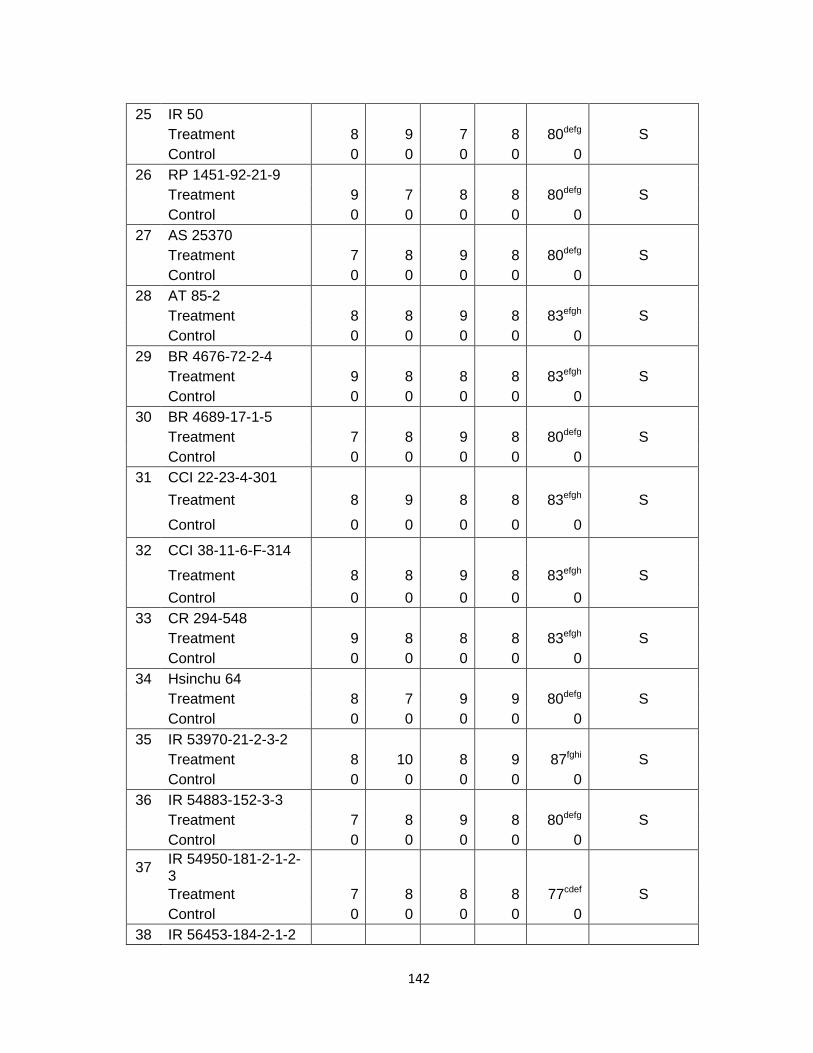
142
25 IR 50 Treatment 8 9 7 8 80defg S Control 0 0 0 0 0
26 RP 1451-92-21-9 Treatment 9 7 8 8 80defg S Control 0 0 0 0 0
27 AS 25370 Treatment 7 8 9 8 80defg S Control 0 0 0 0 0
28 AT 85-2 Treatment 8 8 9 8 83efgh S Control 0 0 0 0 0
29 BR 4676-72-2-4 Treatment 9 8 8 8 83efgh S Control 0 0 0 0 0
30 BR 4689-17-1-5 Treatment 7 8 9 8 80defg S Control 0 0 0 0 0
31 CCI 22-23-4-301 Treatment 8 9 8 8 83efgh S Control 0 0 0 0 0
32 CCI 38-11-6-F-314 Treatment 8 8 9 8 83efgh S Control 0 0 0 0 0
33 CR 294-548 Treatment 9 8 8 8 83efgh S Control 0 0 0 0 0
34 Hsinchu 64 Treatment 8 7 9 9 80defg S Control 0 0 0 0 0
35 IR 53970-21-2-3-2 Treatment 8 10 8 9 87fghi S Control 0 0 0 0 0
36 IR 54883-152-3-3 Treatment 7 8 9 8 80defg S Control 0 0 0 0 0
37 IR 54950-181-2-1-2-3
Treatment 7 8 8 8 77cdef S Control 0 0 0 0 0
38 IR 56453-184-2-1-2

143
Treatment Control
10 0
10 0
10 0
10 0
100i 0
S
39 IR 59682-132-1-1-2 Treatment 8 7 8 8 77cdef S Control 0 0 0 0 0
40 IR 60133-184-3-2-2 Treatment 9 10 7 9 87fghi S Control 0 0 0 0 0
41 IR 60832-187-2-2-2 Treatment 7 8 9 8 80defg S Control 0 0 0 0 0
42 IR 61009-37-2-1-2 Treatment 8 9 8 8 83efgh S Control 0 0 0 0 0
43 IR 62030-18-2-2 Treatment 7 8 9 8 80defg S Control 0 0 0 0 0
44 IR 62164-14-2-2-2-3 Treatment 9 8 8 8 83efgh S Control 0 0 0 0 0
45 MR 123 Treatment 7 8 9 8 80defg S Control 0 0 0 0 0
46 PK 2480-7-31 Treatment 10 10 10 10 100i S Control 0 0 0 0 0
47 PK 2557-24-2-1 Treatment 9 8 8 8 83efgh S Control 0 0 0 0 0
48 RAVI (RP 1664-1529-4254)
Treatment 9 8 9 9 87fghi S Control 0 0 0 0 0
49 RP 1670-1418-2205-1585
Treatment 8 9 8 8 83efgh S Control 0 0 0 0 0
50 S 972B-22-1-3-1-1 Treatment 8 10 9 9 90fghi S Control 0 0 0 0 0

144
51 S 976B-PN-25-1 Treatment 9 7 8 8 80defg S Control 0 0 0 0 0
52 IR 36 Treatment 8 7 8 8 77cdef S Control 0 0 0 0 0
53 Chainan 8 Treatment 8 8 7 8 77cdef S Control 0 0 0 0 0
54 Hsinchu 56 Treatment 9 8 6 8 77cdef S Control 0 0 0 0 0
*LSD (ANOVA at p<0.05) of 54 rice cultivars and lines disease index after inoculation with F. anthophilum (MRC 5806). The values with similar superscript are not significantly different at P< 0.05 (T-test)
S: susceptible 76-100% infected MS: moderately susceptible 51-75% infected MR: moderately resistant 26- 50% infected R: resistant 0- 25% infected

145
Figure 5.2 Cluster Analyses of disease severity of rice cultivars and lines after inoculation with F. anthophilum (MRC 5806)
Figure 5.2 Cluster analyses of disease severity of rice cultivars and lines after inoculation with F. anthophilum (MRC 5806)
Tree Diagram for 54 VariablesWard`s method
Euclidean distances
4 4 4 3 3 2 3 2 4 4 3 2 3 4 2 1 5 2 3 1 5 3 1 8 5 1 7 1 1 1 9 6 4 4 3 2 5 2 2 4 3 2 1 3 5 2 1 1 2 4 3 5 4 1
0
50
100
150
200
250
300
350
Linkage Distance
146
Key to the legend for Figure 5.2
1 AS 34011 2 BG 1165-2 3 BG 915 4 BR 1067-84-1-3-2-1 5 SKY Bonnet 6 E 7034 7 IET 11755 8 IR 29725-117-2-3-3 9 IR 52341-60-1-2-1
10 IR 53292-159-1-2-3 11 IR 53970-100-3-3-2 12 IR 56382-17-3-2 13 IR 56446-94-3-1-2 14 IR 56450-4-2-2 15 IR 57301-195-3-3 16 IR 57311-95-2-3 17 IR 59606-119-3 18 Kalyani II (CR 666) 19 NJ 70507 20 TC 10 (Local) 21 RP2199-16-2-2-1 22 RP 2434-22-3-3 23 ZHONG 83-49 24 BLUE BONNET 25 IR 50 26 RP 1451-92-21-9 27 AS 25370 28 AT 85-2 29 BR 4676-72-2-4 30 BR 4689-17-1-5 31 CCI 22-23-4-301 32 CCI 38-11-6-F-314 33 CR 294-548 34 Hsinchu 64 35 IR 53970-21-2-3-2 36 IR 54883-152-3-3 37 IR 54950-181-2-1-2-3 38 IR 56453-184-2-1-2 39 IR 59682-132-1-1-2

147
40 IR 60133-184-3-2-2 41 IR 60832-187-2-2-2 42 IR 61009-37-2-1-2 43 IR 62030-18-2-2 44 IR 62164-14-2-2-2-3 45 MR 123 46 PK 2480-7-31 47 PK 2557-24-2-1 48 RAVI (RP 1664-1529-4254) 49 RP 1670-1418-2205-1585 50 S 972B-22-1-3-1-1 51 S 976B-PN-25-1 52 IR 36 53 Chainan 8 54 Hsinchu 56

148
CHAPTER 6
COMPARATIVE EFFECTIVENESS OF TWO DIFFERENT FUNGICIDES IN CONTROLLING BAKANAE DISEASE OF RICE
6.1 Abstract
Two fungicides (benomyl 50% wettable powder (WP) and thiram 50% WP) at four
different concentrations (0.10%, 0.15%, 0.20% and 0.25%) were tested for their
effectiveness for controlling bakanae disease of rice caused by Fusarium anthophilum
(MRC 5806) and Fusarium fujikuroi (MRC 5807). Untreated seeds (0%) were used as
the experimental control. Benomyl was found most effective for controlling bakanae
disease when applied as a seed treatment. Thiram was found relatively in effective for
controlling bakanae disease of rice when applied as a seed treatment.
6.2 Introduction
Bakanae disease on rice has been reported in almost all countries where rice (Oryza
sativa) is grown commercially especially in Asian countries such as Malaysia and
Indonesia (Zainudin et al., 2008). Although bakanae disease was first described over
100 years ago in Japan, it is still not clear which Fusarium species are associated with
different symptoms of the disease (Amatulli et al., 2010). Early work in Japan identified
the pathogen as F. moniliforme (current valid name F. verticilioides) in a broad sense
(Ou, 1985; Amatulli et al., 2010).This taxon comprises a number of distinct species,
collectively called as the Gibberella fujikuroi species complex.Three mating populations
of G. fujikuroi complex have been associated with bakanae diseased rice plants
(Desjardins et al., 2000).

149
Mating population-C (anamorph F. fujikuroi) was first identified in 1977 among isolates
from Taiwan rice (Hsieh et al., 1977). Mating population-A (anamorph F. verticillioides)
and mating population-D (anamorph F. proliferatum were isolated from rice in Asia and
mating population-D was also isolated from rice in Africa, Australia and USA (Voight et
al., 1995; Desjardins et al., 1997; Amatulli et al., 2010). Wulff et al. (2010) reported that
F. fujikuroi and F. proliferatum both cause bakanae disease. Carter et al. (2008) showed
that F. fujikuroi as the causal pathogen of bakanae disease of rice (Oryza sativa), water
grass (Echinochloa oryzoidis) and barnyard grass (Echinochloa crus-galli) in California
of USA.
In the present study, both F. anthophilum and F. fujikuroi were isolated from diseased
rice plants with bakanae symptoms. Pathogenicity tests indicated that both F.
anthophilum and F. fujikuroi caused bakanae disease. Bakanae disease has been
reported from most rice producing countries such as Thailand (Kanjanasoon,1965),
Bangladesh (Mia and Zaman,1973), Southern Spain (Marin-Sanchez and Jimenez-
Diaz, 1982), Turkey (Copcu and Karaca, 1983), Nepal (Desjardins et al., 2000), Italy
(Amatulli et al., 2010), Malaysia and Indonesia (Zainudin et al., 2008; Heng et al., 2011),
and Pakistan (Bhalli et al., 2001; Iqbal et al., 2011; Habib et al., 2012; Ghazanfar et al.,
2013).
The control of bakanae disease of rice was suggested by cultivation of resistant
varieties (Li et al., 1993; Lv, 1994; Khokhar and Jaffrey, 2002). These scientists at the
same time complained that only a few rice germplasms have high resistance against
bakanae disease. Similarly in this study, screening of 54 rice cultivars and lines to
bakanae disease caused by Fusarium species in South Africa showed that only 2
cultvars and lines were moderately resistant. Ou (1985) reported that evaluation of
varietal resistance to rice blast, an important disease of rice caused by Pyricularia
oryzae Cavara is complicated because of the high potential variability of the fungus in
terms of pathogenic races in the field, differences in the level and type of resistance in
rice cultivars and environmental factors affecting the expression of resistance. He also
reported that 60 Philippines races or 34 international race groups were detected in the

150
IRRI (International Rice Research Institute) blast nursery during the period of 21 months
when samples were taken.
Bakanae disease is seed-borne and primarily seed transmitted (Desjardins et al., 1997;
Desjardins et al., 2000; Amatulli et al., 2010).The fungus of bakanae disease was
isolated from rice seeds (Hemmi et al., 1931; Kanjanasoon, 1965; Ou, 1985; Amatulli et
al., 2010). Hoschino (1955) emphasized the importance of seed treatment to control
bakanae disease of rice. Sawada and Kurosuwa (1924) recommended brine water for
seed treatment in order to eliminate the light weight diseased seed and formalin was
recommended for seed disinfection. Before 1970, several mercuric compounds such as
Granoson and Cereson lime were widely used in Japan and Taiwan in rice cultivation.
Since the ban on organo-mercurics, other fungicides have been tested, particularly
benomyl and thiram/benomyl combinations.These fungicides have been studied in
Japan (Umehara et al., 1973; Ito et al., 1974) and in Taiwan (Yu and Yang, 1978). They
revealed that benomyl and thiram/benomyl combinations are effective against bakanae
disease of rice caused by F. moniliforme (F. vertcillioides). Sarkar (1986) reported that
thiram reduced bakanae disease significantly in India as seed treatment. Sasaki (1987)
confirmed the control of bakanae disease of rice in Taiwan by treating seeds with
benomyl or thiram/benomyl combinations. Ahmad (1991) also reported that benomyl
had shown good results in inhibiting mycelial growth and sporulation of F. moniliforme
(G. fujikuroi). Bakanae disease of rice was controlled effectively with benomyl (Illyas
and Iftikhar, 1997). Bhalli et al. (2001) evaluated 8 fungicides viz. Apron, Benlate
(benomyl), Derosal, copperoxychloride, Ridomil, Seore, Topaz and Topsin-M to control
the mycelial growth of F. moniliforme.They revealed that the fungicides benomyl and
Derosal caused complete suppression of the growth of F. moniliforme (F. verticillioides),
the causal fungus of bakanae disease of rice when used at 100 ppm. Habib et al. (2012)
also confirmed the fact that benomyl and Derosal had the ability to suppress completely
the growth of the bakanae fungus.
The objective of this investigation was to evaluate the effectiveness of different
treatments of benomyl and thiram as seed treatment to control bakanae disease of rice

151
due to strains of F. anthophilum (MRC 5806) and F. fujikuroi (MRC 5807) under South
African conditions. This investigation was carried out with availalble resources and fund.
These 2 commonly used fungicides in South Africa were selected as they were found
effective to control bakanae disease of rice in other countries such as India, Japan and
Pakistan (Sarkar, 1986; Sasaki, 1987; Ahmad, 1991; Bhalli et al., 2001; Habib et al.,
2012).
6.3 Materials and Methods
Seeds of rice cultivar Hsinchu 56 (known susceptible cultivar in the fields) were pre-
soaked in cold water for 4 hours and soaked in hot water at 54ºC for 15 minutes to
eliminate external micro-organisms.The hot water treated seeds were then dried at
room temperature for future use.The hot water treated seeds were inoculated by
immersing them in the spore suspension of test isolate of F. anthophilum (MRC 5806)
and test isolate of F.fujikuroi (MRC 5807) under reduced pressure 200 mm Hg, so as to
infiltrate the suspension inside the husk following the protocol of Haque et al. (1979),
Ahmed et al. (1986) and Dhingra and Sinclair (1985).
Dry, powdered formulations of the respective fungicides were used to treat the
inoculated seeds with Benomyl 50% WP and Thiram 50% WP at 4 different treatments/
concentrations (0.10%, 0.15%, 0.20% and 0.25%). Inoculated, but untreated seeds
(0%) were used as the experimental control. Five days after treatment, 10 sprouted
seeds were planted into 12.5 cm diameter pots containing sterilized sand. This was
done for each of the different treatment concentrates mentioned above. At least 16
potted seedlings were grown per treatment for each of the two respective test isolates.
Sixteen potted seedlings were grown as controls in a growth chamber. The growth
chamber was set for 14 hours per day photo-period of about 15 000 lux fluorescent
light. Temperature and relative humidity ranged from 20°C to 25°C and 70% to 90%
respectively in the growth chamber as recommended by Marin-Sanchez and Jimenez-
Diaz (1982). A half strength Hoagland’s nutrient solution (Hoagland and Aron, 1950;

152
Dhingra and Sinclair, 1985) was used to provide nutrients for the growth of plants.The
pots were arranged in a randomized block design with four replicates (Mead and
Curnow, 1987; Box et al., 2005). Final counts of diseased and healthy seedlings were
made when plants were 28 days old.
Statistical analysis:
The effect of different concentrations (0.10%, 0.15%, 0.20% and 0.25%) of benomyl
and thiram against F. anthophilum (MRC 5806) and (F. fujikuroi (5807) were measured
and statistically analysed by using the Least Significant Difference (LSD) as an Analysis
of Variance (ANOVA). Least Significant Difference was calculated at p<0.05. Data
analysis was performed using STATISTICA software (Stat soft Inc., version 17.0). The
raw data sets were expressed as percentage of diseased plants. For each
concentration of treatment fungicide, the final disease index was calculated as the mean
of the four pots in each of the four replicates.
6.4 Results
Efficacy results of the different treatments of benomyl and thiram against bakanae
disease caused by F. anthophilum (MRC 5806) have been summarized in Table 6.1
showing statistical significant difference of treatments of two fungicides.This shows that
benomyl gave complete control of the disease at concentrations of 0.15%, 0.20% and
0.25%. Benomyl gave 97% significant control of the disease at concentration of 0.10. At
concentrations of 0.10% and 0.15%, thiram gave only 17% disease control. At 0.20%,
50% disease control was achieved and 75% disease control at a concentration of
0.25%.
Efficacy results of the different treatments of benomyl and thiram against bakanae
disease caused by Fusarium fujikuroi (MRC 5807) have been summerized in Table 6.2
showing statistical significant differences of treatments of two fungicides.This shows

153
that benomyl gave complete control of the disease at concentrations of 0.15; 0.20 and
0.25. It gave 90% disease control at the concentration of 0.10. Thiram completely failed
to control the disease at 0.10% and 0.15%. It gave only 15% disease control at 0.20%
and it gave 40% disease control at 0.25%.
Benomyl was found as an effective seed treatment fungicide to control bakanae disease
of rice. Thiram was found relatively as an ineffective seed treatment fungicide against
both test isolates.
6.5 Discussion and conclusion
The control measures of bakanae disease of rice have been complicated due to the
involvement of different species of Fusarium with the disease. Due to lack of proper
control measures, bakanae disease incidence threat is an increasing trend and this has
resulted in a serious disease problem in the main rice growing countries of the world
(Wahid et al., 1991; Ma et al., 2008; Iqbal et al., 2011; Ghazanfar et al., 2013). In our
previous experiments on the reactions of 54 rice cultivars/lines to bakanae disease,
none of the cultivars and lines were found resistant against the disease caused by F.
anthophilum (MRC 5806) or F. fujikuroi (MRC 5807). For this reason, there was a need
to investigate other method of disease control such as seed treatment with fungicides
such as benomyl and thiram to control bakanae disease of rice.
The conclusion might be drawn from the results that seed treatment with benomyl prior
to planting is highly significant in controling bakanae disease of rice caused by both
isolates of F. anthophilum (MRC 5806) and F. fujikuroi (MRC 5807). The results of this
study on the effectiveness of benomyl as seed treatment fungicide to control bakanae
disease corroborate the conclusions of Sasaki (1987), Ahmad (1991), Illyas and Iftikhar
(1997), Bhalli et al. (2001), Iqbal et al. (2011) and Habib et al. (2012) who suggested
benomyl as good seed treatment fungicide to control bakanae disease of rice. It is also
clear that thiram was relatively ineffective as a seed treatment against both test isolates.

154
In this investigation, thiram at 0.25% reduced bakanae disease incidence to a certain
extent under controlled environmental conditions but the disease control was not very
effective. Sarkar (1986) reported that thiram reduced bakanae disease incidence
significantly in India as a seed treatment fungicide at 0.25% in field conditions, but it did
not give complete control.The findings of the present study do not support these
findings. Good protection against isolates of F. moniliforme (F. verticillioides) causing
bakanae disease of rice in Japan was obtained by dipping seeds for 24 hours in
ipconazole (Tateishi and Chida, 2000). In this study Benomyl was found as an effective
fungicide for controlling bakanae disease of rice. Benomyl was also found effective to
control bakanae disease of rice in Turkey (Surek and Gumustekin, 1994). Sinohin
(2011) reported that benomyl or thiophanate methyl as seed treatment is very effective
to control bakanae disease of rice in the Philippines. Thiram-benomyl wettable powder
(active ingredient: thiram 20%, benomyl 20%) was found as an effective seed treatment
fungicides to control loose smut a seed borne disease of wheat caused by F. roseum
Lk. Emend. Snyd. & Hans. Pro parte [S&H] in Japan (Nakagawa and Yamaguchi,
1989). They also suggested that same fungicides would be useful to control loose smut
of barley caused by F. roseum. However, an isolate of G. fujikuroi the causal agent of
bakanae disease of rice has been reported as highly resistant to benomyl and was first
discovered in 1980. In 1987 most of the isolates of G. fujikuroi were highly resistant to
benomyl (Ogawa and Takeda, 1990).This development of resistance was suspected
due to successive applications of benomyl as a seed treatment (Ogawa and Takeda
(1990). Low and high levels of resistance to the benzimidazole fungicides benomyl and
thiophanate-methyl were observed in the field isolates of Monilinia fructicola (G. Wint.)
Honey, the causal agents of brown rot of stone fruits (Ma et al., 2003).
Fusarium fujikuroi the causal agent of bakanae disease of rice is highly diverse on the
basis of DNA fingerprinting analysis and due to high diversity the fungus poses a risk in
terms of the development of fungicide resistance and susceptibility of new cultivars
(Cumagun, 2012). The fungus has been reported to produce a considerable amount of
fumonisins in corn but fortunately is minimal in rice (Cumagun, 2012). However, the
minimal risk can not be underestimated, because rice is one of the world’s most

155
important crops.There is a need to screen more fungicides to be used as seed
treatment in South Africa. There is a need to screen more rice varieties/cultivars to find
resistant one to control bakanae disease. Development of resistant varieties must be
based on the information of genetic diversity, aggressiveness and the production of
mycotoxins by Fusarium spp. The control strategy should also include biological option.
Bacterization of naturally infected rice seeds reduced bakanae disease incidence in
seed box and seedbed tests (Rosales and Mew, 1997).

156
Reference
Ahmad, M.I. 1991. Incidence of Fusarium moniliforme Sheldon in different rice seed
samples and some physiological studies on the fungus. M.Sc.Thesis, Univ Agric.,
Faisalabad, Pakistan. P115.
Ahmed, H.U., Mia, M.A. and Miah, S.A. 1986. Standardized test tube inoculation for
bakanae disease. International Rice Research Newsletter 11 (2): 21-22.
Amatulli, M.T., Spadaro, D., Gullino, M.L. and Garibaldi,A. 2010. Molecular identification
of Fusarium spp. associated with bakanae disease of rice in Italy and assessment of
their pathogenicity. Plant Pathology 59: 839-844.
Anonymous, 1975. Co-operative studies on control of bakanae, brown spot and
damping off of rice plant. Report of the Kinki-Chugoku National Agricultural Experiment
Station, Japan. 1-77.
Bhalli, J.A., Aurangzeb and Ilyas, M.B. 2001. Chemical control of bakanae disease of
rice caused by Fusarium moniliforme. Journal of Biological Science 1: 483-484.
Box, G.E.P., Hunter, J.S. and Hunter, W.G. 2005. Statistics for Experimenters: Design,
Innovation and Discovery. Pubisher: John Wiley and Sons, Inc., ISBN-13 978-0471-
71813-0 Hoboken, New Jersey, USA. 633p.
Copcu, M. and Karaca, I. 1983. Investigation of rice disease caused by fungi, their
distribution, over wintering in the Aegean region of Turkey, 1. Determination of rice
diseases causal agents and distribution, prevalence and incidence. Journal ofTurkish
Phytopathology, 12: 61-72.

157
Desjardins, A.E., Plattner, R.D. and Nelson, P.E. 1997. Production of fumonisin B1 and
moniliformin by Gibberella fujikuroi from rice from various geographic areas. Applied
and Environmental Microbiology 63: 1838-1842.
Desjardins, A.E., Manandhan, H.K., Plattner, R.D., Manandhar, G.G., Poling, S.M. and
Maragos, C.M. 2000. Fusarium species from Nepalese rice and production of
mycotoxins and gibberellic acids by selected species. Applied and Environmental
Microbiology 66 (3): 1020-1025.
Dhingra, O.D. and Sinclair, J.B. 1985. Basic Plant Pathology Methods. Publisher: CRC
Press, Inc. ISBN 0-8493-5921-X, Florida, USA. p355.
Ghazanfar, M.U., Javed, N., Wakil, W. and Iqbal, M. 2013. Screening of some fine and
coarse rice varieties against bakanae disease. Journal of Agricultural Research 51(1):
41-49.
Habib, A., Javed, N., Sahi, S.T. and Waheed, M. 2012. Detection of seed borne
mycoflora of different coarse and fine rice varieties and their management through seed
treatments. Pakistan Journal of Phytopathology 24 (2): 133-136.
Haque, M.A., Rahman, M.M. and Miah, S.A. 1979. A rapid method of screening rice
varieties for resistance to bakanae disease. International Rice Research Newsletter 28
(2): 34-37.
Hemmi, T. Seto, T. and Ikaya, J. 1931. Studies on the bakanae disease of the rice plant
II. On the infection of rice by Lisea fujikuroi Sawada and Gibberella saubinetti (Mont.)
Sacc. in the flowering period. Forschungen aus dem Gebiet der Pflanzenkrankheiten 1:
99-100. [Ja, en].

158
Heng, M.H., Baharuddin, S. and Latiffah, Z. 2011. Molecular identification of Fusarium
species in Gibberella fujikuroi species complex from rice, sugarcane and maize from
Peninsular Malaysia. International Journal of Molecular Science 12: 6722-6732.
Hoagland, D.R. and Aron, D.I. 1950. The culture method for growing plants without soil.
Californian Agricultural Station Circular 347: 1-34.
Hoschino, Y. 1955. A study on the rice seed disinfection with special reference to
bakanae disease (In Japanese, English summary) Jubilee Publication Commemoration
the 60th Birthdays of Professors Y. Tochinai and T. Fukushi: 290-299. Sapporo. Japan.
Hsieh, W.H., Smith, S.N. and Snyder, W.C.1977. Mating groups in Fusarium
moniliforme. Phytopathology 67: 1041-1043.
Illyas, M.B. and Iftikhar, K. 1997. Screening of rice germplasm and fungitoxicants
against bakanae disease of rice. Pakistan Journal of Plant Pathology 9(1): 67-73.
Iqbal, M., Javed, N., Sahi, S.T. and Cheema, N.M. 2011. Genetic management of
bakanae disease of rice and evaluation of various fungicides against Fusarium
moniliforme in vitro. Pakistan Journal of Phytopathology 23 (2): 103-107.
Ito, H., Hirayama, S., Miura, H., Tokairin, H. and Tanaka, T. 1974. Studies on
epidemiology and control of bakanae disease of rice plants. Bulletin of the Yamagata
Prefecture Agricultural Experiment Station. 8: 97-117 [Japanese].
Kanjanasoon, P. 1965. Studies on the bakanae disease of rice in Thailand. Doc.
Agric.Thesis. Tokyo University, Japan. 120p.

159
Khokhar, L.K. and Jaffrey, A.H. 2002. Identification of sources of resistance against
bakanae and foot rot disease in rice. Pakistan Journal of Agricultural Research 17: 176-
177.
Li, D.J., Luo, K. and Chen, Z. 1993. Studies on resistance of rice materials to bakanae
disease and pathogenicity of pathogen (Fusarium moniliforme) Acta Phytopathology
Sinica 23: 315-319.
Lv, B. 1994. Preliminary study of identification for resistance to rice bakanae disease in
rice resource. Plant Protection. 3: 20-21.
Ma, Z., Yoshimura, M.A. and Michailides, T. 2003. Identification and Characterization of
Benzimidazole Resistance in Monilinia fructicola from Stone Fruit Orchards in California.
Applied and Environmental Microbiology 69 (12): 7145-7152.
Ma, L.Y., Ji, Z., Bao, J., Mu, X., Zhuang, J. Yang, C. and Xia, Y. 2008. Responses of
rice genotypes carrying different dwarf genes to Fusarium moniliforme and gibberellic
acid.
Marin-Sanchez, J.P. and Jimenez-Diaz, R.M. 1982. Two new Fusarium species in
Southern Spain. Plant Disease 66 (4): 332-334.
Mia, S.A. and Zaman, M.Q. 1973. Studies on the reaction of high yielding dwarf to foot
rot and bakanae in Bangladesh. Bangladesh Journal of Botany 2: 73-81
Mead, R. and Curnow, R.N 1987. Statistical Methods in Agriculture and Experimental
Biology, Publisher: Chapman and Hall, ISBN 0 412 2430 2, London, UK. 335p.

160
Nakagawa, A. and Yamaguchi, T. 1989. Seed Treatment for Control of Seed-Borne
Fusarium roseum on Wheat. Journal of Agricultural Research and Quarantine 23 (2):
94-99.
Ogawa, K. and Takeda, S. 1990. Population of benomyl-resistant rice “bakanae” fungus
in paddy fields. Annals of the Phytopathological Society of Japan 56 (2): 247-249.
Ou, S.H. 1985. Rice diseases. Publisher: Commonwealth Mycological Institute, ISBN 0
85198 545 9, Kew, Surrey, UK. 380p.
Rosales, A.M. and Mew, T.W. 1997. Suppression of Fusarium moniliforme in Rice by
Rice- Associated Antagonistic Bacteria. Plant Disease 81 (1): 49-52.
Sarkar, B.B. 1986. Controlling bakanae and foot rot disease with fungicide seed
treatments International Rice research Newsletter 11(3): 18.
Sasaki, T.1987. Epidemiology and control of rice bakanae disease. Bulletin of the
Tohoku National Agricultural Experiment Station, Japan, 74: 1-47.
Sawada, K. and Kurosawa, E.1924. Control of bakanae disease (In Japanese).
Formosan Agriculture Department Special Bulletin 21: 1-11.
Sinohin, A.M. 2011. Diagnosis and management of diseases in rice.
http://www.slideshare.net/macky75/common-rice-diseases-am-sinohin
Surek, H. and Gumustekin, H. 1994. Research activities on controlling rice bakanae and
foot rot disease (Fusarium moniliforme) in Turkey. Cahiers Options Mediterraneennes 8
(2): 27-30.

161
Tateishi, H., and Chida, T. 2000. Sensitivity of Fusarium moniliforme Isolates to
Ipconazole. Journal of General Plant Pathology 66 (4): 353-359.
Thomas, K.M. 1931. A new paddy disease in Madras. Madras Agricultural Journal 19:
34-36.
Umehara, Y. Wakamatsu, T., Komatsu, M. and Yumo, I. 1973. On the control of the
bakanae disease of rice plant by seed disinfectants. Proceedings of the Association of
Plant Protection, Hokuriku 21: 92-95.
Voight, K., Schleier, S. and Bruckner, B. 1995. Genetic variability in Gibberella fujikuroi
and some related species of the genus Fusarium based on random amplification of
polymorphic DNA (RAPD). Current Genetics 27: 528-535.
Wahid, A.M., Khan M.U., Tariq, A.R. and Saleem, A. 1991. Seed mycoflora of rice.
Pakistan Journal of Agricultural Research 31: 95-100.
Wulff, E.G., Sørensen, J.L., Lübeck, M., Nielsen, K.F., Thrane, U. and Torp, J. 2010.
Fusarium spp. associated with rice Bakanae: Ecology, genetic diversity, pathogenicity
and toxigenicity. Environmental Microbiology 12: 649-657.
Yu, C.M. and Yang, H.K. 1978. Effect of seed treatment fungicides on the incidence of
rice bakanae disease. Plant Protection Bulletin, Taiwan 20: 73-76. [ch, en].
Zainudin, N.A.I.M., Razak, A.A. and Salleh, B. 2008. Bakanae disease of rice in
Malaysia and Indonesia: Etiology of the causal agent based on morphological,
physiological and pathogenicity characteristics. Journal of Plant Protection Research 48
(4): 475-48.
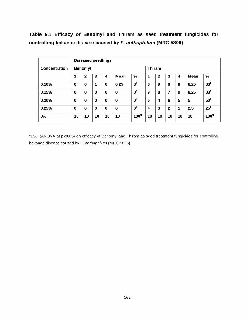
162
Table 6.1 Efficacy of Benomyl and Thiram as seed treatment fungicides for controlling bakanae disease caused by F. anthophilum (MRC 5806)
Diseased seedlings
Concentration Benomyl Thiram
1 2 3 4 Mean % 1 2 3 4 Mean %
0.10% 0 0 1 0 0.25 3a 8 9 8 8 8.25 83f
0.15% 0 0 0 0 0 0a 9 8 7 9 8.25 83f
0.20% 0 0 0 0 0 0a 5 4 6 5 5 50d
0.25% 0 0 0 0 0 0a 4 3 2 1 2.5 25c
0% 10 10 10 10 10 100g 10 10 10 10 10 100g
*LSD (ANOVA at p<0.05) on efficacy of Benomyl and Thiram as seed treatment fungicides for controlling
bakanae disease caused by F. anthophilum (MRC 5806).

163
Table 6.2 Efficacy of Benomyl and Thiram as seed treatment fungicides for controlling bakanae disease caused by F. fujikuroi (MRC 5807)
Diseased seedlings
Concentration Benomyl Thiram
1 2 3 4 Mean % 1 2 3 4 Mean %
0.10% 1 1 2 0 1 10b 10 10 10 10 10 100g
0.15% 0 0 0 0 0 0a 10 10 10 10 10 100g
0.20% 0 0 0 0 0 0a 9 8 9 8 8.5 85f
0.25% 0 0 0 0 0 0a 5 6 7 6 6 60e
0% 10 10 10 10 10 100g 10 10 10 10 10 100g
*LSD (ANOVA at p<0.05) on efficacy of Benomyl and Thiram as seed treatment fungicides for controlling
bakanae disease caused by F. fujikuroi (MRC 5807).

164
CHAPTER 7
STUDIES ON THE PRODUCTION OF MYCOTOXINS BY SELECTED FUSARIUM SPECIES ISOLATED FROM RICE (ORYZA SATIVA)
7.1 Abstract
The ability of 3 representative isolates of Fusarium anthophilum (MRC 5519, MRC 5520
and MRC 5806) and 3 isolates of Fusarium fujikuroi (MRC 5807, MRC 5808 and MRC
5809) to produce mycotoxins were studied. All test isolates of Fusarium species were
obtained from rice plants exhibiting bakanae disease in the fields of South Africa. All 3
isolates of F. anthophilum tested produced the mycotoxin moniliformin in quantities
ranging from 32μg/g to 51μg/g. All 3 isolates of F. fujikuroi produced mycotoxin
moniliformin in quantity ranging from 572μg/g to731 μg/g. None of the representative
isolates of F. anthophilum and F. fujikuroi produced fumonisins (FB1, FB2, and FB3).
This is the first report on the production of moniliformin by F. anthophilum and F.
fujikuroi isolated from rice plants with bakanae disease under South African field
conditions.
7.2 Introduction
Fusarium species are found worldwide in geographical distribution and are well known
as plant pathogenic and toxigenic fungi (Smith, 1971; Nelson et al., 1983; Marasas et
al., 1984; Leslie et al., 1996; Desjardins et al., 2000; Desjardins, 2006; Zainudin, 2008;
Nayaka et al., 2011; Ghazanfar et al., 2013). They produce mycotoxins that negatively
affect animal and human health (Scott et al., 1987; Peraica et al., 1999; Desjardins et
al., 2000; Hussein and Brasel, 2001; Desjardins, 2006; Glenn, 2007; Nayaka et al.,

165
2011; Van Rensburg, 2012). Murray et al. (2005) reported that mycotoxins have been
implicated in a variety of illnesses and clinical syndromes in humans and animals.
Mycotoxins are secondary metabolites which are produced by various fungi and pose a
continuous challenge to the safety and quality of food commodities in South Africa
(Lezar and Barros, 2010). These toxins can cause acute or chronic toxicity in humans
and animals that eat contaminated foods or crops. Manifestation of these symptoms
however, depend on the quantities produced and consumed (Barrett, 2000; Lezar and
Barros, 2010). Mycotoxins that adversely affect human or animal health are found on
virtually any food or animal feeds which support fungal growth including cereal grains
such as maize, rice, sorghum, wheat and oil seeds, ground nuts, cotton seeds, oil-palm
kernels and copra (Marasas et al., 1984; Marasas and Nelson, 1987; Sydenham and
Thiel, 1996; Desjardins et al., 2000; Glenn, 2007; Lezar and Barros, 2010; Van
Rensburg, 2012).
The economic impact of mycotoxins include loss of human and animal life, increased
health care and veterinary care costs, reduced livestock production, disposal of
contaminated food and feeds, and investment in research and applications to reduce
severity of the mycotoxin problem (Hussein and Brasel, 2001; Glenn, 2007).It is
estimated that 25% of the world’s food crops are affected by mycotoxins each year,
which means that Fusarium species are playing an important role and substantially
contribute to food contamination (Charmley et al., 1994; Mellor, 2003; Lezar and Barros,
2010; Van Rensburg, 2012).
The mycoflora are well known to produce various mycotoxins such as aflatoxin,
beauvericin, chlamydosporol, enniatins, equistin, fumonisins, moniliformin, neosolaniol,
trichothecene, ochratoxin A and zearalenone that negatively affect human and animal
health (Marasas et al., 1984; Marasas and Nelson, 1987; Peraica et al., 1999;
Desjardins et al., 2000; Desjadins et al., 2006; Van Rensburg, 2012).
Fumonisins and Moniliformin Production

166
Fumonisins are a group of mycotoxins originally characterized from culture material of
Fusarium verticillioides (F. moniliforme) strain MRC 826) (Bezuidenhout et al., 1988;
Gelderblom et al., 1988). The strain was isolated from maize in the Transkei region of
South Africa and was known to be hepatotoxic to ducklings and rats (Kriek et al., 1981;
Colvin and Harrison, 1992). Fumonisins B1 and B2 have toxicological significance and
while others (B3, B4, A1 and A2) have low concentrations and are less toxic (Peraica et
al., 1999). It was shown that fumonisin B1 is responsible for leukoencephalomalacia
(ELEM) disease in horses (Marasas et al., 1988; Kellerman et al. 1990; Wilson et al.,
1990; Thiel et al., 1991; Gelderblom et al., 1992; Rheeder et al., 2002) and pulmonary
edema in pigs (Harrison et al., 1990; Ross et al., 1990; Gelderblom et al., 1992; Nelson
et al., 1994; Sala et al., 1994; Maheshwar et al., 2009). Fumonisins have been
associated with oesophageal cancer in humans (Marasas et al., 1981; Marasas et al.,
1982; Sydenham et al., 1991; Thiel et al., 1992; Dutton, 1996; Van Rensburg, 2012).
Fumonisins have been associated with a high incidence of oesophageal cancer in
people living in South Africa, China and Italy (Murray et al., 2005). The fungi that
produce fumonisins are well known pathogens of a variety of plants (Bhargava et al.,
1978; Nelson et al., 1983; Cartwright et al., 1995; Abbas et al., 1998; Desjardins et al.,
2000; Desjardins, 2006). Fumonisins have been isolated in Fusarium spp. and
Alternaria spp. (Abbas and Riley, 1996; Deasjardins, 2006). Frisvads et al. (2007)
detected fumonisin B2 in cultures of Aspergillus niger for the first time. Fumonisin B2
production by strains of A. niger was shown again by Noonim et al. (2009) in Thai coffee
beans. Morgensen et al. (2010) showed the production of fumonisin FB2 and FB4 by A.
niger on grapes and raisins. Recently Soares et al. (2013) showed the production of
FB2 by A. niger aggregate strains isolated from harvested maize in three Portuguese
regions.
The presence of fumonisins has been confirmed in at least twenty five countries of the
world (Mazzani et al., 2001; Van Rensburg, 2012). Fumonisin B1 accounts for 70-80%
of total fumonisins produced, FB2 usually makes up to 15-25% and FB3 3-8% (Dilkin et
al., 2002; Rheeder et al., 2002; Van Rensburg, 2012). International Agency for
Research on Cancer (IARC) evaluated fumonisin B1 (FB1) as a possible carcinogen

167
(Group 2B) to humans (Domijan et al., 2005). International Agency for Research on
Cancer grouped carcinogens into different categories such as Group 1(carcinogenic to
humans), Group 2A (probably carcinogenic to humans), Group 2B (possibly
carcinogenic to humans), Group 3 (not classifiable as to its carcinogenicity to humans)
and Group 4 (probably not carcinogenic to humans). Carcinogens are classified as 2B
on the basis of strong evidence from mechanistic and other relevant data (IARC, 1993;
IARC, 2011).
Moniliformin production was reported from infected rice plants and rice seeds with
bakanae disease caused by Fusarium spp. (Desjardins et al., 1997; 2000; Desjardins,
2006). Moniliformin production in food and feed products has been reported from
various Fusarium spp. world wide (Nelson et al., 1993; Rheeder et al., 2002; Sagaram
and Shim, 2007; Saremi et al., 2008). Moniliformin was first reported from natural
occurrence in mouldy maize from the Transkei region of South Africa (Thiel et al.,
1982). In South Africa, moniliformin contamination of maize was associated with the
presence of F. subglutinans (Wollenw. & Reinking) P.E. Nelson, Toussoun & Marasas
(Thiel et al., 1982; Desjardins, 2006). Moniliformin is produced by F. verticillioides a
member of the section Liseola (Cole et al., 1973; Rabie et al, 1982; Marasas et al.,
1984; Marasas et al., 1986; Leslie et al., 1996; Sydenham et al., 1997). There is
however, considerable controversy surrounding its production as summarized by
Marasas et al. (1986).The mycotoxin moniliformin (sodium or potassium salt of 1-
hydroxycyclobut-1-ene-3, 4- Dione) is a highly toxic compound which was first isolated
by Cole et al. (1973) from corn that had been inoculated with F. verticillioides. The
mycotoxin caused the development of pathological lesions on the tissues of the hearts
of experimental animals and subsequent death (Kriek et al., 1977; Burmeister et al.,
1980; Allen et al., 1981; Zhao et al., 1993). Chelkowski et al. (1990) showed the
production of moniliformin by F. anthophilum but they could not show production of
moniliformin by strains of F. chlamydosporum, F. compactum and F. equiseti.
Desjardins (2006) however reported that although the isolation of moniliformin was
based on toxicity to 1 day old chicks, the mycotoxin has not been associated with any
chronic or fatal animal disease outbreak. A strain of F. fujikuroi that produced

168
moniliformin but no fumonisin was crossed with a strain of F. proliferatum that produced
fumonisin but no moniliformin (Desjardins, 2006).
Due to the regular isolation of F. anthophilum and F. fujikuroi during this investigation in
South Africa, this study aimed to determine the ability of 3 representative isolates of F.
anthophilum (MRC 5519, MRC 5520 and MRC 5806) and F. fujikuroi (MRC 5807, MRC
5808 and MRC 5809) obtained from rice plants with bakanae disease to produce the
mycotoxins fumonisins (FB1, FB2 and FB3) and moniliformin with available resources
and fund. Apart from risks assessment, to determine the ability of isolates of F.
anthophilum and F. fujikuroi to produce the mycotoxins fumonisins and moniliformin
may strengthen their identification as an individual biological species (Leslie et al.,
1992a, Leslie et al., 1992b; Leslie et al., 1996).
7.3 Materials and Methods
Three representative isolates of F. anthophilum (MRC 5519, MRC 5520 and MRC 5806)
and 3 isolates of F. fujikuroi (MRC 5807, MRC 5808 and MRC 5809) obtained from
diseased rice plants and preserved in the culture collection of the Programme on
Mycotoxins and Experimental Carcinogenesis (PROMEC), Medical Research Council
(MRC), Tygerberg, South Africa, were used in this study. The isolates of F. anthophilum
and F. fujikuroi were obtained from diseased rice plants exhibiting bakanae symptoms
in the fields of North West province and Northern Cape province of South Africa.
Lyophilised conidia of the respective Fusarium isolates were suspended in sterile water
and used to inoculate moistened yellow corn kernels (400g of kernels and 400 ml of
water) in 2 liter glass fruit jars previously autoclaved at 121°C for 1 hour on each of 2
consecutive days prior to use. The fungal cultures on corn were incubated at 25°C for
21 days, dried overnight at 45°C, ground to a fine meal in a laboratory mill, and stored
at 0°C until analysed as recommended by Thiel et al. (1982, 1991) and Marasas et al.

169
(1986). Corn was chosen as substrate for the production of fumonisins and moniliformin
for the following reasons:
• Both Fumonisins and moniliformin are prevalent on corn and major contaminant
of corn (Marasas et al., 1984; Sala et al., 1994; Dutton, 1996).
• Both mycotoxins were isolated from corn for the first time (Cole et al., 1973;
Bezuidenhout et al., 1988; Gelderblom et al., 1988).
• Corn is considered as one of the crops more susceptible to mycotoxin in the
world (Soares et al., 2013).
• It was expected that corn as a substrate would provide better condition for
production of fumonisins and moniliformin for the fungal cultures.
Moniliformin Analyses:
Moldy meal prepared from Fusarium cultures were analysed by High Performance
Liquid Chromatography (HPLC) as described by Thiel et al. (1982, 1991) and Marasas
et al. (1986).The results obtained by employing a strong anion-exchange column was
substantiated by separation and quantification of moniliformin using a paired ion
chromatography (PIC) technique on a reverse phase column. HPLC analyses were
carried out on both water extracts of the meal samples and water extracts prepurified on
columns of DEAE-Sephadex. Sample extracts were prepared by shaking 3g of the dry
meal with 40 ml of distilled water on a rotary shaker for 2h at room temperature in
centrifuge tubes.The contents were then centrifuged for 20 min at 10 000g and the
supernatant extracts were filtered through Millipore filters (0.45 μM) and retained for
HPLC analyses or for pre-purification on a DEAE-Sephadex column.
Fumonisins Analyses:
Test samples were analysed for fumonisins (FB1, FB2, FB3) by HPLC according to the
method of Shepard et al. (1990) and Thiel et al. (1991). Briefly, samples were extracted

170
with methanol: water (3:1, vol/vol), centrifuged, and filtered. The filtrate was applied to a
Bond-Elut SAX cartridge and eluted with 0.5% acetic acid in methanol. The solution was
evaporated, and the residue was derivatized with o-phthaldihyde (OPA) and injected
onto the HPLC column. The detection limit was 0.5μg/g.
7.4 Results
Results of the production of mycotoxin by all the test isolates of F. anthophilum (MRC
5519, MRC 5520 and MRC 5806) and F. fujikuroi (MRC 5807, MRC 5808 and MRC
5809) are summarized in Table 7.1. All representative isolates of both Fusarium species
produced moniliformin.The isolates of F. anthophilum such as MRC 5519 produced
32µg/g, MRC 5520 produced 48µg/g and MRC 5806 produced 51µg/g of moniliformin
respectively.The isolates of F. fujikuroi such as MRC 5807 produced 731µg/g, MRC
5808 produced 669µg/g and MRC 5809 produced 572 µg/g of mycotoxin moniliformin
respectively. Isolates of Fusarium fujikuroi produced much higher levels (10 fold) of
moniliformin than F. anthophilum. The highest producer of moniliformin was F. fujikuroi
(MRC 5807).The lowest producer of moniliformin was F. anthophilum (MRC 5519). All
the representative 3 isolates of F. anthophilum produced moniliformin in quantities
ranging from 32μg/g to 51μg/g.Three isolates of F. fujikuroi produced moniliformin in
quantities ranging from 572μg/g to 731μg/g. None of the representative isolates of F.
anthophilum and F. fujikuroi produced any chemically detectable fumonisins (FB1, FB2
and FB3).
7.5 Discussion and conclusion
Moniliformin
The present study showed that all 3 strains of F. anthophilum (MRC 5519, MRC 5520
and MRC 5806) and 3 strains of F. fujikuroi (MRC 5807, MRC 5808 and MRC 5809)
isolated from diseased rice plants showing bakanae symptoms in the fields of North

171
West and Northern Cape provinces, South Africa could produce moniliformin. Marasas
et al. (1986) reported that the largest quantities of moniliformin (up to 11717 mg/kg)
were produced by isolates of Fusarium species isolated from rice in Taiwan, Japan and
Philippines with bakanae disease that resembled F. fujikuroi (Nirenberg, 1976). The
mycotoxin moniliformin was found to be toxic to ducklings. Marasas et al. (1986) also
reported the production of moniliformin by isolates of F. anthophilum isolated from soil
debris in New Guinea (up to 830 mg/kg) and from wheat in USA (up to 510 mg/kg).The
moniliformin was found to be toxic to ducklings. Fusarium proliferatum isolated from
various crops in different countries produced moniliformin up to 293 mg/kg (Marasas et
al., 1986). Desjardins et al. (1997) showed high levels of moniliformin production (up to
7000μg/g) by isolates of F. fujikuroi obtained from rice. However, these isolates of F.
fujikuroi could not produce fumonisins. External factors or environment conditions seem
to play a lesser role in the production of fumonisins.
The mycotoxin moniliformin production in food and feed product has been reported from
Fusarium spp. isolated from various countries of the world. (Nelson et al., 1993;
Desjardins et al., 2000; Rheeder et al., 2002; Glenn, 2007; Sagaram and Shim, 2007;
Saremi et al., 2008). Desjardins (2006) showed the production of moniliformin by
isolates of F. anthophilum, F. chlamydosporum, F. equiseti, F. fujikuroi and F.
semitectum.The production of moniliformin has also been reported by strains of F.
chlamydosporum, F. equiseti and F. semitectum (Rabie et al., 1978, 1982; Marasas et
al., 1984; Scott et al., 1987; Farber et al., 1988; Lezar and Barros, 2010).
Most studies on the investigation of mycotoxin from bakanae diseased rice plants and
rice seeds confirmed the production of moniliformin by F. fujikuroi (Marasas et al., 1986;
Desjardins et al., 1997; Desjardins et al., 2000; Desjardins, 2006).This finding is in good
agreement with my observation with three isolates of F. fujikuroi isolated diseased rice
plants with bakanae symptoms in South Africa.Three isolates of F. fujikuroi from
diseased rice plants produced much higher levels (10 folds) of moniliformin than three
isolates of F. anthophilum.

172
Three isolates of F. anthophilum tested for production of moniliformin confirmed the
previous report (Marasas et al., 1986) on moniliformin production by isolates of F.
anthophilum isolated from soil debris in New Guinea and from wheat in USA.This is the
first report of production of moniliformin by F. anthophilum and F. fujikuroi strains
isolated from rice (Oryza sativa) showing symptoms of bakanae disease in South Africa.
Fumonisins
In this study isolates of F. anthophilum (MRC 5519, MRC 5520 and MRC 5806) and F.
fujikuroi (MRC 5807, MRC 5808 and MRC 5809) were not able to produce fumonisins.
The production of fumonisins by Fusarium anthophilum has been reported by many
researchers (Chelkowski and Lew, 1992; Nelson et al., 1992; Sydenham et al., 1997;
Riley et al., 1998; Rheeder et al., 2002). Fumonisins have been reported by isolates of
F. anthophilum, F. fujikuroi and F. konzum (Desjardins, 2006). Lezar and Barros (2010)
also reported the production of fumonisins by isolates of F. anthophilum, F. compactum
and F. semitectum. However, a strain of F. anthophilum isolated from rice grain (Oryza
sativa) in Australia did not produce fumonisins (Nelson et al., 1992). Six isolates of F.
anthophilum from paddy (Oryza sativa) in India did not produce fumonisins (Maheshwar
et al., 2009). The sole culture of F. anthophilum isolated from corn did not produce
fumonisins (Sala et al., 1994).Thiel et al. (1991) reported that isolates of F. anthophilum,
F. compactum, F. equiseti and F. semitectum failed to produce fumonisins. Therefore, it
is clear that not all strains of F. anthophilum will produce fumonisins. Various factors
operate interdependently to affect fungal colonization and or production of mycotoxins
(Hussein and Brasel, 2001). D’Mello and MacDonald (1997) categorized the factors as
physical factors including environmental conditions conducive to fungal colonization and
mycotoxin productions such as temperature, relative humidity and insect infestation,
chemical factors include the use of pesticide and or fertilizers and biological factors
based on the interactions between the colonizing toxigenic fungal species and
substrates. Therefore, it is not only just the inherent ability of the fungus to produce the
specific metabolite.

173
Similarly the failure of production of fumonisins by the isolates of F. fujikuroi confirmed
the findings of Desjadins et al. (2000) in which 5 isolates of F. fujikuroi obtained from
rice in Nepal could not produce fumonisins. Recently, Cruz et al. (2013) showed the
failure of production of fumonisin B1 by 19 of the 32 strains of F. fujikuroi isolated from
rice in the Philippines. Hussein and Brasel (2001) suggested that the production of
mycotoxins by fungi is depended on the surrounding intrinsic and extrinsic environment.

174
References
Abbas H.K. and Riley, R.T. 1996. Short Communication: The presence and
Phytotoxicity of Fumonisins and AAL- Toxin in Alternaria alternata. Toxicon 34(1): 133-
136.
Abbas, H.K., Cartwright, R.D., Shier, W.T., Abouzied, M.M. and Bird, C.B., Rice, L.G.,
Ross, P.F., Sciumbato, G.L. and Meredith, F.I. 1998. Natural occurrence of Fumonisins
in Rice with Fusarium Sheath Rot Disease. Plant Disease 82(1): 22-25.
Allen, N.K., Burmeister, H.R., Weaver, G.A. and Mirocha, C.J. 1981. Toxicity of dietary
and intravenously administered moniliformin to broiler chickens. Poultry Science 60:
1415- 1417.
Barrett, J.R. 2000. Mycotoxins of molds and maladies. Environmental Health
Perspectives 108: A20-A27.
Bezuidenhout, S.C., Gelderblom, W.C.A., Gorst-Allman, C.P., Horak, R.M., Marasas,
W.F.O., Spiteller, G. and Vleggaar, R. 1988. Structure elucidation of the fumonisins,
mycotoxins from Fusarium moniliforme. Journal Chemical Society, Chemical
Communication 1988: 743-745.
Bhargava, S.N., Shukla, D.N. and Singh, N. 1978. Fusarium moniliforme causing
panicle rot of rice. Indian Phytopathology 31: 367-369.
Burmeister, H.R., Grove, M.D. and Kwolek, W.F. 1980. Moniliformin and butenolide:
Effect on mice of high level, long term oral intake. Applied and Environmental
Microbiology 40: 1142-1144.

175
Cartwright, R.D., Correll, J.C. and Crippen, D.L. 1995. Fusarium sheath rot of rice in
Arkansas. (Absstr.) Phytopathology 85:1199.
Charmley, L.L., Rosenberg, A. and Trenholm, H.L. 1994. Factors responsible for
economic losses due to Fusarium mycotoxin contamination of grain, foods and
feedstuffs. In: Mycotoxins in Grains. Miller J.D. and Trenholm, H.L. (eds.) St. Paul, M.N:
Eagan press. 471p.
Chelkowski. J. and Lew, H. 1992. Fusarium species of Liseola section occurrence in
cereals and ability to produce fumonisins. Microbiologie, Alimentents, Nutrition 10: 49-
53.
Cole, R.J., Kirksey, J.W., Cutler, H.G., Doupnic, B.L. and Peckham, J.C. 1973. Toxin
from Fusarium moniliforme: Effects on plants and animals. Science (USA) 179: 1324-
1326.
Colvin, B.M. and Harrison, L. R. 1992. Fumonisin induced pulmonary edema and
hydrothorax in swine. Mycopathologia 117: 79-82.
Cruz, A., Marin, P., Gonzalez-Jean, M.T., Aguilar, K.G.I. and Cumagun, C.J.R. 2013.
Phylogenetic analysis, fumonisin production and pathogenicity of Fusarium fujikuroi
strains isolated from rice in the Philippines. Journal of the Science of Food and
Agriculture 93 (12): 3032-3039.
Desjardins, A.E., Plattner, R.D. and Nelson, P.E. 1997. Production of fumonisin B1 and
moniliformin by Gibberella fujikuroi from rice from various geographic areas. Applied
and Environmental Microbiology 63: 1838-1842.
Desjardins, A.E., Manandhar, H.K., Plattner, R.D., Manandhar, G.G., Poling, S.M. and
Maragos, C.M. 2000. Fusarium species from Nepalese rice and Production of

176
mycotoxins and gibberellic acids by selected species. Applied and Environmental
Microbiology 66 (3): 1020-1025.
Desjardins, A.E. 2006. Fusarium mycotoxins. Chemistry, Genetics and Biology.
American Phytopathological Society Press, ISBN- 13:978-0 89054-335-1, 260p.
D’Mello, J.P.F. and MacDonald, A.M.C. 1997. Mycotoxins. Animal Feed Science and
Technology 69: 155-166.
Domijan, A., Maja, M., Peraica, Z., Jurjevic, E., Ivic, D. and Cvjetkovic, B. 2005.
Fumonisin B1, fumonisin B2, zearalenone and ochratoxin a contamination of maize in
Croatia. Food Additives and Contaminants 22 (7): 677-680.
Dutton, M.F. 1996. Fumonisins, Mycotoxins of Increasing Importance: Their Nature and
Their Effects.Pharmacol 70 (2): 137-161.
Farber, J.M., Sanders, G.W., Lawrence, G.A. and Scott, P.M. 1988. Production of
moniliformin by Canadian isolates of Fusarium. Mycopathologia 101: 187-190.
Frisvad, J.C., Smedsgaards, J., Samson, R.A., Larsen, T.O. and Thrane, U. 2007.
Fumonisin B2 production by Aspergillus niger. Journal of Agricultural Food Chemistry
14: 9727-9732.
Gelderblom, W.C.A., Jaskiewicz, K., Marasas, W.F.O., Thiel, P.G., Horak, R.M.,
Vleggaar, R. and Kriek, N.P.J. 1988. Fumonisins novel mycotoxins with cancer
promoting activity produced by Fusariam moniliforme. Applied and Environmental
Microbiology 54: 1806- 1811.

177
Gelderblom, W.C.A., Marasas, W.F.O., Vleggaar, R., Thiel, P.G. and Cawood, M.E.
1992. Fumonisins: Isolation, chemical characterization and biological effects.
Mycopathology 117: 11-16.
Ghazanfar, M.U., Javed, N., Wakil, W. and Iqbal, M. 2013. Screening of some fine and
coarse rice varieties against bakanae disease. Journal of Agricultural Research 51(1):
41-49.
Glenn, A.E. 2007. Mycotoxigenic Fusarium species in animal feed. Animal Feed
Science and Technology 137: 213-240.
Harrison, L.R., Colvin, B.N., Greene, J.T., Newman, L.E. and Cole, R.J. 1990.
Pulmonary edema and hydrothorax in swine produced by fumonisin B1, a toxic
metabolite of Fusarium moniliforme. Journal of Veterinary Diagnostic Investigation 2:
217-221.
Hussein, H.S. and Brasel, J.M. 2001. Toxicity, metabolism and impact of mycotoxins on
humans and animals. Toxicology 167 (2): 101-134.
IARC (International Agency for Research on Cancer), 1993. Some naturally occurring
substances: Food items and constituents, heterocyclicaromatic amines and mycotoxins.
IARC monographs on the evaluation of carcinogenic risks to humans. Geneva, 489-521.
IARC, 2011. IARC (International Agency for Research on Cancer) classifies radio
frequency electromagnetic fields as possibly carcinogenic to humans. Press Release.
31 May, No. 208, 1-6.
Kellerman, T.S., Marasas, W.F.O., Thiel, P.G., Gelderblom, W.C.A., Cawood, M. and
Coetzer, J.A.W. 1990. Leukoencephalomalacia in two horses induced by oral dosing of
fumonisin B1. Onderstepoort Journal of Veterinary Research 57: 269-275.

178
Kriek, N.P.J., Marasas, W.F.O, Steyn, P.S., Van Rensburg, S.J. and Steyn, M. 1977.
Toxicity of a moniliformin producing strain of Fusarium moniliforme var. subglutinans
isolated from maize. Food and Cosmetics Toxicology 15: 579-587.
Kriek, N.P.J., Marasas, W.F.O. and Thiel, P.G. 1981. Hepato and cardio toxicity of
Fusarium verticillioides (F. moniliforme) isolates from Southern African maize. Food and
Chemical Toxicology 19: 447-456.
Leslie, J.F., Doe, F.J., Plattner, R.D., Shackelford, D.D. and Jonz, J. 1992a. Fumonisin
B1 production and vegetative compatability of strains from Gibberella fujikuroi mating
population A (Fusarium moniliforme). Mycopathologia 117: 37-45.
Leslie, J.F., Plattner, R.D., Desjardins, A.E. and Klittich, C.J.R. 1992b. Fumonisin B1
production by strains from different mating populations of Gibberella fujikuroi (Fusarium
moniliforme). Phytopathology 82: 341-345.
Leslie, J.F, Marasas, W.F.O, Shephard, G.S., Sydenham, E.W., Stockentröm, S. and
Thiel, P.G. 1996. Duckling toxicity and the production of fumonisin and moniliformin by
isolates in the A and F mating populations of Gibberella fujikuroi (Fusarium
moniliforme). Applied and Environmental Microbiology 62: 1182-1187.
Lezar, S. and Barros, E. 2010. Oligonucleotide microarray for the identification of
potential mycotoxigenic fungi. BMC Microbiology 10: 87.
Maheshwar, P.K., Moharram, S.A. and Janardhana, G.R. 2009. Detection of fumonisin
producing Fusarium verticillioides in paddy (Oryza sativa L.) using Polymerase Chain
Reaction (PCR). Brazilian Journal of Microbiology 40: 134-138.

179
Marasas, W.F.O., Wehner, F., Van Rensberg, S. and Schlkwyk, D. 1981. Mycoflora of
corn produced in human esophageal cancer areas in Transkei, Southern Africa.
Phytopathology 71:792-796.
Marasas, W.F.O. 1982. Mycotoxicological investigations on corn produced in
esophageal cancer areas in Transkei. In: Cancer of the Esophagus, Vol. 1, 29-40, C.J.
(ed.) CRC Press Inc, Boca Raton.
Marasas, W.F.O., Nelson, P.E. and Toussoun, T.A. 1984. Toxigenic Fusarium species,
identity and mycotoxicology. Pennsylvania State University Press, University Park and
London, ISBN-0-271-00348-0, 328p.
Marasas, W.F.O, Thiel, P.G., Rabie, C.J, Nelson, P.E. and Tousson, T.A. 1986.
Moniliformin production in Fusarium section Liseola. Mycologia 78 (2): 242-247.
Marasas, W.F.O. and Nelson, P. E. 1987. Mycotoxicology: Introduction to
Mycotoxicology, Plant Pathology, Chemistry, Toxicology of Naturally Occurring
Mycotoxicoses in Animals and Man. The Pennsylvania State University Press,
University Park and London.ISBN 0-271-00442-8. p 102.
Marasas, W.F.O., Kellerman, T.S., Gelderblom, W.C.A., Coetzer, J.A.W., Thiel, P.G.
and Van der Lugt, J.J. 1988. Leukoencephalomalacia in a horse fumonisin B1 isolated
from Fusarium moniliforme. Onderstepoort Journal of Veterinary Research 55: 197-203.
Mellor, S. 2003. Problem of mycotoxins and some solutions. Pig progress 5: 12-15.
Morgensen, J.M., Frisvad, J.C., Thrane, U. and Nielsen, K.F. 2010. Production of
fumonisin B2 and B4 by Aspergillus niger on grapes and raisins. Journal of Agricultural
Food Chemistry 27: 954-958.

180
Murray, P.R., Rosenthal, K.S. and Pfaller, M.A. 2005. Medical Microbiology. Publisher
Elsevier Mosby, ISBN 0-323-03303-2, 963p.
Nayaka, S.C, Wulff, E.G, Udayashankar, A.C, Nandini, B.P, Niranjana, S.R., Mortensen,
C.N. and Prakash, H.S. 2011. Prospects of molecular markers in Fusarium species
diversity. Applied and Microbiological Biotechnology 90: 1625-1639.
Nelson, P.E., Toussoun, T.A. and Marasas, W.F.O. 1983. Fusarium species. An
illustrated manual for identification. Pennsylvania State University Press, University
Park. Pennsylvania, USA. 173 p
Nelson, P.E., Plattner, R.D., Shackelford, D.D., Desjardins, A.E. 1992. Fumonisin B1
production by Fusarium species other than F. moniliforme in section Liseola and by
some related species. Applied and Environmental Microbiology 58: 984-989.
Nelson, P.E., Toussoun, T.A. and Plattner, R.D. 1993. Fumonisins mycotoxins
produced by Fusarium species. Biology, chemistry and significance. Annual Review
Phytopathology 31: 233-252.
Nelson, P.E., Dignani, M.C. and Anaissie, E.J. 1994. Taxonomy, biology and clinical
aspects of Fusarium species. Clinical Microbiological Reviews 7 (4): 479-504.
Nirenberg, H. 1976. Untersuchungen uber die morphologische und biologische.
Differenzierung in-der Fusarium-Sektion Liseola. Mitt. Biol. Bundesanst.Land
Forstwirtsch. Berlin- dahlem. 169: 1-117.
Noonim, P., Mahakarnchanakul, W., Nielsen, K.F., Frisvad, J.C. and Samson, R.A.
2009. Fumonisin B2 production by Aspergillus niger in Thai coffee beans. Food
additives and contaminants 26: 94-100.

181
Peraica, M., Radic, B., Lucic, A. and Pavlovic, M. 1999. Toxic effects of mycotoxins in
humans. Bulletin of the World Health Organization77 (9): 754-766.
Rabie, C.J., Lubben, A., Louw, E.B., Rathbone, E.B., Steyn, P.S. and Vleggaar, R.
1978. Moniliformin, a mycotoxin from Fusarium fusarioides. Journal of Agriculture and
Food Chemistry 26: 375-379.
Rabie, C.J., Marasas, W.F.O., Thiel, P.C., Lubben, A. and Vleggaar, R. 1982.
Moniliformin production and toxicity of different Fusarium species from Southern Africa.
Applied and Environmental Microbiology 43: 517-521.
Rheeder, J.P., Marasas, W.F.O. and Vismer, H.F. 2002. Production of fumonisin
analogs by Fusarium species. Applied and Environmental Microbiology 68 (5): 2101-
2105.
Riley, R.T., Voss, K.A., Norred, W.P., Sharma, R.P., Wang, E. and Merrill, A.E. 1998.
Fumonisins: mechanism of mycotoxicity. Revista de Medicina Veterinaria 149: 617-626.
Ross, P.F., Nelson, P.E., Richard, J.L., Osweiler, G.D., Rice, L.G., Plattner, R.D. and
Wilson, T.M. 1990. Production of fumonisins by Fusarium moniliforme and Fusarium
proliferatum isolates associated with equine leukoencephalomalacia and a pulmonary
edema syndrome in swine. Applied and Environmental Microbiology 56: 3225-3226.
Sagaram, U.S. and Shim, W.B., 2007. Fusarium verticillioides GBB1, a gene encoding
heterotrimeric G protein beta subunit, is associated with fumonisin B1 biosynthesis and
hyphal development but not with fungal virulence. Molecular Plant Pathology 8: 375-
384.

182
Sala, N., Sanchis, V., Vilaro, P., Viladrich, R., Torres, M., Vinas, I. and Canela, R. 1994.
Fumonisin producing capacity of Fusarium strains isolated from cereals in Spain.
Journal of Food Protection 57 (10): 915-917.
Saremi, H., Ammarellou, A., Marefat, A. and Okhvvat, S.M. 2008. Binam a Rice
Cultivar, Resistant for Root Rot Disease on Rice Caused by Fusarium moniliforme in
Northwest, Iran. International of Botany 4: 383-389.
Scott, P.M., Abbas, H.K., Mirocha, C.J., Lawrence, G.A. and Webber, D. 1987.
Formation of moniliformin by Fusarium sporotrichioides and Fusarium culmorum.
Applied and environmental Microbiology 53 (1): 196-197.
Shephard, G.S., Sydenham, E.W., Thiel, P.G., and Gelderblom, W.C.A. 1990.
Qualitative determination of fumonisins B1 and B2 by high performance liquid
chromatography with fluorescence detection. Journal of Liquid Chromatography 13 (10):
2077-2087.
Smith, S.N. 1971. Relationship of inoculums density and soil types to severity of
Fusarium wilt of sweet potato. Phytopathology 61: 1049-1051.
Soares, C., Calado, T. and Venancio, A. 2013. Mycotoxin production by Aspergillus
niger aggregate strains isolated from harvested maize in three Portuguese regions.
Revista Iberoamericana de Micologia 30 (1): 9-13.
Sydenham, E.W., Shephard, G.S., Thiel, P.G., Marasas, W.F.O. and Stockentröm, S.
1991. Fumonisin contamination of commercial corn based human foodstuffs. Journal of
Agriculture and Food Chemistry 39: 2014-2018.

183
Sydenham, E.W. and Thiel, P.G. 1996. Physiochemical data for some selected
Fusarium toxins. Journal of Association of Analytical Chemist (AOAC) International 79
(6): 1365-1379.
Sydenham, E.W., Shephard, G.S., Stockenstrom, S., Rheeder, J.P., Marasas, W.F.O.
and Vander-Merwe, M.J. 1997. Production of fumonisins B analogues and related
compounds by Fusarium globosum, a newly described species from corn. Journal of
Agriculture and Food Chemistry 45: 404-410.
Thiel, P.G., Meyer, C.J. and Marasas, W.F.O. 1982. Natural occurrence of moniliformin
together with deoxynivalenol and zearalaenone in Transkeian corn. Journal of
Agriculture and Food Chemistry 30:308-312.
Thiel, P.G., Shephard, G.S., Sydenham, E.W., Marasas, W.F.O., Nelson, P.E. and
Wilson, T.M. 1991. Levels of fumonisin B1 and B2 in feeds associated with confirmed
cases of equine leukoencephalomalacia. Journal of Agriculture and Food Chemistry 39:
109-111.
Thiel, P.G., Marasas, W.F.O., Sydenham, E.W., Shephard, G.S. and Gelderblom,
W.C.A. 1992. The implications of naturally occurring levels of fumonisins in corn for
human health. Mycopathologia 117: 3-9.
Van Rensburg, B.J. 2012. Modelling the incidence of Fusarium and Aspergillus toxin
producing species in maize and sorghum in South Africa. PhD thesis in Plant Sciences,
University of the Free State, Bloemfontein, South Africa. p150.
Wilson, T.M., Ross, P.F., Rice, L.G., Osweiler, G.D., Nelson, H.A., Owens, D.L.,
Plattner, R.D., Reggiardo, C., Noon, T.H. and Pickrell, J.W. 1990. Fumonisin B1 levels
associated with an epizoctic of equine leukoencephalomalacia. Journal of Veterinary
Diagnostic Investigation 2: 213-216.

184
Zainudin, N.A.I.M., Razak, A.A. and Salleh, B. 2008. Bakanae disease of rice in
Malaysia and Indonesia: Etiology of the causal agent based on morphological,
physiological and pathogenicity characteristics. Journal of Plant Protection Research 48
(4): 475-485.
Zhao, D., Feng, Q., Yah, X., Li, C., Pan, Y. and Cui, Q. 1993. Ultrastructural study of
moniliformin induced lesions of mycocardium in rats and mice. Biomedical
Environmental Science 6: 37-44.

185
Table 7.1 Production of mycotoxins moniliformin and fumonisins by isolates of Fusarium anthophilum and Fusarium fujikuroi from bakanae diseased rice plants
Fungal isolates Toxin’s concentraion (μg/g)
F. anthophilum Mon FB1 FB2 FB3
MRC 5519 32 ND ND ND MRC 5520 48 ND ND ND MRC 5806 51 ND ND ND
F. fujikuroi
MRC 5807 731 ND ND ND MRC 5808 669 ND ND ND MRC 5809 572 ND ND ND Mon = Moniliformin
FB1=Fumonisin B1
FB2= Fumonisin B2
FB3=Fumonisin B3
ND = Not detected

186
CHAPTER 8
GENERAL DISCUSSION
This research study was undertaken to investigate pathogenic and toxigenic mycoflora
associated with rice plants and rice seeds in South Africa and their negative impact.
There is very scant information about the mycoflora associated with rice (Oryza sativa)
under South African conditions thus prompting a study of this nature. Gorter (1977) only
reported some fungal diseases of rice in South Africa but there was no recommendation
on the control measures. Moreover, there was no information at all on the toxicity
caused by these fungi associated with rice in South Africa. Fungi not only cause
disease to plants, they can produce mycotoxins which are hazardous to human and
animal health (Lezar and Barros, 2010), and constitute a factor for economic losses in
food products worldwide (Omidbeygi et al., 2007; Pitt and Hocking, 2009).
Accurate species identification of plant pathogenic and toxigenic fungi is important for
the development of effective disease control, management and quarantine policy.The
ultimate goal being the protection of agricultural crops and natural resources for food
security (Rossman and Palm Hernandez, 2008; Heng et al., 2011). Any error can have
far reaching consequences impacting on biodiversity assessment, ecological studies
and management decision (Bortolus, 2008). Even though rice cultivation is not well
adapted in South Africa, rice is a staple food of many countries across the world. The
main purpose of this research study was therefore to accurately isolate and identify
fungi associated with diseased rice plants and rice seeds, their control measures,
production of mycotoxins and to prevent their harmful impact.
Different species of fungi were isolated from diseased rice plants and rice seeds from
different regions of South Africa. A total of six species of Fusarium were isolated and

187
identified as F. anthophilum, F. chlamydosporum, F. compactum, F. equiseti, F. fujikuroi
and F. semitectum. The isolates of Fusarium species were initially identified on the
basis of their morphological characters and the identifications of the representative
isolates of Fusarium spp. were confirmed based on the translation elongation factor 1-α
(TEF-1-α) gene (O’Donnell et al., 1998).
Fusarium species isolated from bakanae diseased rice plants in the warm areas of
North West province and Northern Cape province were identified as F. anthophilum
(MRC 5519, MRC 5520 and MRC 5806) and F. fujikuroi (MRC 5807, MRC 5808 and
MRC 5809). Bakanae disease of rice had been described as early as 1828 by Konishi a
semiliterate farmer in Japan (Ito and Kimura, 1931). The causal fungus was identified as
F. heterosporum Nees by Hori (Hori, 1898). Later Fujikuroi discovered the perfect stage
and this was described as Lisea fujikuroi by Sawada (Sawada, 1917). Sawada was
uncertain as to whether the imperfect stage of Lisea fujikuroi was identical with F.
heterosporum. Ito and Kimura (1931), after careful comparison of this fungus with
Gibberella moniliforme, concluded that it was indeed Gibberella fujikuroi (Sawada,
1917).The anamorph was considered to be F. moniliforme by Booth (1971) but
Nirenberg (1976) differentiated it as a separate species, F. fujikuroi. Nelson et al. (1983)
did not accept F. fujikuroi as a separate species but included it in F. moniliforme as the
“short chained” type of F. moniliforme. Marasas et al. (1986) reported the presence of
polyphialides in Fusarium isolates from rice with bakanae disease (“bakanae strains”)
and excluded them from F. moniliforme. These authors concluded, however that the use
of the name F. fujikuroi (Nirenberg, 1976) for these cultures would create the problem of
separating this species from F. proliferatum only on the basis of the host plant. On the
basis of studies on the ultrastructure of collarette formation, Tiedt and Jooste (1988)
concluded that F. fujikuroi could be clearly differentiated from F. moniliforme (F.
verticillioides). However, this fungus could not be differentiated from F. proliferatum.
Phylogenetically, F. proliferatum is closely related to the rice pathogen F. fujikuroi,
teleomorph, G. fujikuroi; mating population-C (O’Donnell and Cigelnik, 1997; O’Donnell
et al., 1998b; Malonek et al., 2005; Glenn, 2007). Both F. fujikuroi and F. proliferatum
have been reported to be interfertile, though the level of fertility is generally reduced

188
compared to intraspecific crosses (Desjardins et al., 1997; Leslie et al., 2004; Glenn;
2007). In terms of biological species concept, such interfertility suggests the two species
may actually represent variants of the same species (Leslie et al., 2004). The mycotoxin
profiles of F. fujikuroi and F. proliferatum are very distinct. Some strains of F. fujikuroi
produce low levels of fumonisins but most strains do not produce fumonisins while
these (non fumonisins producers) strains of F. fujikuroi produce significant quantities of
moniliformin (Desjardins et al., 1997; Desjardins et al., 2000; Proctor et al., 2004; Glenn,
2007). Some strains of F. proliferatum were shown to produce significantly greater
quantities (6000μg/g) of fumonisins (Leslie et al., 2004; Desjardins, 2006). Based on
phylogenetic analysis, the strains of F. fujikuroi were found distinct from F. proliferatum
strains isolated from rice in the Philippines (Cruz et al., 2013). These studies showed
the production of fumonisin B1 by 13 of 32 strains of F. fujikuroi and found no
relationship between fumonisin production and pathogenicity. Where as, Glenn et al.
(2008) adviced that fumonisin production by F. verticillioides is required for development
of foliar disease symptoms on maize seedlings.
Marasas of PROMEC Medical Research Council, Tygerberg, South Africa confirmed the
identity of the isolates of Fusarium spp. from rice in South Africa and expressed a
concern that the three isolates MRC 5807, MRC 5808 and MRC 5809 identified as F.
fujikuroi could not be differentiated morphologically from F. proliferatum.Thus these
three strains could either be called “F. fujikuroi” (because they were isolated from rice
plants with bakanae disease) or “short-chained strains of F. proliferatum”.
Desjardins et al. (2000) reported that bakanae disease of rice is caused by one or more
Fusarium species and is a complex of disease symptoms that include root and crown
rot, abnormal elongation of stems, wilting, stunting and the formation of adventitious
roots at nodes on the lower portions of stems (Yamanaka and Honkura, 1978; Ou,
1985; Webster and Gunnell, 1992). Carter et al. (2008) reported F. fujikuroi as the
causal pathogen of bakanae disease of rice (Oryza sativa), water grass (Echinochloa
oryzoidis) and barnyard grass (Echinochloa cruss- galli) in California of USA. Apart from
F. fujikuroi some strains of F. verticillioides and F. proliferatum have been isolated from

189
bakanae infected rice seedlings from various geographical areas (Desjardins et al.,
1997). Fusarium fujikuroi has been reported as the only Fusarium species involved in
bakanae disease of rice in Malaysia and Indonesia (Zainudin et al., 2008). Amatulli et al.
(2010) also reported that F. fujikuroi was the only fungus could cause bakanae disease
of rice in Italy. Fusarium fujikuroi, F. proliferatum and F. verticillioides of G. fujikuroi
species complex have showed varying ability to cause symptoms of bakanae disease
on rice plants (Wulff et al., 2010; Heng et al., 2011). These studies found F. fujikuroi as
being more pathogenic than others. Fusarium fujikuroi the causal pathogen of bakanae
disease of rice can produce the plant hormone gibberellins (Sun and Snyder, 1981;
Desjardins, 2006; Zainudin et al., 2008; Bomke et al., 2009; Amatulli et al., 2010; Wulff
et al., 2010) which play a role in causing bakanae symptoms. Bomke et al. (2008)
showed the presence of the gibberellic acid (GA) gene cluster and the ability to produce
GAs in the G. fujikuroi species complex.These studies could not find any GA- non
producing F. fujikuroi strain isolated from rice, while almost all the other Fusarium
species of G. fujikuroi species complex with different host plants have lost the ability to
produce GAs due to multiple mutations in some GA cluster genes. Other genes in these
clusters still encode functional enzymes, as they were able to complement the
corresponding mutants of F. fujikuroi and restore the GA biosynthesis capability.
Fusarium anthophilum has been isolated from Copra in Trinidad (Gordon, 1956) and
from oats in South Africa (Thiel et al., 1991). Nelson et al. (1992) reported the isolation
of F. anthophilum from rice in Australia and pearl millet in Zambia. Fusarium
anthophilum was found in paddy grains in Egypt (Abdel-Hafez et al., 1987) and isolated
from cultivated rice (Oryza sativa) and wild rice (Zizania palustris) with head blight
symptoms in Minnesota of USA (Nyvall et al., 1999), but pathogenicity has not been
reported. It has also been found to reduce the height of wheat seedlings in Argentina
(Mantecon et al., 1984) and cause wilting and damping off on sunflower in Pakistan
(Sharfun and Mushtaq, 2006).
In pathogenicity test in the present study, all the representative strains of F. anthophlum
(MRC 5519, MRC5520, MRC 5806) and F. fujikuroi (MRC 5807, MRC 5808, MRC

190
5809) isolated from rice plants caused bakanae disease in four different rice cultivars/
lines. The disease incidence caused by the strains of F. anthophilum varied from 70 to
95% on a 0-100 disease index scales and the disease incidence caused by the strains
of F. fujikuroi varied from 78 to 100 % on a 0-100 disease index scales.This is the first
occurrence of bakanae disease of rice in South Africa. Both F. anthophilum and F.
fujikuroi are new records as pathogens of rice in South Africa, although Gorter (1977)
reported “F. moniliforme/Gibberella fujikuroi to be associated with foot rot of rice in
South Africa.To the best of my knowledge, F. anthophilum has not been reported before
from any other country in the world as a pathogen of rice (Oryza sativa) and or as
causative fungus of bakanae disease.
All the Fusarium species isolated from rice in this research are well known as mycotoxin
producers. Representative isolates of F. anthophilum (MRC 5519, MRC 5520 and MRC
5806) and of F. fujikuroi (MRC 5807, MRC 5808 and MRC 5809) produced mycotoxin
moniliformin.The isolates of F. fujikuroi (MRC 5807, MRC 5808 and MRC 5809)
produced much higher levels (10 fold) of mycotoxin moniliformin than the isolates of F.
anthophilum.None of the isolates of F. anthophilum and F. fujikuroi produced fumonisins
(FB1, FB2 and FB3).The production of mycotoxins by fungi is depended on the
surrounding intrinsic and extrinsic environments (Hussein and Brasel, 2001).This is the
first report on the production of mycotoxin moniliformin by F. anthophilum and F.
fujikuroi isolated from rice with bakanae disease in South Africa.
Fifty four rice cultivars and lines were tested against the most pathogenic bakanae
strains (MRC 5806) of F. anthophilum and (MRC 5807) of F. fujikuroi for resistance and
susceptibility of each cultivar/line as a part of economic and environmental friendly
method of disease control of rice in South Africa. Most of the rice cultivars and lines
were found susceptible to moderately susceptible against bakanae disease caused by
the pathogenic strain (MRC 5806) of F. anthophilum. The rice cultivar E7034 was the
only cultivar found to be moderately resistant to bakanae disease caused by the
pathogenic strain (MRC 5806) of F. anthophilum. None of the test cultivars/lines were
found to be resistant against this strain. The disease incidence caused by this strain

191
varied from 40 to 100%. The cultivar E7034 showed 40% disease incidence caused by
the strain MRC 5806 of F. anthophilum. Other rice cultivars and lines showed disease
incidence from 60 to 100%.
Most of the rice cultivars and lines were found susceptible or moderately susceptible to
bakanae disease caused by the most pathogenic strain MRC 5807 of F. fujikuroi. Only
cultivar E7034 and rice line RP2199-16-2-2-1 were found to be moderately resistant to
bakanae disease caused by the strain MRC 5807 of F. fujikuroi. None of the test rice
cultivars and rice lines was found to be resistant to bakanae disease caused by the
strain MRC 5807 of F. fujikuroi. The disease incidence caused by the strain MRC 5807
of F. fujikuroi varied from 43 to 100%. Cultivar E7034 and rice line RP2199-16-2-2-1
showed 43% disease incidence caused by the strain MRC 5807 of F. fujikuroi. Other
cultivars and lines showed disease incidence from 53 to 100%.
Comparative effectiveness of two fungicides (benomyl 50% WP and thiram 50% WP) at
different concentrations (0%, 0.10%, 0.15%, 0.20% and 0.25%) were evaluated as seed
treatment to control bakanae disease of rice caused by one of the most pathogenic
strains of F. anthophilum (MRC 5806) and the pathogenic strains of F. fujikuroi (MRC
5807). Benomyl was found most effective to control bakanae disease against the strain
MRC 5806 of F. anthophilum and against strain MRC 5807 of F. fujikuroi. Benomyl is
well known as systemic fungicide and comparatively environmental friendly. Thiram was
not found effective as a seed treatment fungicide to control bakanae disease caused by
both F. anthophilum (MRC 5806) and F. fujikuroi (MRC 5807).
Fusarium chlamydosporum (MRC 7368) and F. compactum (MRC 7369 and MRC
7370) were isolated from rice seeds of warm area of Mpumalanga province. This is the
first report of the isolation and identification of these fungi from rice in South Africa.
These fungi are well known as both pathogenic and toxigenic to plants. They have a
negative impact on human and animal health.

192
Fusarium equiseti (MRC 5817, MRC 5818 and MRC 5819) was also isolated from
diseased rice plants, but those with sheath rot symptoms. The fungus was isolated from
rice plants in the warm areas of North West Province, Northern Cape Province and cool
areas of Free State Province. The fungus is also well known as being both pathogenic
and toxigenic. Fusarium equiseti is a member of the F. incarnatum-equiseti Species
Complex (FIESC). Of the 28 species within the FIESC, 9 species within the equiseti
clade and 11 within the Incarnatum clade were recovered from mycotic infections.
Species rich FIESC comprises at least 20 mycoses-associated species among the 28
reciprocally monophyletic lineages resolved by multilocus molecular phylogenetics (O’
Donnell et al., 2009).The South African isolates from rice were clustered together in a
single clade with the F. equiseti and F. incarnatum isolates forming two separate sub-
clades.The South African isolates of F. equiseti from rice clustered with none of 28
phylogenetic lineages in the FIESC (O’Donnell et al., 2009).The fungus has been
reported to produce type A trichothecene (T2), zearalenone and moniliformin (Hussein
et al., 1991).There have been no previous findings of this fungus in rice in South Africa.
The fungus F. semitectum (MRC 7363, MRC 7364, MRC 7365, MRC 7366 and MRC
7367) was isolated from rice seeds in the warm areas of Mpumalanga Province of
South Africa. It has previously been shown to exist in rice seeds in Argentina (Broggi
and Molto, 2001). Fusarium semitectum has also been reported by Sergio et al. (1997)
in Argentina and Paraguay and found to have ability to produce fumonisins and
moniliformin (Lezar and Barros, 2010). No previous studies have identified this fungus
from rice in South Africa.
Among other fungi, Alternaria alternata, Alternaria longipes, Cochliobolus miyabeanus,
Nigrospora sphaerica, Phoma eupyrena, Phoma jolyana, Phoma sorghina and
Pithomyces species that were found in association with diseased rice plants and rice
seeds in South Africa. Except, Phoma eupyrena and Phoma jolyana, all other fungi are
also regarded as pathogenic and toxic to plant and human and animal health.

193
There are risks of contamination of rice with mycotoxins both pre-harvest and in post-
harvest storage in South Africa. In this research, a total of six toxigenic Fusarium spp.
were isolated from diseased rice plants in the fields and from rice seeds in storage.
Desjardins et al. (2000) was able to isolate 11 Fusarium spp. from paddy samples from
fields at the foot hills of the Himalayas in Nepal. Apart from Fusarium species, other
toxigenic fungi were also isolated from the diseased rice plants in the fields and from
rice seeds in storage in South Africa.
Mycotoxin contamination in agricultural commodities has been a serious concern for
human and animal health.The contamination of foods and feeds with mycotoxins is a
significant problem and studies have shown extensive mycotoxin contamination in both
developing and developed countries (Hussein and Brasel, 2001). During a review (Fink-
Gremmels, 1999; Hussein and Brasel, 2001), it was estimated that 25% of the world’s
crop may be contaminated with mycotoxins. Surveillance studies (Placinta et al., 1999;
Hussein and Brasel, 2001; Lezar and Barros, 2010) showed worldwide contamination of
cereal grains and other feed with Fusarium mycotoxins and which is a global concern.
Mycotoxin contamination is less commonly reported for rice than many other cereal
crops (Reddy et al., 2008), but rice represents a very good substrates for fungal growth
and toxinogenesis since it is used as an ideal culture medium to test the toxigenic
potential of fungal isolates (Bars and Bars, 1992; Reddy et al., 2008). Among the rice
mycotoxins, aflatoxin B1, fumonisin B1 and ochratoxin A are the most toxic for mammals
and have hepatotoxic, teratogenic and mutagenic activity, causing toxic hepatitis,
hemorrhage, edema, immunosuppresssion, hepatic carcinoma equine leukoencephalo
malacia, oesophageal cancer and nephrotoxicity (Altuntas et al., 2003; Reddy et al.,
2008). Aflatoxin B1 has been classified as a group 1 human carcinogen (The agent is
carcinogenic to human) and fumonisin B1 and B2 as group 2 B carcinogens (The agent
is possibly carcinogenic to humans) by the international Agency for research on Cancer
(IARC, 1993; IARC, 2011). Several mycotoxicoses in humans and animals have been
reported due to the consumption of mycotoxin-contaminated food and feed (Peraica and
Domijan, 2001; Reddy and Raghavender, 2007; Reddy et al., 2008).
194
In this investigation, isolates of both F. anthophilum and F. fujikuroi produced
moniliformin which has been implicated in hepatic carcinoma equine leukoencephalo
malacia and acute neurotoxicity (Gelderblom and Snyman, 1991; Placinta et al., 1999;
Hussein and Brasel, 2001). These two diseases were attributed to consumption of corn
contaminated with fumonisin B1 and moniliformin and have been implicated in hepatic
tumor formation in rats (Gelderblom and Snyman, 1991; Hussein and Brasel, 2001).
Mere isolation and confirmation of mycotoxigenic fungal species in foods or feeds does
not indicate the presence of mycotoxins. Researchers have found that various factors
operate interdependently to affect fungal colonization and/or production of mycotoxins
(Hussein and Brasel, 2001). D’Mello and MacDonald (1997) categorized the factors as
physical, chemical and biological. Physical factors include the environmental conditions
conducive to fungal colonization and mycotoxin production such as temperature,
relative humidity and insect infestation. Chemical factors include the use of fungicides
and/or fertilizers. Stress such as drought, an increase in temperature, and an increase
in relative humidity may selectively alter colonization and metabolism of mycotoxigenic
fungi and alter mycotoxin production (Hussein and Brasel 2001; Russel et al., 1991).
These researchers also indicated that unseasonable conditions may render crops and
forages susceptible to mycotoxin production.The biological factors are based on the
interactions between the colonizing toxigenic fungal species and substrates. While
some plant species are more susceptible to colonization, environmental conditions may
increase the vulnerability of other more resistant plant species (Hussein and Brasel,
2001; Russel et al., 1991).
Mycotoxin contamination of foods or feeds may result from inadequate storage and/or
handling of harvested products. Prevention and control methods have been prescribed
for mitigating mycotoxin contamination of feeds (Harris, 1997; Hussein and Brasel,
2001). These methods require that feed handlers and grain mill operators keep grains at
less than 14% moisture. Feed ingredients must be dry, oxygen free, fermented or
treated with mold growth inhibitors. With regard to silage crops, harvesting at the
appropriate moisture content and both packing and sealing the silo (to exclude oxygen

195
and allow for desirable anaerobic fermentation) are essential for reducing mycotoxin
contamination potential. Wet and dry milling processes as well as heat in the cooking
process have been shown to reduce aflatoxin in foods (Scott, 1984). Heating and
roasting have been shown to significantly decrease aflatoxin content in corn (Conway et
al., 1978; Hale and Wilson, 1979).

196
References
Abdel-Hafez, S.I.I., El-Kady, I.A., Mazen, M.B. and El-Maghraby, O.M.O. 1987.
Mycoflora and trichothecene toxins of paddy grains from Egypt. Mycopathologia 100:
103-112.
Altuntas, H.D., Hamurcu, Z., Imamoglu, N. and Liman, B.C. 2003. Effects of ochratoxin
A on micronucleus frequency in human lymphocytes. Nahrung/Food 47 (1): 33-35.
Amatulli, M., Spadaro, D., Gullino, M.L. and Garibaldi, A. 2010. Molecular identification
of Fusarium spp. associated with bakanae disease of rice in Italy and assessment of
their pathogenicity. Plant Pathology 59: 839-844.
Bars, L. and Bars, L.P. 1992. Fungal contamination of aromatic herbs, aflatoxinogenesis
and residues in infusions. Microbiologie, Aliments, Nutrition 10: 267-271.
Bomke, C., Rojas, M.C., Hedden, P. and Tudzynski, B. 2008. Loss of gibberellin
production in Fusarium verticillioides (Gibberella fujikuroi MP-A) is due to a deletion in
the gibberellic acid gene cluster. Applied and Environmental Microbiology 74 (24): 7790-
7801.
Bomke, C. and Tudzynski, B. 2009. Diversity, regulation, and evolution of the gibberellin
biosynthetic pathway in fungi compared to plants and bacteria. Phytochemistry 1876-
1893.
Booth, C. 1971. The genus Fusarium. Commonwealth Mycological Institute, Kew.
Surrey, England. ISBN 0 85198 395 2, 237p.

197
Bortolus, A. 2008. Error cascades in the biological Sciences: the unwanted
consequences of using bad taxonomy in ecology. American Biology 37: 114-118.
Broggi, L.F. and Molto, G.H. 2001. Fungi associated with rice at entre Rios Province,
Argentina. Toxigenic capacity of Fusarium graminearum and Microdochium nivale.
Mycotoxin Research 17: 96-107.
Carter, L.L.A., Leslie, J.F. and Webster, R.K. 2008. Population structure of Fusarium
fujikuroi from California rice and water grass. Phytopathology 98 (9): 992-998.
Conway, H.F., Anderson, R.A., Bagley, E.B. 1978. Detoxification of aflatoxin
contaminated corn by roasting. Cereal Chemistry 55: 115-118.
Cruz, A., Marin, P., Gonzalez-Jaen, M.T., Aguilar, K.G.I. and Cumagun, C.J.R. 2013.
Phylogenetic analysis, fumonisin production and pathogenicity of Fusarium fujikuroi
strains isolated from rice in the Philippines. Journal of the Science of Food and
Agriculture 93(12): 3032-3039.
Desjardins, A.E., Plattner, R.D. and Nelson, P.E. 1997. Production of fumonisin B1 and
moniliformin by Gibberella fujikuroi from rice from various geographic areas. Applied
and Environmental Microbiology 63: 1838-1842.
Desjardins, A.E., Manandhar, H.K., Plattner, R.D., Manandhar, G.G., Poling, S.M. and
Maragos, C.M. 2000. Fusarium species from Nepalese rice and production of
mycotoxins and gibberellic acids by selected species. Applied and Environmental
Microbiology 66 (3): 1020-1025.
Desjardins, A.E. 2006. Fusarium Mycotoxins Chemistry, Genetics and Biology,
American Phytopathological Society Press, ISBN-13:978-0-89054-335-1, 260p.

198
D’Mello, J.P.F. and MacDonald, A.M.C. 1997. Mycotoxins. Animal Feed Science and
Technology 69: 155-166.
Fink-Gremmels, J. 1999. Mycotoxins: their implications for human and animal health.
Veterinary Quarterly 21: 115-120.
Gelderblom, W.C.A., Snyman, S.D. 1991. Mutagenicity of potentially carcinogenic
mycotoxins produced by Fusarium moniliforme. Mycotoxin Research 7: 46-52.
Glenn, A.E. 2007. Mycotoxigenic Fusarium species in animal feed. Animal Feed
Science and Technology 137: 213-240.
Glenn, A.E., Zitomer, N.C., Zimeri, A.M., Williams, L.D., Riley, R.T. and Proctor, R.H.
2008. Transformation- mediated complementation of a FUM gene cluster deletion in
Fusarium verticillioides restores both fumonisin production and pathogenicity on maize
seedlings. Molecular Plant – Microbe Interactions 21 (1): 87-97.
Gordon, W.L. 1956. The Texonomy and habitats of Fusarium species in Trinidad, B.W.I.
Canadian Journal of Botany 34: 847-864.
Gorter, G.J.M.A. 1997. Index of Plant Pathogens and the disease they cause in
cultivated plants in South Africa. South African Department of Agricultural Technical
Services, Scientific Bulletin 392: 1-177.
Hale, O.M. and Wilson, D.M. 1979. Performance of pigs on diets containing heated or
unheated corn with or without aflatoxin. Journal of Animal Science 48: 1394-1400.
Harris, B. Jr. 1997. Minimizing mycotoxin problems. Feed Management 48: 27-28.

199
Heng, M.H., Baharuddin, S. and Latiffah, Z. 2011. Molecular identification of Fusarium
species in Gibberella fujikuroi species complex from rice, sugarcane and maize from
Peninsular Malaysia. International Journal of Molecular Science 12: 6722-6732.
Hori. S. 1898. Researches on bakanae disease of the rice plant. Nojishikenjyo Seiseki
12, 110-119 (Ja).
Hussein, H.M., Baxter, M., Andrew, I.G. and Franich, R.A. 1991. Mycotoxin production
by Fusarium species isolated from New Zealand maize fields. Mycopathologia 113: 34-
40.
Hussein, H.S. and Brasel, J.M. 2001. Toxicity, metabolism and impact of mycotoxins on
humans and animals. Toxicology 167 (2): 101-134.
IARC (International Agency for Research on Cancer), 1993. Some naturally occurring
substances: Food items and constituents, heterocyclicaromatic amines and mycotoxins.
IARC monographs on the evaluation of carcinogenic risks to humans. Geneva, 489-521.
IARC (International Agency for Research on Cancer), 2011. IARC Classifies
radiofrequency electromagnetic fields as possibly carcinogenic to humans. Press
Release No. 208: 6p.
Ito, S. and Kimura, J. 1931. Studies on the “bakanae” disease of the rice plant. Report
of Hokkaido National Agricultural Experiment Station. 27, 1-95+5. (J, en).
Leslie, J.F., Zeller, K.A., Wohler, M. and Summerell, B.A. 2004. Interfertility of two
mating populations in the Gibberella fujikuroi species complex. European Journal of
Plant Pathology 110: 611-618.

200
Lezar, S. and Barros, E. 2010. Oligonucleolide microarray for the identification of
potential mycotoxigenic fungi. BMC Microbiology 10: 87.
Malonek, S., Bomke, C., Bornberg-Bauer, E., Rojas, M.C., Hedden, P., Hopkins, P. and
Tudzynski, B. 2005. Distribution of gibberellin biosynthetic genes and gibberellin
production in the Giberella fujikuroi species complex. Phytochemistry 66: 1296-1311.
Mantecon, J.D., Melegari, M.L. and Escade, A.R. 1984. Pathogenicity of Fusarium
strains on wheat seeds and seedlings. Fitopathologia 19 (2): 85-88.
Marasas, W.F.O., Thiel, P.G., Rabie, C.J., Nelson, P.E. and Tousson, T.A. 1986.
Moniliformin production in Fusarium section Liseola. Mycologia 78 (2): 242-247.
Nelson, P.E., Toussoun, T.A. and Marasas, W.F.O. 1983. Fusarium species. An
illustrated manual for identification. Pennyslvania State University Press, University
Park. Pennyslvania, U.S.A. 173p.
Nelson, P.E., Plattner, R.D., Shackelford, R.D. and Desjardins, A.E. 1992. Fumonisin
B1 production by Fusarium moniliforme in section Liseola and by some related species.
Applied and Environmental Microbiology 58: 984-989.
Nirenberg, H. 1976. Untersuchungen uber die morphologische und biologische
Differenzierung in der Fusarium Section Liseola. Mitteilungen Biologische
Bundesanstalt Fur Land und Fortwirsts – Chaft Berlin 169: 1-117.
Nyvall, R.F. Percich, J.A. and Mirocha, C.J. 1999. Fusarium head blight of cultivated
and natural wild rice. (Zizania palustris) in Minnesota caused by Fusarium graminearum
and associated Fusarium spp. Plant disease 83 (2): 159-164.

201
O’Donnell, K. and Cegelnik, E. 1997. Two divergent intragenomic rDNA ITS2 types
within a monophyletic lineage of the fungus Fusarium nonorthologous. Molicular
Phylogenetics Evolution 7: 103-116.
O’ Donnell, K., Kistler, H.C., Cigelnik, E., Ploetz, R.C. 1998a. Multiple evolutionary
origins of the fungus causing panama disease of banana: Concordant evidence from
nuclear and mitochondrial gene genealogies. Proceedings National Academy of
Sciences (United States of America) 95: 2044-2049.
O’Donnell, K. and Cigelnik, E. and Nirenberg, H.I. 1998b. Molecular systematics and
phylogeography of the Giberella fujikuroi species complex. Mycologia 90: 465-493.
O’Donnell, K., Sutton, D.A., Rinaldi, M.G., Gueidan, C., Crous, P.W. and Geiser, D.M.
2009. Human pathogenic member of the Fusarium Incarnatum-equiseti and Fusarium
chlamydosporum species complexes within the U.S. Journal of Clininical Microbiology
47 (12):3851-3861.
Omidbeygi, M., Barzegar, M., Hamidi, Z. and Naghdibadi, H. 2007. Anti-fungal activity of
thyme, summer savory and clove essential oils against Aspergillus flavus in liquid
medium and tomato paste. Food Control 18: 1518-1523.
Ou, S.H. 1985. Rice Diseases. Commonwealth Mycological Institute, ISBN 0 85198 545
9, Kew. Surrey, England. 380p.
Placinta, C.M., D’Mello, J.P.F., MacDonald, A.M.C. 1999. A review of worldwide
contamination of cereal grains and animal feed with Fusarium mycotoxins. Animal Feed
Science and Technology 78: 21-37.
Peraica, M. and Domijan, A.M. 2001. Mycotoxins in food and human health. Archives of
Hygiene and Toxicology 52: 23-35.

202
Pitt, J.I. and Hocking, A.D. 2009. Fungi and Food spoilage. Sringer Science and
Business media, New York.
Proctor, R.H., Plattner, R.D., Brown, D.W., Seo, J.A. and Lee, Y.W. 2004. Discontinous
distribution of fumonisin biosynthesis genes in the Giberella fujikuroi species complex.
Mycological Research 108:815-822.
Reddy, B.N., and Raghavender, C.R. 2007. Outbreaks of aflatoxicoses in India. African
Journal of food, Agriculture, Nutrition and Development 7(5).http:www.ajfand.net/Issue
16/PDFs/Reddy 2750. Pdf.
Reddy, K.R.N., Reddy, C.S., Abbas, H.K., Abel, C.A. and Muralidharan, K. 2008.
Mycotoxigenic fungi, mycotoxins and management of rice grains. Toxin Reviews
27:287-317.
Rossman, A.Y. and Palm-Hernandez, M.E. 2008. Systematics of plant pathogenic fungi.
Why it matters. Plant Disease 92: 1377-1386.
Russel, L., Cox, D.F., Larsen G, Bodwell, K., Nelson, C.E. 1991. Incidence of molds and
mycotoxins in commercial animal feed mills in seven Midwestern states 1988-1989.
Journal of Animal Science 69: 5-12.
Sawada, K. 1917. Contributions of Formosan fungi. Transactions of the Natural History
Society of Formosa 31: 31-133 (Ja).
Scott, P.M., 1984. Effects of processing on mycotoxins. Journal of Food Protection 41:
489-492.

203
Sergio, A.T., Raul, S.M., Gladis, J. and Alicia, G. 1997. Mycoflora of paddy and milled
rice produced in the region of Northeastern Argentina and Southern Paraguay.
International Journal of food Microbiology 37: 231-235.
Sharfun, N. and Mushtaq, M. 2006. Pathogenicity and Transmission studies of seed
borneFusarium species (Section Liseola and sporotrichiella) in sunflower. Pakistan
Journal of Botany 38 (2): 487-492.
Sun, S.K. and Snyder, W.C. 1981. The bakanae disease of the rice plant. In: Nelson,
P.E., Toussoun, T.A., Cook, R.J. (Eds.), Fusarium: Diseases, Biology and Taxonomy.
The Pennsylvania State University Press, University Park, PA. p. 104-113.
Tanaka, K. Sago, Y., Zheng, Y., Nakagawa, H., and Kushiro, M. 2007. Mycotoxins in
rice. International Journal of Food Microbiology 119: 59-66.
Thiel, P.G., Marasas, W.F.O., Sydenham, E.W., Shepard, G.S., Gelderblom, W.C.A.
and Nieuwenhuis, J.J. 1991. Survey of fumonisin production by Fusarium species.
Applied and environmental Microbiology 57 (4): 1089-1093.
Tiedt, L.R. and Jooste, W.J. 1988. Ultrastructure of collarette formation in Fusarium
Sect. Liseola and some taxonomic implications. Transactions of the British Mycological
Society 90: 531-536.
Webster, R.K. and Gunnell, P.S., eds. 1992. Compendium of Rice Diseases. American
Phytological Society, St. Paul, M.N. 62p.
Wulff, E.G., Sorensen, J.L. Lubeck, M., Nielsen, K.F., Thrane, U. and Torp, J. 2010.
Fusarium spp. associated with rice bakanae: Ecology, genetic diversity, pathogenicity
and toxigenicity. Applied and Environmental Microbiology 12: 649-657.

204
Yamanaka, S. and Honkura, R. 1978. Symptoms of rice seedlings inoculated with
bakanae disease fungus Fusarium moniliforme Sheldon. Annals of the
Phytopathological Society of Japan. 44: 57-58. (Ja, en).
Zainudin, N.A.I.M., Rozak, A.A. and Salleh, B. 2008. Bakanae Disease of rice in
Malaysia and Indonesia: Etiology of the causal agent based on morphological,
physiological and pathogenicity characteristics. Journal of Plant Protection Research
48: (4): 475.

205
CHAPTER 9
CONCLUSIONS, RECOMMENDATIONS AND FURTHER AREAS OF RESEARCH
In this research, a total of six species of Fusarium were isolated from diseased rice
plants and rice seeds and identified as, F. anthophilum, F. chlamydosporum, F.
compactum, F. equiseti, F. fujikuroi and F. semitectum. All the Fusarium species are
well known as plant pathogens. However pathogenicity of F. compactum with rice
(Oryza sativa or Oryza glaberrima) has not been reported from any other country in the
world. Therefore, it is recommended that further research on the pathogenicity of F.
compactum with rice should be conducted. In the pathogenicity test in this study,
representative isolates F. anthophilum and F. fujikuroi evidently caused bakanae
disease. This is the first report of such an occurrence in South Africa. Therefore both F.
anthophilum and F. fujikuroi are new records as pathogens of rice in South Africa.
All the Fusarium species isolated from rice in the present investigation are well known
as toxin producers which could have negative impact on human and animal health.
Representative isolates of F. anthophilum (MRC 5519, MRC 5520 and MRC 5806) and
F.fujikuroi (MRC5807, MRC5808 and MRC 5809) produced mycotoxin moniliformin.The
isolates of F. fujikuroi produced much higher levels (10 fold) of moniliformin than the
isolates of F. anthophilum. There is no substantial report of production of gibberellic acid
by F. anthophilum. However, F. anthophilum (as moniliforme var. anthophilum) has
been reported to produce substances with gibberellin-like biological properties (Gordon,
1960; Marasas et al., 1984). Gibberellic acid is associated with bakanae symptoms.
Therefore, there is a need for further research on the molecular biology of F.
anthophilum and the potential to produce gibberellic acid. None of the isolates of F.
anthophilum and F. fujikuroi produced fumonisins (FB1, FB2, and FB3). Once again, this

206
is the first revelation on the production of mycotoxin monilifomin by F. anthophilum and
F. fujikuroi isolated from rice with bakanae disease in the fields of South Africa.
Fifty four rice cultivars and lines were tested by the standardized test tube inoculation
method for resistance and susceptibility against bakanae isolate MRC 5807 of Fusarium
fujikuroi and bakanae isolate MRC 5806 of Fusarium anthophilum. No rice cultivars and
rice lines were found resistant against bakanae isolates of Fusarium spp. Therefore, it is
recommended that further research be conducted to find disease resistant rice cultivars
suitable for South African conditions. NERICA (New Rice for Africa) is well known rice
cultivar for disease and insect control. The ability of NERICA against bakanae disease
needs to be established in local field trails.
Benomyl 50% WP was found most effective for controlling bakanae disease of rice due
to isolates of F. anthophilum and F. fujikuroi. Thiram was found most ineffective for
controlling bakanae disease of rice due to both the Fusarium spp.Therefore, it is
recommended to use benomyl 50% WP as seed treatment fungicide for controlling
bakanae disease of rice. Even though bakanae disease can be controlled by the correct
application of Benomyl, the risk of pesticide use on the environment can be mitigated by
investigating alternative methods for disease control such as Integrated Pest
Management or biological control.
In the translation elongation factor (TEF) data set, Fusarium equiseti isolates from rice
in South Africa grouped together within the F. incarnatum - F. equiseti Species
Complex, FIESC. The South African isolates from rice cluster together in a single clade
with the F. equiseti and F. incarnatum isolates forming two separate subclades. The
South African isolates clustered with none of 28 phylogenetic lineages in the FIESC
(O’Donnell et al., 2009). The isolates proved to present a new phylogenetically distinct
species in FIESC. The species-rich FIESC comprises at least 20 mycoses associated
species among 28 reciprocally monophyletic lineages resolved by the multilocus
molecular phylogenetics.Therefore, it is important to do further molecular biology

207
research on the strains of F. equiseti isolated from rice in South Africa and their
production of mycotoxins.
Numerous other species of fungi apart from Fusarium species that were also important
fungi under investigation might also play a significant role in rice disease and toxin
production in storage. These aspects require further investigation an example being well
known fungal pathogen Cochliobolus miyabeanus.The fungus Cochliobolus miyabeanus
that causes brown spot disease of rice.
Since South Africa imports large quantities of rice from foreign countries for
consumption, the risks associated with contamination of rice with mycotoxins under
storage can be ameliorated when further research is undertaken to understand the
biological/biochemical processes that takes place under storage.

208
References
Gordon, W.L. 1960. Distribution and prevalence of Fusarium moniliforme Sheldon
(Gibberella fujikuroi (Saw.) WR. Producing substances with gibberellins- like biological
properties. Nature 186: 698-700.
Marasas, W.F.O., Nelson, P.E. and Toussoun, T.A. 1984. Toxigenic Fusarium species,
identity and mycotoxicology. Pennsylvania State University Press, University Park.
ISBN 0-271-00348-0, 328p.
O’Donnell, K., Sutton, D.A., Rinaldi, M.G., Gueidan, C., Crous, P.W. and Geiser, D.M.
2009. Novel Multilocus Sequence Typing Scheme Reveals High Genetic Diversity of
Human Pathogenic Members of the F. incarnatum- F. equiseti and F. chlamydosporum
Species Complexes within the United States. Journal of Clinical Microbiology 47(12):
3851- 3861.