Studies of Oestrogen and Progesterone Receptors in the...
48
Studies of Oestrogen and Progesterone Receptors in the Sow Uterus With Special Emphasis on the Oestrous Cycle and Early Pregnancy Sayamon Sukjumlong Faculty of Veterinary Medicine and Animal Sciences Department of Anatomy and Physiology Uppsala Doctoral thesis Swedish University of Agricultural Sciences Uppsala 2005
-
Upload
dangkhuong -
Category
Documents
-
view
221 -
download
2
Transcript of Studies of Oestrogen and Progesterone Receptors in the...
Studies of Oestrogen and Progesterone Receptors in the Sow Uterus
With Special Emphasis on the Oestrous Cycle and
Early Pregnancy
Sayamon Sukjumlong Faculty of Veterinary Medicine and Animal Sciences
Department of Anatomy and Physiology Uppsala
Doctoral thesis Swedish University of Agricultural Sciences
Uppsala 2005

Acta Universitatis Agriculturae Sueciae 2005: 35 ISSN 1652-6880 ISBN 91-576-7034-x © 2005 Sayamon Sukjumlong, Uppsala Tryck: SLU Service/Repro, Uppsala 2005

Abstract
Sukjumlong, S. 2005. Studies of oestrogen and progesterone receptors in the sow uterus. With special emphasis on the oestrous cycle and early pregnancy. Doctor’s dissertation. ISSN 1652-6880 ISBN 91-576-7034-x Ovarian steroid hormones and their respective receptors are essential for normal reproductive physiology. The aim of the present thesis was to investigate immunolocalization of oestrogen (ER) and progesterone (PR) receptors and levels of their mRNAs as well as proliferative activities (Ki-67 protein) in the sow uterus during the oestrous cycle and at early pregnancy.
Fifteen non-pregnant cyclic sows and eighteen inseminated/early pregnant sows were used. The cyclic sows were slaughtered at different stages of the oestrous cycle (prooestrus; oestrus; early dieostrus; dioestrus and late dioestrus). The inseminated sows were slaughtered at 5-6 h after AI, 20-25 h and 70 h after ovulation, d 11 and d 19 after the start of standing oestrus. After slaughter, uterine samples were fixed in 10% formaldehyde, embedded in paraffin and sectioned. Immunohistochemistry was used to investigate the localization of ERα, PR and Ki-67. The uterine tissues were studied according to different compartments; surface epithelium (SE), glandular epithelium (GE, superficial and deep layer), subepithelial connective tissue (CNT) and myometrium (MYO). The evaluation was done by manual scoring of intensity (ERα in all compartments except connective tissue), cell counting (ERα in connective tissue and Ki-67 cells) and image analysis (PR). Frozen tissue samples were subject to solution hybridization, measuring the levels of mRNAs for ERα, ERβ and PR in the endometrium and myometrium.
The immunolocalization of steroid receptor proteins and proliferating cells was exclusively found in the nuclei of uterine cells, except for cytoplasmic ERα staining in the SE at early dioestrus/70 h after ovulation. The staining patterns varied with stage of the oestrous cycle/pregnancy as well as with uterine tissue compartment.
In the SE, the most prominent staining for ERα was found at early dioestrus, suggesting a delayed effect from high levels of oestradiol-17β at oestrus while insemination was related to lower levels already at 20-25 h after ovulation. For PR in the SE, there was no obvious difference between cyclic and inseminated/early pregnant sows.
In the deep GE, high presence of ERα at early dioestrus/70 h after ovulation may be related to the high proliferation found at that stage. A positive correlation was found between oestradiol-17β and PR in the GE while the correlation with progesterone was negative as represented by the lower levels of glandular PR at dioestrus/early pregnancy being downregulated by high levels of progesterone.
In the CNT, both ERα and PR were high when oestrogen was high and lower when progesterone levels increased. PR levels were relatively more uniform than in other tissues, indicating a role in mediating hormonal influence when the receptor levels are low in those tissues. A mediating role by the CNT is also suggested by the significant correlation found between numbers of ERα cells in the CNT and Ki-67 cells in the epithelia of both cyclic (SE and GE) and inseminated sows (SE), whereby oestrogens indirectly may stimulate proliferation.
In the MYO, ERα and PR showed the same pattern of localization with high presence around oestrus for both experiments. ERα is suggested to mediate myometrial activity whereas PR may be needed for myometrial quiescence after ovulation.
In the endometrium, similar variations in ERα mRNA and PR mRNA levels were found, indicating common regulatory mechanisms. The expression of ERβ mRNA in both endometrium and myometrium was consistently low in cyclic sows, but higher after insemination implying that ERβ is less necessary for reproductive processes during the oestrous cycle but is more important during pregnancy. Also lower ratios of mRNA for ERα/ERβ in inseminated compared to cyclic sows, indicate a more important role for ERβ in pregnant sows. A higher ratio of mRNA for ERα/ERβ found at early dioestrus/70 h after

ovulation may represent a potentially higher sensitivity to oestrogen at these stages which in turn may be related to the high proliferation found in the uterine epithelia. Keywords: Sow-uterus, Oestrogen receptor alpha (ERα), Oestrogen receptor beta (ERβ), Progesterone receptors (PR), Oestrous cycle, Insemination, Pregnancy.
Author’s address: Sayamon Sukjumlong, Department of Anatomy and Physiology, SLU, Box 7011, SE-750 07 Uppsala, Sweden.

Contents
General introduction, 9 The porcine uterus, 9 Early pregnancy in sows, 10 Actions of steroid hormones, 10
Actions of oestrogen, 11 Actions of progesterone, 11
Steroid receptors, 11 Oestrogen receptors (ER), 12 Progesterone receptors (PR), 12
Determination of steroid receptor proteins, 13 Determination of steroid receptor messenger ribonucleic acids (mRNA), 13 Determination of proliferative activity, 14 Aims of the present investigation, 15 Material and Methods, 16 Animals and general management, 16
Clinical observations, 16 Blood collection and hormone analyses, 16
Tissue collection, 17 Confirmation of pregnancy, 17 Immunohistochemical procedure, 17
Manual overall scoring, 18 Cell counting, 19 Image analysis, 19
Solution hybridization, 20 Statistical analyses, 20 Results, 21 General observations and hormone levels, 21 Immunohistochemistry, 21
Surface epithelium, 23 Glandular epithelium, 23 Subepithelial connective tissue, 24 Myometrium, 24
Solution hybridization of mRNAs for ERα, ERβ and PR, 24 Endometrium, 25 Myometrium, 25 Ratio of ERα mRNA/ERβ mRNA, 25
Correlations, 25

General discussion, 27 Methodological considerations, 27
Animals, 27 Immunohistochemistry, 27
Solution hybridization, 28 Immunohistochemistry, 28 Surface epithelium, 29
Steroid receptors: ERα and PR, 29 Proliferative activity, (Ki-67), 30
Glandular epithelium, 30 Steroid receptors: ERα and PR, 30 Proliferative activity (Ki-67), 31
Subepithelial connective tissue, 32 Steroid receptors: ERα and PR, 32 Proliferative activity (Ki-67), 32
Myometrium, 33 Steroid receptors: ERα and PR, 33 Proliferative activity (Ki-67), 33
Steroid receptor mRNAs, 33 Conclusions, 36 References, 38 Acknowledgements, 47

Appendix
Papers I-IV The present thesis is based on the following papers, which will be referred to by their Roman numerals: I. Sukjumlong, S., Kaeoket, K., Dalin, A.-M. & Persson, E. 2003. Immunohistochemical studies on oestrogen receptor alpha (ERα) and the proliferative marker Ki-67 in the sow uterus at different stages of the oestrous cycle. Reproduction in Domestic Animals 38, 5-12. II. Sukjumlong, S., Persson, E., Kaeoket, K. & Dalin, A.-M. 2004. Immunohistochemical Studies on Oestrogen Receptor Alpha (ERα) and the Proliferative Marker Ki-67 in the Sow Uterus at Oestrus and Early Pregnancy. Reproduction in Domestic Animals 39, 361-369. III. Sukjumlong, S., Dalin, A.-M., Sahlin, L. & Persson, E. 2005. Immunohistochemical studies on the progesterone receptor (PR) in the sow uterus during the oestrous cycle and in inseminated sows at oestrus and early pregnancy. Reproduction 129, 349-359. IV. Sukjumlong, S., Persson, E., Dalin, A.-M., Janson, V. & Sahlin, L. Comparative studies of mRNAs for oestrogen receptors α and β and progesterone receptors in the sow uterus during the oestrous cycle and in inseminated sows at oestrus and early pregnancy. Submitted for publication. Papers I-III are reproduced with permission of the journals concerned.

9
General introduction
Reproductive functions in female mammals, including the pig, involve a complexity of interactions regulated by the endocrine system. Under the control of pituitary gonadotropin hormones, the ovarian sex steroid hormones oestradiol (E2) and progesterone (P4) are released primarily from the follicle and corpus luteum respectively. During the oestrous cycle, which is defined as the time from one oestrus to another, the levels of oestrogen increase and reach a peak approximately at the start of standing oestrus. The high level of oestradiol stimulates the preovulatory surge of the luteinizing hormone (LH) which in turn stimulates the final maturation of the follicle and ovulation.
The plasma progesterone level is high during dioestrus, and unless pregnancy occurs, the corpora lutea will begin to regress at late dioestrus due to a luteolytic effect by prostaglandin F2α produced by the endometrium. This is followed by a drop in plasma progesterone levels and concomitantly, the follicles start to grow with an increasing production of oestradiol-17β as a result, and a new cycle begins. On the other hand, if a sow becomes pregnant, maternal recognition of pregnancy occurs and luteal regression is prevented in order to maintain the production of progesterone that is required for a successful pregnancy.
The ovarian steroid hormones are involved in many reproductive functions, including proliferation in the uterus. Therefore, studies on the steroid receptor proteins and their mRNAs as well as the presence of proliferative activities in the sow uterus, may illustrate some of the regulatory mechanisms carried out by the ovarian steroid hormones during the oestrous cycle and at early pregnancy. The porcine uterus The porcine uterus is bicornuate with a short uterine body and two long slightly convoluted uterine horns. The uterine wall is composed of an endometrium towards the uterine lumen, a myometrium and the perimetrium as the serosa towards the abdominal cavity. The endometrium consists of a surface epithelium, coiled uterine glands and a stroma with connective tissue and blood vessels. The myometrium is mainly composed of smooth muscle cells with two thin longitudinal layers and a thicker circular layer in between them. Histogenesis of the pig uterus begins prenatally. After birth, further development of the uterine wall occurs until approximately day 120 (Bartol et al., 1993). This morphological event is ovary-independent for the first 60 days of postnatal life (Wu, Shin & Dziuk, 1988; Tarleton et al., 1998). Crossbred gilts reach puberty at about 6-7 months of age and the uterus is not considered to be fully morphologically mature until the second oestrus after puberty (Schnurrbusch & Erices, 1979). During the oestrous cycle as well as pregnancy, morphology and physiology of the uterus change under the influence of oestradiol-17β and progesterone. For example, the height of surface and glandular epithelia as well as mitotic activity and presence of immune cells change significantly during the different stages of the oestrous cycle (Kaeoket, Persson & Dalin, 2001) well as at early pregnancy (Kaeoket, Persson & Dalin, 2002). Also, the differences in

10
morphology of uterine epithelia and their secretory activities have been discussed by Stroband et al., (1986). Secretory activity is one of the most important functions by the endometrial glands. The uterine secretions, often termed histotroph or histiotroph, are believed to play a crucial role in the nutritional and developmental support of the conceptuses, particularly during early pregnancy (Roberts et al., 1987). In pigs, there are proteins synthesized and secreted by the glandular epithelial cells of the maternal uterus that are involved in iron transport to the fetus (Roberts, Raub & Bazer, 1986b). In addition, the porcine uterus can secrete protease inhibitors which may serve to protect the uterus from proteases released by the porcine trophoblast (Fazleabas, Bazer & Roberts, 1982), suggested to prevent invasion of the endometrium and resulting in an epitheliochorial placenta. These secretory activities of the uterus are under the influence of steroid hormones (Roberts et al., 1987) which change dynamically throughout the oestrous cycle and pregnancy. Early pregnancy in sows Fertilization occurs in the oviduct, at the ampullary-isthmic junction and the embryos are mainly in the 20-24 h long 4-cell stage when entering the uterus (Flint, 1981). The pig embryos reach the blastocyst stage about d 5-6 and they remain free in the uterine lumen up to day 13 (Dantzer, 1985). The second week of pregnancy is a particularly critical period in pigs when many crucial events occur (for review see Roberts, Xie & Trout, 1993). From days 11-12, pig blastocysts undergo elongation, resulting in up to 100 cm long filamentous structures (Stroband & Van der Lende, 1990). Pig conceptuses secrete oestrogens and the release start at about d 11 of pregnancy (Heap et al., 1981). This secretion of oestrogen by pig conceptuses correlates with a transient increase in concentrations of oestrogens (oestradiol and oestrone) in maternal plasma (Robertson, Dwyer & King, 1985). Moreover, the conceptus oestrogens are clearly involved in the establishment of pregnancy. Bazer & Thatcher suggested already in 1977 that embryonic oestrogens prevent luteolysis in the sow through reorientation of endometrial prostaglandin release, i.e. into the uterine lumen rather than into the uterine vasculature. In addition to oestrogens, other factors such as progesterone and growth factors are involved in the establishment and maintenance of pregnancy in pigs (for review see Bazer, Simmen & Simmen, 1991). Actions of steroid hormones Steroid hormones are lipophilic, and they therefore pass through the target cell membrane by normal diffusion. Within the target cells, steroid hormones bind to their receptors in the nucleus (Tsai & O'Malley, 1994). After binding to the target gene, the steroid-receptor complex initiates synthesis of specific messenger ribonucleic acid (mRNA) molecules from deoxyribonucleic acids (DNA) in the chromatin. The mRNA is then translocated to the cytoplasm where synthesis of new proteins occurs. Consequently, these synthesized proteins are thereafter actively transported to the nucleus (Guiochon-Mantel et al., 1996) where they are responsible for the biological activities of a steroid hormone in its target tissues.

11
Actions of oestrogen One of the major functions of oestrogens is regulation of cellular proliferation and growth of reproductive tissues as well as stimulation of the endometrial glands, and an increase in uterine secretory activity (Tarleton, Wiley & Bartol, 1999). The effects of oestrogen exposure on developmental responses by neonatal uterine tissues have been studied in pigs. At neonatal age, exposure to exogenous oestrogen increased endometrial and myometrial thickness (Spencer, Wiley & Bartol, 1993). In immature animals, oestrogen stimulated protein synthesis and mitosis in the uterus (Stack & Gorski, 1985). Oestrogens are suggested to mediate the control of proliferation indirectly by increasing the production of growth factors as well as stimulating the expression of growth factor receptor genes (Sahlin et al., 1990; Sahlin, Norstedt & Eriksson, 1994; Alvarez-Rodriguez & Baiza-Guzman, 1996). Langendijk et al. (2002) showed that myometrial activity was high during oestrus and that intrauterine infusion of oestrogens increased the activity. Thereby, oestrogens in both blood plasma and boar seminal plasma are suggested to affect uterine activity around mating. Moreover, it is widely accepted that in pigs, embryonic oestrogens establish the signal for maternal recognition of pregnancy (Spencer et al., 2004b). Actions of progesterone Progesterone promotes hypertrophy and secretory activity of endometrial glands and large amounts of proteins are secreted which play a role in the nutritional and developmental support of the conceptuses, particularly during early pregnancy (Roberts et al., 1987). In pregnant gilts, it has been shown that treatment with progesterone on days 2 and 3 of pregnancy accelerated the changes in uterine protein secretion into the conditions that would normally occur at the time of maternal recognition of pregnancy (d 11) (Vallet et al., 1998). In the myometrium, progesterone lowers myometrial activities (Morishita, 1986) as shown by a reduction of both amplitude and frequency of intrauterine pressure as well as by a decreased responsiveness of the myometrium to oxytocin (Porter & Watts, 1986). Steroid receptors Biochemical characterization has shown that steroid receptors are intracellular and belong to the nuclear superfamily of ligand-activated transcription factors. The nuclear receptor proteins are composed of multiple functional domains which are a DNA-binding domain, a steroid binding domain and an N-terminal domain. The DNA-binding domain is highly conserved and it targets the receptor to specific DNA sequences known as hormone response elements (HREs) (Mangelsdorf et al., 1995). The action of steroids is mediated through this binding to HREs (Beato & Klug, 2000). The steroid binding domain is responsible for protein dimerization and a transcriptional activation function (AF-2) whereas the N-terminal domain is involved in steroid independent transcriptional activation (AF-1). The activities of AF-1 and AF-2 vary with different hormone responsive gene promoters and cell types (Parker, 1995; Kumar & Thompson, 1999).

12
Oestrogen receptors (ER) Although early studies indicated that the oestrogen receptors (ER) were
cytoplasmic and became localized to the nucleus only upon hormone binding (Jensen & DeSombre, 1973), it is now accepted that the ER is predominantly a nuclear protein regardless of oestradiol binding (Welshons, Lieberman & Gorski, 1984). However, as it is synthesized in the cytoplasm, at least some of the ER should be present there, and it also appears that ERs constantly shuttle between the cytoplasm and the cell nucleus (Guiochon-Mantel et al., 1996). Two subtypes of ER have been described, ERα and ERβ. The latter was first reported in the rat prostate and ovary (Kuiper et al., 1996). These two ER subtypes have been shown to have structural dissimilarities in the ligand binding domain and the N-terminal transactivation domain (Kuiper et al., 1997) as well as different biological activities related to tissue distribution (Kuiper et al., 1997; Shughrue et al., 1998; Wang et al., 1999; Pelletier, Labrie & Labrie, 2000; Pavao & Traish, 2001). However, in a review article by Muramatsu and Inoue (2000), it was suggested that ERβ may recognize and bind with the oestrogen responsive element at the same site as ERα since the both subtypes have an amino acid identity of 96% in the DNA-binding domain. Moreover, homologies of ERα and ERβ have been identified in species such as human (Mosselman, Polman & Dijkema, 1996; Enmark et al., 1997; Cowley & Parker, 1999), mice (Tremblay et al., 1997) and fish (Karels & Brouwer, 2003).
Several studies have reported organ and/or tissue specific differences in the localization of ERα and ERβ. In the rat uterus, it has been shown that ERα was the dominating subtype (Hiroi et al., 1999; Wang et al., 1999; Pelletier, Labrie & Labrie, 2000) and the same pattern was also observed for their mRNAs (Kuiper et al., 1997; Shughrue et al., 1998; Brandenberger et al., 1999; Wang et al., 1999; Wang, Eriksson & Sahlin, 2000). Therefore, it has been suggested that ERβ plays a minor role in the regulation of uterine physiology. However, though ERα is the main subtype localized in the uterus, only ERβ was present in endothelial cells in the primate endometrium (Critchley et al., 2001), suggesting that the actions of E2 on the vascular endothelium of the uterus are mediated predominantly by ERβ. These different expressions of ERα and β indicate that the respective ER subtypes play distinct physiological roles in the uterus. Progesterone receptors (PR) PR share several features with ER such as a highly conserved DNA-binding domain and a poorly conserved N-terminal region. PR is detectable as two proteins, PR-A and PR-B, with dissimilar molecular weights of 79-94 K and 100-120 K respectively. By use of western blot analysis, it has been demonstrated that the pig endometrium contains both PR-A and PR-B (Geisert et al., 1994) as also reported for the endometrium in other species (Duffy et al., 1997; Bethea & Widmann, 1998; Mote et al., 1999). In mammals, many studies have reported that PR, like ER, is up-regulated by oestrogens and down-regulated by progesterone (Batra & Iosif, 1989; Geisert et al., 1994; Bouchard, 1999; Vermeirsch et al., 2000). The mechanisms of these events are complex and depend on cell type of the uterus as well as timing of regulation. Many earlier studies have shown that stromal and myometrial PR in the
13
uterus are less sensitive to the downregulation by progesterone compared to epithelial PR (Brenner, West & McClellan, 1990; Hild-Petito, Verhage & Fazleabas, 1992), thereby demonstrating that effects of progesterone on PR levels vary among uterine cell types. During early pregnancy, a loss of epithelial PR has been reported in many species, such as in ewes (Spencer & Bazer, 1995), cows (Boos et al., 2000; Robinson et al., 2001), pigs (Geisert et al., 1994), primates (Hild-Petito, Verhage & Fazleabas, 1992), rats (Ohta, Sato & Iguchi, 1993) and rabbits (Gutierrez-Sagal et al., 1993). Downregulation of PR in the epithelia related to plasma levels of progesterone, was found already during attachment between the embryonic trophoblast and the uterine membranes in gilts (Geisert et al., 1994). Moreover, it has recently been suggested that a loss of PR in the glandular epithelium is required for the onset of differentiated glandular function during pregnancy (Spencer et al., 2004b). Determination of steroid receptor proteins Identification and quantification of steroid receptor proteins have been done by help of different methods as reviewed by Andersen, (1992); for example, ligand binding assay which traditionally is a dextran-coated charcoal assay (DCC), enzyme immunoassay (EIA), and immunohistochemistry (IHC). While ligand binding assay and EIA include homogenization of a whole tissue sample (cell types not identified), immunohistochemistry allows the possibility to investigate the distribution of the steroid receptor proteins in different compartments of the uterus. In studies on women, these methods of steroid receptor determination were compared and it was shown that the quantitative methods (EIA, DCC) and the semiquantitative IHC method correlated closely on frozen tissue from the human breast (Andersen, Bentzen & Poulsen, 1988; Aasmundstad et al., 1992) and uterus (al Saati et al., 1993) as well as on formalin fixed paraffin-embedded tissue from the breast (Zafrani et al., 2000; Fiets et al., 2002) and endometrium (Ravn, Havsteen & Thorpe, 1998). A problem involved in steroid receptor analysis on paraffin-embedded tissue is a considerable loss of immunoreactivity due to the effects of fixatives (De Rosa et al., 1987) compared with sections from frozen tissue. However, the use of enzyme preincubation (Hiort, Kwan & DeLellis, 1988) or microwave treatment (Shi et al., 1995) has been applied to increase antigenic exposure of receptor proteins in formaldehyde-fixed tissue. Denaturation of proteins, leading to an exposure of binding sites for the antibodies, is the mechanism which makes it possible to use immunodetection on microwave oven-treated slides (Shi et al., 1995; Shi, Cote & Taylor, 1997). In addition, microwave boiling of tissue sections in citrate buffer has clearly improved the immunoreactivity for some antigens including the oestrogen and progesterone receptors (Taylor et al., 1994). Determination of steroid receptor messenger ribonucleic acids (mRNA) The level of potential steroid receptor protein synthesis in a tissue can be reflected by the presence of messenger ribonucleic acids (mRNAs) for the respective proteins. Methods that can be used for studies of mRNAs are solution
hybridization and in situ hybridization. Solution hybridization is a quantitative technique with high sensitivity that can be used to analyze many samples of homogenized tissue at the same time. However, to show tissue localization, in situ hybridization is required. Determination of proliferative activity During the oestrous cycle and pregnancy, uterine proliferation is needed in order to facilitate growth and development of the uterus including an increased surface epithelial area for placental development, secretory activities especially by the glands, vascular expansion for nutritional support and myometrial contractility. These events are known to be under hormonal regulation and also depend on different functions of uterine tissues.
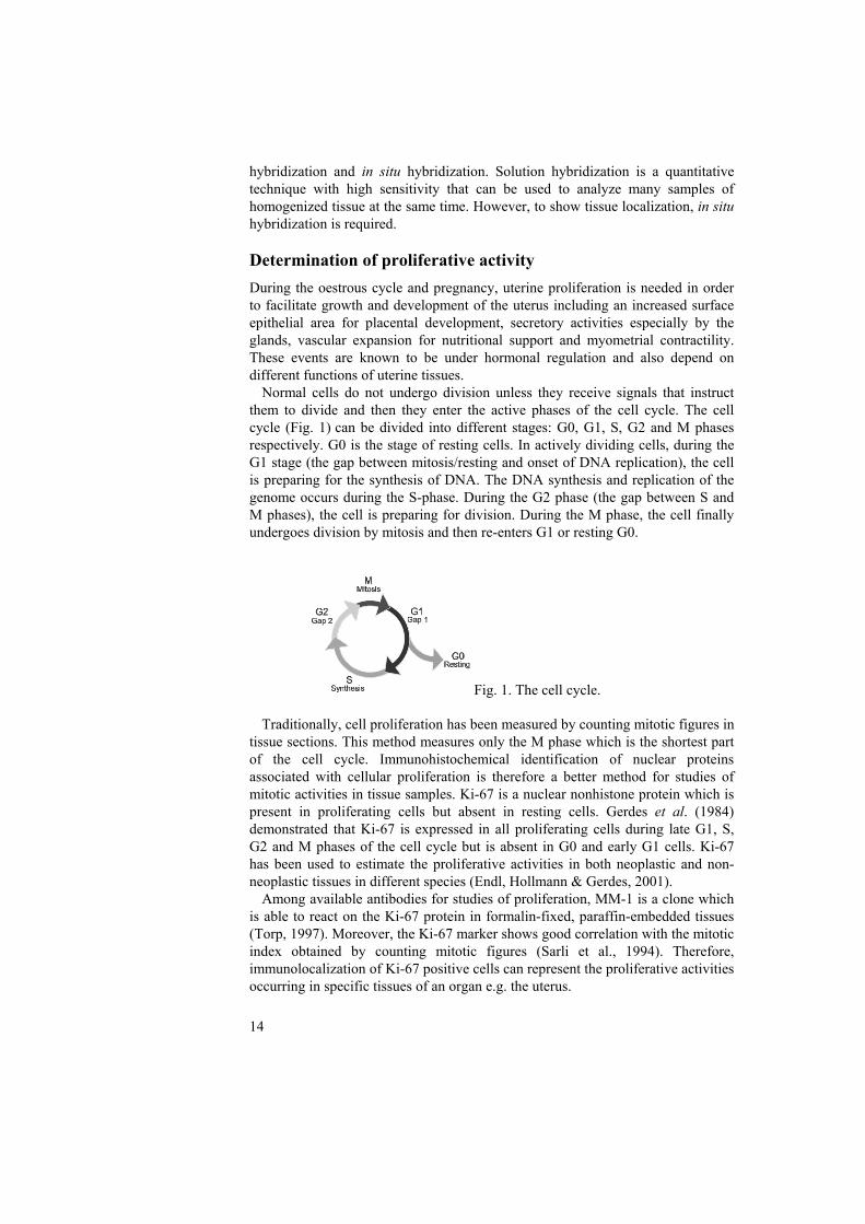
Normal cells do not undergo division unless they receive signals that instruct them to divide and then they enter the active phases of the cell cycle. The cell cycle (Fig. 1) can be divided into different stages: G0, G1, S, G2 and M phases respectively. G0 is the stage of resting cells. In actively dividing cells, during the G1 stage (the gap between mitosis/resting and onset of DNA replication), the cell is preparing for the synthesis of DNA. The DNA synthesis and replication of the genome occurs during the S-phase. During the G2 phase (the gap between S and M phases), the cell is preparing for division. During the M phase, the cell finally undergoes division by mitosis and then re-enters G1 or resting G0.
Fig. 1. The cell cycle.
Traditionally, cell proliferation has been measured by counting mitotic figures in tissue sections. This method measures only the M phase which is the shortest part of the cell cycle. Immunohistochemical identification of nuclear proteins associated with cellular proliferation is therefore a better method for studies of mitotic activities in tissue samples. Ki-67 is a nuclear nonhistone protein which is present in proliferating cells but absent in resting cells. Gerdes et al. (1984) demonstrated that Ki-67 is expressed in all proliferating cells during late G1, S, G2 and M phases of the cell cycle but is absent in G0 and early G1 cells. Ki-67 has been used to estimate the proliferative activities in both neoplastic and non-neoplastic tissues in different species (Endl, Hollmann & Gerdes, 2001). Among available antibodies for studies of proliferation, MM-1 is a clone which is able to react on the Ki-67 protein in formalin-fixed, paraffin-embedded tissues (Torp, 1997). Moreover, the Ki-67 marker shows good correlation with the mitotic index obtained by counting mitotic figures (Sarli et al., 1994). Therefore, immunolocalization of Ki-67 positive cells can represent the proliferative activities occurring in specific tissues of an organ e.g. the uterus.
14

15
Aims of the present investigation The aims of this thesis was to study: • tissue-specific immunolocalization of ERα and PR proteins. • levels of messenger ribonucleic acids (mRNA) of the steroid receptors ERα,
ERβ and PR by solution hybridization. • proliferative activities as illustrated by immunopresence of the Ki-67-protein in
relation to the steroid receptor results. Differences between uterine tissue compartments and different stages of the oestrous cycle as well as after insemination/during early pregnancy were studied and related to changes in plasma levels of oestradiol-17β and progesterone.
16
Material and Methods
Animals and general management The research plan was approved by the ethical committee for experimentation with animals. Altogether 33 crossbred sows (Swedish Landrace x Swedish Yorkshire) were used and the study was performed in two experimental groups of sows, 15 non pregnant cyclic sows (experiment A) (Papers I, III and IV) and 18 inseminated/early pregnant sows (experiment B) (Paper II-IV). The mean parity numbers of these experimental groups were 3.4 + 0.7 and 3.4 + 0.6 respectively for each experimental group. Their body weights ranged from 174 to 268 kg and their average age was around 2 years.
The sows had shown normal reproductive performance before selection to the experiment. They were brought to the faculty clinic directly after weaning and kept in individual pens with boars housed in the same stable throughout the experimental period. The sows were fed according to the Swedish breeding stock standard for dry sows (Simonsson, 1994). The feed allowance was 4-5 kg/day (barley-based sow diet, 14.5% protein and 12.5 MJ/kg metabolisable energy) until the first oestrus after weaning, and thereafter about 2-2.5 kg/day. Water was available ad libitum. Clinical observations After weaning (5 weeks of lactation), careful oestrus detection was done and the ovulation time was checked by ultrasonography as described earlier (Soede, Noordhuizen & Kemp, 1992; Dalin et al., 1995).
All the sows in experiment B (Papers II-IV) were inseminated once by the same person (Kaeoket K.) at 20-15 h before expected ovulation in their second oestrus after weaning (estimated from the first oestrus after weaning). A dose of pooled semen (two boars of proven fertility) containing 10 x 109 spermatozoa in 100 ml BTS (Beltsville Thawing Solution; Pursel and Johnson, 1976) was used for each artificial insemination. Blood collection and hormone analyses Within one hour before slaughter, blood samples were collected from the external jugular vein for analysis of oestradiol-17β and progesterone (Papers I-IV). The plasma oestradiol-17β level was determined by radioimmunoassay (double antibody oestradiol, Diagnostic Products Corporation, Los Angeles, USA) as previously described for analysis of bovine plasma (Duchens et al., 1994) and validated for oestradiol-17β analyses in the pig (Mwanza et al., 2000). The progesterone (P4) level was determined by a luminescence immunoassay (Amerlite, Kodak Clinical Diagnostics Ltd., Amersham, England). The kit was used according to the manufacturer’s instructions and the method had earlier been validated for progesterone analyses in the pig (Rojkittikhun et al., 1993).
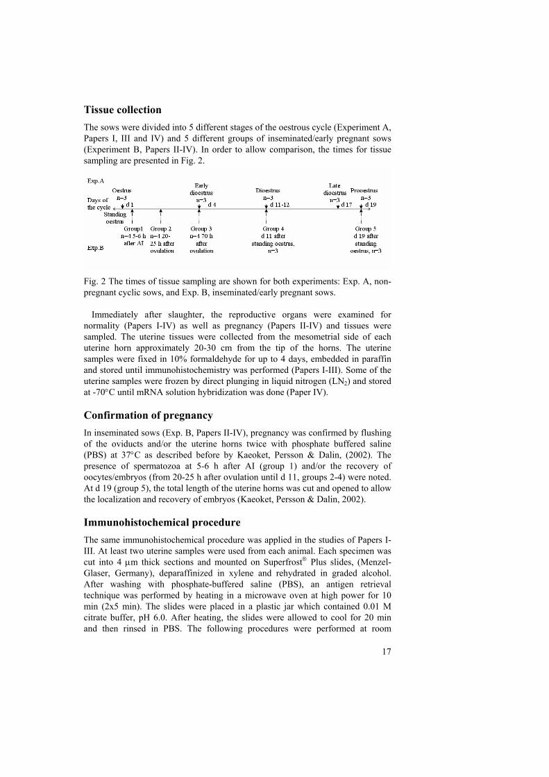
Tissue collection The sows were divided into 5 different stages of the oestrous cycle (Experiment A, Papers I, III and IV) and 5 different groups of inseminated/early pregnant sows (Experiment B, Papers II-IV). In order to allow comparison, the times for tissue sampling are presented in Fig. 2.
Fig. 2 The times of tissue sampling are shown for both experiments: Exp. A, non-pregnant cyclic sows, and Exp. B, inseminated/early pregnant sows.
Immediately after slaughter, the reproductive organs were examined for normality (Papers I-IV) as well as pregnancy (Papers II-IV) and tissues were sampled. The uterine tissues were collected from the mesometrial side of each uterine horn approximately 20-30 cm from the tip of the horns. The uterine samples were fixed in 10% formaldehyde for up to 4 days, embedded in paraffin and stored until immunohistochemistry was performed (Papers I-III). Some of the uterine samples were frozen by direct plunging in liquid nitrogen (LN2) and stored at -70°C until mRNA solution hybridization was done (Paper IV). Confirmation of pregnancy In inseminated sows (Exp. B, Papers II-IV), pregnancy was confirmed by flushing of the oviducts and/or the uterine horns twice with phosphate buffered saline (PBS) at 37°C as described before by Kaeoket, Persson & Dalin, (2002). The presence of spermatozoa at 5-6 h after AI (group 1) and/or the recovery of oocytes/embryos (from 20-25 h after ovulation until d 11, groups 2-4) were noted. At d 19 (group 5), the total length of the uterine horns was cut and opened to allow the localization and recovery of embryos (Kaeoket, Persson & Dalin, 2002). Immunohistochemical procedure The same immunohistochemical procedure was applied in the studies of Papers I-III. At least two uterine samples were used from each animal. Each specimen was cut into 4 μm thick sections and mounted on Superfrost® Plus slides, (Menzel-Glaser, Germany), deparaffinized in xylene and rehydrated in graded alcohol. After washing with phosphate-buffered saline (PBS), an antigen retrieval technique was performed by heating in a microwave oven at high power for 10 min (2x5 min). The slides were placed in a plastic jar which contained 0.01 M citrate buffer, pH 6.0. After heating, the slides were allowed to cool for 20 min and then rinsed in PBS. The following procedures were performed at room
17
18
temperature. Endogenous peroxidase activity was blocked with 3% hydrogen peroxide in methanol, followed by rinsing in PBS. Prior to incubation with the primary antibody, the sections were pretreated with normal horse serum for 30 minutes. The primary mouse monoclonal antibodies used were: • ERα (C-311:sc-787, Santa Cruz Biotechnology Inc., USA, dilution of 1:25)
(Papers I and II) • PR (PR-2C5, Zymed Laboratories Inc., dilution of 1:200) (Paper III) • Ki-67, proliferative activities (MM1, Novocastra Laboratories Ltd., United
Kingdom, dilution of 1:200) (Papers I and II) The antibodies were added and the sections were incubated for 1 h. Negative
controls were obtained by replacing the primary antibodies to ERα, PR or Ki-67 with normal mouse IgG (sc-2025, Santa Cruz Biotechnology Inc., USA) (Papers I-III) in a dilution of 1:200.
After primary antibody binding, the sections were washed in PBS and incubated with the secondary antibody, a biotinylated horse anti-mouse IgG (Vectastain® ABC kit, Vector Laboratories, Inc., USA). Thereafter, another washing with PBS was done and the tissue sections were incubated with a horseradish peroxidase-avidin biotin complex (Vectastain® ABC kit, Vector Laboratories, Inc., USA). After a final wash with PBS, 3,3′-diaminobenzidine (DAB, Dakopatts AB, Älvsjö, Sweden), a chromogen, was added to visualize the bound enzyme (brown color). All sections were counterstained with Mayer’s hematoxylin followed by mounting in glycerine-gelatin before investigation under light microscope.
As the uterus is composed of different compartments, evaluation of the immunohistochemistry was done separately for each tissue compartment (Papers I-III). The results are presented for the different uterine compartments as follows: surface epithelium, glandular epithelium, subepithelial connective tissue and myometrium. In addition, superficial and deep-laying endometrial glands were evaluated separately for ERα and PR since the staining patterns between these compartments differed (Papers I-III). In the myometrium, only smooth muscle cells were studied. The blood vessels were not studied in detail, only general appearance was described.
The results of the immunostaining were evaluated in semiquantitative ways, by manual overall scoring (ERα, epithelia and myometrium, Papers I -II; and PR, all tissue compartments, Paper III), cell counting (ERα, connective tissue, and Ki-67, all tissues, Papers I-II) and for PR, also image analysis (Paper III). Manual overall scoring The evaluations of positively stained cells were carried out as blinded preparation by the same person (S.Sukjumlong). For ERα immunostaining in cyclic sows (Papers I), almost all the uterine cells in the epithelia and myometrium were positively stained but with variation in intensity for the different tissue compartments. Therefore, the staining intensity (average of all cells) was described as weak, moderate or strong, represented by +, ++, +++ respectively. For the immunostaining of ERα in inseminated/early pregnant sows (Paper II) and PR in both experimental groups of sows (Paper III), the same evaluation of intensity was carried out, but as not all cells stained positively in some compartments of the uterus, i.e. the surface epithelium and the myometrium, also
19
the proportion of positive to negative cells was included for these tissues. The proportions were estimated into 4 different levels (marked A-D); low proportion (< 30% of positive cells, A); moderate proportion (30 - 60% of positive cells, B); high proportion (60 - 90% of positive cells, C) and almost all cells positive (> 90%, D). Cell counting In the subepithelial connective tissue of the endometrium, not all cells were positively stained for ERα. Therefore, instead of intensity assessment, the number of clearly ERα positive cells (moderate to strong staining) (per mm2) was identified in each sample. Five arbitrarily chosen microscopic fields were counted in each of two sections obtained from each sow. The counting was performed by use of an ocular reticule (ocular micrometer, 0.25x0.25 mm, with 100 squares) placed in the eyepieces of the light microscope and by movement of the ocular micrometer along the subepithelial connective tissue. The results of the counting were presented as a mean value for each stage of the oestrous cycle (Paper I) respectively for each group of inseminated/early pregnant sows (Paper II). For PR staining in the subepithelial connective tissue, no manual cell counting was done but the image analysis method was applied (Paper III).
For the proliferative activities (Ki-67) (Papers I-II), positive cells were counted in the same manner as for ERα in the connective tissue but regardless of the intensity since only few uterine cells were positively stained. Therefore, the results were presented as mean numbers of positively stained cells per mm length (surface epithelium) or per mm2 (glandular epithelium, stroma and myometrium). In the glandular epithelium, the intensity of positively stained cells in the superficial and deep glands was similar, which is why the results are presented as means of positive glandular cells per mm2 of endometrial tissue without separating superficial and deep laying glands. Image analysis A difference between ERα and PR immunostaining was that most of the cells always stained positively for ERα whereas for PR, more negative cells were found at some stages of the oestrous cycle and during early pregnancy. Moreover, as some cytoplasmic staining for ERα was found at some stages in the surface epithelium, it could interfere with the evaluation of nuclear staining by the image analysis system. Therefore, only PR immunostaining was evaluated by the image analysis method in addition to manual scoring (Paper III). In order to assess the PR immunostaining quantitatively in each tissue compartment, a Leica microscope and a Sony video camera (Park Ridge, NJ, USA) connected to a computer with an image analysis system (Leica Imaging System Ltd., Cambridge, UK) was applied. Quantification of the immunostaining was performed on five randomly selected fields in each compartment. For the subepithelial connective tissue, non-stromal elements (superficial endometrial glands, blood vessels) were interactively removed. Also in other compartments, proper areas were selected and blood vessels were excluded interactively for the image analysis measurement. By use of a colour discrimination software, the nuclear staining intensity was divided into three different levels; weak, moderate and strong. However, it is difficult for the

20
system to distinguish a faint background from weak staining in the cell nuclei and therefore, to avoid false positive staining, only moderate to strong intensity was included for the image analysis measurement. The results are presented as mean ratio of total area of positive nuclear staining per total area of cell nuclei. Solution hybridization Solution hybridization of mRNA levels for ERα, ERβ and PR is described in Paper IV. In brief, the frozen tissues were thawed and the endometrium was manually separated from the myometrium. These tissues were used for total nucleic acid (TNA) preparation as well as for fluorometrical measurement of DNA concentration in the TNA samples. To measure specific mRNAs, probes were synthesized in vitro and radiolabelled which was followed by hybridization to the TNA samples, digestion of the non-hybridized RNA by RNase, precipitation of labelled hybrids, collection on filters, washing, and measurement of radioactivity in a scintillation counter. The results are expressed as amol (10-18) mRNA/μg DNA in the TNA samples. Statistical analyses The statistical analyses were performed on counts of immunopositive cells (Ki-67 in all compartments and ERα in the subepithelial connective tissue, Papers I and II), on the image analysis results (Paper III) and on levels of mRNA from solution hybridization (Paper IV) by using the SAS statistical package (SAS Institute Inc., 1989). One-way analysis of variance was performed for each tissue compartment using general linear model (GLM) procedure (Papers I, II and IV). The stage of oestrous cycle in experiment A or groups of inseminated/early pregnant animals in experiment B were regarded as independent variables. Mean values for each class of the independent variables were compared by using Student’s t-test (Papers I-II) or by Scheffé’s test (Paper IV) only when overall significance was found. In paper III, NPAR1WAY was used to analyse the differences among stages of the oestrous cycle (Exp. A) and groups of inseminated sows (Exp. B). Spearman rank correlations (Papers I-III) or Pearson correlations (Paper IV) between plasma levels (oestradiol-17β, progesterone) and ratio of positive cells or mRNA levels were performed in each experiment by using CORR procedure. P-values < 0.05 were regarded as statistically significant.

21
Results
In Papers I, II and III, non-pregnant cyclic sows at different stages of the oestrous cycle (Experiment A) were studied while the inseminated/early pregnant sows (Experiment B) were included in papers II, III and IV. General observations and hormone levels Before the experiments, all sows used in the present studies had shown normal reproductive performance. In cyclic sows, the average weaning to oestrous interval was 4.0 + 0.8 days (mean + S.D.) and the ovulation took place 35.3 + 6.4 h (range 20-42 h) after the start of standing oestrus (Kaeoket, Persson & Dalin, 2001). In inseminated/early pregnant sows, the average weaning to oestrous interval was 4.1 + 0.6 days (mean + S.D., range 3-6 days) and ovulation took place at 40.7 + 1.9 h and 41.4 + 2.5 h after onset of the first and second oestrus after weaning respectively (range 36-48 h). The average insemination time before ovulation was 18.6 h (range 15-23) (Kaeoket, Persson & Dalin, 2003).
The plasma levels of oestradiol-17β and progesterone changed during the oestrous cycle as well at early pregnancy. The cyclic sows (Papers I, III and IV) showed high plasma levels of oestradiol-17β at prooestrus and oestrus and a high level of progesterone during dioestrus. In the inseminated/early pregnant sows (Papers II-IV), the highest level of plasma oestradiol-17β was found at oestrus (group 1) while the plasma progesterone levels were high at days 11 and 19 after the onset of standing oestrus (early pregnancy, groups 4 and 5).
In Experiment B, the sows slaughtered before ovulation (group 1), had spermatozoa in the uterus, utero-tubal junction, isthmus and ampulla. All sows slaughtered after ovulation in Experiment B were pregnant (groups 2-5) as embryos were observed (Kaeoket, Persson & Dalin, 2002). Immunohistochemistry Immunohistochemical staining of ERα, PR and Ki-67 (Papers I-III) in the uteri showed positive reddish brown nuclear staining in all tissue compartments. No specific staining was found in the negative controls. The intensity of the nuclear staining varied not only between the stages of the oestrous cycle (Papers I and III) and with time after insemination/during early pregnancy (Papers II and III) but also between the different uterine compartments. Vascular cells in the endometrium and the myometrium occasionally appeared positive for ERα and PR immunostaining (Papers I and III) while no positive cells were found when immunolabelling for Ki-67. All results are shown according to different tissue compartments. A summary of the ERα and PR immunostaining by manual scoring in the epithelia and the myometrium, and at different stages of the oestrous cycle as well as after insemination are shown in Table 1, page 22. In order to allow comparison in that table, proportions (D at all stages) of ERα positive cells in the cyclic sows were added.

Tabl
e1 Im
mun
osta
inin
g of
ERα
and
PR
(man
ual s
corin
g) a
ccor
ding
to u
terin
e co
mpa
rtmen
ts (S
E; su
rfac
e ep
ithel
ium
, GE;
gla
ndul
ar e
pith
eliu
m
and
MY
O; m
yom
etriu
m),
A: c
yclic
sow
s, B
: ins
emin
ated
/ear
ly p
regn
ant s
ows.
Cyc
lic so
ws
Inse
min
ated
/ear
ly p
regn
ant s
ows
Tiss
ue
com
partm
ent
Ster
oid
rece
ptor
pr
otei
n
Pro-
oest
rus
Oes
trus
Ea
rlydi
oest
rus
Dio
estru
s La
tedi
oest
rus
Gro
up 1
G
roup
2
Gro
up 3
G
roup
4
Gro
up 5
ERα
+/
++D
++
D
+++D
++
/+++
D
++D
+/
++C
+A
+A
+A
+B
SE
PR
-/+
A
++/+
++D
++
/+++
D
-/+A
-/+
A
+/++
D
+/++
D
++/+
++D
-/+
A
-/+A
ER
α
++D
++
D
++/+
++D
++
D
+/++
D
+D
+/++
D
++/+
++D
++
D
++D
G
E (s
uper
- fic
ial l
ayer
) PR
+
A
+/
++B
++
D
+D
+D
+/++
D
+ D
++
D
+/++
D
+ D
ER
α
++/+
++D
++
D
+++D
++
/+++
D
++D
++
D
++D
++
+D
+++D
++
D
GE
(dee
p la
yer)
PR
+/
++ D
++
/+++
D
+/++
D
++ D
+/
++ D
++
/+++
D
++/+
++D
++
D
+/++
D
+ D
ER
α
++/+
++D
++
/+++
D
+D
+/++
D
++D
++
/+++
D
+/++
D
+B
+D
+A
MY
O
PR
++/+
++ D
++
/+++
D
+/++
D
+/++
D
+/++
D
++/+
++D
++
/+++
D
+/++
D
+ D
+
D
Stai
ning
inte
nsity
: - =
neg
ativ
e, +
= w
eak,
++
= m
oder
ate,
+++
= st
rong
A
= lo
w p
ropo
rtion
(<30
%),
B =
mod
erat
e pr
opor
tion
(30-
60%
), C
= h
igh
prop
ortio
n (>
60-9
0%),
D =
alm
ost a
ll ce
lls (>
90%
) are
pos
itive
G
roup
1, a
t oes
trus,
5-6
h af
ter A
I G
roup
2, 2
0-25
h a
fter o
vula
tion
Gro
up 3
, 70
h af
ter o
vula
tion
Gro
up 4
, d 1
1 (d
ay 1
= fi
rst d
ay o
f sta
ndin
g oe
stru
s)
Gro
up 5
, d 1
9 (d
ay 1
= fi
rst d
ay o
f sta
ndin
g oe
stru
s)
22
Surface epithelium In cyclic sows (Paper I), almost all epithelial cells were positively stained for ERα with highest intensity at early dioestrus. For PR (Paper III), the strongest intensity as well as high proportion was found at oestrus and early dioestrus while weak immunostaining and low proportion was observed at other stages, which is in accordance with the results from image analysis (Paper III, Table 1, P<0.001).
As shown in Paper II, low proportions of ERα-positive epithelial cell nuclei were found except at 5-6 h after AI (group 1) when a slightly stronger intensity as well as higher proportion was observed. For PR immunostaining, the highest intensity and high proportion was observed at 70 h after ovulation (group 3). The results from image analysis (Table 2, Paper III) showed that the mean ratios of positive cells were higher also at oestrus, in addition to 70 h after ovulation (groups 1 and 3) compared with the other groups (groups 2, 4 and 5) (p <0.001). Additionally, for ERα immunohistochemistry, light-brown staining was observed in the basal part of the cytoplasm of surface epithelial cells at early dioestrus (Paper I) and at 70 h after ovulation (group 3, Paper II), while no cytoplasmic staining was found at any occasion for PR or Ki-67.
For Ki-67 in cyclic sows (Paper I, Table 2), significantly higher numbers of positively stained cells per mm surface epithelium, were found at oestrus and early dioestrus compared with the other stages (P<0.05). In the inseminated sows (Paper II, Table 2), the numbers of Ki-67 positive cells were low or none, and not significantly different between groups due to high individual variations within some groups (groups 1-3). Glandular epithelium In both cyclic and inseminated sows, all glandular epithelial nuclei stained positively but with different intensity for ERα (Paper I and II) and PR (Paper III), except for PR in the superficial glands of cyclic sows at prooestrus and oestrus when the proportion of positive cells varied as well. The staining intensity also differed between superficial and deep glands at some stages and in some groups, why the results were presented separately.
In cyclic sows, the strongest intensity of ERα (Table 1, Paper I) in both superficial and deep glands was found at early dioestrus compared with the other stages. PR immunostaining (Table 1, Paper III) by manual scoring showed its highest intensity at early dioestrus for superficial glands and at oestrus for deep glands. However, the image analysis results showed the highest mean ratio of positive nuclei at oestrus in both layers of glandular epithelium (P<0.001).
In inseminated/early pregnant sows, the most prominent ERα (Table 1, Paper II) staining was found at 70 h after ovulation (group 3) in both superficial and deep laying glands. The latter also showed prominent staining at d 11 (group 4). For PR (Table 2, Paper III), the highest intensity (moderate) was observed at 70 h after ovulation (group 3) in the superficial glands and at oestrus and 20-25 h after ovulation (moderate/strong, groups 1 and 2) in the deep glands. The image analysis (Table 2, Paper III) differed somewhat from that, showing for both layers of uterine glands, a highest mean ratio of PR positive cells at oestrus (group 1) while the lowest was found at d 19 (P<0.001). The latter is in accordance with the manual scoring.
23

A high variation in proliferation was seen in the glandular epithelium. For Ki-67 in cyclic (Table 2, Paper I) and especially in inseminated sows (Table 2, Paper II), the numbers of positive cells were apparently higher in the glandular epithelium compared with the other uterine compartments at early dioestrus/group 3, also differing from other stages/groups (P<0.05, respectively P<0.001). The numbers of positively stained cells were none or low during dioestrus and late dioestrus respectively in groups 4 and 5. Subepithelial connective tissue In cyclic sows, the significantly highest count of ERα positive connective tissue cells (moderate to strong intensity) (Fig. 2 Paper I) was observed at oestrus (P<0.05). For PR immunostaining (Table 1, Paper III), most of the cells stained positively at all stages of the oestrous cycle and with strong intensity at oestrus. The image analysis results showed a significant difference between oestrus and all other stages (P<0.001).
In inseminated/early pregnant sows (Fig. 2, Paper II), the highest number of ERα positive cells was found at oestrus and early after ovulation (groups 1 and 2) which differed significantly compared with days 11 and 19 (groups 4 and 5) (P<0.05). Group 2 also showed significantly higher levels than group 3. A high ratio of PR positive cells was found at oestrus (group 1), differing significantly from all other groups (P<0.001, image analysis, Table 2, Paper III).
For Ki-67 immunostaining, the numbers of positive cells were low during all stages of the oestrous cycle (Table 1, Paper I) as well as after insemination/during early pregnancy (Table 1, Paper II) with no significant differences. Myometrium In the myometrium of cyclic sows (Papers I and III), the strongest intensity for both ERα (Table 1, Paper I) and PR (Table 1, Paper III, image analysis, P<0.001) immunostaining was observed during prooestrus and oestrus while weaker staining was found at all other stages.
In inseminated sows, the strongest intensity and highest proportion of positively stained cell nuclei was found in group 1 for ERα (Table 1, Paper II) and in groups 1 and 2 for PR (P<0.01, image analysis, Table 2, Paper III) while weaker staining intensity was found in all other groups (Papers II and III). In group 4, although the intensity of ERα immunostaining was weak, a high proportion of positive cells was observed (Table 1, Paper II).
Proliferative activity (Ki-67) in the myometrium was low or none during the oestrous cycle (Table 1, Paper I) as well as after insemination/during pregnancy (Table 1, Paper II) with no significant differences. Solution hybridization of mRNAs for ERα, ERβ and PR The results on the expression of mRNAs for ERα, ERβ and PR (Fig. 2, Paper IV) were presented according to the uterine compartments that could be separated before tissue homogenization i.e. endometrium and myometrium for cyclic respectively inseminated/early pregnant sows.
24

Endometrium The lowest level of ERα mRNA in the endometrium of cyclic sows was observed at late dioestrus which was significantly different to the other stages of the oestrous cycle except oestrus (p<0.01) while the level of ERβ mRNA was constantly low at all stages examined. For PR mRNA, the expression in the endometrium was low at all stages examined with a significantly higher level observed at early dioestrus compared with dioestrus and late dioestrus (P<0.05).
In inseminated sows, the levels of both ERα mRNA and PR mRNA in the endometrium were highest at 70 h after ovulation which was significantly different to the lowest at d19 (group 5) for ERα (group 3) (P<0.001) and at days 11 and 19 (groups 4 and 5) (P<0.01) for PR. Significantly higher expression of ERβ mRNA in the endometrium was found at oestrus (group 1) compared with d 19 (group 5) (P<0.01). Myometrium The ERα mRNA levels in the myometrium were significantly higher at prooestrus and oestrus compared with levels at dioestrus and late diestrus (p <0.001) and PR mRNA was highest at prooestrus compared with all other stages (P<0.01). For ERβ mRNA, the expression was similarly low at all stages of the oestrous cycle. In inseminated/early pregnant sows, the highest levels of mRNAs for ERα and PR were found at oestrus (group 1) which was significantly different to all other stages (P<0.001 for ERα, P<0.05 for PR). The ERβ mRNA level was similar at all stages but at higher levels than found in the cyclic sows. Ratio of ERα mRNA/ERβ mRNA In the endometrium, a higher ratio of ERα to ERβ mRNA (Fig. 3, Paper IV) was found at early dioestrus (P<0.01) and at 70 h after ovulation (group3, P<0.001) respectively, compared with all other stages for cyclic sows and with all other groups except 20-25 h after ovulation (group 2) of inseminated/early pregnant sows.
In the myometrium of cyclic sows, a higher ratio was found at prooestrus compared with dioestrus and late dioestrus (P<0.01). For inseminated/early pregnant sows, oestrus (group 1) differed compared to all other stages (P<0.001). Correlations For cyclic sows (Papers I, III and IV), significant positive correlations were found between the plasma levels of oestradiol-17β and :
• levels of PR immunolabelling (Paper III) in superficial glands (P<0.001), deep glands (P<0.001), connective tissue (P<0.001) and myometrium (p<0.001).
• numbers of Ki-67 positive cells in the myometrium (P<0.01). • levels of ERα mRNA in the myometrium (P<0.05).
Negative correlations were observed in the cyclic sows between plasma levels of progesterone and:
25

• levels of PR immunolabelling (Paper III) in superficial glands (P<0.001), deep glands (P<0.001), connective tissue (P<0.001) and myometrium (p<0.001).
• numbers of Ki-67 positive cells in the myometrium (P<0.001). • levels of ERα mRNA (P<0.05) and PR mRNA (P<0.01) in the
myometrium. For inseminated/early pregnant sows (Papers II, III and IV), significant positive correlations were found between plasma levels of oestradiol-17β and:
• levels of PR immunolabelling in superficial glands (P<0.001), deep glands (P<0.001), connective tissue (P<0.001) and myometrium (P<0.001).
• levels of ERα mRNA (P<0.001) and PR mRNA (P<0.001) in the myometrium.
Negative correlations were found for inseminated/early pregnant sows between the levels of plasma progesterone and:
• numbers of ERα positive cells in the subepithelial connective tissue (P<0.001).
• numbers of Ki-67 positive cells in the surface epithelium (P<0.001). • levels of PR immunolabelling in superficial glands (P<0.001), deep glands
(P<0.001), connective tissue (P<0.001) and myometrium (P<0.001). • levels of ERα mRNA (P<0.05) and PR mRNA (P<0.05) in the
endometrium. • levels of and ERα mRNA (P<0.01) and PR mRNA (P<0.01) in the
myometrium. Concerning the numbers of ERα and Ki-67 immunolabelled cells (Papers I and II), positive correlation was found between:
• Ki-67 positive cells in the surface epithelium and the glandular epithelium in both cyclic (Paper I) (P<0.01) and inseminated/early pregnant sows (Paper II) (P<0.01).
• ERα positive cells in the subepithelial connective tissue and Ki-67 labelled cells in the surface epithelium of cyclic (P<0.01) (Paper I) and inseminated sows (P<0.001) (Paper II).
• ERα positive cells in the subepithelial connective tissue and Ki-67 labelled cells in the glandular epithelium (P<0.05) of cyclic sows (Paper I).
26

General discussion
Physiological and morphological changes in the sow uterus occur during the oestrous cycle and at early pregnancy as has been shown earlier for the same sows (Kaeoket, Persson & Dalin, 2001; 2002) as the ones used in the present study. In order to further clarify the mechanisms behind the physiological variations in response to ovarian steroid hormone levels, the localization of oestrogen and progesterone receptors was investigated. Also the expression of their mRNAs was studied in order to better understand the regulation and role of steroid receptors in the uterus. In addition, proliferative activities, as represented by the protein Ki-67, were related to hormonal and steroid receptor changes. Methodological considerations Animals The sows had a reproductive history without complications and at clinical examination, all sows (Papers I-IV) showed normal reproductive performance i.e. interval from weaning to oestrus, oestrus signs and ovulation time (Kaeoket, Persson & Dalin, 2001; 2002). After slaughter, post mortem examination showed that the genital organs were normally developed and the numbers of corpora lutea were within normal range. In experiment B, all sows slaughtered after ovulation were pregnant as properly developed early embryos were observed (Kaeoket, Persson & Dalin, 2002). The presented normality of the animals ensures as representative results as possible, especially as the number of sows per stage/group was small. Immunohistochemistry An advantage of immunohistochemistry (IHC) is the ability to visualize the localization of a protein in different tissue compartments which is not possible through other methods of steroid receptor protein investigation. However, with IHC, variations in sensitivity and specificity of the antibodies used i.e. different antibodies to the same protein as well as different protocols may give diverging results. This is a likely explanation to why the results from our studies in this thesis differ when compared with other studies in pigs (Geisert et al., 1993; 1994). Moreover, by the semiquantitative methods used in the present studies, the exact levels of steroid receptors can not be measured, which may also account to the diverging results. The overall manual scoring method used has the advantage that it represents the average results of a whole tissue compartment. However, as some compartments differed in both staining intensity and proportion of positive cells, image analysis would be useful. On the other hand, a problem when using the image analysis system is that the differences between unspecific faint staining and specific weak staining can not be distinguished by the system, which is why the method was not used for the ERα-studies (Papers I-II) where some cytoplasmic staining was found and why weak staining was excluded from the image analysis measurements in Paper III. When comparing these two methods (manual scoring and image
27

analysis), most of the results are in agreement. However, some discrepancies could be observed, probably due to the exclusion of weak staining intensity in the image analysis and that the manual scoring method evaluated the whole tissue area of a section while the image analysis was limited to five randomly selected areas. For these reasons, both methods (manual scoring and image analysis) were included in the evaluation of the PR immunostaining (Paper III).
For Ki-67, the staining intensity was similar in all cells but not the proportion of positive cells and therefore, all cells stained were counted through the ocular reticle. ERα was evaluated by the manual scoring for all compartments except the subepithelial connective tissue where cell counting through the ocular reticule was used. Therefore, correlations between different tissues could only by done for the connective tissue in comparison with the proliferative marker, Ki-67, and significant differences were found. Solution hybridization The expression of messenger ribonucleic acids was studied in order to better understand the regulation of the ovarian steroid hormone receptor proteins at mRNA level (Paper IV). As the results were evaluated according to two main different tissue compartments, the endometrium and the myometrium, the results are discussed for the respective compartment. Comparisons with the results from immunohistochemistry are possible for the myometrium but as the endometrium consists of several different tissues, further studies, using in situ hybridization would be needed in order to illustrate tissue specific mRNA expression. Immunohistochemistry The steroid receptors ERα and PR, as well as the proliferative marker, Ki-67 are discussed separately for each tissue compartment as the different tissues are considered to have specific roles in the reproductive processes. In general, immunostaining of the steroid receptor proteins and the proliferative marker Ki-67, was confined to the nuclei. However, in the studies of ERα (Papers I and II), cytoplasmic staining was observed in the surface epithelium at some stages of both experimental groups of sows (Paper I and II) while no cytoplasmic staining was found when immunolabelling for PR (Paper III) or proliferative activity (Papers I and II). The cytoplasmic ERα staining in the surface epithelium was unique as it was found only at early dioestrus/70 h after ovulation in cyclic and pregnant sows respectively (Papers I and II). This could represent a surplus of receptor protein being degraded at that reproductive time and the cytoplasmic staining is therefore considered to represent real cytoplasmic ERα protein rather than the unspecific staining seen in other compartments. Surface epithelium The surface epithelium is important for the reproductive physiology by secreting substances into the lumen of the uterus in order to enhance embryo survival and development as well as for trophoblast attachment and establishment of the placenta. The interactions between embryos and the surface epithelium are specifically needed for maintaining pregnancy. For instance, administration of
28
exogenous oestrogen on d 9 and 10 to early pregnant gilts altered endometrial surface epithelium and caused conceptus mortality (Blair et al., 1991). Conceptus oestrogens, the signal for maternal recognition of pregnancy, are believed to cause a reroute of PGF2α into uterine lumen instead of to the circulation, and thus luteolysis cannot occur (Geisert & Yelich, 1997). Steroid receptors: ERα and PR In cyclic sows (Papers I and III), the most prominent staining for ERα and PR in the surface epithelium was observed at early dioestrus. As both ERα and PR are considered to be upregulated by oestrogens (Ing & Tornesi, 1997; Bouchard, 1999), these results could be interpreted as a delayed effect from the high levels of plasma oestradiol-17β at oestrus. However, the levels of ERα and PR proteins may also depend on other factors such as locally produced substances, e.g. growth factors (Ignar-Trowbridge et al., 1992; Stoica et al., 2000; Klotz et al., 2002) from epithelial or stromal cells. A dependence on oestradiol-17β via paracrine regulation is supported by the present study, showing high levels of ERα in the subepithelial connective tissue already at oestrus, which could be responsible for the following upregulation of both ERα and PR at early dioestrus in the surface epithelium.
In inseminated sows (Papers II-III), the pattern of ERα-staining in the surface epithelium was markedly different to that of the cyclic sows (Paper I) as a lower level of ERα was observed as early as 20-25 h after ovulation and with low levels also in groups 3-5. At days 11 and 19 (groups 4-5), both ERα and PR were found to be at low levels. The lower levels of ERα in the inseminated sows compared with cyclic could be effects from insemination and pregnancy, i.e. semen and viable embryos. Even though the general view is that oestrogens upregulate ERα, it has also been shown that oestradiol can down-regulate the expression of its own receptor, ERα-mRNA, in the endometrium of ovariectomized gilts (Sahlin et al., 1990) and the ERα protein in the uterus of immature ewes (Meikle et al., 2000). Therefore, it may be the high oestrogen levels in boar semen (Claus et al., 1987) together with embryonic oestrogens at later stages (Claus et al., 1987) that cause a decrease of ERα in the surface epithelium. Moreover, when comparing the presence of ERα in the surface and glandular epithelia, the downregulation of ERα was more obvious in the surface epithelium which is supporting the suggested influence by semen and/or embryos from the luminal environment. An effect by semen/embryos on PR in the surface epithelium was not found. When comparing levels of ERα and PR in cyclic sows, differences could be observed indicating different regulatory mechanisms of these receptor proteins at the same plasma steroid levels. Relatively high proportions of positive ERα cells (Table 1, page x) were found at all stages whereas PR positive cells were hardly observed at dioestrus, late dioestrus and prooestrus suggesting the need of an absence or low level of PR when progesterone is high, in order to regulate reproductive functions. It was proposed by Geisert et al.(1994) that the downregulation of PR in the epithelia may be involved in the timing of luteolysis in cyclic gilts as well as having a role for conceptus growth and placentation at early pregnancy.
29

Furthermore, an interesting finding was the cytoplasmic ERα staining observed in the surface epithelium of cyclic and inseminated sows at one comparable stage, i.e. at early dioestrus/70 h after ovulation. The nucleocytoplasmic shuttling of ERα described by Guiochon-Mantel et al, (1996) supports our suggestion on cytoplasmic presence resulting from large amounts of nuclear protein at oestrus being degraded in the cytoplasm at later stages when hormone levels were low, i.e. low levels of the ligand that can occupy the receptor. Although it has been shown that the PR-protein also undergoes nucleocytoplasmic shuttling (Guiochon-Mantel et al., 1989), no cytoplasmic staining was found in any uterine compartment when immunolabelling for PR was carried out as in the present study. An explanation may be that the PR protein was not synthesized in such large amounts at the studied stages and in relation to access to the ligand for the receptor. Proliferative activity, (Ki-67) Proliferation in the surface epithelium of cyclic sows (Paper I) may be influenced by oestrogen acting via ERα in the subepithelial connective tissue since significant correlation was observed between Ki-67 cells in the surface epithelium and ERα cells in the connective tissue. However, also PR may be involved as progesterone have been shown to promote proliferation via the regulation through growth factors and/or growth factor receptors (Reynolds et al., 1990; Taketani & Mizuno, 1991). However, these regulatory mechanisms merit further studies. Among the inseminated sows (Paper II), proliferative activity in the surface epithelium was low or none without any significant differences. Moreover, no increase in uterine weight of the early pregnant sows was observed. Glandular epithelium In the present investigations, the two types of epithelia (surface and glandular epithelium) differed in steroid hormone receptor localization (Papers I-III). Therefore different secretions and functions of these compartments could be expected as also shown by other studies (Fazleabas et al., 1985; Zhang, Paria & Davis, 1991; Davis & Blair, 1993; Blackwell, Speth & Mirando, 2003).
Especially in pigs, proliferation of the uterine glands is necessary as they synthesize, transport and secrete substances which are essential for conceptus survival and development (Roberts et al., 1986a; Roberts, Raub & Bazer, 1986b; Roberts & Bazer, 1988). Extensive endometrial gland hyperplasia and hypertrophy occur during gestation (Spencer et al., 2004a), and this is presumed to increase secretory activities for support of pregnancy. In pregnant ewes, Gray et al., (2002) showed that pregnancy could not be established without uterine glands. Glandular functions are therefore expected to be under the influence of the ovarian steroid hormones and depending on expression of their respective receptors. Steroid receptors: ERα and PR For ERα in the present studies, the staining intensity always appeared slightly stronger in the deep-laying glands than in the superficial with the highest level found at early dioestrus/70 h after ovulation in cyclic (Paper I) respectively inseminated sows (Paper II). The patterns of ERα presence in cyclic and
30

inseminated sows did not differ, indicating that the presence of ERα in endometrial glandular epithelium is independent of pregnancy, at least during the first three weeks. Thereby, embryonic oestrogens do not appear to be the main regulators of ERα-presence in the glandular epithelium.
The pattern of PR staining in the glandular epithelium (Paper III) was similar for cyclic and inseminated sows, indicating an independence of pregnancy. The highest level of PR was observed at oestrus and the lowest at late dioestrus/d 19. However, even though the patterns were similar, the results showed that oestrogen from boar semen may have some effects on the presence of PR in especially the superficial glands as mean ratio (image analysis) as well as a higher proportion (manual scoring) of positive cells were observed in inseminated sow at 5-6 h after AI compared to cyclic sows at oestrus. In addition to effects by plasma oestrogens, semen could act locally on the endometrium and result in the slightly higher presence of glandular PR found after insemination.
When comparing ERα and PR immunopresence in the deep glandular epithelium, higher presence of ERα was found at early dioestrus than at oestrus while PR immunopresence was vice versa. This shows that a high level of PR presence in the glandular epithelium occurred before ERα and that the appearance of ERα and PR are differently regulated at the same reproductive stage. Moreover, the need of ERα seemed to be prolonged as strong staining could be found at dioestrus/d 11 in both cyclic and early pregnant sows while PR was already low at these stages. An explanation could be that maintained ERα expression constitutes a potential response to oestrogens in semen as well as from embryos while PR has to be present and able to respond to the increasing progesterone levels in plasma early after ovulation. Thereafter PR decreases to low levels that can balance the high levels of plasma progesterone. The lower presence of PR was co-occurring with the highest amount of secretory vesicles found at dioestrus in studies of the same groups of sows (Kaeoket, Persson & Dalin, 2001). In conclusion, high secretory activity of the glandular epithelium may need a downregulation of PR early after ovulation by help of increasing progesterone levels and a maintained high level of ERα able to respond to luminal oestrogens. Proliferative activity (Ki-67) The present studies demonstrate a high proliferative activity in the glandular epithelium compared with other compartments, and at markedly higher levels in inseminated sows compared with cyclic (Papers I and II). The highest proliferative activity in the glandular epithelium was found at early dioestrus/70 h after ovulation in the respective experimental groups of sows (Paper I and II) which coincided with the highest level of ERα in the glandular epithelium. This indicates that endometrial glands increase its proliferation in order to facilitate secretory functions, in response to oestrogenic influence mediated through the high presence of ERα. Moreover, high proliferation in the glandular epithelium may also be due to the high presence of PR at oestrus whereafter the receptors become occupied by low but increasing levels of progesterone and resulting in an induction of proliferation at the following stages. That view is supported by a high correlation between PR expression and endometrial cell proliferation observed in early pregnant goats (Flores et al., 2001). Alternatively, proliferation in the glandular
31

epithelium may also be indirectly mediated via steroid receptors in the stroma resulting in paracrine effects from e.g. growth factors (Rider, 2002). In addition, since the proliferative activity was much higher in pregnant sows, it is likely that there are several ways to ensure the secretory functions that are crucial for embryonic survival. However, these regulatory mechanisms and their relative importance need to be elucidated by further studies. Subepithelial connective tissue Steroid receptors: ERα and PR During the oestrous cycle as well as at early pregnancy, levels of the ERα and PR proteins in the connective tissue, as shown by the results of cell counting (Papers I and II) and image analysis (Paper III), correlated positively with the levels of plasma oestradiol-17β and negatively with progesterone. It has been shown that many reproductive processes in the uterus are mediated in a paracrine manner via receptors in the stroma (Kurita et al., 2000a; 2000b; 2001). As described above, the present studies support this concept as correlations between ERα presence in the connective tissue and Ki-67 cells in the epithelia were found for both cyclic and inseminated sows. Furthermore, it has been suggested that stromal cells with maintained levels of steroid receptor proteins can play a balancing role in regulating the physiological changes in the uterus when these receptors are absent or decrease in other compartments (Brenner, West & McClellan, 1990; Hild-Petito, Verhage & Fazleabas, 1992; Spencer & Bazer, 1995). It was clearly shown in the study of PR (Paper III) that PR positive cells were maintained in the connective tissue when absent or low in other compartments during the oestrous cycle as well as at early pregnancy. Moreover, this was also found for ERα in inseminated sows (Paper II), where the presence of ERα was low and almost absent in the surface epithelium at some stages of early pregnancy while it was maintained in the connective tissue at all stages examined. Therefore, a potential effect on the endometrial epithelia by the steroid hormones might be mediated via their respective receptors in the connective tissue as also suggested for other species (Perrot-Applanat et al., 1994; Spencer & Bazer, 2002).
Regarding a potential downregulation of steroid receptor proteins by plasma progesterone during pregnancy, the findings from the present studies (Papers II and III) support the concept that uterine epithelia are more sensitive to a downregulating effect by progesterone than stromal cells (Lessey et al., 1988; Hild-Petito, Verhage & Fazleabas, 1992). A relatively uniform appearance of the steroid receptors ERα and PR in the endometrial subepithelial connective tissue appear to be necessary at least until d 11 after insemination in pregnant sows. Proliferative activity (Ki-67) Proliferative activities in the connective tissue were similarly low but could be observed throughout the cycle (Paper I) as well as during early pregnancy (Paper II). The results imply that uterine growth may not require a high proliferation in the connective tissue as shown for the glands but a low level of proliferation should be maintained in order to provide stromal cells able to mediate other reproductive functions in a paracrine manner as suggested above. On the other
32
hand, in pregnant rodents, proliferation has been shown to switch from the surface and glandular epithelia to the endometrial stroma at d 4 of pregnancy (Rider & Psychoyos, 1994). However, as mentioned above, that process was not observed in the sows of the present studies as the level of proliferation was similar in the stroma throughout the oestrous cycle as well as during the different stages of early pregnancy examined. The difference between the two species may be related to the process of decidualization in rodent species which is not found in sows, and therefore, there may be no need for high proliferation in the connective tissue of the porcine species at early pregnancy. However, a higher level of proliferation could be expected at later stages as the porcine uterus has been shown to increase its weight from three weeks of pregnancy (Wu, Shin & Dziuk, 1988). Myometrium Steroid receptors: ERα and PR The pattern of steroid receptor localization for ERα and PR in the myometrium was similar with high levels around oestrus for both cyclic (Papers I and III) and inseminated sows (Papers II and III). However, the mechanisms requiring the presence of these receptors may be different. In the sow myometrium, uterine contractions have been shown to increase around oestrus (Scheerboom, Van Adrichem & Taverne, 1987; Langendijk et al., 2002) and ERα is suggested to mediate the oestrogenic influence that stimulate myometrial contractions. On the other hand, progesterone action via PR may be needed to oppose the effect by oestrogen as high levels of progesterone have been shown to provide a quiescent uterus with low myometrial activity (Porter & Watts, 1986; Ding et al., 1994). Therefore, high levels of myometrial PR at oestrus (Paper III) may be a result of up-regulation by high levels of plasma oestrogen in order to prepare the myometrium for the increasing levels of progesterone at following stages. Thereafter, the lower levels of PR seemed to be enough for maintenance of the required quiescence in response to high levels of plasma progesterone. On the other hand, if the sows were not pregnant, the appearance of PR in the myometrium should increase again, as shown for prooestrus in Paper III, when the level of progesterone was decreased. Proliferative activity (Ki-67) As low levels of proliferation were observed at all stages for both cyclic and inseminated sows (Papers I and II), it can be concluded that during the oestrous cycle as well as early pregnancy, no growth of the myometrium is needed in order to support the uterine functions. However, an increased proliferation could be expected at later stages of pregnancy. Steroid receptor mRNAs The expression of ERα mRNA and PR mRNA in the homogenized endometrium was highest at early dioestrus/70 h after ovulation. The lowest levels were found at late dioestrus/d 19 for ERα mRNA and at dioestrus/d 11 and late dioestrus/d 19 for PR mRNA in cyclic and inseminated sows respectively. These results indicate
33
a similar regulation of ERα mRNA and PR mRNA in the endometrium at the same plasma levels of ovarian steroid hormones. However, the localization and levels of both ERα and PR proteins (Papers I-III) varied with tissue compartment during the oestrous cycle. The explanation to the difference may be that the mRNA levels were determined in homogenized tissue, including several cell types i.e. both epithelia, connective tissue and vessels, that could not be illustrated by the solution hybridization method. However, the low levels of both ERα mRNA and PR mRNA around dioestrus/days 11-19 were in agreement with the studies on the ER and PR proteins by use of immunohistochemistry (Papers I-III) showing a downregulated transcription resulting in a lower presence of the proteins in most compartments of the uterus.
For ERβ mRNA in cyclic sows, the expression of its mRNA in the uterus (both endometrium and myometrium) was low and not significantly different though it was detectable. This indicates that ERβ may be constantly translated to protein only at a low level. Moreover, this low expression of ERβ in cyclic sows, together with higher expression in the inseminated sows at all corresponding stages implies that ERβ is neither necessary nor principally regulating reproductive physiology during the oestrous cycle but is more important during pregnancy. A role of ERβ in mediating myometrial function in pregnant uteri has been shown by Wu et al., (2000), illustrating a switch from ERα to ERβ expression in the myometrium of the rhesus monkey. Moreover, as ERβ has been shown to be highly present in blood vessels, it might be the main subtype of ER in mediating effects of oestrogen on the vascular wall (Makela et al., 1999; Critchley et al., 2001). This higher expression of ERβ mRNA during early pregnancy could therefore be a preparation for the extensive vascular growth and development needed at pregnancy in the endometrium as well as the myometrium.
Oestrogen sensitivity has been suggested to be related to the ratio of ERα and ERβ. As ERβ can bind with the oestrogen responsive element at the same site as ERα (Muramatsu & Inoue, 2000), thus it may block the binding and thereby the physiological effects of ERα. According to Spencer & Bazer, 2002, the organs that have higher ratios of ERα/ERβ mRNA display higher sensitivity to oestrogenic effects. A higher ratio of mRNA for ERα/ERβ in the sow endometrium at early dioestrus, when the plasma level of oestrogen was low, may result in a potentially higher sensitivity to oestrogen than expected. The ratio may also be related to the high proliferation (Ki-67 cells) found in the uterine epithelia at early dioestrus/70 h after ovulation as shown in Papers I and II though the plasma level of oestrogen was low at that stage. Moreover, the more important role of ERβ during pregnancy was also reflected by lower ratios of mRNA for ERα/ERβ in inseminated compared to cyclic sows. However, studies on the localization of the ERβ protein in specific uterine tissues also need to be investigated for a better understanding of its role in regulation of reproductive processes. Though the expression of PR mRNA was low during the oestrous cycle and at early pregnancy in the present studies, it has been shown that PR, under indirect or direct influence of oestrogen and progesterone, regulates many reproductive functions in the uterus (Spencer & Bazer, 2002). Therefore, the low level of PR mRNA during the oestrous cycle was not interpreted as a minor role in uterine regulation as suggested for ERβ. Instead, the explanation may be that a high
34
expression of PR mRNA was not crucially needed and that high levels of the PR protein already existed at the studied stages. To support this, it was shown by our results in anoestrous sows (Sukjumlong, Dalin & Persson, 2004) which had low levels of steroid hormones, that the presence of PR was high in all compartments except in the glandular epithelium. To further understand the role and regulation of PR, more information on the expression of PR mRNA in different uterine cell types of the sow endometrium would be needed by help of in situ hybridization. In the myometrium, the pattern of mRNA expression for ERα and PR was similar with the highest expression observed around oestrus in both cyclic and inseminated sows, i.e. when the plasma level of oestradiol-17β was high. This indicates an upregulation by oestrogens followed by a downregulation by progesterone. Moreover, a high presence of ERα protein at oestrus (Paper I) together with high expression of ERα mRNA and a high ratio of the mRNAs for ERα/ERβ (Paper III) could be related to the uterine contractions needed for semen transportation. However, high levels of PR mRNA (Paper IV) in addition to high levels of PR proteins, also at prooestrus and oestrus (Paper III), may precede the increasing levels of progesterone mediating the low myometrial activity that have been shown for later stages of the oestrous cycle/early pregnancy in ewe and human respectively (Rexroad, 1981; Bygdeman & Swahn, 1990). When comparing the endometrium and myometrium, higher expression of mRNAs for ERα and PR in the myometrium around oestrus, indicates that ERα and PR genes may be required for myometrial functions at these periods, e.g. ERα may by needed for myometrial contractions in order to transport semen and PR presence may be a preparation for the progesterone effects at the following stages.
35

Conclusions
Steroid receptors in the sow uterus ERα and PR immunolabelling was exclusively found in the nuclei of
uterine cells except for the cytoplasmic ERα staining that was observed in the surface epithelium at early dioestrus/70 h after ovulation in cyclic respectively inseminated sows, indicating that a surplus of the protein at that stage is being degraded in the cytoplasm.
The presence of ERα and PR was generally related to the plasma levels of ovarian steroid hormones, being high around oestrus when the level of oestradiol-17β was or had been high and low when progesterone was high. However, some differences were found with respect to uterine compartment and physiological status of the uterus i.e. during the oestrous cycle and at early pregnancy.
As ERα and PR proteins were found in almost all cells of some tissues compartments, it can be concluded that the two proteins can be co-localized in the same cell.
When comparing cyclic and inseminated/early pregnant sows, some differences in immunopresence of the steroid receptors were found in the surface epithelium and superficial glands, indicating effects by insemination/semen and early embryos.
In the subepithelial connective tissue, PR protein levels were relatively more uniform than in other tissues, indicating a role in mediating hormonal influence when the receptor levels are low in those tissues. A mediating role by the subepithelial connective tissue is also suggested by the significant correlation found between numbers of ERα cells in the connective tissue and Ki-67 cells in the epithelia of both cyclic and inseminated sows, whereby oestrogens indirectly may stimulate proliferation.
The presence of ERα and PR in the subepithelial connective tissue as well as in the myometrium was similar for cyclic and inseminated sows with highest levels around oestrus. High presence of ERα in the myometrium is suggested to be related to increased contractility at oestrus while the high presence of PR could be needed for myometrial quiescence after ovulation. Proliferation in the sow uterus
Proliferative activity, as shown by nuclear Ki-67-immunopresence, in surface and glandular epithelia, was found around oestrus but low or none at dioestrus/days 11 and 19 of early pregnancy, suggesting that proliferation at oestrus/a few days afterwards provides cells for secretory activity and morphological reorganization at following stages. The level of proliferation was markedly higher in the glands than in the other tissue compartments.
36
In the subepithelial connective tissue of the endometrium and in the myometrium, low levels of proliferation were found with no significant differences, neither during the oestrous cycle nor at early pregnancy up to three weeks. Growth in these compartments could, however, be expected at later stages of pregnancy. mRNAs for ERα, ERβ and PR in the sow uterus
The mRNA expression patterns for ERα and PR in the endometrium varied in similar ways for cyclic and inseminated sows, suggesting the same regulatory mechanisms, e.g. in response to circulating levels of oestradiol-17β and progesterone.
mRNAs for ERα and PR were much higher in the myometrium than in the endometrium around oestrus which indicates a higher potential in the myometrium to produce the respective steroid receptor proteins, which in turn are expected to be involved in the regulation of myometrial contractility.
The expression of ERβ was similarly low in both endometrium and myometrium during the oestrous cycle but a generally higher expression was found after insemination and during early pregnancy, suggesting a role in uterine preparations for pregnancy, e.g. vascular development.
Differences in ratio of mRNAs for ERα/ERβ were found in both endometrium and myometrium, especially during the oestrous cycle, indicating differences in sensitivity to oestrogenic effects. Lower ratios in the inseminated sows compared with the cyclic implies a more important role for ERβ after insemination/during early pregnancy.
37

References
Aasmundstad, T. A., Haugen, O. A., Johannesen, E., Hoe, A. L. & Kvinnsland, S. 1992. Oestrogen receptor analysis: correlation between enzyme immunoassay and immunohistochemical methods. Journal of Clinical Pathology 45, 125-129.
al Saati, T., Clamens, S., Cohen-Knafo, E., Faye, J. C., Prats, H., Coindre, J. M., Wafflart, J., Caveriviere, P., Bayard, F. & Delsol, G. 1993. Production of monoclonal antibodies to human estrogen-receptor protein (ER) using recombinant ER (RER). International Journal of Cancer 55, 651-654.
Alvarez-Rodriguez, C. & Baiza-Guzman, L. A. 1996. Regulation of uterine cellular proliferation with estrogens and growth factors. Ginecologia y Obstetricia de Mexico 64, 406-410.
Andersen, J. 1992. Determination of estrogen receptors in paraffin-embedded tissue. Techniques and the value in breast cancer treatment. Acta Oncologica 31, 611-627.
Andersen, J., Bentzen, S. M. & Poulsen, H. S. 1988. Relationship between radioligand binding assay, immunoenzyme assay and immunohistochemical assay for estrogen receptors in human breast cancer and association with tumor differentiation. European Journal of Cancer & Clinical Oncology 24, 377-384.
Bartol, F. F., Wiley, A. A., Spencer, T. E., Vallet, J. L. & Christenson, R. K. 1993. Early uterine development in pigs. Journal of Reproduction and Fertility. Supplement 48, 99-116.
Batra, S. & Iosif, C. S. 1989. Tissue specific effects of progesterone on progesterone and estrogen receptors in the female urogenital tract. Journal of Steroid Biochemistry 32, 35-39.
Bazer, F. W., Simmen, R. C. & Simmen, F. A. 1991. Comparative aspects of conceptus signals for maternal recognition of pregnancy. Annals of the New York Academy of Sciences 622, 202-211.
Bazer, F. W. & Thatcher, W. W. 1977. Theory of maternal recognition of pregnancy in swine based on estrogen controlled endocrine versus exocrine secretion of prostaglandin F2alpha by the uterine endometrium. Prostaglandins 14, 397-400.
Beato, M. & Klug, J. 2000. Steroid hormone receptors: an update. Human Reproduction Update 6, 225-236.
Bethea, C. L. & Widmann, A. A. 1998. Differential expression of progestin receptor isoforms in the hypothalamus, pituitary, and endometrium of rhesus macaques. Endocrinology 139, 677-687.
Blackwell, D. M., Speth, R. C. & Mirando, M. A. 2003. Morphometric analysis of the uterine endometrium of swine on days 12 and 16 postestrus. The Anatomical Record 270A, 59-66.
Blair, R. M., Geisert, R. D., Zavy, M. T., Yellin, T., Fulton, R. W. & Short, E. C. 1991. Endometrial surface and secretory alterations associated with embryonic mortality in gilts administered estradiol valerate on days 9 and 10 of gestation. Biology of Reproduction 44, 1063-1079.
Boos, A., Kohtes, J., Stelljes, A., Zerbe, H. & Thole, H. H. 2000. Immunohistochemical assessment of progesterone, oestrogen and glucocorticoid receptors in bovine placentomes during pregnancy, induced parturition, and after birth with or
38
without retention of fetal membranes. Journal of Reproduction and Fertility 120, 351-360.
Bouchard, P. 1999. Progesterone and the progesterone receptor. The Journal of Reproductive Medicine 44, 153-157.
Brandenberger, A. W., Lebovic, D. I., Tee, M. K., Ryan, I. P., Tseng, J. F., Jaffe, R. B. & Taylor, R. N. 1999. Oestrogen receptor (ER)-alpha and ER-beta isoforms in normal endometrial and endometriosis-derived stromal cells. Molecular Human Reproduction 5, 651-655.
Brenner, R. M., West, N. B. & McClellan, M. C. 1990. Estrogen and progestin receptors in the reproductive tract of male and female primates. Biology of Reproduction 42, 11-19.
Bygdeman, M. & Swahn, M. L. 1990. Uterine contractility during pregnancy and the effect of abortifacient drugs. Bailliere's Clinical Obstetrics and Gynaecology 4, 249-261.
Claus, R., Hoang-Vu, C., Ellendorff, F., Meyer, H. D., Schopper, D. & Weiler, U. 1987. Seminal oestrogens in the boar: origin and functions in the sow. Journal of Steroid Biochemistry 27, 331-335.
Cowley, S. M. & Parker, M. G. 1999. A comparison of transcriptional activation by ER alpha and ER beta. The Journal of Steroid Biochemistry and Molecular Biology 69, 165-175.
Critchley, H. O., Brenner, R. M., Henderson, T. A., Williams, K., Nayak, N. R., Slayden, O. D., Millar, M. R. & Saunders, P. T. 2001. Estrogen receptor beta, but not estrogen receptor alpha, is present in the vascular endothelium of the human and nonhuman primate endometrium. The Journal of Clinical Endocrinology and Metabolism 86, 1370-1378.
Dalin, A. M., Nanda, T., Hulten, F. & Einarsson, S. 1995. Ovarian activity at naturally attained oestrus in the sow. An ultrasonographic and LH study. Acta Veterinaria Scandinavica 36, 377-382.
Dantzer, V. 1985. Electron microscopy of the initial stages of placentation in the pig. Anatomy and Embryology 172, 281-293.
Davis, D. L. & Blair, R. M. 1993. Studies of uterine secretions and products of primary cultures of endometrial cells in pigs. Journal of Reproduction and Fertility. Supplement 48, 143-155.
De Rosa, C. M., Ozzello, L., Greene, G. L. & Habif, D. V. 1987. Immunostaining of estrogen receptor in paraffin sections of breast carcinomas using monoclonal antibody D75P3 gamma: effects of fixation. The American Journal of Surgical Pathology 11, 943-950.
Ding, Y. Q., Zhu, L. J., Bagchi, M. K. & Bagchi, I. C. 1994. Progesterone stimulates calcitonin gene expression in the uterus during implantation. Endocrinology 135, 2265-2274.
Duchens, M., Forsberg, M., Edqvist, L. E., Gustafsson, H. & Rodriguez-Martinez, H. 1994. Effect of induced suprabasal progesterone levels around oestrus on plasma concentrations of progesterone, estradiol-17β and LH in heifers. Theriogenology 42, 1159-1169.
Duffy, D. M., Wells, T. R., Haluska, G. J. & Stouffer, R. L. 1997. The ratio of progesterone receptor isoforms changes in the monkey corpus luteum during the luteal phase of the menstrual cycle. Biology of Reproduction 57, 693-699.
39

Endl, E., Hollmann, C. & Gerdes, J. 2001. Antibodies against the Ki-67 protein: assessment of the growth fraction and tools for cell cycle analysis. Methods in Cell Biology 63, 399-418.
Enmark, E., Pelto-Huikko, M., Grandien, K., Lagercrantz, S., Lagercrantz, J., Fried, G., Nordenskjold, M. & Gustafsson, J. A. 1997. Human estrogen receptor beta-gene structure, chromosomal localization, and expression pattern. The Journal of Clinical Endocrinology and Metabolism 82, 4258-4265.
Fazleabas, A. T., Bazer, F. W., Hansen, P. J., Geisert, R. D. & Roberts, R. M. 1985. Differential patterns of secretory protein localization within the pig uterine endometrium. Endocrinology 116, 240-245.
Fazleabas, A. T., Bazer, F. W. & Roberts, R. M. 1982. Purification and properties of a progesterone-induced plasmin/trypsin inhibitor from uterine secretions of pigs and its immunocytochemical localization in the pregnant uterus. The Journal of Biological Chemistry 257, 6886-6897.
Fiets, W. E., Blankenstein, M. A., Struikmans, H., Ruitenberg, H. M. & Nortier, J. W. 2002. The prognostic value of hormone receptor detection by enzyme immunoassay and immunohistochemistry: a prospective study in patients with early breast cancer. The International Journal of Biological Markers 17, 24-32.
Flint, A. P. 1981. A unifying hypothesis for the control of blastocyst growth based on observations of the pig. Journal of Reproduction and Fertility. Supplement 29, 215-227.
Flores, J. M., Sanchez, M. A., Nieto, A., Sanchez, B., Gonzalez, M. & Garcia, P. 2001. Detection of estrogen alpha and progesterone receptors and cell proliferation in the uterus during early pregnancy in the goat. Theriogenology 56, 341-355.
Ford, S. P. 1982. Control of uterine and ovarian blood flow throughout the estrous cycle and pregnancy of ewes, sows and cows. Journal of Animal Science 55 Suppl 2, 32-42.
Geisert, R. D., Brenner, R. M., Moffatt, R. J., Harney, J. P., Yellin, T. & Bazer, F. W. 1993. Changes in oestrogen receptor protein, mRNA expression and localization in the endometrium of cyclic and pregnant gilts. Reproduction, Fertility, and Development 5, 247-260.
Geisert, R. D., Pratt, T. N., Bazer, F. W., Mayes, J. S. & Watson, G. H. 1994. Immunocytochemical localization and changes in endometrial progestin receptor protein during the porcine oestrous cycle and early pregnancy. Reproduction, Fertility, and Development 6, 749-760.
Geisert, R. D. & Yelich, J. V. 1997. Regulation of conceptus development and attachment in pigs. Reproduction, Fertility, and Development 52, 133-149.
Gerdes, J., Lemke, H., Baisch, H., Wacker, H. H., Schwab, U. & Stein, H. 1984. Cell cycle analysis of a cell proliferation-associated human nuclear antigen defined by the monoclonal antibody Ki-67. Journal of Immunology 133, 1710-1715.
Gray, C. A., Burghardt, R. C., Johnson, G. A., Bazer, F. W. & Spencer, T. E. 2002. Evidence that absence of endometrial gland secretions in uterine gland knockout ewes compromises conceptus survival and elongation. Reproduction 124, 289-300.
Guiochon-Mantel, A., Delabre, K., Lescop, P. & Milgrom, E. 1996. The Ernst Schering Poster Award. Intracellular traffic of steroid hormone receptors. The Journal of Steroid Biochemistry and Molecular Biology 56, 3-9.
40

Guiochon-Mantel, A., Loosfelt, H., Lescop, P., Sar, S., Atger, M., Perrot-Applanat, M. & Milgrom, E. 1989. Mechanisms of nuclear localization of the progesterone receptor: evidence for interaction between monomers. Cell 57, 1147-1154.
Gutierrez-Sagal, R., Perez-Palacios, G., Langley, E., Pasapera, A. M., Castro, I. & Cerbon, M. A. 1993. Endometrial expression of progesterone receptor and uteroglobin genes during early pregnancy in the rabbit. Molecular Reproduction and Development 34, 244-249.
Heap, R. B., Flint, A. P., Hartmann, P. E., Gadsby, J. E., Staples, L. D., Ackland, N. & Hamon, M. 1981. Oestrogen production in early pregnancy. Journal of Endocrinology 89 Suppl, 77P-94P.
Hild-Petito, S., Verhage, H. G. & Fazleabas, A. T. 1992. Immunocytochemical localization of estrogen and progestin receptors in the baboon (Papio anubis) uterus during implantation and pregnancy. Endocrinology 130, 2343-2353.
Hiort, O., Kwan, P. W. & DeLellis, R. A. 1988. Immunohistochemistry of estrogen receptor protein in paraffin sections. Effects of enzymatic pretreatment and cobalt chloride intensification. American Journal of Clinical Pathology 90, 559-563.
Hiroi, H., Inoue, S., Watanabe, T., Goto, W., Orimo, A., Momoeda, M., Tsutsumi, O., Taketani, Y. & Muramatsu, M. 1999. Differential immunolocalization of estrogen receptor alpha and beta in rat ovary and uterus. Journal of Molecular Endocrinology 22, 37-44.
Ignar-Trowbridge, D. M., Nelson, K. G., Bidwell, M. C., Curtis, S. W., Washburn, T. F., McLachlan, J. A. & Korach, K. S. 1992. Coupling of dual signaling pathways: epidermal growth factor action involves the estrogen receptor. Proceedings of the National Academy of Sciences of the United States of America 89, 4658-4662.
Ing, N. H. & Tornesi, M. B. 1997. Estradiol up-regulates estrogen receptor and progesterone receptor gene expression in specific ovine uterine cells. Biology of Reproduction 56, 1205-1215.
Jensen, E. V. & DeSombre, E. R. 1973. Estrogen-receptor interaction. Science 182, 126-134.
Kaeoket, K., Persson, E. & Dalin, A. M. 2001. The sow endometrium at different stages of the oestrous cycle: studies on morphological changes and infiltration by cells of the immune system. Animal Reproduction Science 65, 95-114.
Kaeoket, K., Persson, E. & Dalin, A. M. 2002. The influence of pre- and post-ovulatory insemination on sperm distribution in the oviduct, accessory sperm to the zona pellucida, fertilisation rate and embryo development in sows. Animal Reproduction Science 71, 239-248.
Kaeoket, K., Persson, E. & Dalin, A. M. 2003. Influence of pre-ovulatory insemination and early pregnancy on the infiltration by cells of the immune system in the sow endometrium. Animal Reproduction Science 75, 55-71.
Karels, A. A. & Brouwer, M. 2003. Cloning, sequencing and phylogenetic/phenetic classification of an estrogen receptor alpha (alpha) subtype of sheepshead minnow (Cyprinodon variegatus). Comparative Biochemistry and Physiology. Part B, Biochemistry & Molecular Biology 135, 263-272.
Klotz, D. M., Hewitt, S. C., Ciana, P., Raviscioni, M., Lindzey, J. K., Foley, J., Maggi, A., DiAugustine, R. P. & Korach, K. S. 2002. Requirement of estrogen receptor-alpha in insulin-like growth factor-1 (IGF-1)-induced uterine responses and in vivo evidence for IGF-1/estrogen receptor cross-talk. The Journal of Biological Chemistry 277, 8531-8537.
41

Kuiper, G. G., Carlsson, B., Grandien, K., Enmark, E., Haggblad, J., Nilsson, S. & Gustafsson, J. A. 1997. Comparison of the ligand binding specificity and transcript tissue distribution of estrogen receptors alpha and beta. Endocrinology 138, 863-870.
Kuiper, G. G., Enmark, E., Pelto-Huikko, M., Nilsson, S. & Gustafsson, J. A. 1996. Cloning of a novel receptor expressed in rat prostate and ovary. Proceedings of the National Academy of Sciences of the United States of America 93, 5925-5930.
Kumar, R. & Thompson, E. B. 1999. The structure of the nuclear hormone receptors. Steroids 64, 310-319.
Kurita, T., Lee, K. J., Cooke, P. S., Lydon, J. P. & Cunha, G. R. 2000a. Paracrine regulation of epithelial progesterone receptor and lactoferrin by progesterone in the mouse uterus. Biology of Reproduction 62, 831-838.
Kurita, T., Lee, K. J., Cooke, P. S., Taylor, J. A., Lubahn, D. B. & Cunha, G. R. 2000b. Paracrine regulation of epithelial progesterone receptor by estradiol in the mouse female reproductive tract. Biology of Reproduction 62, 821-830.
Kurita, T., Wang, Y. Z., Donjacour, A. A., Zhao, C., Lydon, J. P., O'Malley, B. W., Isaacs, J. T., Dahiya, R. & Cunha, G. R. 2001. Paracrine regulation of apoptosis by steroid hormones in the male and female reproductive system. Cell Death and Differentiation 8, 192-200.
Langendijk, P., Bouwman, E. G., Soede, N. M., Taverne, M. A. & Kemp, B. 2002. Myometrial activity around estrus in sows: spontaneous activity and effects of estrogens, cloprostenol, seminal plasma and clenbuterol. Theriogenology 57, 1563-1577.
Lessey, B. A., Killam, A. P., Metzger, D. A., Haney, A. F., Greene, G. L. & McCarty, K. S., Jr. 1988. Immunohistochemical analysis of human uterine estrogen and progesterone receptors throughout the menstrual cycle. The Journal of Clinical Endocrinology and Metabolism 67, 334-340.
Makela, S., Savolainen, H., Aavik, E., Myllarniemi, M., Strauss, L., Taskinen, E., Gustafsson, J. A. & Hayry, P. 1999. Differentiation between vasculoprotective and uterotrophic effects of ligands with different binding affinities to estrogen receptors alpha and beta. Proceedings of the National Academy of Sciences of the United States of America 96, 7077-7082.
Mangelsdorf, D. J., Thummel, C., Beato, M., Herrlich, P., Schutz, G., Umesono, K., Blumberg, B., Kastner, P., Mark, M., Chambon, P. & et al. 1995. The nuclear receptor superfamily: the second decade. Cell 83, 835-839.
Meikle, A., Bielli, A., Masironi, B., Pedrana, G., Wang, H., Forsberg, M. & Sahlin, L. 2000. An immunohistochemical study on the regulation of estrogen receptor alpha by estradiol in the endometrium of the immature ewe. Reproduction, Nutrition, Development 40, 587-596.
Morishita, S. 1986. Prompt effect of progesterone on the adrenergic response of smooth muscles. Japanese Journal of Pharmacology 42, 289-296.
Mosselman, S., Polman, J. & Dijkema, R. 1996. ER beta: identification and characterization of a novel human estrogen receptor. FEBS letters 392, 49-53.
Mote, P. A., Balleine, R. L., McGowan, E. M. & Clarke, C. L. 1999. Colocalization of progesterone receptors A and B by dual immunofluorescent histochemistry in human endometrium during the menstrual cycle. The Journal of Clinical Endocrinology and Metabolism 84, 2963-2971.
42

Muramatsu, M. & Inoue, S. 2000. Estrogen receptors: how do they control reproductive and nonreproductive functions? Biochemical and Biophysical Research Communications 270, 1-10.
Mwanza, A. M., Madej, A., Kindahl, H., Lundeheim, N. & Einarsson, S. 2000. Plasma levels of cortisol, progesterone, oestradiol-17 beta and prostaglandin F2 alpha metabolite after ACTH (Synacthen Depot) administration in ovariectomized gilts. Journal of Veterinary Medicine. A, Physiology, Pathology, Clinical Medicine 47, 193-200.
Ohta, Y., Sato, T. & Iguchi, T. 1993. Immunocytochemical localization of progesterone receptor in the reproductive tract of adult female rats. Biology of Reproduction 48, 205-213.
Parker, M. G. 1995. Structure and function of estrogen receptors. Vitamins and Hormones 51, 267-287.
Pavao, M. & Traish, A. M. 2001. Estrogen receptor antibodies: specificity and utility in detection, localization and analyses of estrogen receptor alpha and beta. Steroids 66, 1-16.
Pelletier, G., Labrie, C. & Labrie, F. 2000. Localization of oestrogen receptor alpha, oestrogen receptor beta and androgen receptors in the rat reproductive organs. Journal of Endocrinology 165, 359-370.
Perrot-Applanat, M., Deng, M., Fernandez, H., Lelaidier, C., Meduri, G. & Bouchard, P. 1994. Immunohistochemical localization of estradiol and progesterone receptors in human uterus throughout pregnancy: expression in endometrial blood vessels. The Journal of Clinical Endocrinology and Metabolism 78, 216-224.
Porter, D. G. & Watts, A. D. 1986. Relaxin and progesterone are myometrial inhibitors in the ovariectomized non-pregnant mini-pig. Journal of Reproduction and Fertility 76, 205-213.
Pursel, V. G. & Johnson, L. A. 1976. Frozen boar spermatozoa: methods of thawing pellets. Journal of Animal Science 42, 927-931.
Ravn, V., Havsteen, H. & Thorpe, S. M. 1998. Immunohistochemical evaluation of estrogen and progesterone receptors in paraffin-embedded, formalin-fixed endometrial tissues: comparison with enzyme immunoassay and immunohistochemical analysis of frozen tissue. Modern Pathology 11, 709-715.
Rexroad, C. E., Jr. 1981. Estrogen and progestogen binding in the myometrium of the ewe. I. During the estrous cycle. Journal of Animal Science 53, 1057-1069.
Reynolds, R. K., Talavera, F., Roberts, J. A., Hopkins, M. P. & Menon, K. M. 1990. Regulation of epidermal growth factor and insulin-like growth factor I receptors by estradiol and progesterone in normal and neoplastic endometrial cell cultures. Gynecologic Oncology 38, 396-406.
Rider, V. 2002. Progesterone and the control of uterine cell proliferation and differentiation. Frontiers in Bioscience 7, d1545-1555.
Rider, V. & Psychoyos, A. 1994. Inhibition of progesterone receptor function results in loss of basic fibroblast growth factor expression and stromal cell proliferation during uterine remodelling in the pregnant rat. Journal of Endocrinology 140, 239-249.
Roberts, R. M., Baumbach, G. A., Saunders, P. T., Raub, T. J., Renegar, R. H. & Bazer, F. W. 1986a. Possible function of carbohydrate on glycoproteins secreted by the pig uterus during pregnancy. Molecular and Cellular Biochemistry 72, 67-79.
Roberts, R. M. & Bazer, F. W. 1988. The functions of uterine secretions. Journal of Reproduction and Fertility 82, 875-892.
43

Roberts, R. M., Murray, M. K., Burke, M. G., Ketcham, C. M. & Bazer, F. W. 1987. Hormonal control and function of secretory proteins. Advances in Experimental Medicine and Biology 230, 137-150.
Roberts, R. M., Raub, T. J. & Bazer, F. W. 1986b. Role of uteroferrin in transplacental iron transport in the pig. Federation Proceedings 45, 2513-2518.
Roberts, R. M., Xie, S. & Trout, W. E. 1993. Embryo-uterine interactions in pigs during week 2 of pregnancy. Journal of Reproduction and Fertility. Supplement 48, 171-186.
Robertson, H. A., Dwyer, R. J. & King, G. J. 1985. Oestrogens in fetal and maternal fluids throughout pregnancy in the pig and comparisons with the ewe and cow. Journal of Endocrinology 106, 355-360.
Robinson, R. S., Mann, G. E., Lamming, G. E. & Wathes, D. C. 2001. Expression of oxytocin, oestrogen and progesterone receptors in uterine biopsy samples throughout the oestrous cycle and early pregnancy in cows. Reproduction 122, 965-979.
Rojkittikhun, T., Einarsson, S., Zilinskas, H., Edqvist, L. E., Uvnas-Moberg, K. & Lundeheim, N. 1993. Effects of insulin administration at weaning on hormonal patterns and reproductive performance in primiparous sows. Zentralblatt fur Veterinarmedizin. Reihe A 40, 161-168.
Sahlin, L., Norstedt, G. & Eriksson, H. 1994. Estrogen regulation of the estrogen receptor and insulinlike growth factor-I in the rat uterus: a potential coupling between effects of estrogen and IGF-I. Steroids 59, 421-430.
Sahlin, L., Rodriguez-Martinez, H., Stanchev, P., Dalin, A. M., Norstedt, G. & Eriksson, H. 1990. Regulation of the uterine expression of messenger ribonucleic acids encoding the oestrogen receptor and IGF-I peptides in the pig uterus. Zentralblatt fur Veterinarmedizin. Reihe A 37, 795-800.
Sarli, G., Benazzi, C., Preziosi, R. & Marcato, P. S. 1994. Proliferative activity assessed by anti-PCNA and Ki67 monoclonal antibodies in canine testicular tumours. Journal of Comparative Pathology 110, 357-368.
SAS, Institute Inc. (1989) SAS User's Guide, Cary, NC. USA. Scheerboom, J. E., Van Adrichem, P. W. & Taverne, M. A. 1987. Uterine motility of the
sow during the oestrous cycle and early pregnancy. Veterinary Research Communications 11, 253-269.
Schnurrbusch, U. & Erices, J. 1979. [Histomorphological changes in the uterus of gilts during the first 3 sexual cycles]. Archiv fur Experimentelle Veterinarmedizin 33, 527-546.
Shi, S. R., Cote, R. J. & Taylor, C. R. 1997. Antigen retrieval immunohistochemistry: past, present, and future. The Journal of Histochemistry and Cytochemistry 45, 327-343.
Shi, S. R., Imam, S. A., Young, L., Cote, R. J. & Taylor, C. R. 1995. Antigen retrieval immunohistochemistry under the influence of pH using monoclonal antibodies. The Journal of Histochemistry and Cytochemistry 43, 193-201.
Shughrue, P. J., Lane, M. V., Scrimo, P. J. & Merchenthaler, I. 1998. Comparative distribution of estrogen receptor-alpha (ER-alpha) and beta (ER-beta) mRNA in the rat pituitary, gonad, and reproductive tract. Steroids 63, 498-504.
Simonsson, A. 1994 Naringsrekommendationer och fodermedel till svin. Research Information Centre, Husdjur. 75, 71.
44
Soede, N. M., Noordhuizen, J.P.T.M. & Kemp, B. 1992. The duration of ovulation in pigs, studied by transrectal ultrasonography, is not related to early embryonic diversity. Theriogenology 38, 653-666.
Spencer, T. E. & Bazer, F. W. 1995. Temporal and spatial alterations in uterine estrogen receptor and progesterone receptor gene expression during the estrous cycle and early pregnancy in the ewe. Biology of Reproduction 53, 1527-1543.
Spencer, T. E. & Bazer, F. W. 2002. Biology of progesterone action during pregnancy recognition and maintenance of pregnancy. Frontiers in Bioscience 7, d1879-1898.
Spencer, T. E., Burghardt, R. C., Johnson, G. A. & Bazer, F. W. 2004a. Conceptus signals for establishment and maintenance of pregnancy. Animal Reproduction Science 82-83, 537-550.
Spencer, T. E., Johnson, G. A., Burghardt, R. C. & Bazer, F. W. 2004b. Progesterone and placental hormone actions on the uterus: insights from domestic animals. Biology of Reproduction 71, 2-10.
Spencer, T. E., Wiley, A. A. & Bartol, F. F. 1993. Neonatal age and period of estrogen exposure affect porcine uterine growth, morphogenesis, and protein synthesis. Biology of Reproduction 48, 741-751.
Stack, G. & Gorski, J. 1985. Relationship of estrogen receptors and protein synthesis to the mitogenic effect of estrogens. Endocrinology 117, 2024-2032.
Stoica, A., Saceda, M., Fakhro, A., Joyner, M. & Martin, M. B. 2000. Role of insulin-like growth factor-I in regulating estrogen receptor-alpha gene expression. Journal of Cellular Biochemistry 76, 605-614.
Stroband, H. W., Taverne, N., Langenfeld, K. & Barends, P. M. 1986. The ultrastructure of the uterine epithelium of the pig during the estrous cycle and early pregnancy. Cell and Tissue Research 246, 81-89.
Stroband, H. W. & Van der Lende, T. 1990. Embryonic and uterine development during early pregnancy in pigs. Journal of Reproduction and Fertility. Supplement 40, 261-277.
Sukjumlong, S., Dalin, A. M. & Persson, E. (2004) In 15th International Congress on Animal ReproductionPorto Seguro, Brazil, pp. 8.
Taketani, Y. & Mizuno, M. 1991. Evidence for direct regulation of epidermal growth factor receptors by steroid hormones in human endometrial cells. Human Reproduction 6, 1365-1369.
Tarleton, B. J., Wiley, A. A. & Bartol, F. F. 1999. Endometrial development and adenogenesis in the neonatal pig: effects of estradiol valerate and the antiestrogen ICI 182,780. Biology of Reproduction 61, 253-263.
Tarleton, B. J., Wiley, A. A., Spencer, T. E., Moss, A. G. & Bartol, F. F. 1998. Ovary-independent estrogen receptor expression in neonatal porcine endometrium. Biology of Reproduction 58, 1009-1019.
Taylor, C. R., Shi, S. R., Chaiwun, B., Young, L., Imam, S. A. & Cote, R. J. 1994. Strategies for improving the immunohistochemical staining of various intranuclear prognostic markers in formalin-paraffin sections: androgen receptor, estrogen receptor, progesterone receptor, p53 protein, proliferating cell nuclear antigen, and Ki-67 antigen revealed by antigen retrieval techniques. Human Pathology 25, 263-270.
Torp, S. H. 1997. Proliferative activity in human glioblastomas: evaluation of different Ki67 equivalent antibodies. Molecular Pathology 50, 198-200.
45

Tremblay, G. B., Tremblay, A., Copeland, N. G., Gilbert, D. J., Jenkins, N. A., Labrie, F. & Giguere, V. 1997. Cloning, chromosomal localization, and functional analysis of the murine estrogen receptor beta. Molecular Endocrinology 11, 353-365.
Tsai, M. J. & O'Malley, B. W. 1994. Molecular mechanisms of action of steroid/thyroid receptor superfamily members. Annual Review of Biochemistry 63, 451-486.
Vallet, J. L., Christenson, R. K., Trout, W. E. & Klemcke, H. G. 1998. Conceptus, progesterone, and breed effects on uterine protein secretion in swine. Journal of Animal Science 76, 2657-2670.
Vermeirsch, H., Simoens, P., Hellemans, A., Coryn, M. & Lauwers, H. 2000. Immunohistochemical detection of progesterone receptors in the canine uterus and their relation to sex steroid hormone levels. Theriogenology 53, 773-788.
Wang, H., Eriksson, H. & Sahlin, L. 2000. Estrogen receptors alpha and beta in the female reproductive tract of the rat during the estrous cycle. Biology of Reproduction 63, 1331-1340.
Wang, H., Masironi, B., Eriksson, H. & Sahlin, L. 1999. A comparative study of estrogen receptors alpha and beta in the rat uterus. Biology of Reproduction 61, 955-964.
Welshons, W. V., Lieberman, M. E. & Gorski, J. 1984. Nuclear localization of unoccupied oestrogen receptors. Nature 307, 747-749.
Wu, M. C., Shin, W. J. & Dziuk, P. J. 1988. Influence of pig embryos on uterine growth. Journal of Animal Science 66, 1721-1726.
Wu, W. X., Ma, X. H., Smith, G. C. & Nathanielsz, P. W. 2000. Differential distribution of ERalpha and ERbeta mRNA in intrauterine tissues of the pregnant rhesus monkey. American Journal of Physiology. Cell Physiology 278, C190-198.
Zafrani, B., Aubriot, M. H., Mouret, E., De Cremoux, P., De Rycke, Y., Nicolas, A., Boudou, E., Vincent-Salomon, A., Magdelenat, H. & Sastre-Garau, X. 2000. High sensitivity and specificity of immunohistochemistry for the detection of hormone receptors in breast carcinoma: comparison with biochemical determination in a prospective study of 793 cases. Histopathology 37, 536-545.
Zhang, Z., Paria, B. C. & Davis, D. L. 1991. Pig endometrial cells in primary culture: morphology, secretion of prostaglandins and proteins, and effects of pregnancy. Journal of Animal Science 69, 3005-3015.
46

47
Acknowledgements
This study was carried out at the Department of Anatomy and Physiology, in co-operation with Division of Comparative Reproduction, Obstetrics and Udder health, Faculty of Veterinary Medicine and Animal Science, Swedish University of Agricultural Sciences, Uppsala, Sweden. Financial supports wee provided by the Swedish Foundation for International Cooperation in Research and Higher Education (STINT) and Swedish Meat Producers Research and Development Foundation and the Swedish Swine Research Foundation. I would like to express my sincere gratitude to the following persons: Professor Stig Drevemo, head of the Department of Anatomy and Physiology for giving me the opportunity to study and be a member of the department as well as providing the necessary facilities not only for my MSc but also for the PhD. Associate Prof. Elisabeth Persson, my main supervisor, for a lot of things that you have been doing for me; encouraging on academic, social and family life; persuading and giving me the opportunity to study on steroid receptors. I would also like to thank you for understanding my Thai personality, respect and accept my Asian views and you are the only one who completely understands my Thai-English language. Associate Prof. Anne-Marie Dalin, my co-supervisor, for neverending support on thesis work, impressive ideas on how pig reproduction should be as well as helpful comments on my manuscripts. I am also grateful for your expert guidance on how to interprete and show the results on scientific papers in a proper way. Associate Prof. Payattra Tuntilipikara, former head of the Department of Anatomy, Chulalongkorn Univeristy, Bangkok, Thailand, for being my co-supervisor and giving me the opportunity to further my studies. Thank you for your endless support since I am a member of the department. Prof. Leif Ploen, for being another supervisor of mine. Though you are not in this world anymore, you still remain in my thoughts. Associate Prof. Nils Lundeheim, for your patient and helpful suggestions. You never hestitate to listen and give me a solution on statistic problems. Lena Sahlin, my co-author, for all the comments on steroid receptors proteins and mRNAs. Special thanks for putting the facilities of your division to my disposal as well as for comments on statistical issues and guidance for solving these problems. Karin Östensson, Director of the Swedish International Programme on Animal Reproduction (SIPAR), for giving me the opportunity to be one of the STINT students. This gives me a good chance to further my study.

48
Dr. Kampon Kaeoket, my co-author and classmate, for carrying out the clinical part of all studies. Special thanks to your wife, Chosita Kaeoket for sharing a good time in Uppsala, Sweden. Veronica Janson, my co-author, for nice friendship and help whenever I asked for it. Marianne and Hans Ekwall, for your technical assistance not only for the lab work but also for computer problems as well as your precious friendship to me. Åsa Jansson, for the best technical support on immunohistochemistry. IHC would never be so easy without your help. Britt Masironi, for invaluable support with the image analysis system, and Monica Lindberg for performing the solution hybridization in my studies. Many thanks to both of you for nice conversations during the period of my image analysis evaluation. Hong Wang for the good examples of immunohistochemistry on ERα as well as ERβ. Though we have only a small chance to meet but your thesis is very valuable for me as a guidance. Persson/Hultgren and Dalin families for always inviting me to visit your houses, making me experience how Swedish family life is and most important, for all the friendships you give, not only for me but also for all Thai people around me. Former and present staff members at the Department of Anatomy and Physiology, for the conversations and for sharing special occasions during the whole period I was in Sweden. P’Tor, Naruepon and his lovely family, Ae and Then Then, for all the helping hands you gave during the whole period I stayed in Sweden. All my Thai friends, for being my source of happiness especially when I was homesick. My husband, P’Seng for sharing life and love. My beloved family, dad, mom and my brother. Without your support, I could not have come as far as this.