
FP7 Projects in Rare Anaemias : DEEP - Deferiprone Evaluation in Paediatrics
STUDIES IN THE ANAEMIAS OFINFANCY AND EARLY CHILDHOOD
(From the Children's Hospital and the Department of Diseases ofChildren of the University, Birmingham).
Part IV. Trhe hamolytic (erythronoclastic)anaemias of the neonatal period; with specialreference to erythroblastosis of the new born
BY
LEONARD G. PARSONS, M.D., F.R.C.P.,J. C. HAWKSLEY, M.D., M.R.C.P.,
AND
ROBERT GITTINS, M.D., M.R.C.P., D.T.M. & H.
In Part I of this series we have emphasized the fact that ansemia mustbe regarded as a disease of the erythron, that is, as a disease which not onlymay affect the red cells in the circulating blood, but also their precursors inhaemopoietic centres of the marrow and elsewhere, although both portions ofthe erythron do not necessarily suffer to the same extent nor even at the sametime. Such a conception is of particular importance in the consideration ofthe destructive diseases of the erythron. These disorders are usually spokenof as the haemolytic anaemias, and although we shall make use of this term,it is, in our view, not sufficiently embracing. It stresses the destruction ofred cells in the blood stream (haemolysis), whereas often there is alsoevidence of damage to, or sometimes paralysis or destruction of, thehaemopoietic centres (aplastic anaemia). Therefore we suggest that thesedestructive diseases of the erythron should be called ' erythronoclasticanaemias ' as a more correct, if less euphonious, title than haemolyticanaemia. These anaemias are of the hyperchromic type and show markedanisocytosis and poikilocytosis. If the haemopoietic centres are not tooseverely damaged to respond to the call for new cells, reticulocytosis,polychromasia, megaloblastosis and normoblastosis occur. The presence ofmegaloblasts and normoblasts in the peripheral circulation has not the samesignificance as in the deficiency anamias, because in them the maturation ofthe erythrocyte cannot take place unless the missing factors are supplied,whereas in the erythronoclastic anvemias these immature cells appear inthe peripheral circulation only because blood formation is extremely active.
A
on 5 June 2019 by guest. Protected by copyright.
http://adc.bmj.com
/A
rch Dis C
hild: first published as 10.1136/adc.8.45.159 on 1 June 1933. Dow
nloaded from

160 ARCHIVES OF DISEASE IN CHILDHOO1)For the sake of convenience and clarity we have divided the erythrono-
clastic anaemias into two main groups: -(a) those occurring in the neonatalperiod (first four weeks of life) which are described in this paper, and(b) those occurring in later infancy and childhood (see Part V). Damage tothe marrow, and to the extra-medullary haemopoietic portions of the erythronif still in existence, is more frequent in the latter group, but it occurs in both.
The erythronoclastic anaemias of the neonatal period may be classifiedas follows -
Group 1. Haemolytic anaemia of the new born.A. With hydrops foetalis;B. With icterus gravis;C. Without cedema or icterus gravis.
Group 2. Haemolytic anemia later in the neonatal period withoutcedema or icterus gravis.
In the following pages these groups are first described clinically, afterwhich a description is given of the pathological changes found in associationwith each group.
Group IL Haemolytic anaemia of the new born.In many examples of this form of anaemia, whether associated with
dropsy or grave jaundice or not, there is a high degree of erythroblastsemia;for this reason they have recently been grouped under the head of' erythroblastosis of the new born,' and the suggestion has been made thatthey are clinical varieties of the same underlying ' erythroblastic process.'The evidence in favour of this view has been based upon: -(a) the similarityof the pathological appearances and the blood picture in the three varieties;(b) the presence at birth of jaundice in some cases of hydrops foetalis; (c) theoccurrence of a mild degree of oedema in some cases of icterus gravis; (d) theincidence in the same family both of hydrops feetalis and icterus gravis.This hypothesis will be discussed later, but here it may be stated that some-times hydrops foetalis shows little or no pathological evidence of an'erythroblastic process '; that in some cases of anamia of the new born, inour opinion haemolytic in nature, erythroblastaemia does not occur at anyrate until recovery begins; and that grave familial jaundice may not showerythroblast2emia at birth.
A. Haemolytic anaemia with congenital hydrops fetalis (Erythroblastosisfoetalis). -The infant suffering from this condition is most frequently still-born; it may, however, be born alive to survive only a few hours or days.The degree of dropsy is variable, and although perhaps more marked inone part of the body than another, it is usually generalized. Ascites isusually present and may be of such degree as to obstruct labour; indeed,sometimes the infant's abdomen has to be perforated before delivery canoccur. Not infrequently fluid in considerable amounts is present in thepleural and pericardial cavities, and the placenta and umbilical cord may becedematous. There is marked enlargement of the liver and spleen accounting
on 5 June 2019 by guest. Protected by copyright.
http://adc.bmj.com
/A
rch Dis C
hild: first published as 10.1136/adc.8.45.159 on 1 June 1933. Dow
nloaded from

STUDIES 1N ANAEMIA-PART IV 161
in part for the abdominal distension, and Clifford and Hertig' found in theircases that the heart was enlarged to twice its normal size, an observationwe can confirm. Jaundice may be present but only to a slight degree, andthere is marked pallor. Examination of the blood shows a severe anaemia, thered blood count having been recorded as under half a million cells per c.mm.,and there is a striking erythroblast2emia, the number of nucleated red andwhite cells found by different observers varying from 5,000 to 297,000 perc.mm., of which the greater number are nucleated red cells. In someinstances the myelogenous series of cells is also increased, and myeloblastsand myelocytes have been reported as present in abnormally large numbers.In view of these findings the condition has also been called ' erythroleuko-blastosis foetalis.' The cedema is usually regarded as secondary to theanaemia, but it has been suggested that the heart hypertrophies as theresult of the anaemia and that later cardiac failure leads to a generalizedcedema.
We have only seen one case of hydrops foetalis during recent years.Case 1.-B. M., male, was admitted to hospital on the day of his birth and
died in a few hours. He was the third child in the family the second having beena premature child who lived six hours. The patient weighed 5 lb. 9 oz., and wassaid to be seven weeks before term. He was pale, showed rapid respirations andgreat distension of the abdomen.
B. Haemolytic anaemia with icterus gravis (Erythroblastosis of the newborn).-Icterus gravis is a severe, usually fatal, jaundice occurring in thenew-born baby, and should be sharply differentiated from those forms ofsevere or fatal icterus which are associated with infection, syphilis, sepsisor congenital malformation. It is often familial, and the infant sufferingtherefrom is not infrequently slightly jaundiced at birth. Icterus usuallyoccurs a few hours after birth although it may not be present until the endof the first or beginning of the second day. It deepens rapidly and reachesan intense degree so that by the third day the child's skin has become almosta mahogany brown colour. Later the child becomes drowsy, the respirationsrapid, and subcutaneous haemorrhages or petechise, convulsions, cyanotic andcollapse attacks may develop. Unconsciousness supervenes and deepens,and death usually occurs on the fourth or fifth day. If the child recoversthe jaundice gradually disappears but takes three to four weeks or evenlonger to clear completely. During this period of decreasing jaundice thecolour becomes more and more of a lemon yellow tint, and from theappearance of the child it is obvious that a severe degree of anaemia is present.Examination at or shortly after birth may reveal slight enlargement of theliver and sometimes of the spleen, and the latter organ may increase in sizeduring the period of intense jaundice. The urine shows urobilinogen,urobilin in excess, and bile pigments, but the stools are not always acholic.The placenta is not infrequently hypertrophied.
A high degree of erythroblastaemia directly after birth has beenreported: thus, Buhrman and Stanford2 have recorded two cases. In onethe blood five hours after birth showed: -haemoglobin 50 per cent., red cells2,950,000, and nucleated red cells 213,300 per c.mm. In the other the blood
A2
on 5 June 2019 by guest. Protected by copyright.
http://adc.bmj.com
/A
rch Dis C
hild: first published as 10.1136/adc.8.45.159 on 1 June 1933. Dow
nloaded from
162 ARCHIVES OF DISEASE IN CHILDHOOD
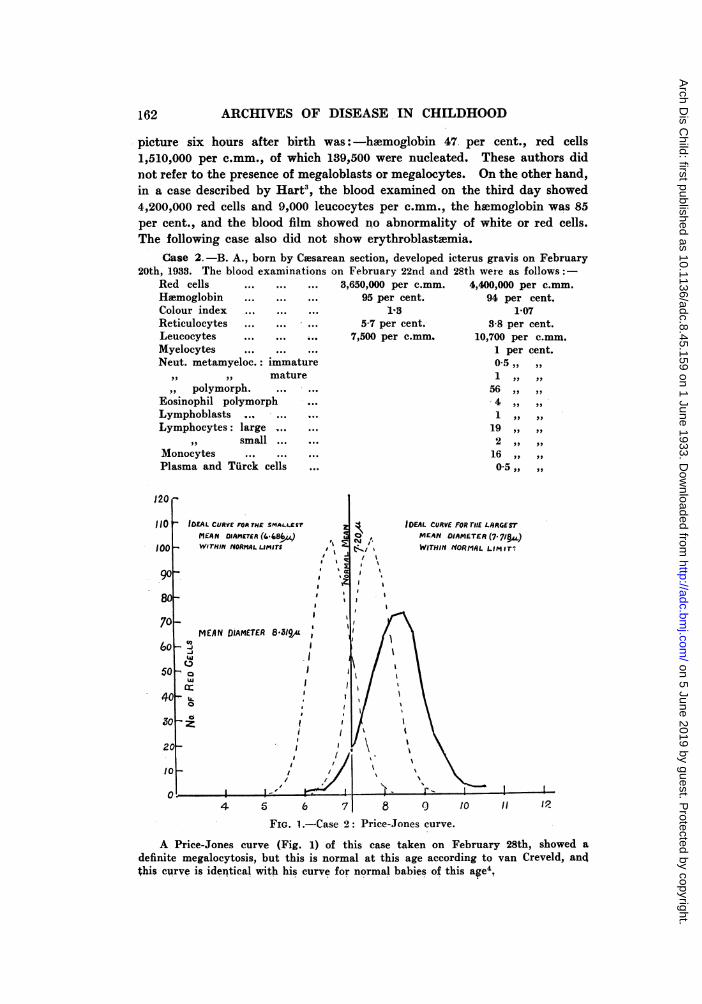
picture six hours after birth was: -haemoglobin 47 per cent., red cells1,510,000 per c.mm., of which 139,500 were nucleated. These authors didnot refer to the presence of megaloblasts or megalocytes. On the other hand,in a case described by Hart3, the blood examined on the third day showed4,200,000 red cells and 9,000 leucocytes per c.mm., the hwmoglobin was 85per cent., and the blood film showed no abnormality of white or red cells.The following case also did not show erythroblasteemia.
Case 2. -B. A., born by Cmsarean section, developed icterus gravis on February20th, 1933. The blood examinations on February 22nd and 28th were as follows:-
Red cells ... ... ... 3,650,000 per c.mm. 4,400,000 per c.mm.Hlemoglobin ... ... ... 95 per cent. 94 per cent.Colour index ... ... ... 13 1-07Reticulocytes ... ... ... 5-7 per cent. 3-8 per cent.Leucocytes.... ... 7,500 per c.mm 10,700 per c.mm.Myelocytes ... ... ... 1 per cent.Neut. metamyeloc.: immature 0,5 ,,
,, ,, mature 1 ,, ..polymorph. 56
Eosinophil polymorph 4Lymphoblasts . ... 1Lymphocytes: large ... 19
,,9 small ... ,...2Monocytes ... ... 16Plasma and Turck cells ... 0-5 ,,
J20 -
110 IDEAL CURVE FOR THE SMAL.T2r IDEAL CURVE FOR rTE LARGESTMEAN DIAMETEA (4.-84) , O, MEAN DIAMETER (7.7I98)
1oo WITHINrVORMALLIMITS E WITHIH NYORMAL LIMar.
90 Ia
70 -
MEAN DIAMETER 8.319, j
40 U.
30aa
2 i I g ' / 2'I\I\'20
10 /
4 5 6 7 8 9 10 I1 1?FIG. 1.-Case 2: Price-Jones curve.
A Price-Jones curve (Fig. 1) of this case taken on February 28th, showed adefinite megalocytosis, but this is normal at this age according to van Creveld, andthis curve is identical with his curve for normal babies of this age4,
on 5 June 2019 by guest. Protected by copyright.
http://adc.bmj.com
/A
rch Dis C
hild: first published as 10.1136/adc.8.45.159 on 1 June 1933. Dow
nloaded from

STUDIES IN ANA,MIA-PAtT IV 16a
Examination of the blood when the jaundice is fading as a rule showsa profound ansemia of the hyperchromic type, the red cells beingdiminished out of proportion to the haemoglobin, and the colour index beingunity or higher. A considerable degree of polychromasia and anisocytosisis present, as are normoblasts and reticulocytes. Megaloblasts may-also befound. The platelet count may be normal, but usually is slightly reduced.The white cell count may be raised, but this is not a constant feature nor ofgreat importance. It is interesting that an eosinophilia is present, especiallyduring recovery. As the child improves the normoblasts quickly disappear,and the blood picture gradually returns to normal.
The haematological details in a typical case of anaemia following icterus gravis,18 days after the onset of jaundice, were as follows:Red cells ... ... 2,147,200 per c.mm. Lymphocytes ... ... 35 per cent.Haemoglobin ... 36 per cent. Monocytes ... ... 7,1 ,.Reticulocytes ... 28 ,, ,, Plasma cells ... 0-8 ,, ,
Platelets ... 320,000 per c.mm. NormoblastsLeucocytes ... 23,420 ,, ,, (per 250 leucocytes) 16Premyelocytes ... 11 per cent. MacronormoblastsMyelocytes ... 3 ,, ,, (per 250 leucocytes) 2Neut. metamyeloc. 2 ,, ,, Anisocytosis ... ... +
,, polymorph. 48-2 ,, ,, Poikilocytosis. ... +Eosinophils ... 28 ,, ,, Polychromasia ... + ++
The presence of abnormal megalocytosis, or of megaloblastosis, is to beregarded as the response to a great demand on the marrow and extra-medullary haemopoietic centres. All of these still possess embryonic qualitiesand their ability to cope with the demand for new cells is shown by thepresence of a reticulocytosis. In assessing the degree of megalocytosis it isnecessary to bear in mind that, as has been already mentioned, megalocytosisnormally occurs in the new born.
The ansemia following icterus gravis therefore differs clinically fromnutritional anemia of the new born in the following respects:
(1) A severe hyperchromic anaemia becoming obvious as a severe attackof icterus is clearing up.
(2) The presence of a positive indirect van den Bergh reaction in theblood which may not be obtained if the icterus has all disappeared.
(3) The presence of an excess of urobilinogen and of urobilin in the urine,which also may not be found after the disappearance of the icterus.
(4) A spontaneous effort at recovery as shown by marked reticulocytosisand progressive improvement.
(5) A macrocytic response and in some cases the presence of megaloblastsin the peripheral blood, an occurrence almost unknown in nutritional anaemia.
(6) Frequent occurrence of eosinophilia.This late anaemia of icterus gravis does not really call for treatment
because if the child survives until the anemia is obvious spontaneous
on 5 June 2019 by guest. Protected by copyright.
http://adc.bmj.com
/A
rch Dis C
hild: first published as 10.1136/adc.8.45.159 on 1 June 1933. Dow
nloaded from

164 ARCHIVES OF DISEASE IN CHILDHOOD
recovery will occur. The treatment of the anamia present at birth and theprevention of the severest degrees of the later anaemia are best carried outby efficient treatment of the icterus gravis. We have found the method oftreatment outlined by Hampson5, in which intramuscular injections of about10 c.cm. of parental blood serum are given daily for several days, has beenvery successful if commenced sufficiently early. If the administration ofserum is delayed until after the second day of life it is doubtful whetherrecovery will occur; indeed, one of our cases which received treatment beforethe end of the second day ultimately developed a considerable degree ofspasticity (kernicterus). Icterus gravis has also been successfully treated byintramuscular injections of whole blood, but we prefer to give serum.Frequent small blood transfusions have also been given. We have not hadany experience of this form of treatment, but in view of the results we haveobtained in the treatment of acute hoemolytic anaemia of later infancy we arenot surprised that good results have been reported. Hampson's method,however, has the great merit of simplicity. Blood transfusions are likelyto be more helpful than serum in combating the early anaemia, andin hydrops foetalis. In any case the essentials for success are early diagnosisand treatment, and to this end it should be pointed out that Clifford andHertig lay stress on the peculiar yellow colour of the vernix caseosain erythroblastosis of the new born.
C. Heemolytic aneemia without cedema or icterus gravis.-These casesshould perhaps be classified under Group 2, and in any case they form aconnecting link with that group. Infants which can without hesitation beclassified under this heading are examples of erythroblastosis of the new born,but may not show any erythroblastaemia. This condition must be one ofextreme rarity, but we believe that we have seen one case, and havepathological material from two.
Case 3.-B. O., male, aged 6 days, died within an hour of admission to hospitaland before any extensive examinations had been carried out. He was the third childin the family; no miscarriages. He was born at term and was very pale at birth;the umbilicus was healthy, no blecding from the cord. On the 3rd day he wasfaintly jaundiced, but this was not evident by artificial light when he was seen. Thespleen was enlarged; respirations were 40 and pulse 120 per minute and temperaturewas 970. He died without further examination, but blood drawn from the heart bya syringe ten minutes after death was pale pink of the colour of dilute strawberryjam. This fluid gave a red cell count of 320,000 and leucocyte count of 15,200 perc.mm. The hemoglobin was too low to estimate, being under 20 per cent. The serumwas bile-stained. Films showed an excess of large lymphocytes, but the stainingproperties of the cells were very poor. Erythroblastiemia was not a feature of thefilm. A differential leucocyte count gave the following percentages:Neut. polymorph ... ... ... 2 Basophil polymorph. ... 0 5
,, metamyeloc., mature 5 Lymphocytes, large ... ... 82-5,,,, immature 3 ,, small
... ... 05Eosin. polymorph . ... ... ... 2 Monocytes ... . .. ... ... 1-5
, metamyeloc., immature ... 1-5 Monocytes ( ?) ... ... 1-5
At autopsy slight bile-staining of the lungs, pleural exudate and liver was observed,
on 5 June 2019 by guest. Protected by copyright.
http://adc.bmj.com
/A
rch Dis C
hild: first published as 10.1136/adc.8.45.159 on 1 June 1933. Dow
nloaded from

STUDIES IN ANIEMIA-PART IV 165
The other case was that of a child who was born in one of theBirmingham municipal maternity homes and is to be reported in detailelsewhere by Dr. Mary Crosse. The following is a brief note of it:-
Case 4.-B. W., developed slight jaundice on the 2nd day of life. This soondisappeared. At 7 days the child was noticed to be pale, and the following day thespleen was palpable, the red cell count 2,600,000 per c.mm., and hsemoglobin 42per cent. A day later the red cell count was below a million and the hemoglobin20 per cent. The child died on the 12th day.
Group 2. Hsemolytic aniemia later in the neonatal period without cedemaor icterus gravis.
In the course of our investigations we have found a few examples ofsevere anaemia of the hyperchromic type in infants varying in age from a fewweeks to two or three months. The history and examination may or maynot reveal jaundice, and even when jaundice has occurred it may have madesuch a slight impression on the mother's mind that to obtain any history ofit a direct question has to be asked; certainly there is never any historysuggesting the occurrence of icterus gravis. Usually the child is normal atbirth and during the subsidence of icterus neonatorum it is noticed to bepale. Advice may be sought at this time, or not until some weeks later. Onexamination the child is found to be extremely pale and, if in the neonatalperiod, a slight icteric tiint inay still be noticeable. Frequently someenlargement of the spleen is found. Its degree is, however, rarely markedand in any event slight enlargement of the spleen is no great help inclassifying the anaemias of infancy. Urobilinogen and urobilin are in excessin the urine.
The following case illustrates the history and clinical findings in thistype of haemolytic ansemia:-
Case 5.-W. S., a male breast-fed child aged 4 weeks, was brought to hospitalfor anzemia with a history of jaundice of five days' duration during the first weekof life. Since that time he was said to have become increasingly pale. On admissionhe was obviously very anoemic. There was no pyrexia, and no trace of jaundice; theliver was slightly and the spleen considerably enlarged; the stools were deeplypigmented and urobilinogen was present in the urine in considerable quantities. Theblood examination corresponded exactly with that already described in the anaemiafollowing icterus gravis; there was an anaemia of the hyperchromic type showing aconsiderable degree of polychromasia, anisocytosis, macrocytosis, reticulocytosis andmegaloblastosis. The white cells were very slightly increased and showed someimmaturity of the myeloid series.
From the time of admission to hospital this child showed a steady progress tocomplete recovery and during this time an eosinophilia developed. Recovery wascomplete and spontaneous; indeed the blood picture when first seen indicated thatthe marrow was active and healthy. The details of the blood counts are given inthe accompanying list,
on 5 June 2019 by guest. Protected by copyright.
http://adc.bmj.com
/A
rch Dis C
hild: first published as 10.1136/adc.8.45.159 on 1 June 1933. Dow
nloaded from
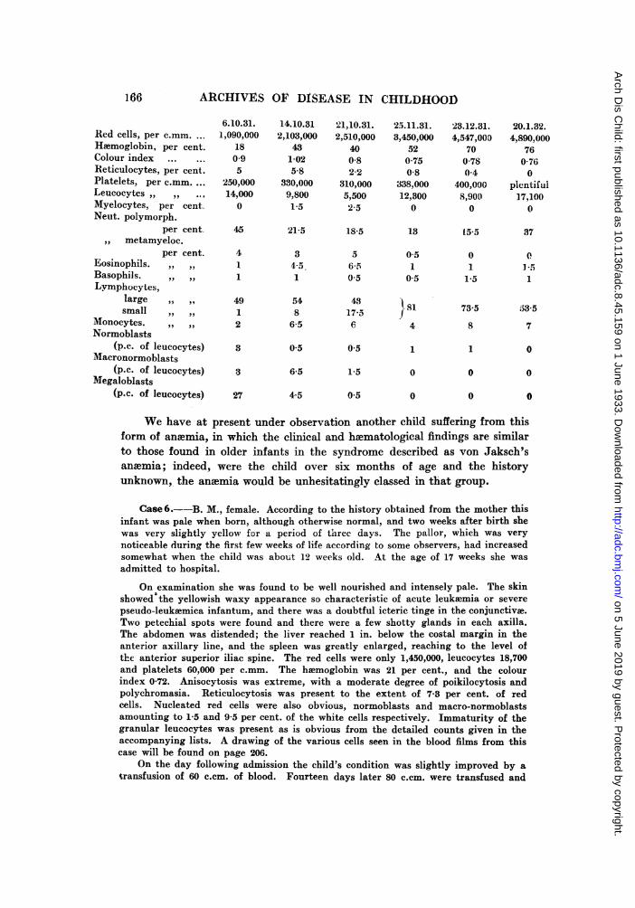
166 ARCHIVrES OF DISEASE IN CHILDHOOD
6.10.31. 14.10.31 2'1,10.31. 25.11.31. 23.12.31. 20.1.32.Red cells, per c.mm. 1,090,000 2,103,000 2,510,000 3,450,000 4,547,000 4,890,000Hiemoglobin, per cent. 18 43 40 52 70 76Colour index ... 0 9 1-02 0-8 0 75 0-78 0 76Reticulocytes, per cent. 5 5-8 2-2 0-8 0 4 0Platelets, per c.mm. 250,000 330,000 310,000 338,000 400,000 plentifulLeucocytes ,, ,, 14,000 9,800 5,500 12,300 8,901 17,100Myelocytes, per cent, 0 1-5 25 0 0 0Neut. polymorph.
per cent 45 21-5 18-5 13 [5.5 37,, metamyeloc.
per cent. 4 3 5 05 0 QEosinophils. ,, ,, 1 45. 6 5 1 1 1J5Basophils. ,, ,, 1 1 0 5 0 5 1-5 1Lymphocytes,
large ,, ,, 49 54 43 7small ,, ,, 1 8 17a5 81 73-5 J;3-5
Monocytes. ,, ,, 2 6-5 6 4 8 7Normoblasts
(p.c. of leucocytes) 3 0-5 0 5 1 1 0Macronormoblasts
(p.c. of leucocytes) 3 6-5 1-5 0 0 0Megaloblasts
(p.c. of leucocytes) 27 4-5 0 5 0 0 0
We have at present under observation another child suffering from thisform of anaemia, in which the clinical and haematological findings are similarto those found in older infants in the syndrome described as von Jaksch'sanaemia; indeed, were the child over six months of age and the historyunknown, the anawmia would be unhesitatingly classed in that group.
Case 6.--B. M., female. According to the history obtained from the mother thisinfant was pale when born, although otherwise normal, and two weeks after birth shewas very slightly yellow for a period of three days. The pallor, which was verynoticeable during the first few weeks of life according to some observers, had increasedsomewhat when the child was about 12 weeks old. At the age of 17 weeks she wasadmitted to hospital.
On examination she was found to be well nourished and intensely pale. The skinshowed the yellowish waxy appearance so characteristic of acute leukaemia or severepseudo-leukaemica infantum, and there was a doubtful icteric tinge in the conjunctivae.Two petechial spots were found and there were a few shotty glands in each axilla.The abdomen was distended; the liver reached 1 in. below the costal margin in theanterior axillary line, and the spleen was greatly enlarged, reaching to the level ofthe anterior superior iliac spine. The red cells were only 1,450,000, leucocytes 18,700and platelets 60,000 per c.mm. The hwemoglobin was 21 per cent., and the colourindex 0-72. Anisocytosis was extreme, with a moderate degree of poikilocytosis andpolychromasia. Reticulocytosis was present to the extent of 7-3 per cent. of redcells. Nucleated red cells were also obvious, normoblasts and macro-normoblastsamounting to 1-5 and 9-5 per cent. of the white cells respectively. Immaturity of thegranular leucocytes was present as is obvious from the detailed counts given in theaccompanying lists. A drawing of the various cells seen in the blood films from thiscase will be found on page 206.
On the day following admission the child's condition was slightly improved by atransfusion of 60 c.cm. of blood. Fourteen days later 80 c.cm. were transfused and
on 5 June 2019 by guest. Protected by copyright.
http://adc.bmj.com
/A
rch Dis C
hild: first published as 10.1136/adc.8.45.159 on 1 June 1933. Dow
nloaded from
STUDIES IN AN2EMIA-PART IV 167
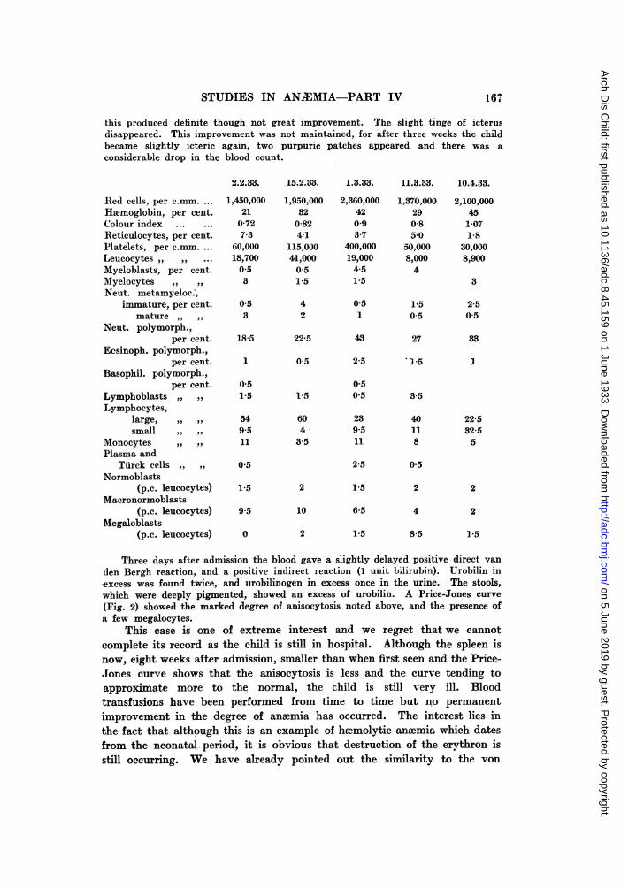
this produced definite though not great improvement. The slight tinge of icterusdisappeared. This improvement was not maintained, for after three weeks the childbecame slightly icteric again, two purpuric patches appeared and there was aconsiderable drop in the blood count.
2.2.33. 15.2.33. 1.3.33. 11.3.33. 10.4.33.
Ited cells, per c.mm. 1,450,000 1,950,000 2,360,000 1,370,000 2,100,000Haemoglobin, per cent. 21 32 42 29 45Colour index ... ... 0-72 0-82 0-9 0-8 107iReticulocytes, per cent. 7-3 4-1 3.7 50 18Platelets, per c.mm ... 60,000 115,000 400,000 50,000 30,000Leucocytes ,, ,, ... 18,700 41,000 19,000 8,000 8,900Myeloblasts, per cent. 0 5 05 4-5 4Myelocytes ,, ,, 3 1-5 1-5 3Neut. metamyeloc;,
immature, per cent. 0 5 4 0 5 1-5 2-5mature ,, ,, 3 2 1 0-5 0 5
Neut. polymorph.,per cent. 18-5 22-5 43 27 33
Eesinoph. polymorph.,per cent. 1 0-5 2-5 ' -5 1
Basophil. polymorph.,per cent. 0-5 0-5
Lymphoblasts ,, ,, 1-5 1V5 05 3-5Lymphocytes,
large, ,, ,, 54 60 23 40 22-5small ,, ,, 9.5 4 9.5 11 32-5
Monocytes ,, ,, 11 3-5 11 8 5Plasma and
Tiirck cells ,, ,, 0-5 2-5 0-5Normoblasts
(p.c. leucocytes) 1.5 2 1-5 2 2Macronormoblasts
(p.c. leucocytes) 9-5 10 6-5 4 2Megaloblasts
(p.c. leucocytes) 0 2 1-5 8-5 1.5
Three days after admission the blood gave a slightly delayed positive direct vanden Bergh reaction, and a positive indirect reaction (1 unit bilirubin). Urobilin inexcess was found twice, and urobilinogen in excess once in the urine. The stools,which were deeply pigmented, showed an excess of urobilin. A Price-Jones curve(Fig. 2) showed the marked degree of anisocytosis noted above, and the presence ofa few megalocytes.
This case is one of extreme interest and we regret thatwe cannotcomplete its record as the child is still in hospital. Although the spleen isnow, eight weeks after admission, smaller than when first seen and the Price-.Jones curve shows that the anisocytosis is less and the curve tending toapproximate more to the normal, the child is still very ill. Bloodtransfusions have been performed from time to time but no permanentimprovement in the degree of anaemia has occurred. The interest lies inthe fact that although this is an example of haemolytic anaemia which datesfrom the neonatal. period, it is obvious that destruction of the erythron isstill occurring. We have already pointed out the similarity to the von
on 5 June 2019 by guest. Protected by copyright.
http://adc.bmj.com
/A
rch Dis C
hild: first published as 10.1136/adc.8.45.159 on 1 June 1933. Dow
nloaded from

168 ARCHIVES OF DISEASE IN CHILDHOOD
Jaksch's syndrome presented by the clinical picture of this child; it isprobable that the clinical course of the case indicates the mode of develop-ment of that syndrome and so lends support to the view that the aneemia ofvTon Jaksch is a sub-chronic haemolytic anaemia.
120
110 IAL. CURVF FOR 71f SA14LL.Xr DCAL. CURVE FO THrC LARGesTME01 DIAMErE. (6.64) CEAN DIAMETER (7 7/p)WITHIN ('ORMrAL LIMITS 'i1 WITHII#NtOAMAL. r rlrs
90 I
44 IL z f Sl~~I
70 (EI/A DIAMETUR6799C g
6olE
20
3 4 5 6 7 8 9. tO 11 12
Fic. 2.-Case 6: Price-Jones curve.
Although in our experience in this group of cases recovery after avarying time is the rule, yet death occurred in one instance.
Ca<> 7-R. L., male, was aged 3 months at the time of his death and had beennoticeably pale from the first week or so of life, but had never suffered from jaundiceor oedema. A week before his admission to hospital he had become paler. Whenexamined it was obvious that he had severe anaemia; there were some purpuric patcheson the head; the spleen and liver were enlarged but the lymphatic glands werenormal. Examination of the blood revealed the presence of a very severe anaemia,the red cell count being under one million, and the haemoglobin 20 per cent.
Red blood cells per c.mm ... 810,00) Eosinoph. polymorph. ... ... 2-5Haemoglobin per cent. ... 20 Basoph. polymorph. ... ... 10Colour index ... ... ... 125 Large lymphocytes. ... ... 18Leuc3cytes per c.mm. ... 15,330 Small lymphocytes ... ... 475Reticulocytes per cent. ... 1-0 Lymphoblasts ... ... 1-5Myeloblasts ... ... ... 0-5 Tilrck cells ... ... ... ... 0 5Neut. polym., segmrrented Monocytes ... 1-5
per cent. 20 Normoblasts ) per 100 3non-segmented Megaloblasts leucocytes ... 5 5
per cent. 7 Polychromasia ... .. +. +Metamyelocytes, mature Anisocytosis .++ +
per cent. 10 Poikilocytosis ... . .. ... t
on 5 June 2019 by guest. Protected by copyright.
http://adc.bmj.com
/A
rch Dis C
hild: first published as 10.1136/adc.8.45.159 on 1 June 1933. Dow
nloaded from

STUDIES IN ANAlMIA-PART IV 169
Fig. 3 is a composite drawing and Fig. 4 is a photograph of the cells present inthe blood films. He died five days after admission and the details of the autopsy aregiven in the section on pathology (page 176).
1'
FIG. 3.-Case 7:. Composite drawing show ing large numbers of megaloblasts.1. Megaloblasts. 2. Basophilic lymphocytes, 3. Lymphocyte.4. Lymphoblast, possibly myeloblast. 5. Chromasoines inmitotic red cell. 6. Mast cell. 7. Matute metamyelocyte8. Mature neutrophilic polymorpIioiiuclear. 9. Polychroirrasia
10. Platelets.
~~~~~~~~~~~~~~~~~~~~~~FIG. 4.-Case 7 Blood film ( x 390): mitosis in
erythroblast on right, pre mtyelocyte belowlymphocyte to left,
on 5 June 2019 by guest. Protected by copyright.
http://adc.bmj.com
/A
rch Dis C
hild: first published as 10.1136/adc.8.45.159 on 1 June 1933. Dow
nloaded from

170 ARCHIVtES OF DISEASE IN CHILDHOOD
Pathological changes in hemolytic anaemia of the new born.
Pathological changes in icterus gravis.-We have had material from fourfatal cases, and the following were the chief macroscopic abnormalities found.All the organs, with the exception of the central nervous system and cerebro-spinal fluid which were only slightly tinted, showed general icteric staining,often of an extreme degree. Deep bile staining of the basal ganglia(kernicterus) and degeneration of the nerve cells are described in theliterature, but unfortunately we have not had an example of this in ourseries. The liver was slightly enlarged, deeply bile stained, and did notshow any cirrhosis. The gall-bladder contained either a normal or smallamount of bile; sometimes little more than mucus. The spleen was slightlyenlarged The bone marrow was of a rich red colour throughout, tingedwith the general icterus, which sometimes stained the epiphyseal line agreenish hue. A little oozing of blood from the umbilical stump has beennoted but without any evidence of infection.
HISTOLOGICAL EXAMINATION: LIVER (Fig. 5, 6 and 7). The principalfinding has been the presence of embryonic blood formation in the hepaticsinusoids which produced a striking low power picture. The liver cellcolumns were broken up by wide sinusoids, in which were found many smallclumps, of fairly deeply staining round and oval cells possessing prominentnuclei somewhat vesicular in type. Much bile pigment was present, chieflylying free in the sinusoids, but also occurring in the liver cells, in phagocytichistiocytes in the sinusoids and sometimes as small thrombi in the bilecanaliculi. There was no obvious fatty degeneration in the central areasof the lobules which is so common in many toxic affections in children. Thispicture of the packing of the liver sinusoids with immature cells closelysimulates a leukaemic process and careful examination of the cells wastherefore required. This was carried out on well-cut sections with thetwelfth-inch immersion objective. Most of the larger cells were about 8 or0Lin their longest diameter, and possessed round or oval nuclei of about
7,u in size and surrounded by a moderate amount of cytoplasm. The nucleiwere vesicular, a well defined nuclear margin enclosing a pale interior inwhich one or two nucleoli might be found; the knots of karyomitone wererather distinct although not numerous and tended to lie mostly near thenuclear membrane. Such characters are very similar to those of themyeloblast and premyelocyte when these cells are seen in histologicalsections. A careful comparison of our specimens (stained with hsematoxylinand eosin; eosin and methylene blue; Giemsa) with those of myeloidleukaemia has shown that cytoplasm, nuclear margin and chromatin networktend to be definitely darker in staining reaction than those of the myeloidcells; the cytoplasm too is more basophilic. Moreover the real nature ofthese cells was revealed by the large number of erythroblasts scattered intheir vicinity and throughout the sinusoids. Extruded erythroblastic nucleiwere common and erythroblasts of larger size with karyorrhectic polymorphic
on 5 June 2019 by guest. Protected by copyright.
http://adc.bmj.com
/A
rch Dis C
hild: first published as 10.1136/adc.8.45.159 on 1 June 1933. Dow
nloaded from

STUDIES IN ANAMIA-PART IV 171
Fio. 5.-Liver in icterus gravis; projection drawinIg ( x 1000): showingpro.erythroblasts with cytoplasm darker than in mye]ocyrtes; darkersmall cell w-ith them appears tO be a type of megaloblast, the nucleusis larger than in the usual normoblast.
FIG. 6-Liver in icterus gratins ( x 90!: chowing w~idQsinusoids with h em~opoietic foci.
on 5 June 2019 by guest. Protected by copyright.
http://adc.bmj.com
/A
rch Dis C
hild: first published as 10.1136/adc.8.45.159 on 1 June 1933. Dow
nloaded from

172 ARCHIVES OF DISEASE IN CHILDHOODlnuclei were not uncommon and required differentiation from polymorpho-nuclear granulocytes, which they superficially resemble in sections. Theseprominent dark cells are in fact erythropoietic in character being pro-erythroblasts*. Further search of the sections revealed cells some of whichappeared to be less and other more mature than pro-erythroblasts. Theless mature consisted of a few larger and paler cells with similar, but lightervesicular nuclei (probably haemohistioblasts), which appeared to be derivedfrom swollen endothelial cells either partially or wholly detached (pro-liferated Kupffer cells). On the other hand, cells of similar generalcharacters, about 8,u in size, but with a darker, strongly and evenly stipplednucleus throughout, were seen, and these were probably megaloblasts.Nuclear forms and types between these and the recognizable erythroblastshave been found, though many of the erythroblasts have, on the whole,rather large nuclei. Other cells observed included some polymorphonucleargranulocytes and some large phagocytic histiocytes. Sections stainedby the Turnbull-Hueck process revealed the presence of a considerableamount of haemosiderin.
SPLEEN.-The presence of the above described cells and erythropoiesiswas variable: recently one of our cases showed these, but the next case didnot. The recticular cells always showed evidence of activity: proliferation insome cases and phagocytosis in others. Lymphocytes were numerous.
MARROW.-In our cases the marrow has shown considerable activity,especially on the myeloid side, but it does not appear to have any distinctivecharacters. One marrow, which was decalcified and hence did not revealits cellular characters well, showed a diminution in normoblasts, and a smearmade from this marrow revealed many immature granulocytes but nonormoblasts. The significance of this finding is not understood, but it ispossible that the embryonic haemopoietic function of the liver is maintainedin activity partly because of some failure of the erythropoietic functionof the developing marrow.
Pathological changes in hydrops foetalis.-In hydrops foetalis verysimilar changes have been recorded, and the erythroblastosis evident in theorgans has been regarded as one proof of an inherent relationship betweenicterus gravis and hydrops foetalis. De Lange and Arntzenius10 and othershave described a breaking up of liver cell columns by erythroblasts and cellslike lymphocytes (? megaloblasts), with and without a few bile thrombi,and a positive iron reaction by the Turnbull-Hueck process. We have hadthe opportunity of examining only one case in recent years (Case 1 above).
*' Erythroblast ' designates any form of nucleated red cell (i.e., eithermegaloblast or normoblast). ' Pro-erythroblast ' is a more immature cell whichappears to be developing into a nucleated red cell. In Sabin's terminology,' erythroblast ' does not include ' megaloblast.'
on 5 June 2019 by guest. Protected by copyright.
http://adc.bmj.com
/A
rch Dis C
hild: first published as 10.1136/adc.8.45.159 on 1 June 1933. Dow
nloaded from

STUDIES IN AN2FMIA-PART IV 17.
Fic.. 7.-Liver in icterus gravis (x 850) PE, pro- FiG. 8 -Case 1 Liver in hydrops foetahis (x 75):erythrocytes with lighter nuclei anid dark showing sex ere toxic change and necrosis in livercytoplasm (tlhe cytoplasmn is darker and the cells E, erytbroblasts.chromatin miasses in the nuclei slightly darkertlani in inyeloblasts) :M, niegaloblasts withdarker, heavily dotted nuclei N, normoblasts.
FIG 9.-Case 1 Liver in hydrops foe-talis (x 700' FIG. 10.-Case 4 Liver (x 700) :showing moreshowing great disorganization, only a few mature erythroblasts, mostly with nuclei ofhepatic cells (H) being recognizable ;the scat- nori-noblastic type.teied dark round '_ells arc normoolastic nucleior small erythroblasts ; PE, ? large pro-erythroblast.
on 5 June 2019 by guest. Protected by copyright.
http://adc.bmj.com
/A
rch Dis C
hild: first published as 10.1136/adc.8.45.159 on 1 June 1933. Dow
nloaded from
174 ARCHIVES OF DISEASE IN CHILDHOOD
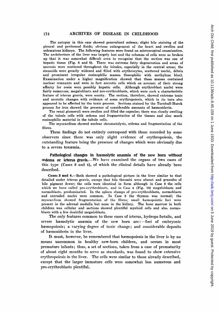
The autopsy in this case showed generalized cedema; slight bile staining of thepleural and peritoneal fluids; obvious enlargement of the heart and swollen andcedematous kidneys. The following features were found on microscopical examination.The architecture of the liver was largely lost and the columns of cells were so brokenup that it was somewhat difficult even to recognize that the section was one ofhepatic tissue (Fig. 8 and 9). There was extreme fatty degeneration and areas ofnecrosis were scattered throughout the lobules, especially in the central areas; thesinusoids were greatly widened and filled with erythrocytes, scattered nuclei, debrisand prominent irregular eosinophilic masses (basophilic with methylene blue).Examination under a higher magnification showed that these masses containednuclear remnants and were in fact necrotic cells which on account of their strongaffinity for eosin were possibly hepatic cells. Although erythroblast nuclei werefairly numerous, megaloblasts and pro-ervthroblasts, which were suich a characteristicfeature of icterus gravis, were scanty. The section, therefore, showed extreme toxicand necrotic changes with evidence of some erythropoiesis, which in its turn alsoappeared to be affected by the toxic process. Sections stained by the Turnbull-Hueckprocess for iron showed the presence of considerable amounts of h.mosiderin.
The renal glomeruli were swollen and filled the capsules; there was cloudy swellingof the tubule cells with cedema and fragmentation of the tissues and also mucheosinophilic material in the tubule cells.
The myocardium showed nuclear chromatolysis, cedema and fragmentation of thefibres.
These findings do not entirely correspond with those recorded by someobservers since there was only slight evidence of erythropoiesis, theoutstanding feature being the presence of changes which were obviously dueto a severe toxaemia.
Pathological changes in hoemolytic anoemia of the new born withoutedema or icterus gravis.-We have examined the organs of two cases ofthis type (Cases 3 and 4), of which the clinical details have already beendescribed.
Cases 3 and 4.-Both showed a pathological picture in the liver similar to thatdetailed under icterus gravis, except that bile thrombi were absent and granules ofbile pigment fewer; the cells were identical in form although in Case 3 the cellswhich we have called pro-erythroblasts, and in Case 4 (Fig. 10) megaloblasts andnormoblasts, predominated. In the spleen clumps of pro-erythroblasts, normoblastsand extruded nuclei were common. In Case 3 the thymus was normal; themyocardiurn showed fragmentation of the fibres; small biemopoietic foci werepresent in the adrenal medulla but none in the kidney. The bone marrow in bothchildren was cellular and sections showed plentiful myeloid cells and also normo-blasts with a few doubtful megaloblasts.
The only features common to these cases of icterus, hydrops feetalis, andsevere hwemolytic anaemia of the new born are: -foci of embryonichsemopoiesis; a varying degree of toxic change; and considerable depositsof hwemosiderin in the liver.
It must, however, be remembered that haemopoiesis in the liver is by nomeans uncommon in healthy new-born children, and occurs in mostpremature infants; thus, a set of sections, taken from a case of prematurityof about eight months to serve as standards, was found to show extensiveerythropoiesis in the liver. The cells were similar to those already described,except that the larger immature cells were somewhat less numerous andpro-erythroblasts plentiful,
on 5 June 2019 by guest. Protected by copyright.
http://adc.bmj.com
/A
rch Dis C
hild: first published as 10.1136/adc.8.45.159 on 1 June 1933. Dow
nloaded from
STUDIES IN ANAEMIA-PART IV 175
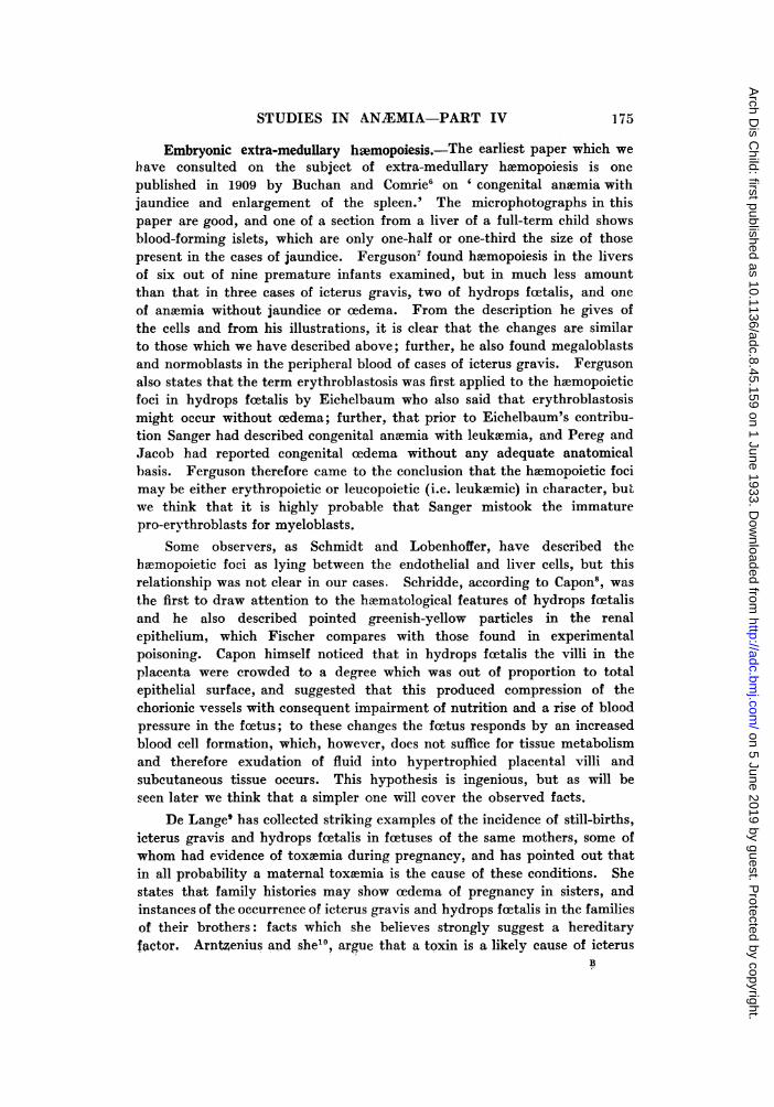
Embryonic extra-medullary haemopoiesis.-The earliest paper which wehave consulted on the subject of extra-medullary haemopoiesis is onepublished in 1909 by Buchan and Comrie6 on ' congenital anaemia withjaundice and enlargement of the spleen.' The microphotographs in thispaper are good, and one of a section from a liver of a full-term child showsblood-forming islets, which are only one-half or one-third the size of thosepresent in the cases of jaundice. Ferguson7 found haemopoiesis in the liversof six out of nine premature infants examined, but in much less amountthan that in three cases of icterus gravis, two of hydrops fretalis, and oneof anaemia without jaundice or cedema. From the description he gives ofthe cells and from his illustrations, it is clear that the changes are similarto those which we have described above; further, he also found megaloblastsand normoblasts in the peripheral blood of cases of icterus gravis. Fergusonalso states that the term erythrob]astosis was first applied to the haemopoieticfoci in hydrops foetalis by Eichelbaum who also said that erythroblastosismight occur without cedema; further, that prior to Eichelbaum's contribu-tion Sanger had described congenital anaemia with leukwemia, and Pereg andJacob had reported congenital cedema without any adequate anatomicalbasis. Ferguson therefore came to the conclusion that the haTmopoietic focimay be either erythropoietic or leucopoietic (i.e. leukaemic) in character, butwe think that it is highly probable that Sanger mistook the immaturepro-erythroblasts for myeloblasts.
Some observers, as Schmidt and Lobenhoffer, have described thehbemopoietic foci as lying between the endothelial and liver cells, but thisrelationship was not clear in our cases. Schridde, according to Capon8, wasthe first to draw attention to the haematological features of hydrops foetalisand he also described pointed greenish-yellow particles in the renalepithelium, which Fischer compares with those found in experimentalpoisoning. Capon himself noticed that in hydrops foetalis the villi in theplacenta were crowded to a degree which was out of proportion to totalepithelial surface, and suggested that this produced compression of thechorionic vessels with consequent impairment of nutrition and a rise of bloodpressure in the foetus; to these changes the foetus responds by an increasedblood cell formation, which, however, does not suffice for tissue metabolismand therefore exudation of fluid into hypertrophied placental villi andsubcutaneous tissue occurs. This hypothesis is ingenious, but as will beseen later we think that a simpler one will cover the observed facts.
De Lange' has collected striking examples of the incidence of still-births,icterus gravis and hydrops foetalis in foetuses of the same mothers, some ofwhom had evidence of toxaemia during pregnancy, and has pointed out thatin all probability a maternal toxaemia is the cause of these conditions. Shestates that family histories may show oedema of pregnancy in sisters, andinstances of the occurrence of icterus gravis and hydrops foetalis in the familiesof their brothers: facts which she believes strongly suggest a hereditary,actor. Arntzenius and she10, argue that a toxin is a likely cause of icterus
on 5 June 2019 by guest. Protected by copyright.
http://adc.bmj.com
/A
rch Dis C
hild: first published as 10.1136/adc.8.45.159 on 1 June 1933. Dow
nloaded from
176 ARCHIVES OF DISEASE IN CHILDHOOD
gravis and of hydrops foetalis, and that the haemopoiesis is reparative incharacter. They also say that the presence in the liver in hydrops of apositive action for iron when stained by the Turnbull-Hueck method isevidence of moderate haemolysis; further, that the oedema may be causedby the influence of the toxin on the blood vessels; and they point out that,although extra-medullary blood formation occurs in most cases, it is notpresent in all. In their view, therefore, there is no relationship betweengrave icterus and hydrops, except that they are both due to the action ofa toxin of an unknown nature. Rantmann, on the other hand, holds thatthe erythroblastosis is not a reaction, but a primary factor.
We agree with Salomonsen that the finding of considerable quantitiesof haemosiderin in the liver by the Turnbull-Hueck process cannot beregarded as evidence of an abnormal degree of haemolysis. The livers in ourcases of icterus gravis, hydrops foetalis, and haemolytic anaemia of the newborn showed considerable quantities of iron; nevertheless we have found asgreat a degree of iron present in the liver of a premature child who died whenone day old and in that of a full-term child who died when eighteen days old,although, on the other hand, the liver of a child one month old waspractically free from iron.
Extra-medullary hemopoiesis in later infancy.-In a full review of thesubject of extra-medullary hawmopoiesis, Brannan" reported the case of amale infant of 7- months showing many normoblasts and a few megaloblastsin the peripheral blood with reduction of platelets and changes in the skullbones. This child had tumour-like deposits of haemopoiesis in the hilum ofthe kidneys and in the falx cerebri. He also quoted instances of suchmarrow deposits in the broad ligaments, in the breasts, and in relation tosweat glands in the soles and palms, and considered it probable that thesemyeloid elements are constantly present in the spleen of adults. Weconsider that there is some histological evidence for this process, since insections of splenic tissue we have found scanty cells of myeloid type witherythroblasts, the latter being more numerous than in other organs (exceptthe marrow). In a case of lymphatic leukwemia, recorded in the article onleukeemia (Part VI) a small focus of marrow formation was discovered inthe hilum of a kidney. This type of extra-medullary haemopoiesis has beenreferred to in order to demonstrate how it differs from the embryonic formwhich we have described as occurring notably in the liver of the prematureand new-born baby. It differs in its activity, its occurrence in later infancy,its focal distribution, and its relatively greater activity in the formation ofmyeloid cells.
Pathological changes in haemolytic anaemia later in neonatal periodwithout aedema or grave jaundice.
The chief findings at the autopsy on the only fatal case of this type thatwe have seen were as follows:-
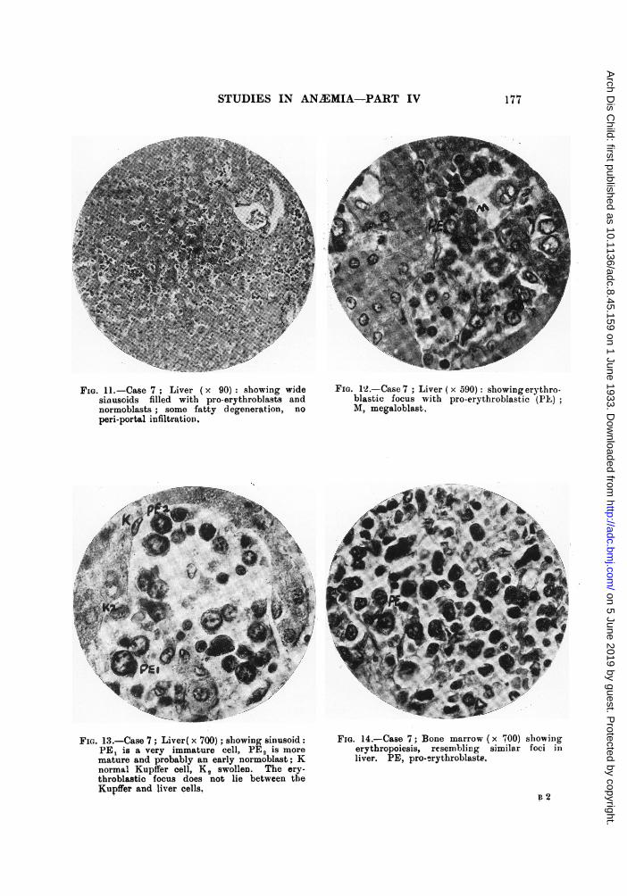
Case 7.-R. L. The organs were pale, especially the liver and kidneys. Theliver weighed 265 grm., the spleen was uniformly enlarged and firm, epicardialecehymoses were present, and the bone marrow of the left femur was red throughout,
on 5 June 2019 by guest. Protected by copyright.
http://adc.bmj.com
/A
rch Dis C
hild: first published as 10.1136/adc.8.45.159 on 1 June 1933. Dow
nloaded from
STUDIES IN ANk:MIA-PART IV 177
FIG. II.-Case 7 Liver (x 90): showing wide FiGt. 12.-Case 7 ;Liver (x 590): showiing erythro-si-nusoids filled with pro-erytbroblasts and blastic focus with pro-erytbroblastic (PFE)normoblasts,; some fatty degeneration, no M, megaloblast,periportal infiltration.
noma Kuife cel swoSllen Th ery_ _FIG 13Cs 7; Lvrx 70_ shwn siuoid FIG 1.Cse 7; Bon mro 70sbwn
mI.1.atureand Livrobbl an early noroblastwid IG.er. Pae7;provery(txr590):shoinertho
throblastic focus does not lie between theKupffer and li ercells.
on 5 June 2019 by guest. Protected by copyright.
http://adc.bmj.com
/A
rch Dis C
hild: first published as 10.1136/adc.8.45.159 on 1 June 1933. Dow
nloaded from
178 ARCHIVES OF DISEASE IN CHILDHOOD
All sections were taken from specimens fixed in formalin; the autopsy wasperformed 8 hours after death in the month of September.
LIVER (Fig. 11, 12 and 13). Sections stained with haematoxylin and eosin whenexamined under the low power showed the liver cell columns broken up by widesinusoids filled with large and small dark cells; some fatty degeneration of thecortical areas was present and the portal tracts were inconspicuous. Under the highpower many of the cells in the sinusoids were large, about 13,u in diameter, and possessedpale oval light vesicular nuclei, 10,u in diameter which showed fine dots of chromatinand sometimes one or more nucleoli. All grades of deeper nuclear staining were foundup to cells which were apparently megaloblasts and normoblasts as already described inthe various forms of heemolytic anaemia of the new born. Swollen Kiippfer cellsand free histiocytes were common. Moreover, the differentiation of the cells appearedto be preponderatingly on the erythrocyte and not on the granulocyte side.
SPLEEN. Low power magnification: -the lymph follicles were rather small, thepulp was very cellular and showed many fairly large round and oval cells; therewas some increase of reticulum in the less cellular parts. High power magnification:-a proliferation of reticular cells was clearly seen and these cells had swollen lightoval and spindle-shaped nuclei. In the meshes of the reticular there were largevesicular nucleated cells, similar to those in the liver, and scattered everywherewere many darker cells differentiating towards normoblasts. Generally distributedalso were lymphocytes of normal type whose nuclei were slightly larger (4-5y) andmore finely dotted and streaked than those of the normoblasts (nuclei of normoblastsare usually about 2N-4j; also the nuclear contour, though fairly regular, tended inmany cases to be slightly polygonal as against the more constantly roundnormoblastic nuclei. The normoblastic cytoplasm was eosinophilic when stainedwith hiematoxylin and eosin. With eosin and methylene blue the nuclear featureswere clearer, but the cytoplasmic process not so well defined. Normoblasticcytoplasm with this stain was bluish with a tinge of pink.
LYMPH GLANDS. In the hilum considerable endothelial proliferation was presentand, as in the liver, amongst apparent histocytes were many large vesicular cellswith accompanying normoblasts. In a mesenteric gland endothelial proliferation wasless marked.
MARROW (Fig. 14). After decalcification the cellular characters did not showwell, but there were many immature cells similar to those already described andmyelocytes were fairly common but not more mature granulocytes. Erythroblastswere also fairly numerous.
The erythropoietic foci seen in the organs of this case were strikinglysimilar to those found in icterus gravis and haemolvtic anaemia of the newborn. Taking into consideration the fact that this child showed markedmegaloblastosis of the peripheral blood stream it appears clear that thesefoci were preponderatingly erythropoietic and were part of a response tothe call for erythropoiesis because of the haemolysis. The haemolysiscommencing before the regression of the embryonic form of the erythropoiesisin the liver had taken place had the effect of retaining this type oferythropoiesis in activity.
Discussion.
From time to time reports of instances of ' anemia of the new born'appear in the literature. We have given reasons in the paper on thehaematopoietic anaemias (Part III) and elsewhere12 for maintaining thatsometimes anaemia of the new born is a congenital nutritional ansemia;
on 5 June 2019 by guest. Protected by copyright.
http://adc.bmj.com
/A
rch Dis C
hild: first published as 10.1136/adc.8.45.159 on 1 June 1933. Dow
nloaded from
STUDIES IN ANAMIA-PART IV 179
nevertheless, in our opinion, the majority of cases of aneemia ofthe new born are haemolytic in nature, being examples either of the presentclass (haemolytic anaemia of the new born or of the later neonatal period,unaccompanied by severe icterus or cedema), or of the anaemia followingicterus gravis.
The clinical manifestations of anaemia of the new born have beendescribed by A. F. Abt13 as follows:-
The infants are born of healthy parents, in normal spontaneous labour, andfrom their history has been excluded all evidence of birth injury, haemorrhage,haemorrhagic disease, infection or pre-natal poisoning. Within the first two weeksof life these infants while thriving and behaving in every way as normal new borninfants, become extremely pale. This pallor is a sheet-like whiteness . . . Theinfants nurse well, are afebrile, have normal periods of sleep and wakefulness, crylustily and vigorously, and aside from the marked and startling pallor, show no signof ill-being. Recovery has been observed, both without and with treatment, andinfants who have been followed for several years have shown no residual effects.
Whilst accepting with considerable reluctance the possible existence ofan anaemia which is neither nutritional nor haemolytic in origin, we never-theless think that Abt has not sufficiently excluded the possibility ofhemolysis, since we cannot understand how a new-born infant can suddenlybecome intensely pale except as a result of hoemorrhage or severe haemolysis.For instance, the infant described by Abt had a pink colour at birth andthe pallor was noted on the eighth day, whereas on the following day withoutany evidence of haemorrhage ' the child seemed as white as the sheet onwhich it lay.' Several of the cases from the literature accepted by himare also open to the same criticism, as is one published by Pasachoff andWilson"4. The case reported by these authors differed from that of Abt inthat pallor was noted on the second day of life, and the evidence of haemolyticanaemia in this particular instance appears to be complete. Nutritionalanaemia of the new born may be distinguished from the hoemolytic group bythe fact that it is a hypochromic anawmia, by its response to treatment byiron, and by the absence of those characters of a haemolytic anemia whichnave already been detailed.
The question may be asked:-Why are some haemolytic anaemiasassociated with severe jaundice, while others are either free from jaundiceor exhibit it only in a slight degree? The same curious difference is seenin the haemolytic anaemias of later infancy and childhood. It is, of course,well known that severe haemolysis may occur without severe jaundice, such,for instance, as that which sometimes follows blood transfusion.
The occurrence or non-occurrence of severe jaundice in association withhaemolysis probably depends on the condition of the liver, and the degreeand suddenness of the hiemolysis. After' severe haemolysis an excessivebilirubinaemia results, and the liver being unable to excrete all this bilirubin,a haemolytic jaundice develops-the so-called ' retention jaundice.' In thisform of jaundice the colour is'not a deep yellow but a lemon tint, the bloodgives a positive indirect van den Bergh reaction, and there is an increase infaecal urobilinogen. Urobilinogen or urobilin, or both, are present in excess
on 5 June 2019 by guest. Protected by copyright.
http://adc.bmj.com
/A
rch Dis C
hild: first published as 10.1136/adc.8.45.159 on 1 June 1933. Dow
nloaded from
180 ARCHIVES OF DISEASE IN CHILDHOOD
in the urine, but neither bilirubin nor bile salts are found in the urine.Retention jaundice is particularly likely to occur when the haemolysis isgradual in onset. If, however, there is necrosis of the liver cells, orobstruction to the outflow of bile, bilirubin after passing through the livercells is regurgitated into the blood stream, and under such conditions amuch deeper jaundice, guinea gold in colour, develops. In ' regurgitationjaundice ' the blood serum gives a positive direct van den Bergh reaction,bilirubin and bile salts appear in the urine, urobilinuria may occur but thefaecal urobilinogen is reduced in amount. This form of jaundice is also likelyto occur when a severe and sudden haemolysis produces an intensebilirubinaemia because the resulting bile is likely to be viscid and producea degree of biliary obstruction. An interesting parallel to these cases occursin acholuric familial jaundice, in which in most of the haemolytic crises thejaundice is only of a moderate degree, but occasionally with a severe crisisthe jaundice becomes greatly increased and bile pigments may appear in theurine, because bilirubin is secreted in such large quantities that the bilebecomes thickened and a degree of biliary obstruction results followed byregurgitation jaundice. In icterus gravis the blood gives an indirect van denBergh reaction; the urine contains increased amounts of urobilin andmoreover also shows bile pigments. In other words, there is evidence ofretention and of regurgitation and, as would be expected, at autopsy bilestasis and in some instances necrosis of the liver cells are found. In thoseexamples of haemolytic anaemia in which there is a history or evidence ofa mild degree of jaundice, or even in those which have become anaemicwithout jaundice, the findings are an indirect van den Bergh reaction inthe blood and an excess of urobilinogen and urobilin in the urine. However,even these findings may be absent if the child does not come underobservation until some time after the occurrence of haemolysis.
Another subject for discussion is the exact relationship existing betweenicterus gravis, hydrops foetalis, and severe haemolytic anaemia of the newborn. Are they, or are they not, all manifestations of an erythroblasticprocess (erythroblastosis of the new born)? For the purposes of thisdiscussion it should be accepted that the term erythroblastosis implies anactive bone marrow and active extra-medullary h.Tmopoietic centres, butthat these may or may not be associated with erythroblastaTmia. From theremarks made in the section on pathology,-it is clear that haemopoietic fociin all three of these conditions are usually to be found in positions where bloodformation occurs in the embryo, notably in the liver and spleen; and it isreasonably certain from histological appearances alone that these foci arechiefly erythropoietic, and not leucopoietic in type. In some cases ofhydrops foetalis, however, such foci are present only to a very limited extent(as in Case 1), or may even be absent (de Lange); thus providing strongevidence that they do not represent the origin of these disorders, but areonly a concomitant feature or response. It is most probable that they doin fact represent a response to an increased call for erythropoiesis. In icterus
on 5 June 2019 by guest. Protected by copyright.
http://adc.bmj.com
/A
rch Dis C
hild: first published as 10.1136/adc.8.45.159 on 1 June 1933. Dow
nloaded from
STUDIES IN ANAEMIA-PART 1V 181
gravis there is no doubt that this call is the result of excessive haemolysis,possibly commencing before birth; and in hydrops foetalis there is evidenceof a hamolyzing process in the occurrence of slight icterus and the presencein the liver of a positive iron reaction by the Turnbull-Hueck process. Aprocess similar as regards haemolysis, though no doubt different as regardsits cause, in that it acts after birth, is at work in those forms of h2emolyticanaemia of the new born which do not show grave jaundice or cedema. Thefactor of haemolysis in varying degree is thus common to these disorders, andthe resultant response is also varying in degree and common to them all.
This response, embryonic erythropoiesis, is hence a symptom and not acause of the disease. Further, the frequent presence of this form oferythropoiesis in premature and some full-term infants is evidence againstthe assumption that the cells produced by such hawmopoiesis are soabnormal in their action or structure as to initiate haemolysis and its effects.Extra-medullary haemopoiesis is, in short, a process which in young infantsmay readily be retained in activity and may be intensified by theprovocation of increased haemolysis. Given the existence of embryonicblood-forming tissue in such organs as the liver and spleen, its activity willbe governed by the extent of the demand for its function. If by the timea haemolytic process in post-natal life occurs the embryonic blood-formingtissue in the liver and spleen has atrophied, then this form of response tothe increased demand for hawmopoiesis cannot occur in these organs, andthe marrow has to shoulder the burden alone. We have seen this responseafter haemolysis occur as late as the eighth month of life. Believing, as wedo, that von Jaksch's anawmia is a chronic haemolytic process occurring inlater infancy, the presence of extra-medullary haemopoietic foci in thiscondition will depend on whether the embryonic blood-forming foci haveatrophied or not at the time when the haemolysis begins. The consequenthistological variations in the response to haemolysis may account for theconfusion of some instances of this syndrome with leukaemic metaplasia, acondition which, as we have pointed out, may closely resemble embryoniccrythropoiesis.
The nature of the haemolyzing factor in the haemolytic anaemias of thenew born, and the relationship between the haemolysis thus produced and thehaemolysis which normally occurs at birth, are both unknown. Haemolysisoccurs constantly in intra- and extra-uterine life, but normally is kept withinbounds by some mechanism. Hampson has suggested that haemolysis maybe controlled in intra-uterine life by something produced by the mother andpassed on by her to the foetus, which in extra-uterine life is elaboratedby the infant itself; for at birth a considerable degree of haemolysis occursowing to alterations in the oxygen tension of its surroundings, which isprevented from becoming excessive by the anti-haemolytic agent. If thisfactor is absent or insufficient, excessive hawmolysis with the production ofanaemia, and perhaps severe jaundice may occur; this may, however, beprevented if the anti-haemolytic factor is given by administration of blood
on 5 June 2019 by guest. Protected by copyright.
http://adc.bmj.com
/A
rch Dis C
hild: first published as 10.1136/adc.8.45.159 on 1 June 1933. Dow
nloaded from
182 ARCHIVES OF DISEASE IN CHILDHOOD
serum. This is the basis of the treatment of icterus gravis by injections ofblood serum, a treatment which we owe to Hampson, and which has proveda great success. On this hypothesis it is possible that in icterus gravis thematernal anti-ha!molytic factor supplied to the foetus becomes deficienttowards the end of intra-uterine life, and that in extra-uterine life the infantfails to produce sufficient to keep the haemolysis of birth within reasonablelimits, and that the injection of blood serum remedies the deficiency. Inhaemolytic anaemia of the new born, on the other hand, it is possible that thedeficiency is entirely a post-natal one, or that the anaemia is due to a factorsimilar to that which initiates the acute haemolytic anaemia of Lederer seenin older children. The fact that hwmolysis after birth is more severe inthe premature child than in the normal child can be explained on the groundthat the anti-haemolytic factor has not been elaborated to the degree whichit would have been if the child had gone to term. Such an hypothesis wouldfurnish an adequate explanation of the fact that jaundice is more frequentand severe in premature than in full-term children; also of the developmentof haemolytic anaemia in prematures, and of its prevention by bloodtransfusion. If this hypothesis is correct, the hemolytic anaemias of theneonatal period, with the exception of that associated with hydrops foetalis,may be classified as deficiency anemias, although they differ from the otherdeficiency anaemias in that the deficiency is of a temporary character only.
This hypothesis does not cover the whole ground because it leaves outof account any damage to the haemopoietic portion of the erythron whichmay, and quite possibly does, occur. It thereby fails adequately to explainbydrops foetalis and those cases of icterus gravis which develop spasticdiplegia in later life and those which show necrotic changes in the liver.Heemolysis occurs in hydrops, and in addition there are extensive toxicchanges in the parenchvmatous cells and the capillary walls, which stronglysuggest that the condition is due to a toxin which has some haemolyzinginfluence, and the changes which occur in the liver and central nervoussystem in some cases of icterus gravis also strongly suggest that a toxinplays some part in the causation of that disease.
Summary.
In this paper the clinical and pathological characters of theerythronoclastic (haemolytic) anaemias of the new born are described andthe view put forward that the embryonic erythropoiesis (erythroblastosis ofthe new born) present in them is a result and not a cause of these anaemias.Attention is also drawn to the existence of a group of erythronoclastic(haemolytic) anaemias which arise late in the neonatal period; areunconnected with severe jaundice or cedema; and- may not be recognizeduntil the children are some weeks old. The similarity between the clinicalpicture presented by some of the older children of this group to that of theanamia of von Jaksch, and the light that this may throw upon the causationof the latter syndrome, is commented upon.
on 5 June 2019 by guest. Protected by copyright.
http://adc.bmj.com
/A
rch Dis C
hild: first published as 10.1136/adc.8.45.159 on 1 June 1933. Dow
nloaded from

STUDIES IN ANAMIA-PART IV 183
REFERENCES.
1. Clifford, S. H., & Hertig, A. T., New England Med. J., Boston, 1932, II, 105.2. Burman, W. L., & Stanford, H. N., Am. J. Dis. Child., Chicago, 1931, XLI, 225.3. Hart, A. P., Selected Art. from the Dept. of Paed., Univ. of Toronto, 418.4. Parsons, L. G., Arch. Dis. Childh., London, 1933, VIII, 87.5. Hampson, A. C., Lancet, London, 1929, i, 429.6. Buchan, A. H., & Comrie, T. D., J. Path. and Bact., Edinb., 1909, XIII, 398.7. Ferguson, J., Am. J. Path., Boston, 1931, VII, 277.8. Capon, N. B., J. Obstet., 4 Gyne., Manchester, 1922, XXIX, 239.9. de Lange, C., Acta Pxd., Upsala, 1932, XIII, 292.
10. de Lange, C., & Arntzenius, A. K. W., Jahrb. f. Kinderh., Berlin, 1929, CXXIV, J.11. Brannan, O., Bull. Johns Hopkins Hosp., Baltimore, 1927, XLI, 104.12. Parsons, L. G., Acta Paed., Upsala, 1932, XIII, 378.13. Abt, A. F., Am. J. Dis. Child., Chicago, 1932, XLIII, 337.14. Pasachoff, H. D., & Wilson, L., Ibid., 1931, XLII, 111.15. Naegeli, O., Blutkrankheiten u. Blutdiagnostik, Berlin, 1923.
ERRATA (PART III).
Page 128, line 19: ' 1 mgrm. per cent.' read ' 01 mgrm. per cent.'line 22: for ' 11-25 grn.' read ' 11-25 grm.'line 23: for ' 1 grn.' read ' 1 grm.'
on 5 June 2019 by guest. Protected by copyright.
http://adc.bmj.com
/A
rch Dis C
hild: first published as 10.1136/adc.8.45.159 on 1 June 1933. Dow
nloaded from


