Structure function analysis of pneumococcal DprA protein ... · Structure–function analysis of...
10
Structure–function analysis of pneumococcal DprA protein reveals that dimerization is crucial for loading RecA recombinase onto DNA during transformation Sophie Quevillon-Cheruel a,1,2 , Nathalie Campo b,c,1 , Nicolas Mirouze b,c,d,1 , Isabelle Mortier-Barrière b,c , Mark A. Brooks a , Marion Boudes a , Dominique Durand a , Anne-Lise Soulet b,c , Johnny Lisboa a , Philippe Noirot d , Bernard Martin b,c , Herman van Tilbeurgh a , Marie-Françoise Noirot-Gros d , Jean-Pierre Claverys b,c , and Patrice Polard b,c,2 a Institut de Biochimie et de Biophysique Moléculaire et Cellulaire, Université de Paris-Sud, Centre National de la Recherche Scientifique, Unité Mixte de Recherche 8619, Bât 430, 91405 Orsay Cedex, France; b Centre National de la Recherche Scientifique, LMGM-UMR5100, F-31000 Toulouse, France; c Université de Toulouse, UPS, Laboratoire de Microbiologie et Génétique Moléculaires, F-31000 Toulouse, France; and d Institut Micalis, Institut National de la Recherche Agronomique, Unité Mixte de Recherche 1319, Bât 440, Domaine de Vilvert, 78352 Jouy en Josas Cedex, France Edited by Stephen C. Kowalczykowski, University of California, Davis, CA, and approved July 20, 2012 (received for review April 14, 2012) Transformation promotes genome plasticity in bacteria via RecA- driven homologous recombination. In the Gram-positive human pathogen Streptococcus pneumoniae, the transformasome a multi- protein complex, internalizes, protects, and processes transforming DNA to generate chromosomal recombinants. Double-stranded DNA is internalized as single strands, onto which the transforma- tion-dedicated DNA processing protein A (DprA) ensures the load- ing of RecA to form presynaptic filaments. We report that the structure of DprA consists of the association of a sterile alpha motif domain and a Rossmann fold and that DprA forms tail-to- tail dimers. The isolation of DprA self-interaction mutants revealed that dimerization is crucial for the formation of nucleocomplexes in vitro and for genetic transformation. Residues important for DprA– RecA interaction also were identified and mutated, establishing this interaction as equally important for transformation. Position- ing of key interaction residues on the DprA structure revealed an overlap of DprA–DprA and DprA–RecA interaction surfaces. We propose a model in which RecA interaction promotes rearrange- ment or disruption of the DprA dimer, enabling the subsequent nucleation of RecA and its polymerization onto ssDNA. bacterial transformation | genetic exchange | recombinase loader | recombination mediator protein | horizontal gene transfer H omologous recombination (HR) plays essential roles in ge- nome maintenance, the generation of genetic diversity, and proper chromosome segregation in all living organisms. HR is universally catalyzed by recombinases, i.e., RecA in bacteria, Rad51 in eukaryotes, and RadA in archaea (1, 2). HR recom- binases promote the exchange of homologous DNA strands, a prerequisite for which is the formation of a complex between ssDNA and the recombinase called the “presynaptic filament.” Nucleation of the recombinase [i.e., binding of its first subunit(s) to DNA] is the rate-limiting step, hence the need for cofactors. A specialized class of cofactors interacting with ssDNA is rep- resented by the recombination mediator proteins (RMPs), which have evolved to ensure recombinase loading on ssDNA bound by ssDNA-binding proteins (SSBs) (3). Thus, the well-characterized RMPs UvsY of bacteriophage T4, Rec(F)OR of Escherichia coli, and yeast Rad52 alleviate the SSB barrier and promote the loading of UvsX, RecA, and Rad51 on Gp32-, SSB-, and RP-A– precoated ssDNA, respectively (3). The E. coli RecBCD complex represents another category of cofactors that process linear dsDNA into ssDNA onto which they simultaneously ensure the loading of the recombinase (4). Neither the RecBCD equivalent RexAB (5) nor RecFOR is required for genetic transformation of the Gram-positive bacterium Streptococcus pneumoniae (6). However, we recently identified the widespread bacterial protein, DNA processing protein A (DprA), as a transformation-dedicated RecA loader (7). Transformation is believed to play a major role in the genetic plasticity of this human commensal and major pathogen (8, 9). For example, trans- formation allows variation of the polysaccharide capsule (the primary determinant of pneumococcal virulence) through ac- cess to a species gene pool almost equivalent to the size of an individual genome (10). Pneumococcal transformation requires transient differentiation to competence, during which expression of a specific set of genes (11) allows assembly of the transformasome (6). This dynamic machine involves both membrane and cytosolic proteins (including DprA and RecA), and promotes internalization, protection, and processing of transforming DNA into recombinants. Competence (or X-state) (11) is induced in exponentially growing cultures by a peptide pheromone, competence-stimulating peptide (CSP) (12), which ultimately activates the synthesis of an alternative sigma factor specific for competence (σ X ) (13). Induction of compe- tence by some antibiotics and DNA-damaging agents (14) supports the view that CSP functions as an alarmone and that competence/ X-state constitutes an SOS substitute for S. pneumoniae (11). During the course of transformation, DprA may interact with and facilitate the loading of RecA onto two types of substrates, naked and SSB-coated ssDNA. The former could occur close to the membrane immediately after initiation of exogenous dsDNA uptake, which results in the internalization of ssDNA with 3′ → 5′ polarity (6), and may lead to the formation of mixed DprA-RecA nucleofilaments. Such mixed filaments were documented pre- viously in vitro and were shown to be proficient in the catalysis of homology-dependent synapsis (7). Internalized ssDNA also could be complexed with the transformation-dedicated SSB protein, SsbB. Such complexes were detected in vivo (15) and were proposed to represent a reservoir of transforming ssDNA (7); recently, evidence was provided favoring a reservoir-mainte- nance role for SsbB (16). In vitro data revealed the capacity of DprA to promote the loading of RecA on SSB–ssDNA com- plexes (7). Author contributions: S.Q.-C., N.C., P.N., M.-F.N.-G., J.-P.C., and P.P. designed research; S.Q.-C., N.C., N.M., I.M.-B., M.A.B., M.B., D.D., A.-L.S., J.L., B.M., M.-F.N.-G., J.-P.C., and P.P. performed research; S.Q.-C., N.C., N.M., I.M.-B., M.A.B., M.B., D.D., A.-L.S., J.L., P.N., B.M., H.v.T., M.-F.N.-G., J.-P.C., and P.P. analyzed data; and S.Q.-C., N.C., J.-P.C., and P.P. wrote the paper. The authors declare no conflict of interest. This article is a PNAS Direct Submission. Data deposition: Crystallography, atomic coordinates, and structure factors reported in this work for the structure of Sp DprA have been deposited in the Protein Data Bank (PDB), www.pdb.org (PDB ID code 3UQZ). 1 S.Q.-C., N.C., and N.M. contributed equally to this work. 2 To whom correspondence may be addressed. E-mail: [email protected] or [email protected]. See Author Summary on page 14738 (volume 109, number 37). This article contains supporting information online at www.pnas.org/lookup/suppl/doi:10. 1073/pnas.1205638109/-/DCSupplemental. E2466–E2475 | PNAS | Published online August 17, 2012 www.pnas.org/cgi/doi/10.1073/pnas.1205638109
Transcript of Structure function analysis of pneumococcal DprA protein ... · Structure–function analysis of...

Structure–function analysis of pneumococcal DprAprotein reveals that dimerization is crucial for loadingRecA recombinase onto DNA during transformationSophie Quevillon-Cheruela,1,2, Nathalie Campob,c,1, Nicolas Mirouzeb,c,d,1, Isabelle Mortier-Barrièreb,c, Mark A. Brooksa,Marion Boudesa, Dominique Duranda, Anne-Lise Souletb,c, Johnny Lisboaa, Philippe Noirotd, Bernard Martinb,c,Herman van Tilbeurgha, Marie-Françoise Noirot-Grosd, Jean-Pierre Claverysb,c, and Patrice Polardb,c,2
aInstitut de Biochimie et de Biophysique Moléculaire et Cellulaire, Université de Paris-Sud, Centre National de la Recherche Scientifique, Unité Mixte deRecherche 8619, Bât 430, 91405 Orsay Cedex, France; bCentre National de la Recherche Scientifique, LMGM-UMR5100, F-31000 Toulouse, France; cUniversitéde Toulouse, UPS, Laboratoire de Microbiologie et Génétique Moléculaires, F-31000 Toulouse, France; and dInstitut Micalis, Institut National de la RechercheAgronomique, Unité Mixte de Recherche 1319, Bât 440, Domaine de Vilvert, 78352 Jouy en Josas Cedex, France
Edited by Stephen C. Kowalczykowski, University of California, Davis, CA, and approved July 20, 2012 (received for review April 14, 2012)
Transformation promotes genome plasticity in bacteria via RecA-driven homologous recombination. In the Gram-positive humanpathogen Streptococcus pneumoniae, the transformasome a multi-protein complex, internalizes, protects, and processes transformingDNA to generate chromosomal recombinants. Double-strandedDNA is internalized as single strands, onto which the transforma-tion-dedicated DNA processing protein A (DprA) ensures the load-ing of RecA to form presynaptic filaments. We report that thestructure of DprA consists of the association of a sterile alphamotif domain and a Rossmann fold and that DprA forms tail-to-tail dimers. The isolation of DprA self-interaction mutants revealedthat dimerization is crucial for the formation of nucleocomplexes invitro and for genetic transformation. Residues important for DprA–RecA interaction also were identified and mutated, establishingthis interaction as equally important for transformation. Position-ing of key interaction residues on the DprA structure revealed anoverlap of DprA–DprA and DprA–RecA interaction surfaces. Wepropose a model in which RecA interaction promotes rearrange-ment or disruption of the DprA dimer, enabling the subsequentnucleation of RecA and its polymerization onto ssDNA.
bacterial transformation | genetic exchange | recombinase loader |recombination mediator protein | horizontal gene transfer
Homologous recombination (HR) plays essential roles in ge-nome maintenance, the generation of genetic diversity, and
proper chromosome segregation in all living organisms. HR isuniversally catalyzed by recombinases, i.e., RecA in bacteria,Rad51 in eukaryotes, and RadA in archaea (1, 2). HR recom-binases promote the exchange of homologous DNA strands,a prerequisite for which is the formation of a complex betweenssDNA and the recombinase called the “presynaptic filament.”Nucleation of the recombinase [i.e., binding of its first subunit(s)to DNA] is the rate-limiting step, hence the need for cofactors.A specialized class of cofactors interacting with ssDNA is rep-resented by the recombination mediator proteins (RMPs), whichhave evolved to ensure recombinase loading on ssDNA bound byssDNA-binding proteins (SSBs) (3). Thus, the well-characterizedRMPs UvsY of bacteriophage T4, Rec(F)OR of Escherichia coli,and yeast Rad52 alleviate the SSB barrier and promote theloading of UvsX, RecA, and Rad51 on Gp32-, SSB-, and RP-A–
precoated ssDNA, respectively (3). The E. coli RecBCD complexrepresents another category of cofactors that process linear dsDNAinto ssDNA onto which they simultaneously ensure the loadingof the recombinase (4).Neither the RecBCD equivalent RexAB (5) nor RecFOR is
required for genetic transformation of the Gram-positive bacteriumStreptococcus pneumoniae (6). However, we recently identified thewidespread bacterial protein, DNA processing protein A (DprA),as a transformation-dedicated RecA loader (7). Transformation isbelieved to play a major role in the genetic plasticity of this human
commensal and major pathogen (8, 9). For example, trans-formation allows variation of the polysaccharide capsule (theprimary determinant of pneumococcal virulence) through ac-cess to a species gene pool almost equivalent to the size of anindividual genome (10).Pneumococcal transformation requires transient differentiation
to competence, during which expression of a specific set of genes(11) allows assembly of the transformasome (6). This dynamicmachine involves both membrane and cytosolic proteins (includingDprA and RecA), and promotes internalization, protection, andprocessing of transforming DNA into recombinants. Competence(or X-state) (11) is induced in exponentially growing cultures bya peptide pheromone, competence-stimulating peptide (CSP) (12),which ultimately activates the synthesis of an alternative sigmafactor specific for competence (σX) (13). Induction of compe-tence by some antibiotics and DNA-damaging agents (14) supportsthe view that CSP functions as an alarmone and that competence/X-state constitutes an SOS substitute for S. pneumoniae (11).During the course of transformation, DprA may interact with
and facilitate the loading of RecA onto two types of substrates,naked and SSB-coated ssDNA. The former could occur close tothe membrane immediately after initiation of exogenous dsDNAuptake, which results in the internalization of ssDNA with 3′ → 5′polarity (6), and may lead to the formation of mixed DprA-RecAnucleofilaments. Such mixed filaments were documented pre-viously in vitro and were shown to be proficient in the catalysis ofhomology-dependent synapsis (7). Internalized ssDNA also couldbe complexed with the transformation-dedicated SSB protein,SsbB. Such complexes were detected in vivo (15) and wereproposed to represent a reservoir of transforming ssDNA (7);recently, evidence was provided favoring a reservoir-mainte-nance role for SsbB (16). In vitro data revealed the capacity ofDprA to promote the loading of RecA on SSB–ssDNA com-plexes (7).
Author contributions: S.Q.-C., N.C., P.N., M.-F.N.-G., J.-P.C., and P.P. designed research;S.Q.-C., N.C., N.M., I.M.-B., M.A.B., M.B., D.D., A.-L.S., J.L., B.M., M.-F.N.-G., J.-P.C., andP.P. performed research; S.Q.-C., N.C., N.M., I.M.-B., M.A.B., M.B., D.D., A.-L.S., J.L., P.N.,B.M., H.v.T., M.-F.N.-G., J.-P.C., and P.P. analyzed data; and S.Q.-C., N.C., J.-P.C., and P.P.wrote the paper.
The authors declare no conflict of interest.
This article is a PNAS Direct Submission.
Data deposition: Crystallography, atomic coordinates, and structure factors reported inthis work for the structure of SpDprA have been deposited in the Protein Data Bank (PDB),www.pdb.org (PDB ID code 3UQZ).1S.Q.-C., N.C., and N.M. contributed equally to this work.2To whom correspondence may be addressed. E-mail: [email protected] [email protected].
See Author Summary on page 14738 (volume 109, number 37).
This article contains supporting information online at www.pnas.org/lookup/suppl/doi:10.1073/pnas.1205638109/-/DCSupplemental.
E2466–E2475 | PNAS | Published online August 17, 2012 www.pnas.org/cgi/doi/10.1073/pnas.1205638109

DprA possesses all the distinctive properties of RMPs, in-cluding the ability to bind ssDNA and interact with RecA,leading us to propose that it is a member of the RMP familydedicated to natural bacterial transformation (7). However, noevidence was provided that both the ssDNA-binding and RecA-interaction properties of DprA are required for transformation.In addition, the mechanism of the DprA-facilitated loading ofRecA onto ssDNA remained elusive. Here, we report that thestructure of DprA consists of the association of a sterile alphamotif (SAM) domain and a Rossmann fold (RF). We show thatDprA forms tail-to-tail dimers and that dimerization is crucialfor formation of nucleocomplexes in vitro. Taking advantage ofthe previous detection of DprA–DprA and DprA–RecA inter-actions in yeast two-hybrid assays (Y2H) (7), we isolated DprAmutants deficient in either of these interactions. This identifi-cation of key DprA residues allowed us to demonstrate thatdimerization and interaction with RecA are equally importantfor transformation. Structural comparison between S. pneumo-niae DprA and DprA from Rhodopseudomonas palustris [re-cently deposited in the Protein Data Bank (PDB); ID 3MAJ]and with 3D models of DprA from E. coli led us to concludethat (i) the widespread DprA family is structurally defined by theassociation of a SAM and an RF, and (ii) DprA dimerizationpotential and interface residues are evolutionarily conserved. Onthe other hand, the finding that DprA dimerization and RecAinteraction residues partially overlap, together with the puzzlingobservation that RecA interaction residues appear to be evolu-tionarily less conserved, led us to postulate a much larger overlapof the two interaction surfaces. Site-directed mutagenesis of themost conserved DprA dimerization residues then establishedthat these residues also are important for interaction with RecA.We conclude that there is a large overlap between the dimerizationand RecA interaction interfaces, suggesting that, upon interaction,RecA promotes rearrangement or disruption of the DprAdimer, enabling subsequent nucleation and loading of RecA ontotransforming ssDNA fragments.
ResultsOverall Structure of S. pneumoniae DprA. The structure of S. pneu-moniae DprA was solved at 2.7-Å resolution by single-wavelengthanomalous dispersion using seleno-methionine–labeled protein.Refinement statistics are presented in Table S1 and show goodstereochemistry for the final model. Three copies of DprA (chainsA, B, and C) are present per asymmetric unit in the crystal. Thefinal model is complete, containing all residues (1–282) for chainsA and C with the addition of one histidine from the His-tag forchain A. Chain B lacks the last two residues, E281 and F282.A sulfate ion is visible on the surface of each chain, linked viahydrogen bonds with R115, S230, and G229.DprA consists of two domains (Fig. 1A). The N-terminal domain
(residue M1-F71) is composed of five helices (Fig. 1 A and C) andpresents considerable structural similarity to the SAM domainof PA4738, a protein of unknown function from Pseudomonasaeruginosa (PDB code 1YWW; Z score 2.1; rmsd of 2.97 Å).SAM domains frequently are involved in various types of proteininteraction (17).The C-terminal region of DprA, starting approximately from
residue P107, adopts an RF-like topology with a typical three-layer (αβα) sandwich (hereafter called “RF”) (Fig. 1 A and C).The short region connecting the SAM and the RF is composedof two antiparallel β-strands, each followed by α-helices. Theβ-strands of the connecting region are associated by main-chainhydrogen bonds to the main β-sheet of the RF and therefore canbe considered as an extension of the RF. Furthermore, the limitsof Pfam02481, which characterizes the DprA protein family, al-most exactly match the extension plus the RF (Fig. 1B). We referto this entity hereafter as “extended RF” (eRF). The SAM packsclosely onto the eRF, and the whole DprA structure forms a bean-shaped, globular structure.
DprA Dimers in the Crystal. Analysis of crystal packing revealedtwo distinct types of DprA dimers involving RF–RF and SAM–SAM interactions, respectively. RF–RF interactions result in theformation of two nearly identical tail-to-tail (hereafter “C/C”)and antiparallel dimers [rmsd of 0.59 Å and a template-modeling(TM) score of 0.99118 (TM-align server, http://zhanglab.ccmb.
C
α4
α5
N
α1
α2
α3
η1
β1
η2
η3
β2
β3β4β5β6 β7 β8 β9
α12
α11
η5
α6
α7
α8
α9 α10
η4
B
α1
α2α3
α4
α5
β1β2
β3
α6β4
α7β5
α8
β6
α9
β7α10
β8
α11
α12Nter
Cter
η2η3
1 71 107 282COOHNH2
SAM RF extension Rossmann fold
S e RFPfam02481:DNA_processg_A
S e RFS
eRF180°C
A or C B’ or C’
D275
L269
Q264
D275
L269
Q264
α11
α11
α12
α12
α6
α6
DS e RF
S e RF
C/C dimer
C
A
Fig. 1. 3D structure of DprA from S. pneumoniae.(A) Ribbon schematic presentation of the DprA3D structure. The N and C termini and secondarystructure elements are indicated. Color code: red,SAM domain; blue-green, RF; green, eRF extension.S, SAM; e, RF extension; RF, Rossmann fold. (B)Linear representation of DprA with limits ofPfam02481 and structural domains (color code asin A). The Pfam02481 domain is shown in blue. (C)Topological representation of DprA (color code asin A). (D) Ribbon schematic presentation of the3D structure of AB′ or CC′ DprA dimers. (Inset) TheC-terminal dimerization zone is enlarged to showsome of the residues (represented as yellow sticks)involved in the interaction (see also Fig. 2 A and Band Table S2). Hydrogen bonds are represented bydashed lines, and distances between residues areindicated in Å.
Quevillon-Cheruel et al. PNAS | Published online August 17, 2012 | E2467
MICRO
BIOLO
GY
PNASPL
US

med.umich.edu/TM-align/)] (Fig. 1D) (18). Both the first C/Cdimer, composed of molecules A and its symmetric B′, and thesecond dimer, composed of molecules C and C′, are generated bya crystallographic twofold-symmetry axis. An angle of 130–150° isobserved between the two subunits of C/C dimers. Their interfacesbury very similar surface areas: 1,496 Å2 and 1,430 Å2 for the AB′and CC′ dimers, respectively [calculated by addition of the sur-faces of the two monomers in contact; European BioinformaticsInstitute Protein Interactions Surfaces and Assemblies (PISA)service; http://www.ebi.ac.uk/msd-srv/prot_int/pistart.html]. Res-idues that contribute to C/C dimerization in the crystal are listedin Table S2, together with an estimate of accessibility of in-dividual residues in the dimer versus the monomer. Interfaceresidues are located on α6, the loop between β8 and α11, α11, β9,and α12 (Figs. 1D and 2A and Table S2). The interface is mainlyhydrophobic, with the addition of hydrogen bonds between Q264of one monomer and L269–D275 from the opposite monomer(Fig. 1D).The solvent content of DprA crystals is high (70%), and the
crystals are characterized by remarkable major and minor chan-nels in the lattice, with diameters of 130 and 100 Å, respectively(Fig. S1A). DprA forms filament-like packing along these solventchannels. These twisted filaments are composed of C/C dimersinteracting through their N termini, i.e., via SAM/SAM in-teraction, to form multimers (Fig. S1B). [SAM/SAM interactions(Fig. S1C) are referred to as “N/N” interactions hereafter.]
Y2H Screen for DprA-Interaction Mutants. We first sought to estab-lish which C/C or N/N interactions are required for dimerizationin vivo by taking advantage of Y2H, which previously revealedDprA self-interaction (7). Therefore a Y2H screen for DprAmutants that are unable to self-interact but retain full capacityto interact with RecA was launched (Materials and Methods) withthe aim of identifying DprA residues important for self-interaction
in vivo. This genetic screen isolated three double mutants, C234R-F245L, I251V-H260R, and D257G-L269S (Fig. 2B). Interestingly,these mutations affected only C-terminal residues, three of which(I251, H260, and L269) are involved directly in the C/C di-merization interface in the crystal. On the other hand, no variantsupporting the N/N interfacing was isolated, allowing us to con-clude tentatively that in vivo DprA self-interaction primarilyinvolves C/C interactions.
Purified DprA Forms C/C Dimers in Solution. To investigate whetherDprA also forms dimers in solution and to characterize the in-teraction interface, we carried out gel filtration and small-angleX-ray scattering (SAXS) experiments with purified proteins. Wecompared wild-type DprA with the Y2H C-terminal interaction mu-tant DprAI251V-H260R and with the DprAH260A-L269K andDprAH260A-L269R double mutants (hereafter referred to as DprAVR,DprAAK, and DprAAR, respectively). The latter two were designedto combine mutations of residues first identified in the screenfor self-interaction mutants and also engaged in the C/C dimerinterface in the crystal (Fig. 2B and Table S2).Gel filtration experiments indicated that both DprA and
DrpAAR behave as a single homogeneous species, but DprAeluted from the sizing column with an apparent mass between amonomer and a dimer, whereas DprAAR eluted with an apparentmass about half that observed for DprA (Fig. S2). Although theapparent molecular mass of DprAAR was lower than that pre-dicted for DprA (31.885 kDa including the His-tag), these resultswere consistent with the DprAAR mutant protein being mono-meric. Analysis of DprA SAXS data (Fig. 3A) then allowed twoindependent estimates of molecular mass, establishing un-equivocally that DprA is a dimer in solution (SI Materials andMethods, SAXS Data Analysis). Then model reconstruction fromthe DprA experimental curve was carried out and revealed quitesatisfactory superimposition of the C/C crystallographic dimer on
A
B
C
Q264
D275T271L269
H260
L252
I251
α12
α11
G249
α6
P248
Q120
K123
S124
K127
V128
S250
D243
H260
L269
α12
α11
α6
E235
Q264
L277
S124
E265
T233
F280
Y183
D170M151L142
DprA dimeriza�on mutants (Y2H)
282107
I251
H 260
1 71
COOHNH2 S RF
Q26
4RE 2
65G
L 277
P
Y 183
H
D 243
GE2
35G
T 233
P
D 170
G
M15
2 TH1
51R
L142
PS 1
24P
F 280
LL 2
69
Pfam02481:DNA_processg_AS e RF
S124
Q12
0
K123
K127
V128
G24
9
I 247
P248
Q26
4
S250
L252
D275
T271
DprA-RecA interac�on mutants (Y2H)
Dimeriza�on interface residues
H260
R
I251
V
L 269
S
D 257
G
F245
L
C 234
R
I 263
Fig. 2. Location of DprA residues involved in thedimerization interface and in interaction withRecA. (A) Ribbon view with DprA residues involvedin C/C interactions shown in yellow. Green residuesindicate C/C interaction residues mutated to gen-erate the DprAAK and DprAAR monomeric mutants.(B) Linear representation of DprA with DprA in-teraction residues and mutants. C/C dimerizationinterface residues are shown immediately abovethe linear map (see also Table S2). DprA mutantsunable to self-interact but retaining full capacityto interact with RecA are shown in yellow withina gray rectangle above the linear map. Mutationsaffecting interaction with RecA but not dimerizationare shown in purple below the linear map. For eachmutation, the wild-type residue is indicated first,followed by its position, and then the mutant resi-due is shown. (C) Ribbon view with DprA residuesinvolved in interaction with RecA shown in purple.Color code is as in Fig. 1A.
E2468 | www.pnas.org/cgi/doi/10.1073/pnas.1205638109 Quevillon-Cheruel et al.
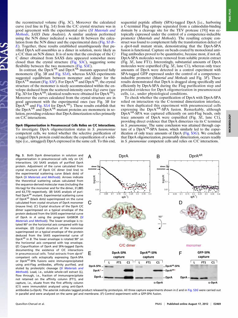
the reconstructed volume (Fig. 3C). Moreover the calculatedcurve (red line in Fig. 3A) from the C/C crystal structure was ingood agreement with the experimental curve (SI Materials andMethods, SAXS Data Analysis). A similar analysis performedusing the N/N dimer indicated a weaker fit between the calcu-lation from the N/N dimer and the experimental data (Fig. S3 B–E). Together, these results established unambiguously that pu-rified DprA self-assembles as a dimer in solution, more likely asa C/C than an N/N dimer. It is of note that the envelope of the C/C dimer obtained from SAXS data appeared somewhat moreclosed than the crystal structure (Fig. S3C), suggesting someflexibility between the two monomers (Fig. S3E).In contrast, the DprAAR and DprAAK mutants appeared fully
monomeric (Fig. 3B and Fig. S3A), whereas SAXS experimentssuggested equilibrium between monomer and dimer for theDprAVR mutant (Fig. S3F). For DprAAR and DprAAK, the crystalstructure of the monomer is nicely accommodated within the en-velope deduced from the scattered-intensity curve I(q) curve (seeFig. 3D for DprAAR; identical results were obtained for DprAAK).Moreover the curves calculated from the crystal structure are ingood agreement with the experimental ones (see Fig. 3B forDprAAR and Fig. S3A for DprAAK). These results establish thatthe DprAAR and DprAAK mutant proteins are monomeric in so-lution, providing evidence that DprA dimerization relies primarilyon C/C interactions.
DprA Oligomerization in Pneumococcal Cells Relies on C/C Interactions.To investigate DprA oligomerization status in S. pneumoniaecompetent cells, we tested whether the selective purification ofa tagged DprA protein could mediate the copurification of a wild-type (i.e., untagged) DprA expressed in the same cell. To this end,
sequential peptide affinity (SPA)-tagged DprA [i.e., harboringa C-terminal Flag epitope separated from a calmodulin-bindingdomain by a cleavage site for the TEV protease (19)] was ec-topically expressed under the control of a competence-induciblepromoter (Materials and Methods). The resulting ectopic con-struct was found to complement fully the transformation defect ofa dprA-null mutant strain, demonstrating that the DprA-SPAfusion is functional. Capture on beads coated by monoclonal anti-Flag antibodies proved to be quantitative, because most, if not all,DprA-SPA molecules were removed from soluble protein extract(Fig. 3E, lane FT1). Interestingly, substantial amounts of DprAmolecules were copurified (Fig. 3E, lane C1), whereas only traceamounts of DprA were detected in a control experiment withSPA-tagged GFP expressed under the control of a competence-inducible promoter (Material and Methods and Fig. 3F). Theseresults demonstrated that DprA is dragged along specifically andefficiently by DprA-SPA during the Flag purification step andprovided evidence for DprA oligomerization in pneumococcalcells, i.e., under physiological conditions.To check whether the copurification of DprA with DprA-SPA
relied on interaction via the C-terminal dimerization interface,we then duplicated this experiment with pneumococcal cellsexpressing the DprAAR-SPA fusion. Remarkably, althoughDprAAR-SPA was captured efficiently on anti-Flag beads, onlytrace amounts of DprA were copurified (Fig. 3E, lane C1),providing direct evidence that DprA dimerizes via its C terminalin S. pneumoniae. The same conclusion was attained through cap-ture of a DprAVR-SPA fusion, which similarly led to the copur-ification of only trace amounts of DprA (Fig. S3G). We concludethat DprA oligomerization, presumably dimerization, occurs readilyin S. pneumoniae competent cells and relies on C/C interactions.
ArpDArpD ARA B
90°
F
L C1FT1
GFP-SPAcapture
E
DprA-SPAL C1FT1
DprA-SPAcapture
DprAAR-SPAL C1FT1
DprAAR-SPAcapture
C/C dimer monomer
DprA
-DprA
DprA
DprA* DprAAR*
-DprA
DprA
C D
Fig. 3. Both DprA dimerization in solution andoligomerization in pneumococcal cells rely on C/Cinteractions. (A) SAXS analysis of purified DprAprotein. Adjustment of the curve calculated fromcrystal structure of DprA C/C dimer (red line) tothe experimental scattering curve (black dots) ofDprA (SI Materials and Methods). Arrows indicatethe theoretical scattering values calculated fromthe sequence-derived molecular mass (including theHis-tag) for the monomer and for the dimer, 31,885and 63,770 respectively. (B) SAXS analysis of puri-fied DprAAR mutant. Experimental scattering curveof DprAAR (black dots) superimposed on the curvecalculated from crystal structure of DprA monomer(mauve line). (C) Crystal structure of the DprA C/Cdimer superimposed on a typical envelope of theprotein deduced from the SAXS experimental curveof DprA in A using the program GASBOR (SIMaterials and Methods). The lower envelope is ro-tated 90° on the horizontal axis compared with topenvelope. (D) Crystal structure of the monomersuperimposed on a typical envelope of the proteindeduced from the SAXS experimental curve ofDprAAR in B. The lower envelope is rotated 90° onthe horizontal axis compared with top envelope.(E) Copurification of DprA and SPA-tagged DprAsdocumenting the existence of C/C interactionsin pneumococcal cells. Total extracts from dprA+
competent cells ectopically expressing DprA-SPAor DprAAR-SPA fusions were immunoprecipitatedusing anti-Flag antibodies, affinity purified, andeluted by proteolytic cleavage (SI Materials andMethods). Load, i.e., soluble whole-cell extract (L),flow through, i.e., fraction of immunoprecipitatenot retained on the affinity column (FT1), andcapture, i.e., eluate from the first affinity column(C1) were immunoblot analyzed using anti-DprAantibodies (α-DprA). The asterisk indicates tagged product released by proteolysis. All three capture experiments shown in E and in Fig. S3G were carried outin parallel and were analyzed on the same gel and membrane. (F) Control experiment with a GFP-SPA fusion.
Quevillon-Cheruel et al. PNAS | Published online August 17, 2012 | E2469
MICRO
BIOLO
GY
PNASPL
US

DprA Oligomerization Is Crucial for Genetic Transformation. Next,the H260A-L269R pair of mutations was introduced at the dprAchromosomal locus to investigate its effects on pneumococcaltransformation. The resulting dprAAR strain was found to expresswild-type protein levels (Fig. 4A, compare lanes L). Interestingly,this mutant presented a more than 2-log decrease in transformationefficiency compared with wild type (Fig. 4B), pinpointing the im-portance of DprA C/C dimerization for genetic transformation.The DprAVR mutant displayed a similar transformation defect(Fig. 4B). Because DprAVR is only partially dimeric in solution(Fig. S3F), we concluded that stable DprA dimerization is crucialfor chromosomal transformation.Although the DprAVR change disrupts DprA dimerization in
Y2H without affecting interaction with RecA, the effect ofDprAAR with respect to the latter had not been documented. Wetook advantage of pull-down experiments with tagged proteins toestablish whether the DprAAR mutations affect the interactionwith RecA in pneumococcal cells. Using a functional RecA-SPAfusion expressed ectopically and specifically in competent cells(Materials and Methods), similar amounts of DprA and DprAAR
were captured (Fig. 4A, compare lanes C1). This experimentestablished that the DprAAR monomeric mutant is not affectedby interaction with RecA and ruled out the possibility that itstransformation deficiency was caused by an inability to interactwith RecA. This conclusion was confirmed by Y2H showing thatDprAAR fails to self-interact but interacts normally with RecA(Table 1).
DprA Dimerization Is Required for Formation of Stable Nucleocomplexes.In search of an explanation for the transformation defect of theDprAAR monomeric mutant, we then investigated its ssDNA-binding activity in vitro using two types of assay. We first usedfluorescence anisotropy titration (FAT), which showed that bothpurified DprA and DprAAR proteins could interact efficientlywith a 5′ fluorescein-labeled dT20 oligonucleotide in solution,with the latter possibly exhibiting a slightly lower affinity (Fig. 4C).However, the difference in apparent Kd was only twofold (Fig.4C), probably too mild to account for DprAAR transformationdefect. In any case, these results suggested the presence of anssDNA binding site on the monomer.We then carried out EMSA, which were used previously
to document the binding of DprA to dT90 oligonucleotide (7).EMSA had revealed that DprA produces nucleoprotein com-plexes (NPC) that barely enter the polyacrylamide gel (7); ssDNAtrapped by DprA in the wells could be released by the addition ofexcess cold competitor ssDNA, demonstrating that NPC are
formed reversibly and do not represent dead-end reactionproducts. Further studies by transmission electron microscopyestablished that NPC consist of a network of several ssDNAmolecules bridged by DprA molecules (7). Fig. 4D shows thatalthough DprA readily formed NPC with a labeled dT100 oli-gonucleotide, the DprAAR monomeric mutant failed to produceany complex. This striking observation strongly suggested that C/C dimerization of DprA is crucial for NPC formation. To un-derstand NPC formation by DprA dimer better, the oligonu-cleotide size limit for NPC detection by EMSA was investigated.Examination of wild-type DprA binding to dT50, dT40, dT30,and dT20 revealed that NPC formed readily only with dT50 (Fig.S4). A significant increase in apparent Kd was observed withdT40, and NPC formation was severely affected with dT30,whereas no NPC could be detected with dT20. This lack of NPCformation with dT20 contrasted with the detection of DprAbinding through FAT. This difference is inherent to the twoassays; FAT involves equilibrium binding in solution, whereasEMSA requires nucleocomplexes stable enough to resist elec-trophoresis. We conclude from these data that primary ssDNAbinding (including binding with the DprAAR monomeric mutant)occurs readily with a 20-nt-long substrate, but that formation ofstable NPC requires a C/C dimer of DprA and ssDNA moleculeslonger than 30 nt, and becomes optimal with 50 nt. We proposethat the transformation deficiency of monomeric mutants of DprAresults from the failure to form stable NPC with incoming ssDNA.
DprA Residues Required for Interaction with RecA in Vivo. Althoughprevious data provided evidence for direct interactions betweenDprA and RecA (7), the residues involved remain unknown.Therefore we used the Y2H screen for DprA interaction mutantsto isolate mutants that are disrupted in their interaction withRecA but retain a full capacity to self-interact. Seven single andthree double mutants were isolated. All mutated residues map inthe RF, several on the surface of DprA and, interestingly, someare very close to the C-terminal dimerization interface (Fig. 2 Band C). To investigate the effect of such mutations in pneumo-coccal cells, we retained four point mutations located at the Cterminal of DprA, namely, E235G, D243G, Q264R, and E265G.When introduced individually at the dprA chromosomal locus,none affected chromosomal transformation (Table 1).Several explanations could account for the differential effect
of single-residue mutations in S. pneumoniae and yeast. First,DprA–RecA interaction may not be required for transformation.Second, the nature of the physical interaction required forproper functioning in the two systems may differ. A slight variant
100
10
1
0.1
0.01DprAVRDprA DprAAR
1 183 ± 23 176 ± 23Fold
deficiency
B
Tra
nsf
orm
ants
(%
)
A
RecA*RecA-SPA
RecA
DprA
RecA-SPA captures
RecA*RecA-SPA
RecA
DprAAR
α-DprA
α-RecAα-RecA
α-DprA
L C1FT1
DprAAR
L C1FT1
DprA
DprA
DprAAR
[DprA] nM
D
dT100
NPC
DprA DprAAR
- 1 2.5 5 10 25 50 100 200 500 - 2.5 5 10 25 50 100 200 500 750 1000nM
C
Fig. 4. DprA dimerization is required for the for-mation of NPC and for genetic transformationbut not for interaction with RecA. (A) DprA di-merization is not required for interaction withRecA in S. pneumoniae. Cocaptures of DprA (Left)and DprAAR (Right) using RecA-SPA expressed ec-topically in recA+ dprA+ or recA+ dprAAR competentcells. RecA and DprA proteins were revealed usingα-RecA and α-DprA antibodies. See legend of Fig. 3Efor details. (B) Comparison of chromosomal trans-formation deficiency in the DprAAR and DprAVR
dimerization mutants and in wild-type DprA cells.(C) Equilibrium binding of DprA and DprAAR tofluorescein-labeled dT20. Fluorescence anisotropyvariation with DprA (black circles) and DprAAR
(open circles) concentrations fit to a single-ligand–binding model with SigmaPlot-calculated (appar-ent) Kd of 220 nM and 567 nM for DprA andDprAAR, respectively. (D) EMSA of DprA and DprAAR
binding to a 32P-dT100 probe. Increasing amountsof purified proteins were incubated with a 0.1-nMprobe as described in SI Materials and Methods.Apparent Kd calculated for DprA, 28 nM.
E2470 | www.pnas.org/cgi/doi/10.1073/pnas.1205638109 Quevillon-Cheruel et al.

of the latter explanation would be that in pneumococcal cells,DprA–RecA interaction is robust and can be abolished onlythrough the simultaneous mutation of several residues. To in-vestigate the latter possibility, we attempted to combine muta-tions of residues important for interaction with RecA in yeast, inthe hope of generating a stronger effect in S. pneumoniae. Wetargeted three acidic residues (E235, D243, and E265) that areclustered on the surface of DprA (Fig. 2C; see also Fig. 7A). Tominimize structural alterations resulting from the accumulationof mutations, we changed these residues into Q, N, and Q, re-spectively. The triple mutant, “DprAQNQ
” hereafter, was char-acterized first in vitro. Gel filtration (Fig. S2 B and C) and SAXSanalysis (Fig. S5A) indicated that purified DprAQNQ is a homo-geneous dimer in solution. DprAQNQ also was found to be in-distinguishable from wild-type DprA with respect to both ssDNAbinding activity (Fig. S5B) and ability to form NPC (Fig. S5C).These three mutations then were introduced at the dprA
chromosomal locus. The strategy used led to the isolation ofa double mutant (mutated for E235 and D243 residues; hereafter“DprAQN
”) in addition to the triple mutant. Expression levelsand solubility of DprAQN and DprAQNQ in S. pneumoniae cellswere found to be identical to those of DprA (Fig. S5D, lanes L).Pull-down experiments revealed that both DprAQN and DprAQNQ
interacted with RecA less efficiently than wild-type DprA (Fig.5A). Quantification of the amount of DprA cocaptured by theRecA-SPA fusion indicated a 1.6-fold and fivefold decrease inDprAQN and DprAQNQ recovery, respectively. Taken together,these results led to the identification of three DprA surfaceresidues that collectively are important for stable physical asso-ciation with RecA in competent cells of S. pneumoniae. It is ofnote that DprA–RecA complexes formed in vivo are quite stable,being maintained through two consecutive stages of purification(SI Materials and Methods; and Fig. S5D, lane C2).
DprA–RecA Interaction Is Required for Genetic Transformation. BothDprAQN and DprAQNQ mutants exhibited a significant reductionin the frequency of chromosomal transformation (Fig. 5B), al-though the triple mutant was clearly more affected (154- versuseightfold reduction). Because DprAQNQ also was more affectedin its capacity to form complexes with RecA (Fig. 5A), we con-clude both that DprA interaction with RecA is required forpneumococcal transformation and that the efficiency of trans-
formation correlates directly with the ability of DprA to interactand form stable complexes with RecA in competent cells.
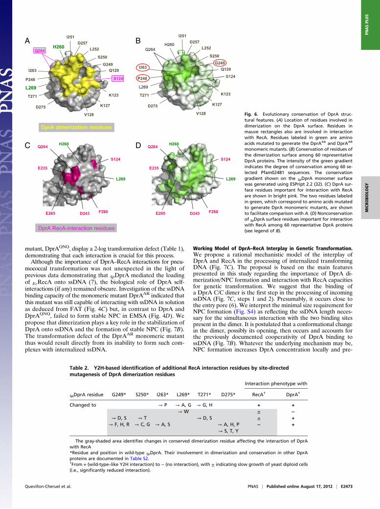
Evolutionary Conservation of DprA Structural Features. To de-termine whether the main structural features established for S.pneumoniae DprA (SpDprA) could be extended to other mem-bers of the family, we took advantage of the recent availability ofthe structure of DprA from R. palustris (RpDprA; PDB ID3MAJ) to launch a comparative analysis. Examination of theRpDprA structure confirmed the presence of an eRF (Fig. S6A)that exhibited excellent structural overlap with that of SpDprA(Fig. S6B), as expected given the conservation of Pfam02481. Inaddition, a C-terminal extradomain folded independently of theeRF in RpDprA (yellow domain in Fig. S6A).Interestingly, dimers were present in the RpDprA crystal, and,
despite the presence of the C-terminal extradomain, theyalso involved RF–RF interactions (Fig. S6C, C/C dimer).Comparison of residues involved in SpDprA and RpDprA di-merization surfaces revealed good sequence conservation (TableS2). Nine of 15 RpDprA interface residues were identical to thosein SpDprA. Stabilization of SpDprA and RpDprA dimers involvespredominantly hydrophobic interactions, together with hydrogenbonds for the former and a combination of hydrogen bonds andsalt bridges for the latter (Table S2). Three dimerization in-terface residues identified in SpDprA (P248, G249, and I263)(Fig. 6A) were identical in RpDprA, E. coli DprA (EcDprA), andBacillus subtilis DprA (BsDprA) (Fig. S7). More generally, theseresidues appeared to be the best-conserved surface residues among60 selected Pfam02481 sequences (Fig. 6B), arguing in favor ofthe evolutionary conservation of the DprA dimerization poten-tial and interfacing residues.In contrast, only 3 of 13 SpDprA residues involved in interaction
with RecA (Fig. 6C) appeared strictly conserved in RpDprA,BsDprA, and EcDprA (Table S3). All three correspond to buriedresidues suggesting they are structurally important rather thandirectly engaged in DprA–RecA interaction. This observationsuggested a rather limited evolutionary conservation, which wasconfirmed by examination of 60 selected Pfam02481 sequences(Fig. 6D).
Overlap Between DprA Dimerization and RecA Interaction Interfaces.The apparent lack of conservation of RecA interaction residuesin the DprA family appeared paradoxical for a protein involved
Table 1. Overview of results regarding DprA structure–function relationships
Interaction withDprA
ssDNAbinding*
Interaction withRecA
Transformation†DprA protein Crystal In vitro‡ Y2H In vivo§ Y2H In vivo¶
Wild type Yes Yes Yes Yes Yes Yes Yes +234R-245L k No ↓ ∼1 log257G-269S No ↓ ∼1 logVR (251V-260R) Mix** No No Yes Yes ↓ 183 ± 23-foldAR (260A-269R) No†† No No No Yes Yes ↓ 176 ± 23-fold235G, 243G, 264R or 265G‡‡ Yes No +QN (235Q-243N) Yes Yes ±§§ ↓ 7.9 ± 0.9-foldQNQ (235Q-243N-265Q) Yes Yes Yes No ↓ 154 ± 18-fold
*Formation of NPC based on EMSA; note that FAT revealed interaction of the DprAAR protein with a 20-mer oligonucleotide (Fig. 4C).†+, wild-type transformation frequency; fold reduction is indicated compared with wild type.‡Based on SAXS analysis (Fig. 3A): yes, dimer; no, monomer.§Revealed by cocapture from S. pneumoniae competent cells using DprA-SPA.¶Revealed by cocapture from S. pneumoniae competent cells using RecA-SPA.kEmpty box, not determined.**SAXS indicated equilibrium between monomer and dimer (Fig. S3F).††Beside SAXS results, in gel filtration experiments DprAAR eluted from the sizing column as a single homogeneous species with anapparent mass about half that observed for DprA and DprAQNQ (Fig. S2).‡‡All individual mutants behave similarly.§§Reduced cocapture of DprAQN with RecA-SPA (Fig. 5A).
Quevillon-Cheruel et al. PNAS | Published online August 17, 2012 | E2471
MICRO
BIOLO
GY
PNASPL
US

primarily in the loading of RecA onto ssDNA. We first consid-ered the possibility that DprA–RecA interaction could involvesurface residues prone to species-specific evolution to adapt totheir corresponding cytosolic environment. In support of thisidea was the finding that DprAE265Q substitution (Table 1)affected interaction with RecA in pneumococcal cells, whereasa Q residue was present at that same position in three wild-typeproteins, RpDprA, BsDprA, and EcDprA (Table S3). On the otherhand, the previous observation that SpDprA interacted withBsRecA in Y2H and promoted the loading of EcRecA ontossDNA (7) provided evidence for the maintenance of functionalinteractions between phylogenetically distant proteins, which isdifficult to reconcile with the idea of a species-specific evolutionof DprA surface residues involved in RecA interaction.These considerations prompted us to consider the alternative
hypothesis of an overlap between DprA dimerization and RecAinteraction interfaces. This hypothesis would be consistent withthe identification of S124 and Q264 as potentially involved inboth types of interaction (Figs. 2B and 6 A and C). According tothis hypothesis, our Y2H screen for DprA mutants selectivelyaffected in their interaction with RecA but retaining full capacityto self-interact (i.e., to dimerize) could have failed to identifymost RecA interaction residues. To check this possibility, sixresidues belonging to the dimerization interface of SpDprA andwell-conserved in the family—G249, S250, I263, L269, T271, andD275—were selected for site-directed mutagenesis (SI Matherialsand Methods). Twenty-four individual residue changes wereobtained with one or more substitutions at each site affectingthe potential of DprA interaction in Y2H (Table 2). Althoughfive changes had no effect, six substitutions significantly re-duced and 13 abolished the DprA–RecA interaction. On theother hand, only one substitution, the L269W change, abol-ished DprA self-interaction. We tentatively attribute the lastobservation to the fact that the DprA dimerization assay in-volved a combination of DprA mutant and wild-type proteins.Most importantly, for each of the selected dimerization resi-dues mutagenized, at least one substitution resulted in a sig-nificant reduction or the complete abolition of the interactionwith RecA. We conclude that there is a large overlap of the twointeraction surfaces in Y2H, suggesting that in S. pneumoniae thetwo interactions could be mutually exclusive (i.e., that interactionwith RecA could promote the discruption of the DprA C/Cdimer) (Fig. 7C).
DiscussionStructural Definition of the DprA Family. The resolution of thefull-length structure of SpDprA, the transformation-dedicatedRecA loader of S. pneumoniae, revealed that it consists of theassociation of an SAM and an eRF (Fig. 1A). Although an eRFalso was present in the structure of the phylogenetically distantRpDprA, its N terminal did not exhibit a SAM fold (Fig. S6A).Nevertheless, the intriguing structural similarity between SpDprAand RpDprA N terminals prompted us to deepen our analysis.Firstly, we observed that the SAM of SpDprA could not be pre-dicted through 3Dmodeling using RpDprA coordinates (Fig. S6D),suggesting that constraints in the RpDprA crystal prohibited SAMfolding. Second, in both cases 3D modeling of the N terminal ofRpDprA and EcDprA using SpDprA coordinates predicted a bonafide SAM fold (Fig. S6E). Taken together, these observationsled us to propose that all members of the DprA family harbora SAM fold at their N terminals. The DprA family, previouslydefined at the primary sequence level by the Pfam02481 (7), nowcan be regarded at the structural level as a larger entity com-prising an eRF, which entirely overlaps Pfam02481, precededby a SAM fold. The SAM-eRF structural association is totallyunrelated to the structure of any other recombinase loaders, i.e.,RecBCD (20), RecFOR (ref.21 and references therein), Rad52(22, 23), and BRCA2 (24, 25).Previous primary sequence analysis indicated that some DprA
orthologs, including RpDprA (Fig. S6A) and EcDprA, containa C-terminal extradomain next to Pfam02481. This extradomaincould modulate properties common to all DprAs or confer dis-tinct additional properties. With respect to the latter possibility,it might be relevant that the C-terminal extradomain of RpDprAexhibited an excellent structural match with the Z-DNA–bindingtumor-associated protein DML-1 (26). This extradomain doesnot prevent the formation of RF-RF dimers (Fig. S6C), stronglysuggesting that dimerization, which is crucial for transformationof S. pneumoniae, is an evolutionarily conserved property ofDprA proteins. This conclusion is fully consistent with a previousreport that BsDprA also displays self-interaction in Y2H (7).
Dimerization/NPC Formation and RecA-Interaction Properties of SpDprAAre Crucial for Transformation. Previous studies provided evidencefor DprA–DprA and DprA–RecA interaction in yeast, as well asfor DprA–RecA interaction in pneumococcal cell extracts (7).Here, our detailed phenotypic characterization of strains harbor-ing point mutations in dprA established that these interactionsare functionally important for genetic transformation. Both theDprA monomeric mutant, DprAAR, and the RecA-interaction
1 7.9 ± 0.9 154 ± 18
A100
10
1
0.1
0.01
DprAQN
DprA DprAQNQ
B
Fold
deficiency
Tran
sfo
rm
an
ts (%
)
DprA
quantification1 0.6 0.2
RecA*
RecA
DprA
α-RecA
α-DprA
RecA-SPA capture
Fig. 5. DprA–RecA interaction is required for genetic transformation. (A) DprAQN and DprAQNQ display reduced stability of complexes with RecA in vivo.Cocaptures of DprA (Left), DprAQN (Center), and DprAQNQ (Right) using RecA-SPA expressed ectopically in recA+ cells harboring the dprA+, dprAQN, or dprAQNQ
alleles were conducted as described in the legend of Fig. 3E but with an additional purification step before elution (SI Materials and Methods). Only C2 finaleluates are shown in this figure, but note that all three capture experiments were carried out in parallel and were analyzed on the same membrane (Fig. S5D).The amount of mutant DprAs captured is indicated relative to that of wild-type DprA. Signals were quantified using a Fujifilm LAS-4000 luminescent imageanalyzer. (B) Chromosomal transformation deficiency of the RecA-interaction mutants DprAQN and DprAQNQ compared with wild-type DprA.
E2472 | www.pnas.org/cgi/doi/10.1073/pnas.1205638109 Quevillon-Cheruel et al.
mutant, DprAQNQ, display a 2-log transformation defect (Table 1),demonstrating that each interaction is crucial for this process.Although the importance of DprA–RecA interactions for pneu-
mococcal transformation was not unexpected in the light ofprevious data demonstrating that SpDprA mediated the loadingof EcRecA onto ssDNA (7), the biological role of DprA self-interactions (if any) remained obscure. Investigation of the ssDNAbinding capacity of the monomeric mutant DprAAR indicated thatthis mutant was still capable of interacting with ssDNA in solutionas deduced from FAT (Fig. 4C) but, in contrast to DprA andDprAQNQ, failed to form stable NPC in EMSA (Fig. 4D). Wepropose that dimerization plays a key role in the stabilization ofDprA onto ssDNA and the formation of stable NPC (Fig. 7B).The transformation defect of the DprAAR monomeric mutantthus would result directly from its inability to form such com-plexes with internalized ssDNA.
Working Model of DprA–RecA Interplay in Genetic Transformation.We propose a rational mechanistic model of the interplay ofDprA and RecA in the processing of internalized transformingDNA (Fig. 7C). The proposal is based on the main featurespresented in this study regarding the importance of DprA di-merization/NPC formation and interaction with RecA capacitiesfor genetic transformation. We suggest that the binding ofa DprA C/C dimer is the first step in the processing of incomingssDNA (Fig. 7C, steps 1 and 2). Presumably, it occurs close tothe entry pore (6). We interpret the minimal size requirement forNPC formation (Fig. S4) as reflecting the ssDNA length neces-sary for the simultaneous interaction with the two binding sitespresent in the dimer. It is postulated that a conformational changein the dimer, possibly its opening, then occurs and accounts forthe previously documented cooperativity of DprA binding tossDNA (Fig. 7B). Whatever the underlying mechanism may be,NPC formation increases DprA concentration locally and pre-
A B
C D
E235
Q264
S124
L269
H260
E265 F280D243
E235
Q264
S124
L269
H260
E265 F280D243
DprA RecA-interaction residues
D275
P248
D257L252H260
Q120
K123
K127
V128
Q264
L269
T271
S250
I251
I263G249
S124
DprA dimerization residues
D275
P248
D257L252
H260
Q120
K123
K127
V128
Q264
L269
T271
S250
I251
I263
S124
G249
Fig. 6. Evolutionary conservation of DprA struc-tural features. (A) Location of residues involved indimerization on the DprA surface. Residues inmauve rectangles also are involved in interactionwith RecA. Residues labeled in green are aminoacids mutated to generate the DprAAK and DprAAR
monomeric mutants. (B) Conservation of residues ofthe dimerization surface among 60 representativeDprA proteins. The intensity of the green gradientindicates the degree of conservation among 60 se-lected Pfam02481 sequences. The conservationgradient shown on the SpDprA monomer surfacewas generated using ESPript 2.2 (32). (C) DprA sur-face residues important for interaction with RecAare shown in bright pink. The two residues labeledin green, which correspond to amino acids mutatedto generate DprA monomeric mutants, are shownto facilitate comparison with A. (D) Nonconservationof SpDprA surface residues important for interactionwith RecA among 60 representative DprA proteins(see legend of B).
Table 2. Y2H-based identification of additional RecA interaction residues by site-directedmutagenesis of DprA dimerization residues
Interaction phenotype with
SpDprA residue G249* S250* I263* L269* T271* D275* RecA† DprA†
Changed to → P → A, G → G, H + +→ W ± −
→ D, S → T → D, S ± +→ F, H, R → C, G → A, S → A, H, P − +
→ S, T, Y
The gray-shaded area identifies changes in conserved dimerization residue affecting the interaction of DprAwith RecA*Residue and position in wild-type SpDprA. Their involvement in dimerization and conservation in other DprAproteins are documented in Table S2.†From + (wild-type–like Y2H interaction) to − (no interaction), with ± indicating slow growth of yeast diploid cells(i.e., significantly reduced interaction).
Quevillon-Cheruel et al. PNAS | Published online August 17, 2012 | E2473
MICRO
BIOLO
GY
PNASPL
US
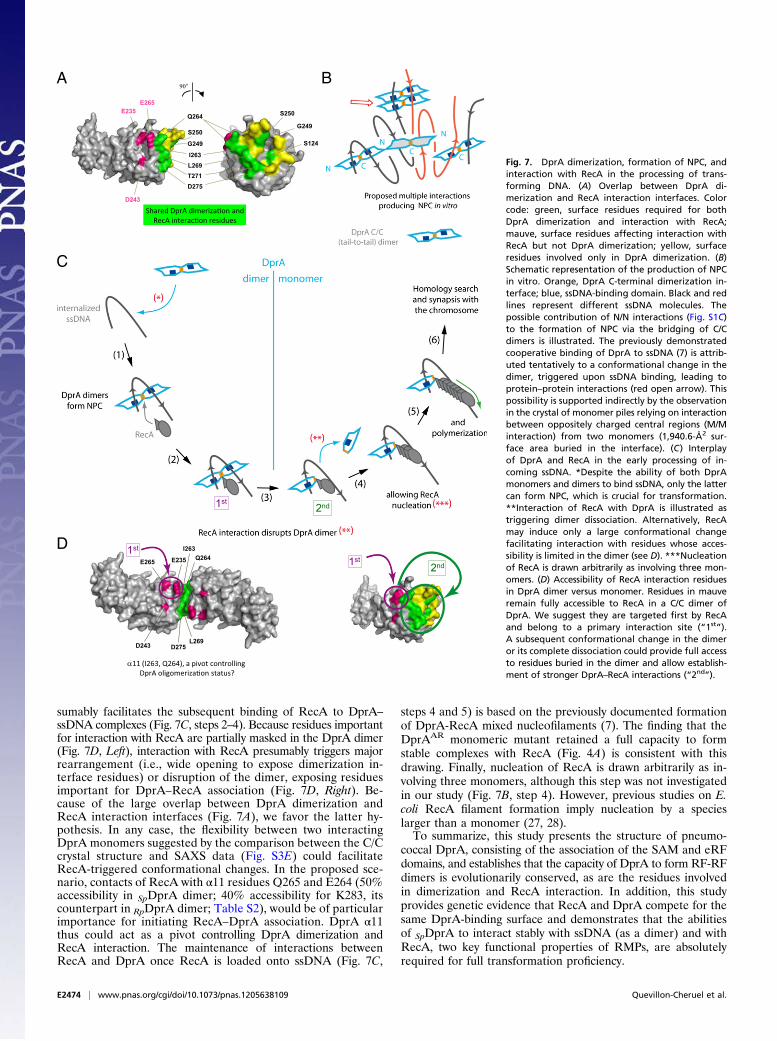
sumably facilitates the subsequent binding of RecA to DprA–ssDNA complexes (Fig. 7C, steps 2–4). Because residues importantfor interaction with RecA are partially masked in the DprA dimer(Fig. 7D, Left), interaction with RecA presumably triggers majorrearrangement (i.e., wide opening to expose dimerization in-terface residues) or disruption of the dimer, exposing residuesimportant for DprA–RecA association (Fig. 7D, Right). Be-cause of the large overlap between DprA dimerization andRecA interaction interfaces (Fig. 7A), we favor the latter hy-pothesis. In any case, the flexibility between two interactingDprA monomers suggested by the comparison between the C/Ccrystal structure and SAXS data (Fig. S3E) could facilitateRecA-triggered conformational changes. In the proposed sce-nario, contacts of RecA with α11 residues Q265 and E264 (50%accessibility in SpDprA dimer; 40% accessibility for K283, itscounterpart in RpDprA dimer; Table S2), would be of particularimportance for initiating RecA–DprA association. DprA α11thus could act as a pivot controlling DprA dimerization andRecA interaction. The maintenance of interactions betweenRecA and DprA once RecA is loaded onto ssDNA (Fig. 7C,
steps 4 and 5) is based on the previously documented formationof DprA-RecA mixed nucleofilaments (7). The finding that theDprAAR monomeric mutant retained a full capacity to formstable complexes with RecA (Fig. 4A) is consistent with thisdrawing. Finally, nucleation of RecA is drawn arbitrarily as in-volving three monomers, although this step was not investigatedin our study (Fig. 7B, step 4). However, previous studies on E.coli RecA filament formation imply nucleation by a specieslarger than a monomer (27, 28).To summarize, this study presents the structure of pneumo-
coccal DprA, consisting of the association of the SAM and eRFdomains, and establishes that the capacity of DprA to form RF-RFdimers is evolutionarily conserved, as are the residues involvedin dimerization and RecA interaction. In addition, this studyprovides genetic evidence that RecA and DprA compete for thesame DprA-binding surface and demonstrates that the abilitiesof SpDprA to interact stably with ssDNA (as a dimer) and withRecA, two key functional properties of RMPs, are absolutelyrequired for full transformation proficiency.
D275
Q264
L269
I263
E235E265
D243
1st
α11 (I263, Q264), a pivot controlling DprA oligomeriza�on status?
1st2nd
A
C
B
1st2nd
D
90°
Q264
D275
I263
L269
Shared DprA dimeriza�on and RecA interac�on residues
E265E235
D243
T271
S250
G249
G249
S250
S124
Fig. 7. DprA dimerization, formation of NPC, andinteraction with RecA in the processing of trans-forming DNA. (A) Overlap between DprA di-merization and RecA interaction interfaces. Colorcode: green, surface residues required for bothDprA dimerization and interaction with RecA;mauve, surface residues affecting interaction withRecA but not DprA dimerization; yellow, surfaceresidues involved only in DprA dimerization. (B)Schematic representation of the production of NPCin vitro. Orange, DprA C-terminal dimerization in-terface; blue, ssDNA-binding domain. Black and redlines represent different ssDNA molecules. Thepossible contribution of N/N interactions (Fig. S1C)to the formation of NPC via the bridging of C/Cdimers is illustrated. The previously demonstratedcooperative binding of DprA to ssDNA (7) is attrib-uted tentatively to a conformational change in thedimer, triggered upon ssDNA binding, leading toprotein–protein interactions (red open arrow). Thispossibility is supported indirectly by the observationin the crystal of monomer piles relying on interactionbetween oppositely charged central regions (M/Minteraction) from two monomers (1,940.6-Å2 sur-face area buried in the interface). (C) Interplayof DprA and RecA in the early processing of in-coming ssDNA. *Despite the ability of both DprAmonomers and dimers to bind ssDNA, only the lattercan form NPC, which is crucial for transformation.**Interaction of RecA with DprA is illustrated astriggering dimer dissociation. Alternatively, RecAmay induce only a large conformational changefacilitating interaction with residues whose acces-sibility is limited in the dimer (see D). ***Nucleationof RecA is drawn arbitrarily as involving three mon-omers. (D) Accessibility of RecA interaction residuesin DprA dimer versus monomer. Residues in mauveremain fully accessible to RecA in a C/C dimer ofDprA. We suggest they are targeted first by RecAand belong to a primary interaction site (“1st”).A subsequent conformational change in the dimeror its complete dissociation could provide full accessto residues buried in the dimer and allow establish-ment of stronger DprA–RecA interactions (“2nd”).
E2474 | www.pnas.org/cgi/doi/10.1073/pnas.1205638109 Quevillon-Cheruel et al.
Materials and MethodsBacterial Strains, Plasmids, Primers, Pneumococcal Transformation, and Y2H.S. pneumoniae strains and plasmids used are listed in Table S4; primers usedare listed in Table S5. Pneumococcal transformation was performed as pre-viously described (29). Random mutagenesis and selection for DprA interaction-defective mutants using Y2H were performed as previously described (30).Details of the Y2H screen and plasmid constructions are described in SIMaterials and Methods.
Protein Purification and Western Blot Analysis. His-tagged wild-type andmutant DprA proteins were purified from E. coli by a two-step procedure,i.e., an Ni-NTA column followed by gel filtration. SPA-tagged proteins werepurified from pneumococcal cells using a protocol developed for B. subtilis(31). Details regarding these protocols and the immunodetection of DprAand RecA by Western blotting are given in SI Materials and Methods.
Crystallization, Structure Determination, Comparison, and 3D Model Building.Native and Selenomethionine-labeled protein crystals were grown inhanging drops by mixing protein and reservoir solution in a 1:1 ratio. Forcrystallization conditions, see SI Materials and Methods. Sorbitol cry-oprotected crystals were flash frozen in liquid nitrogen and exposed on theProxima-1 beamline at the SOLEIL synchrotron, St-Aubin, France. Diffractiondata and refinement statistics are given in Table S1. The structure was de-termined by the SAD method using SeM-labeled protein data. The re-finement was done as described in SI Materials and Methods.
Exploration of 3D structures was performed using the following tools:Dali server, I-TASSER, and SWISS-MODEL servers, and the PyMOL MolecularGraphics System (SI Materials and Methods).
SAXS Measurements and Data Analysis. SAXS experiments with wild-typeDprA and the DprAQNQ mutant were performed using the Nanostar instrumentat the Institut de Biochimie et Biophysique Moléculaire et Cellulaire (Orsay,France). SAXS experiments on DprAAR and DprAVR were performed on the
SWING beamline at the SOLEIL synchrotron. For each sample, data werecollected at two or three protein concentrations: about 1, 2, and sometimes5 mg mL−1, in 50 mM MES (pH 6.5), 2 M NaCl. Data analysis and shape re-construction for the scattering object are described in SI Materials and Methods.
DNA-Binding Assays. EMSAwere performed exactly as described previously (7)with a 32P dT100 as ssDNA substrate. After electrophoresis, the gel wasdried, revealed with a FLA-3000 series fluorescent image analyzer (Fuji), andsignal was quantified with MultiGauge software V 3.0 (FujiFilm). FAT wasmeasured in a Fluoromax-4 (Horiba Scientific) (Fig. 4C) or a CARY Eclipse(Varian) spectrofluorometer (Fig. S5B), at 20 °C, in a final reaction volumeof 200 μL buffered with 25 mM NaCl, 20 mM Tris (pH 7.5), 2.5 mM MgCl2,2.5% (vol/vol) glycerol, and supplemented with 25 nM (Fig. S5B) or 100 nM(Fig. 4C) of a dT20 5′-labeled with fluoresceine (GeneCust). The excitationwavelength was set at 490 nm, and emission was observed at 525 nm (10-nmbandwidth). Protein injections were 0.25–1 μL from a 10-mg/mL stock solu-tion. Each titration curve was made in triplicate. The data were treated withSigmaPlot 12.0 (Systat Software).
ACKNOWLEDGMENTS. We thank Dr. Andrew Thompson and Pierre Legrandfor assistance with beamline Proxima-1; Dr. Javier Pérez for assistance withSWING beamline at the SOLEIL synchrotron facility; Dr. François Lecointe forhelping us adapt the protocol of purification of SPA-tagged proteins forS. pneumoniae; Jérome Cicolari for help with crystallization; Ines Li de laSierra-Gallay for the last refinement steps of the structure; Maud Hertzogfor performing a control fluorescence anisotropy titration experiment; andCalum Johnston for critical reading of the manuscript. The European Synchro-tron Radiation Facility and the SOLEIL synchrotron provided synchrotron radi-ation facilities. This work was supported in part by a grant from the AgenceNationale de la Recherche (projet n_ BLAN06-3_141806 to J.-P. C. and P.P.),by Grant INE20080311761 from the Fondation pour la Recherche Médicale(to P.P.), and by the Programme Incitatifs à Mobilité d’Équipe from theCentre National de la Recherche Scientifique (to P.P.). N.M. was the recipientof PhD Thesis Fellowship 10/2006-09/2007 from the Association pour laRecherche sur le Cancer.
1. Cox MM (2007) Motoring along with the bacterial RecA protein. Nat Rev Mol Cell Biol8:127–138.
2. Haldenby S, White MF, Allers T (2009) RecA family proteins in archaea: RadA and itscousins. Biochem Soc Trans 37:102–107.
3. Beernink HT, Morrical SW (1999) RMPs: Recombination/replication mediator proteins.Trends Biochem Sci 24:385–389.
4. Dillingham MS, Kowalczykowski SC (2008) RecBCD enzyme and the repair of double-stranded DNA breaks. Microbiol Mol Biol Rev 72:642–671.
5. Halpern D, Gruss A, Claverys JP, El-Karoui M (2004) rexAB mutants in Streptococcuspneumoniae. Microbiology 150:2409–2414.
6. Claverys JP, Martin B, Polard P (2009) The genetic transformation machinery:Composition, localization, and mechanism. FEMS Microbiol Rev 33:643–656.
7. Mortier-Barrière I, et al. (2007) A key presynaptic role in transformation for a widespreadbacterial protein: DprA conveys incoming ssDNA to RecA. Cell 130:824–836.
8. Hiller NL, et al. (2010) Generation of genic diversity among Streptococcus pneumoniaestrains via horizontal gene transfer during a chronic polyclonal pediatric infection. PLoSPathog 6:e1001108.
9. Croucher NJ, et al. (2011) Rapid pneumococcal evolution in response to clinicalinterventions. Science 331:430–434.
10. Bentley SD, et al. (2006) Genetic analysis of the capsular biosynthetic locus from all90 pneumococcal serotypes. PLoS Genet 2:e31.
11. Claverys JP, Prudhomme M, Martin B (2006) Induction of competence regulons as ageneral response to stress in gram-positive bacteria. Annu Rev Microbiol 60:451–475.
12. Håvarstein LS, Coomaraswamy G, Morrison DA (1995) An unmodified heptadecapeptidepheromone induces competence for genetic transformation in Streptococcuspneumoniae. Proc Natl Acad Sci USA 92:11140–11144.
13. Lee MS, Morrison DA (1999) Identification of a new regulator in Streptococcuspneumoniae linking quorum sensing to competence for genetic transformation.J Bacteriol 181:5004–5016.
14. Prudhomme M, Attaiech L, Sanchez G, Martin B, Claverys JP (2006) Antibiotic stressinduces genetic transformability in the human pathogen Streptococcus pneumoniae.Science 313:89–92.
15. Morrison DA, Mortier-Barrière I, Attaiech L, Claverys JP (2007) Identification of the majorprotein component of the pneumococcal eclipse complex. J Bacteriol 189:6497–6500.
16. Attaiech L, et al. (2011) Role of the single-stranded DNA-binding protein SsbB inpneumococcal transformation: Maintenance of a reservoir for genetic plasticity. PLoSGenet 7:e1002156.
17. Kim CA, Gingery M, Pilpa RM, Bowie JU (2002) The SAM domain of polyhomeoticforms a helical polymer. Nat Struct Biol 9:453–457.
18. Zhang Y, Skolnick J (2005) TM-align: A protein structure alignment algorithm basedon the TM-score. Nucleic Acids Res 33:2302–2309.
19. Zeghouf M, et al. (2004) Sequential peptide affinity (SPA) system for the identificationof mammalian and bacterial protein complexes. J Proteome Res 3:463–468.
20. Singleton MR, Dillingham MS, Gaudier M, Kowalczykowski SC, Wigley DB (2004)Crystal structure of RecBCD enzyme reveals a machine for processing DNA breaks.Nature 432:187–193.
21. Timmins J, Leiros I, McSweeney S (2007) Crystal structure and mutational study ofRecOR provide insight into its mode of DNA binding. EMBO J 26:3260–3271.
22. Kagawa W, et al. (2002) Crystal structure of the homologous-pairing domain fromthe human Rad52 recombinase in the undecameric form. Mol Cell 10:359–371.
23. Singleton MR, Wentzell LM, Liu Y, West SC, Wigley DB (2002) Structure of thesingle-strand annealing domain of human RAD52 protein. Proc Natl Acad Sci USA 99:13492–13497.
24. Pellegrini L, et al. (2002) Insights into DNA recombination from the structure ofa RAD51-BRCA2 complex. Nature 420:287–293.
25. Yang H, et al. (2002) BRCA2 function in DNA binding and recombination froma BRCA2-DSS1-ssDNA structure. Science 297:1837–1848.
26. Schwartz T, Behlke J, Lowenhaupt K, Heinemann U, Rich A (2001) Structure of theDLM-1-Z-DNA complex reveals a conserved family of Z-DNA-binding proteins. NatStruct Biol 8:761–765.
27. Spies M, Kowalczykowski SC (2006) The RecA binding locus of RecBCD is a generaldomain for recruitment of DNA strand exchange proteins. Mol Cell 21:573–580.
28. Joo C, et al. (2006) Real-time observation of RecA filament dynamics with singlemonomer resolution. Cell 126:515–527.
29. Martin B, PrudhommeM, Alloing G, Granadel C, Claverys JP (2000) Cross-regulation ofcompetence pheromone production and export in the early control of transformationin Streptococcus pneumoniae. Mol Microbiol 38:867–878.
30. Noirot-Gros MF, et al. (2006) Functional dissection of YabA, a negative regulator ofDNA replication initiation in Bacillus subtilis. Proc Natl Acad Sci USA 103:2368–2373.
31. Lecointe F, et al. (2007) Anticipating chromosomal replication fork arrest: SSB targetsrepair DNA helicases to active forks. EMBO J 26:4239–4251.
32. Gouet P, Courcelle E, Stuart DI, Métoz F (1999) ESPript: analysis of multiple sequencealignments in PostScript. Bioinformatics 15:305–308.
Quevillon-Cheruel et al. PNAS | Published online August 17, 2012 | E2475
MICRO
BIOLO
GY
PNASPL
US