Structure and Self-Assembly of the Calcium Binding Matrix Protein of Human Metapneumovirus
13
Structure Article Structure and Self-Assembly of the Calcium Binding Matrix Protein of Human Metapneumovirus Cedric Leyrat, 1 Max Renner, 1 Karl Harlos, 1 Juha T. Huiskonen, 1 and Jonathan M. Grimes 1,2, * 1 Division of Structural Biology, The Wellcome Trust Centre for Human Genetics, University of Oxford, Roosevelt Drive, Oxford OX3 7BN, UK 2 Diamond Light Source Limited, Harwell Science and Innovation Campus, Didcot, Oxfordshire, OX11 0DE, UK *Correspondence: [email protected] http://dx.doi.org/10.1016/j.str.2013.10.013 This is an open-access article distributed under the terms of the Creative Commons Attribution License, which permits unrestricted use, distribution, and reproduction in any medium, provided the original author and source are credited. SUMMARY The matrix protein (M) of paramyxoviruses plays a key role in determining virion morphology by direct- ing viral assembly and budding. Here, we report the crystal structure of the human metapneumovirus M at 2.8 A ˚ resolution in its native dimeric state. The structure reveals the presence of a high-affinity Ca 2+ binding site. Molecular dynamics simulations (MDS) predict a secondary lower-affinity site that cor- relates well with data from fluorescence-based ther- mal shift assays. By combining small-angle X-ray scattering with MDS and ensemble analysis, we captured the structure and dynamics of M in solution. Our analysis reveals a large positively charged patch on the protein surface that is involved in membrane interaction. Structural analysis of DOPC-induced polymerization of M into helical filaments using elec- tron microscopy leads to a model of M self-assembly. The conservation of the Ca 2+ binding sites suggests a role for calcium in the replication and morphogenesis of pneumoviruses. INTRODUCTION Human metapneumovirus (HMPV) is a leading cause of acute respiratory diseases in children, the elderly, and immune- compromised patients worldwide (Boivin et al., 2002; van den Hoogen, 2007; van den Hoogen et al., 2003; Williams et al., 2004; Xepapadaki et al., 2004). Together with respiratory syncy- tial virus (RSV), HMPV is grouped into the Pneumovirinae sub- family of the Paramyxoviridae (van den Hoogen et al., 2002). HMPV is an enveloped virus with an 13-kb, single-stranded () RNA genome that encodes nine proteins in the order 3 0 -N- P-M-F-M2(1)/(2)-SH-G-L-5 0 . HMPV proteins show detect- able levels of sequence identity to RSV, but the order of the genes is different and HMPV lacks the NS1 and NS2 genes pre- sent in RSV. For all paramyxoviruses, the nucleoprotein (N) en- capsidates viral RNA, leading to an N-RNA complex, and forms with the RNA-dependent RNA polymerase (L) and the phospho- protein (P) the viral replication complex. The matrix protein (M) is a major component of the virus, and is thought to form an ordered layer beneath the viral membrane (Battisti et al., 2012; Liljeroos et al., 2013). The M2 gene is specific to the Pneumovir- inae subfamily and possesses two overlapping open reading frames encoding two proteins, the antitermination/transcription elongation factor M2-1, which is required for viral transcription (Fearns and Collins, 1999), and the RNA synthesis regulatory fac- tor M2-2 (Buchholz et al., 2005). HMPV virions bud from the cell surface and form pleiomorphic or filamentous particles (Peret et al., 2002). The viral membrane contains the three viral transmembrane glycoproteins (G, F, and SH), along with the matrix protein (M), which associates with the membrane’s inner surface. M plays a critical role in assembly and budding through interactions with multiple viral and cellular components such as nucleoprotein-RNA oligomers (N-RNA; Ghildyal et al., 2002), lipid membranes (McPhee et al., 2011), and cytoplasmic tails of the viral glycoproteins (Henderson et al., 2002). In addition, viral matrix proteins are known to possess immunomodulatory properties through interactions with nucleic acids and host cell proteins, nucleocytoplasmic traf- ficking, and inhibition of host cell transcription (reviewed in Ghildyal et al., 2006). Among the order Mononegavirales, X-ray crystallographic structures of M proteins have been solved for RSV (Pneumovi- rus-Paramyxoviridae; Money et al., 2009), Newcastle disease virus (NDV; Avulavirus-Paramyxoviridae; Battisti et al., 2012), and Ebola virus (EBOV; Dessen et al., 2000), which belongs to the distantly related Filoviridae family but nevertheless pos- sesses an M protein that is structurally related to RSV M (Money et al., 2009). Additionally, the crystal structure of Borna disease virus (BDV) M (Neumann et al., 2009) from Bornaviridae and the structure of several Ms from members of the Rhabdoviridae fam- ily have been solved (Gaudier et al., 2002; Graham et al., 2008). Contrary to M proteins of Paramyxoviridae and Filoviridae, these proteins possess a single-domain M protein. However, BDV M has been shown to be homologous to the N-terminal domain (NTD) of EBOV VP40, suggesting that Paramyxoviridae and Filo- viridae M proteins evolved by gene duplication Neumann et al. (2009). Interestingly, while EBOV and RSV M have been crystal- lized as monomers, NDV and BDV Ms form dimers and tetra- mers, respectively, with a similar quaternary diamond shape. In this study, we solved the X-ray crystallographic structure of the M protein from HMPV at 2.8 A ˚ resolution. Furthermore, we analyzed the solution structure of intact M using small-angle X-ray scattering (SAXS) combined with classical, microsecond- long, explicit solvent molecular dynamics simulations (MDSs) 136 Structure 22, 136–148, January 7, 2014 ª2014 The Authors
Transcript of Structure and Self-Assembly of the Calcium Binding Matrix Protein of Human Metapneumovirus

Structure
Article
Structure and Self-Assembly of the CalciumBinding Matrix Protein of Human MetapneumovirusCedric Leyrat,1 Max Renner,1 Karl Harlos,1 Juha T. Huiskonen,1 and Jonathan M. Grimes1,2,*1Division of Structural Biology, The Wellcome Trust Centre for Human Genetics, University of Oxford, Roosevelt Drive, Oxford OX3 7BN, UK2Diamond Light Source Limited, Harwell Science and Innovation Campus, Didcot, Oxfordshire, OX11 0DE, UK
*Correspondence: [email protected]://dx.doi.org/10.1016/j.str.2013.10.013
This is an open-access article distributed under the terms of the Creative Commons Attribution License, which permits unrestricted use,
distribution, and reproduction in any medium, provided the original author and source are credited.
SUMMARY
The matrix protein (M) of paramyxoviruses plays akey role in determining virion morphology by direct-ing viral assembly and budding. Here, we report thecrystal structure of the human metapneumovirus Mat 2.8 A resolution in its native dimeric state. Thestructure reveals the presence of a high-affinityCa2+ binding site. Molecular dynamics simulations(MDS) predict a secondary lower-affinity site that cor-relates well with data from fluorescence-based ther-mal shift assays. By combining small-angle X-rayscattering with MDS and ensemble analysis, wecaptured the structure and dynamics ofM in solution.Our analysis reveals a large positively charged patchon the protein surface that is involved in membraneinteraction. Structural analysis of DOPC-inducedpolymerization of M into helical filaments using elec-tronmicroscopy leads to amodel ofMself-assembly.The conservation of theCa2+ binding sites suggests arole for calcium in the replication andmorphogenesisof pneumoviruses.
INTRODUCTION
Human metapneumovirus (HMPV) is a leading cause of acute
respiratory diseases in children, the elderly, and immune-
compromised patients worldwide (Boivin et al., 2002; van den
Hoogen, 2007; van den Hoogen et al., 2003; Williams et al.,
2004; Xepapadaki et al., 2004). Together with respiratory syncy-
tial virus (RSV), HMPV is grouped into the Pneumovirinae sub-
family of the Paramyxoviridae (van den Hoogen et al., 2002).
HMPV is an enveloped virus with an �13-kb, single-stranded
(�) RNA genome that encodes nine proteins in the order 30-N-P-M-F-M2(�1)/(�2)-SH-G-L-50. HMPV proteins show detect-
able levels of sequence identity to RSV, but the order of the
genes is different and HMPV lacks the NS1 and NS2 genes pre-
sent in RSV. For all paramyxoviruses, the nucleoprotein (N) en-
capsidates viral RNA, leading to an N-RNA complex, and forms
with the RNA-dependent RNA polymerase (L) and the phospho-
protein (P) the viral replication complex. The matrix protein (M) is
a major component of the virus, and is thought to form an
136 Structure 22, 136–148, January 7, 2014 ª2014 The Authors
ordered layer beneath the viral membrane (Battisti et al., 2012;
Liljeroos et al., 2013). The M2 gene is specific to the Pneumovir-
inae subfamily and possesses two overlapping open reading
frames encoding two proteins, the antitermination/transcription
elongation factor M2-1, which is required for viral transcription
(Fearns andCollins, 1999), and the RNA synthesis regulatory fac-
tor M2-2 (Buchholz et al., 2005).
HMPV virions bud from the cell surface and form pleiomorphic
or filamentous particles (Peret et al., 2002). The viral membrane
contains the three viral transmembrane glycoproteins (G, F, and
SH), along with the matrix protein (M), which associates with the
membrane’s inner surface. M plays a critical role in assembly
and budding through interactions with multiple viral and cellular
components such as nucleoprotein-RNA oligomers (N-RNA;
Ghildyal et al., 2002), lipid membranes (McPhee et al., 2011),
and cytoplasmic tails of the viral glycoproteins (Henderson
et al., 2002). In addition, viral matrix proteins are known to
possess immunomodulatory properties through interactions
with nucleic acids and host cell proteins, nucleocytoplasmic traf-
ficking, and inhibition of host cell transcription (reviewed in
Ghildyal et al., 2006).
Among the order Mononegavirales, X-ray crystallographic
structures of M proteins have been solved for RSV (Pneumovi-
rus-Paramyxoviridae; Money et al., 2009), Newcastle disease
virus (NDV; Avulavirus-Paramyxoviridae; Battisti et al., 2012),
and Ebola virus (EBOV; Dessen et al., 2000), which belongs to
the distantly related Filoviridae family but nevertheless pos-
sesses an M protein that is structurally related to RSV M (Money
et al., 2009). Additionally, the crystal structure of Borna disease
virus (BDV) M (Neumann et al., 2009) from Bornaviridae and the
structure of several Ms frommembers of theRhabdoviridae fam-
ily have been solved (Gaudier et al., 2002; Graham et al., 2008).
Contrary to M proteins of Paramyxoviridae and Filoviridae, these
proteins possess a single-domain M protein. However, BDV M
has been shown to be homologous to the N-terminal domain
(NTD) of EBOV VP40, suggesting that Paramyxoviridae and Filo-
viridae M proteins evolved by gene duplication Neumann et al.
(2009). Interestingly, while EBOV and RSV M have been crystal-
lized as monomers, NDV and BDV Ms form dimers and tetra-
mers, respectively, with a similar quaternary diamond shape.
In this study, we solved the X-ray crystallographic structure of
the M protein from HMPV at 2.8 A resolution. Furthermore, we
analyzed the solution structure of intact M using small-angle
X-ray scattering (SAXS) combined with classical, microsecond-
long, explicit solvent molecular dynamics simulations (MDSs)

Table 1. Crystallographic Statistics
Data Collection
Beamline Diamond I03
Wavelength (A) 0.97949
Space group P31
Unit cell constants (A) a = b = 62.0, c = 275.4
Resolution limits (A)a 53.7–2.8 (3.0–2.8)
Number of measured reflections 269,370
Number of unique reflections 27,947
Completeness of data (%) 99.5 (99.1)
Rmerge (%)b 19.4 (181.5)
Rpim (%) 6.6 (60)
Multiplicity 9.6 (9.9)
I/s 9.5 (1.7)
Refinement
Rxcpt (%)c 18.5
Rfree (%)d 22.9
Number of atoms (protein/water/
other)
6935, 174, 5
Ramachandran favored/outliers (%) 94.1/1.8
Rmsd bond length 0.012
Rmsd bond angle 1.5
Average B factors (protein/water/
other) (A2)
88, 70, 82
Rpim, precision-indicating merging R factor; Rmsd, root mean square
deviation from ideal geometry.aThe values for the highest resolution shell are given in parentheses.bRmerge = Shkl SijI(hkl;i) � <I(hkl)>j/Shkl SiI(hkl;i), where I(hkl;i) is the inten-
sity of an individual measurement of a reflection and <I(hkl)> is the
average intensity of that reflection.cRxpct = ShkljjFobsj � jFxpctjj/Shkl jFobsj, where jFobsj and jFxpctj are the
observed structure factor amplitude and the expectation of the model
structure factor amplitude, respectively.dRfree = Rxpct of the test set (1%–5% of the data removed prior to
refinement).
Figure 1. Crystal Structure of HMPV M
(A) Structure of the M dimer. One of the monomers is colored from blue (N
terminus) to red (C terminus) with secondary structure elements labeled,
whereas the other one is in gray.
(B) Close-up of the Ca2+ binding site. The Ca2+ ion is represented as a purple
sphere in the electron density from an omit map (contour level = 4s) calculated
using PHASER and omitting the Ca2+ ion. Coordinating water molecules are
displayed as nonbonded spheres. See also Figure S1.
Structure
Structure of the Human Metapneumovirus M Protein
and the ensemble optimization method (EOM). We show that
HMPV M is a dimer, both in the crystal and in solution, and
Ca2+ stabilizes the structure. Similarly to RSV M, HMPV M
assembles into helical filaments in the presence of lipids. An
electron microscopy reconstruction of the ultrastructure of an
M filament allows us to propose a model of M assembly in the
virion. Finally, the similarity with M proteins from other para-
myxoviruses and filoviruses enables evolutionary relationships
between these different viruses to be discerned.
RESULTS
Crystal Structure of HMPV MHMPV M was recombinantly expressed in E. coli, with an N-ter-
minal small ubiquitin-like modifier (SUMO) tag followed by a 3C
protease cleavage site. The tag was essential for soluble
expression and maintaining the protein in solution. The strin-
gent buffer conditions required for untagged M solubility (see
Experimental Procedures) resulted in slow and incomplete
cleavage of the SUMO tag, which further led to additional
degradation of untagged M as observed by SDS-PAGE (data
not shown), presumably in loop regions. HMPV M was crystal-
lized from a mixture of intact and proteolyzed untagged M
purified on gel filtration after prolonged incubation with 3C pro-
tease at 4�C, and this led to some irreproducibility in crystalli-
zation. HMPV M was solved at 2.8 A resolution by molecular
replacement using the structure of RSV M (Protein Data Bank
ID [PDB ID] 2VQP; sequence identity, 38%). Data collection
and refinement statistics are given in Table 1 (Rwork = 0.19;
Rfree = 0.23).
The P31 crystallographic asymmetric unit contains flattened,
diamond-shaped M dimers. The M dimer is stabilized by a large
network of conserved hydrophobic interactions with a buried
interface area of 1,421 A2 per monomer (Figure 1A; see also
Figure S1 available online). Each M subunit is composed of
two similarly folded domains (NTD and C-terminal domain
[CTD]), which are joined by a 14-residue linker (residues
123–137) for which no density is visible. The dimeric interface
involves contacts between the NTD and CTD of the related
monomers. The NTD comprises residues 1–123 and the CTD
residues 137–254. Each domain consists of a twisted b sand-
wich, in which the b strands in the opposing b sheets are approx-
imately orthogonal to each other, and a few short a helices. CTD
Structure 22, 136–148, January 7, 2014 ª2014 The Authors 137

Structure
Structure of the Human Metapneumovirus M Protein
loops (residues 180–188 and residues 208–218) are not visible in
the electron density, most probably due to intrinsic disorder in
these regions. However, one subunit from each dimer shows
no density for residues 170–190, which likely reflects degrada-
tion of the protein prior to crystallization, as evidenced by
SDS-PAGE (data not shown).
A unique feature of HMPV M among all experimentally
solved M proteins so far is the presence of a Ca2+ binding
site located on the solvent-exposed surface of the NTD. The
binding pocket adopts the classic Ca2+ pentagonal bipyra-
midal geometry, with an average coordinating distance of
2.4 A between Ca2+ and interacting oxygens. Binding involves
the side chain carboxyls of Glu24 and Asp26, the backbone
carbonyl of Leu28 and Lys101, and two water molecules
(Figure 1B). The interaction of Asp26 carboxyls is bidentate,
and Glu24 shows direct coordination with Ca2+ with one of
its carboxyl oxygens, whereas the other interacts through a
bridging water molecule.
Dimers pack crystallographically through a relatively large
(624 A2) NTD/NTD interface (Figure S1A) that is stabilized by
polar interactions, propagating a helical arrangement within the
crystal. This suggests that this interaction could be involved in
the formation of higher order forms of M.
Structural Characterization of Dimeric HMPVMby SAXSWe investigated the structure of M in solution using SAXS.
SUMO-3C-M was measured in its native form and after addition
of 1 mM of either Ca2+ or EGTA (Figure 2A; see also Figure S2).
Oligomeric state and low-resolution structure remained
unaffected by the presence of these additives, with Guinier-
determined Rg values of 4.6–4.8 nm (Figure S2B). Next, we stud-
ied untagged M conformation in solution. We first attempted to
use untagged M protein purified in 1 M NaCl, which we concen-
trated directly prior to measurement. However, only a single
high-concentration measurement could be obtained (Figure 2A;
see also Figure S2), due to protein aggregation occurring within a
few hours after concentration. Notably, this aggregation was
independent of the addition of 1 mM Ca2+ or EGTA (Figure S2C).
Because of the intrinsic propensity of HMPV M to both degrade
internal loops and self-aggregate at medium to high concentra-
tions, further attempts to produce intact, unaggregated protein
used 1 M NDSB-201 containing buffers, resulting in a significant
loss of separation power of the S200 column. Thus, M protein
could only be obtained as a (9:1 molar ratio) mixture with undi-
gested SUMO-3C-M, as seen on SDS-PAGE (data not shown).
Gel filtration fractions were used directly for SAXS measure-
ments and concentrated on site to avoid slow concentration-
dependent aggregation of M. Total protein concentrations
ranged from 1 to 4 mg/ml, and Rg values were between 2.6
and 3.0 nm, as determined by Guinier approximation, depending
on the presence or absence of contaminating SUMO-3C-M.
These measured Rg compared well with the Rg value of 2.4 nm
calculated from the crystallographic dimer, the 2 A discrepancy
resulting from the absence of loop regions in the crystal. Interest-
ingly, all SAXS profiles showed a marked change in slope at
intermediate resolution (�0.13–0.15A�1), a feature typically
observed in hollow macromolecular complexes (as observed in
ab initio models built from the SAXS data; Figures S2D and
S2E; Ribeiro Ede et al., 2009).
138 Structure 22, 136–148, January 7, 2014 ª2014 The Authors
Ensemble OptimizationThe observed SAXS profiles were treated as a mixture of two
flexible components (untagged M and SUMO-3C-M). Large
ensembles of both proteins were generated by classical MDS
(Table S1), which were then fitted to experimental data using
EOM in two successive rounds of refinement. The advantage
of this approach to analyze flexible mixtures is the combination
of MDS ability to fold secondary structure elements missing
from the crystal structure with explicit treatment of flexibility in
SAXS, resulting in restoration of missing protein fragments and
a gain in effective information content (Bernado et al., 2007;
Pelikan et al., 2009). By selecting an optimized ensemble (OE)
of MDS models that maximizes the agreement between experi-
mental and simulated SAXS profiles (c), we obtained a refined
model for use in a second round of MDS, using two copies of
the most represented monomer as starting coordinates. As an
indication of the success of the refinement protocol, the second
round of EOM fitting exclusively selected models from the sec-
ond round of MDS over unrefined models from the first round
(data not shown).
Figure 2B shows the Rg distribution of pure untagged M and
the M/SUMO-3C-M mixture, which forms two well-separated
populations. Untagged M represents 90%–100% of the mixture
and samples a narrow Rg range centered around 2.6 nm,
whereas SUMO-M-3C accounts for only 0%–10%of themixture
and samples a wider range of Rg (4.0–5.5 nm), consistent with
the presence of a flexibly linked SUMO tag. The fit is of good
quality and consistent between independent measurements,
with c values comprised between 0.7 and 0.9 (Figure 2A; see
also Figure S2). Careful inspection of models in the OEs and
comparison with the range of conformations sampled in the
pool from MDS indicates that, in solution, M populates only a
specific subset of the available MDS-derived conformers. In
particular, in presence of 1 M NDSB, the majority of selected
models display an interaction between the interdomain linkers,
encompassing residues 123–137 (Figures 2C and 2D; see also
Figure S3). Untagged M measured in 1 M NaCl buffer tends to
show a collapse of the interdomain linkers onto the core of their
respective monomers, rather than interaction between them.
This difference might be attributable to the stabilizing effect of
NDSBs on protein fold (Expert-Bezancon et al., 2003; Vuillard
et al., 1998). Furthermore, OEs are enriched in models in which
residues 169–174 spontaneously folded into a short a-helical
motif during MDS (Figures 2C–2F; see also Figure S3). Notably,
a similar a-helix is also present in the same region of RSV M
(Money et al., 2009). The OEs display flexibility in the CTD loops
connecting b9 and b10 (residues 170–190), and b12 and b13
(residues 208–218), as well as in the interdomain linker (residues
123–137), consistent with the disorder observed in these regions
in the crystal structure and in MDS.
Solution Structure of HMPV M Studied using MDSsIn order to study the impact of bound calcium on the structure,
explicit-solvent MDSs were performed in the presence or
absence of Ca2+ (Table S1) for a total simulation time of
�2.9 ms. Analysis of atomic root mean square fluctuations
(RMSFs) indicated three main flexible regions located in the
CTD loops connecting the b sheets (residues 170–190 and
208–218), as well as in the interdomain linker (residues 123–137;

Figure 2. Small-Angle X-Ray Scattering
(A) Fitted SAXS profiles of the SUMO-3C-M/untagged M mixture (black spheres) and merged data (gray spheres), measured in 1M non-detergent sulfobetaines
201 (NDSB-201) buffer, and SAXS profile of untagged M in 1M NaCl buffer (green spheres). The red lines represent fits from the OEs.
(B) Radius of gyration (Rg) distributions of the initial pool of 11,000 models (black area), composed of models of untagged M and models bearing one or two
N-terminal SUMO-3C tags, and OEs for the SUMO-3C-M/untagged M mixture (gray line) and untagged M (green line).
(C and D) Views of five superimposed representative models from the OE corresponding to the SUMO-3C-M/untagged M mixture measured in 1M NDSB-201
buffer. Residues missing from the crystal structure are shown in red, or in orange for residues that are missing in only one monomer.
(E and F) Similar views of the OE from untagged M in 1M NaCl. See also Figures S2 and S3.
Structure
Structure of the Human Metapneumovirus M Protein
Figure S3), in good agreement with the crystal structure.
Frequently, folding of a short a-helical motif encompassing resi-
dues 169–174 was observed. The interdomain linkers collapsed
onto the core of M, consistent with features present in the RSV
M crystal structure (Money et al., 2009).
In the crystal structure, Lys106 is involved in stabilizing the
relatively large NTD/NTD packing interface through a hydrogen
bond with the side chain of Gln77 from a crystallographically
related partner (Figure S1C). However, MDS performed in the
absence of the bound Ca2+ resulted in the formation of an
Structure 22, 136–148, January 7, 2014 ª2014 The Authors 139

Structure
Structure of the Human Metapneumovirus M Protein
intramolecular salt bridge between Lys106 and Asp26, which
otherwise interacts with the Ca2+ ion (Figure S1D). This interac-
tion seemed to compensate for the absence of bound Ca2+
and suggests that the absence of bound Ca2+ might impact
negatively on M ultrastructure assembly.
MDSs performed in the presence of 150 mM free calcium in
the simulation box predicted binding at a second, lower affinity
site, which is more solvent exposed than the primary site and
is unoccupied in the crystal structure. Binding of Ca2+ in this sec-
ond site induced slight local conformational changes in a3 and
the loop connecting a4 and b13 (Figures 3A and 3B). Additional
simulations starting from the bound state in the absence of free
Ca2+ suggested binding at this low-affinity site was stable
(Figure S3C). The geometry of this second Ca2+ binding site is
shown in Figure 3B, revealing close coordination by Asp97 and
Glu98 side chains, the backbone carbonyl of Val94, and the
carbonyl group from Gln231 side chain.
Ca2+ Increases M StabilityThermal shift assays (TSAs) were performed to determine the
effect of Ca2+ on M stability (Figure S4). Addition of 1 and
5 mM of Ca2+ increased the protein melting temperature (Tm)
from 50.6�C to 68.5�C and 73.8�C, respectively, whereas addi-
tion of similar concentrations of EGTA induced a 5�C drop in
Tm to 45.5�C. The effect was specific to Ca2+, with addition of
5 mM Mg2+ leading to no significant change in Tm (Figure S4).
Because SUMO-3C-M is easier to handle and produce in large
amounts than untagged M, quantitative TSA binding data were
obtained using uncleaved protein. SUMO-3C-M was titrated
using either EGTA or CaCl2, and unfolding transitions weremoni-
tored (Figures 3C and 3E; see also Figure S4), revealing changes
in Tm of the same magnitude as observed for untagged M. It is
important to emphasize that these variations in Tm did not involve
any change in oligomeric state, as evidenced by dynamic light
scattering (data not shown) and confirmed by SAXS (Figure S2).
Titration of SUMO-3C-M with EGTA could be analyzed by
assuming saturation of the crystallographic (high-affinity) Ca2+
binding site using the classical Cheng-Prusoff equation (Cheng
and Prusoff, 1973) in order to yield the affinity constant of
SUMO-3C-M for the first Ca2+ binding site (Kd1). Experiments
performed at protein concentrations of 2 and 4 mM resulted in
values of Kd1 = 1.1 nM (±1.0 nM) or Kd1 = 2.0 nM (±1.8 nM),
respectively (Figure 3D; see also Figure S4).
Titration of SUMO-3C-M using Ca2+ was analyzed by convert-
ing the observed Tm values intoCa2+-induced free energy change
of unfolding (DDGu) following standard procedures (Layton and
Hellinga, 2010). Because the SUMO-3C-M used as a reference
in this experiment has its high-affinity Ca2+ binding site saturated,
the analysis canbe used to extract the affinity for the secondCa2+
binding site (Kd2), yielding a value of Kd2 = 158 mM (±124 mM)
(Figure 3F; see also Figure S4). Interestingly, titrations of
SUMO-3C-M using EGTA/Ca2+ mixtures yielded DDGu versus
free Ca2+ concentration plots that could not be adequately fitted
assuming a single binding site (data not shown), further indicating
the presence of the lower affinity site.
Structure of Lipid-Bound M FilamentsIncubation of purified M with 1,2-dioleoyl-sn-glycero-3-
phosphocholine (DOPC) resulted, after several days, in the
140 Structure 22, 136–148, January 7, 2014 ª2014 The Authors
growth of long, flexible tubules with varying diameters (�37 ±
4 nm), as revealed by electron microscopy (EM; Figure 4).
Attempts to study the effect of EGTA and/or calcium on filament
formation were inconclusive; the addition of EGTA or calcium led
to nonspecific aggregation of the sample over the same time
scale required for filament growth.
Images of computationally straightened tubules were ana-
lyzed for the presence of higher order organization of M. Fourier
transforms of tubule images gave rise to layer lines character-
istic of helical symmetry. One tubule (diameter 34 nm) was
consistent with principal Bessel orders of 6 and �13 at 1/92 A
and 1/114 A, respectively (Figure 4F). Helical, three-dimensional
reconstruction of this tubule revealed an arrangement of sub-
units with a rise of 5.16 A and a turn of �56.6 degrees
(Figure 4G).
To model the higher order organization of M, we fitted the
X-ray structure, as well as SAXS-validated MD models, into
the EM map (Figures 4H and 4I; see also Figure S5) using
Chimera (Goddard et al., 2007; Pettersen et al., 2004). The
fitting revealed that M can only orientate with its dimeric sym-
metry axis orthogonal to the long axis of the filament. Most
importantly, the fitting unambiguously determined the concave
membrane binding surface of the molecule: in all cases, fitting
with the concave face of the protein pointing toward lipids
resulted in significantly better correlation (C) and overlap (O)
between the experimental EM map and a map simulated from
the atomic model than fitting with the concave face pointing
outside (C = 0.94/O = 126 versus C = 0.88/O = 115; Figure 4I).
Furthermore, imposing helical symmetry revealed no clashes
between the symmetry-related copies of M providing indepen-
dent validation for fitting (Figure 4H; see also Figure S4). The
packing with the concave face toward the membrane suggests
that M polymerization involves side-by-side interactions
between lipid-bound subunits (Figure 4H; see also Figure S4),
although the resolution of the EM reconstruction was insufficient
to precisely define the residues involved in forming these
interfaces.
Structural Comparison with Other ss(–)RNA VirusesReveals Conservation of Domain Architecture andInterdomain Interfaces across Paramyxoviridae
We used the Structural Homology Program (SHP) to identify
evolutionary relationships between M proteins on the basis of
structural alignments (Figures 5A and 5B; see also Figure S6).
Structural alignments of HMPV, RSV, NDV, and EBOVM indicate
that the NTDs and CTDs are homologous across Paramyxoviri-
dae and Filoviridae families (Figure 5B; see also Figure S6). The
location of the NTD/CTD interface is similar in pneumoviruses
and avulaviruses, but different in EBOV M, where the CTD
adopts a different orientation relative to the NTD (Figures 5A;
see also Figure S6). The phylogenetic tree obtained from aligning
the five full-length M structures reproduces well the classical
distinction observed between Paramyxoviridae, Filoviridae, and
Bornaviridae, with a clustering of RSV, HMPV, and NDV M
(Figure 5A; see also Figure S6).
Structural comparison at the domain level indicates that BDV
M is most similar to the M CTD of Paramyxoviridae (Figure 5B),
whereas EBOV M NTDs and CTDs cluster with their respective
Paramyxoviridae counterparts. This partition of NTDs and

Figure 3. Fluorescence-Based TSA
(A) Close-up of the second (low-affinity) Ca2+ binding site in the crystal structure.
(B) Model of binding of Ca2+ as observed in MDS.
(C) Unfolding transitions of native SUMO-3C-M (black) in 50 mM HEPES, pH 7.8, and 1.15 M NaCl and with increasing concentrations of EGTA (indicated by an
arrow). Only the titration performed using 4 mM of protein is represented for clarity.
(D) Plots of Tm versus total EGTA concentration, for a protein concentration of 4 mM (black) or 2 mM (red). The data points were fitted using a four-parameter
sigmoidal dose-response function.
(E) Unfolding transitions of native SUMO-3C-M (black) in 50 mM HEPES, pH 7.8, and 1.15 M NaCl and with increasing concentrations of CaCl2 from 0.25 mM to
2 mM (indicated by an arrow).
(F) Plot of DDGu versus total Ca2+ concentration. DDGu values were calculated and fitted using equations from Layton and Hellinga (2010). See also Figure S4.
Structure
Structure of the Human Metapneumovirus M Protein
Structure 22, 136–148, January 7, 2014 ª2014 The Authors 141

Figure 4. Helical Ordering of M in the Presence of Lipids Visualized by Electron Microscopy
(A) Samples of M incubated in the presence of DOPC stained with uranyl acetate revealed tubular and spherical structures with free M in the background. Scale
bar = 100 nm.
(B) A close-up of free M dimers. Inset shows a class average of M calculated from 577 of 840 particles.
(C) A close-up of a tubular filament. M is seen coating the filament surface.
(D) A close-up of an M-coated spherical structure.
(E) A computationally straightened, long tubule. Scale bars in (B)–(E) = 25 nm.
(F) A computational diffraction pattern of the tubule shown in (E) reveals maxima on layer lines, indicating that the tubule has helical symmetry. Lattice indexes
(white numbers) and layer line heights (black numbers) are indicated for clearly visible maxima.
(G) A radially depth-cued isosurface representation of the density map for lipid-bound M is shown. The map was calculated using helical reconstruction from
electron microscopy images of negatively stained samples.
(H) A close-up of the map (gray transparent surface) shows the packing of the fitted M (blue) after imposing helical symmetry. Only the C-alpha trace is shown
for M.
(I) Same rendering as (H), but shown from the side. All isosurfaces were calculated at 2s above the mean value. See also Figure S5.
Structure
Structure of the Human Metapneumovirus M Protein
CTDs implies that the members of Paramyxoviridae and
Filoviridae families evolved from a common ancestor prior to
divergence of the structural interdomain relationships in Filovir-
idae M protein. Interestingly, a structure-based sequence
142 Structure 22, 136–148, January 7, 2014 ª2014 The Authors
alignment of Filoviridae and Pneumovirinae M proteins reveals
a strikingly conserved stretch of residues at the NTD/CTD
interface, despite the absence of overall sequence identity.
Indeed, the interdomain interaction is mediated in part by a
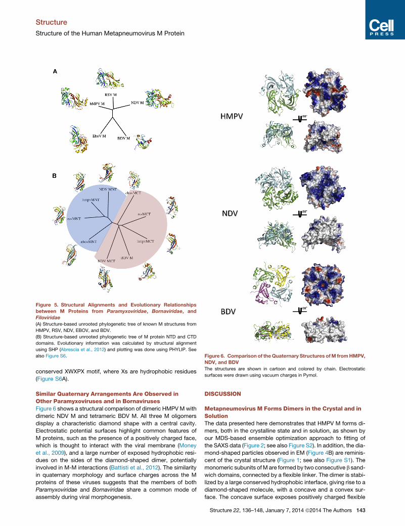
Figure 5. Structural Alignments and Evolutionary Relationships
between M Proteins from Paramyxoviridae, Bornaviridae, and
Filoviridae
(A) Structure-based unrooted phylogenetic tree of known M structures from
HMPV, RSV, NDV, EBOV, and BDV.
(B) Structure-based unrooted phylogenetic tree of M protein NTD and CTD
domains. Evolutionary information was calculated by structural alignment
using SHP (Abrescia et al., 2012) and plotting was done using PHYLIP. See
also Figure S6. Figure 6. Comparison of theQuaternary Structures ofM fromHMPV,
NDV, and BDV
The structures are shown in cartoon and colored by chain. Electrostatic
surfaces were drawn using vacuum charges in Pymol.
Structure
Structure of the Human Metapneumovirus M Protein
conserved XWXPX motif, where Xs are hydrophobic residues
(Figure S6A).
Similar Quaternary Arrangements Are Observed inOther Paramyxoviruses and in BornavirusesFigure 6 shows a structural comparison of dimeric HMPVMwith
dimeric NDV M and tetrameric BDV M. All three M oligomers
display a characteristic diamond shape with a central cavity.
Electrostatic potential surfaces highlight common features of
M proteins, such as the presence of a positively charged face,
which is thought to interact with the viral membrane (Money
et al., 2009), and a large number of exposed hydrophobic resi-
dues on the sides of the diamond-shaped dimer, potentially
involved in M-M interactions (Battisti et al., 2012). The similarity
in quaternary morphology and surface charges across the M
proteins of these viruses suggests that the members of both
Paramyxoviridae and Bornaviridae share a common mode of
assembly during viral morphogenesis.
DISCUSSION
Metapneumovirus M Forms Dimers in the Crystal and inSolutionThe data presented here demonstrates that HMPV M forms di-
mers, both in the crystalline state and in solution, as shown by
our MDS-based ensemble optimization approach to fitting of
the SAXS data (Figure 2; see also Figure S2). In addition, the dia-
mond-shaped particles observed in EM (Figure 4B) are reminis-
cent of the crystal structure (Figure 1; see also Figure S1). The
monomeric subunits ofM are formed by two consecutive b sand-
wich domains, connected by a flexible linker. The dimer is stabi-
lized by a large conserved hydrophobic interface, giving rise to a
diamond-shaped molecule, with a concave and a convex sur-
face. The concave surface exposes positively charged flexible
Structure 22, 136–148, January 7, 2014 ª2014 The Authors 143

Structure
Structure of the Human Metapneumovirus M Protein
loops that are thought to interact with the viral membrane
(Money et al., 2009). Together with the observed structural sim-
ilarity to the previously reported dimeric NDV M (Battisti et al.,
2012) but also for tetrameric BDV M (Neumann et al., 2009),
this indicates that the M dimers are the basic unit for matrix
assembly in these viruses.
Implications of the Structure of M Filaments for ViralAssembly and BuddingThe helical filament of DOPC-bound HMPV M provides experi-
mental evidence that M interacts with lipids via its concave
face. Interestingly, the electrostatic surface of the concave face
of M comprises negatively charged residues that are partially
covered by positively charged loops (Figure 6), resulting in a
surface that is complementary to the zwitterionic choline heads
that harbor a terminal quaternary ammonium followed by a phos-
phate group. Notably, helical assemblies with a diameter of
29 nm were reported for RSV M following prolonged incubation
with 1,2-dipalmitoyl-sn-glycero-3-phosphoethanolamine/DOPC
mixtures (McPhee et al., 2011; versus a diameter of 37 ± 4 nm for
HMPV M filaments), highlighting the similarity in the lipid binding
and self-assembly properties between the two proteins.
Additionally, the uncharged sides of the protein are involved in
formation of contacts betweenM dimers, predominantly through
NTD/NTD interfaces. We note that the positive curvature
observed in the filament structure is likely to be nonphysiological
since the M dimers coat the exterior of the lipid tubules. The
same nonphysiological curvature has been observed with RSV
(Liljeroos et al., 2013; McPhee et al., 2011). Interestingly, the
filaments revealed similar side-to-side contacts as in the crystal
structure of M (Figure S1A). However, the packing of M in the
crystal displays the opposite curvature to that seen in the EM
filaments (Figure S5). This negative curvature perhaps recapitu-
lates more closely the packing and assembly of M in the virion.
The observation that M can form filaments with different curva-
tures suggests that this dynamic plasticity in M packing might
play a role in HMPVmorphogenesis. In RSV virions, the presence
of an inner layer of membrane-associated M protein has been
shown to correlate with partial ordering of the glycoprotein
spikes (Liljeroos et al., 2013), suggesting that in the context of
viral infection, M would bind membranes at sites enriched in viral
glycoproteins and the concave face would be involved in binding
the conserved cytoplasmic tails of F and/or G proteins. This indi-
cates that the formation of flexible, curved, or planar arrays of M
proteins directly controls the localization and impacts the
conformation of F and G proteins within the membrane, allowing
membrane deformations required for budding to take place
without disruption of viral particles. Thus, we could postulate
that the plasticity of M protein self-assemblies enables M to
transduce internal signals from the cell cytoplasm, such as
conformational change induced by nucleocapsid binding, lead-
ing to a change in M array curvature, membrane deformation,
and budding.
Effect of Ca2+ Binding on M StabilityA distinguishing feature of metapneumovirus, and perhaps
pneumovirus M proteins, resides in their ability to bind Ca2+, as
evidenced by the presence of Ca2+ in the X-ray structure,
changes in Tm observed by TSA, and observations from MDS.
144 Structure 22, 136–148, January 7, 2014 ª2014 The Authors
Many viruses have been shown to perturb Ca2+ homeostasis
and utilize Ca2+ and cellular Ca2+-binding proteins in their repli-
cation cycles (reviewed in Zhou et al., 2009). In particular, para-
myxoviruses such as Sendai virus have been reported to
increase cytosolic Ca2+ concentrations, leading to a rounding
of chicken erythrocytes and increased rates of cell fusion (Hallett
et al., 1982; Volsky and Loyter, 1978). RSV replication in cell
culture was also negatively impacted by the absence of Ca2+,
and syncytium formation was inhibited (Shahrabadi and Lee,
1988). Interestingly, the SH protein of RSV, a viroporin specific
to the Pneumovirinae, associates with cellular membranes and
forms pentameric, cation-selective channels in infected cells
(Carter et al., 2010; Gan et al., 2008, 2012), possibly leading to
increased cytosolic Ca2+ levels.
The residues involved in side chain coordination to Ca2+ for
both the high- and low-affinity sites are conserved between
RSV and HMPV (Figure S6B), suggesting a common utilization
of Ca2+ in these viruses. However, the binding sites seem to
have diverged in other pneumoviruses, such as pneumonia virus
of mice and pneumonia virus of dog (data not shown). Intrigu-
ingly, the high-affinity Ca2+ binding pocket in RSV crystal struc-
tures (PDB ID 2VQP and 2YKD) is in an open conformation, and
the a3 helix that forms the binding site is unresolved, raising the
possibility of cleavage or conformational disorder, possibly
induced by the use of chelating agents.
Because the Ca2+ binding sites are located on the convex
face of M, it is possible that the variations in Ca2+ concentra-
tions inside infected cells at various stages of the viral cycle
regulate the assembly of viral nucleocapsids onto M arrays at
viral budding sites, but also perhaps intracellular transport of
M proteins to the membrane. Additionally, exposure of viral
particles to low calcium concentrations after cell entry could
play a role in the uncoating of nucleocapsids from the inner
matrix layer of the virion. Finally, the observed 25�C difference
in the thermal stability of unbound and Ca2+-bound HMPV M
at 1 mM Ca2+ suggests that Ca2+ is involved in stabilizing virions
in the Ca2+-rich extracellular environment, thus improving virion
lifetime and infectivity.
Evolution of Mononegavirales M ProteinsAnalysis of structural relationships among M proteins from
members of Paramyxoviridae, Bornaviridae, and Filoviridae
reveals structural similarity of the NTDs and CTDs across these
families and provides direct evidence that EBOV, NDV, and
HMPV/RSV evolved from a common ancestor prior to diver-
gence of the families and changes to the quaternary structure
of M. Gene duplication took place prior to separation of these
viruses, suggesting that BDV, which possesses a single-domain
M protein, might be more similar to the common ancestor of
these three Mononegavirales families. Additionally, the clus-
tering of BDV M with CTDs of the other M proteins implies
that the CTD was the originator and that the NTD appeared later
through duplication. This observation is consistent with the fact
that membrane binding of EBOV matrix protein VP40 (EBOV M)
occurs primarily through its CTD (Ruigrok et al., 2000). Indeed,
EBOV M assembles into ring-like structures that bind lipid
membranes (Ruigrok et al., 2000; Scianimanico et al., 2000;
Timmins et al., 2003) and induces budding of virus-like particles
when expressed in the absence of any other viral protein

Structure
Structure of the Human Metapneumovirus M Protein
(Timmins et al., 2001). Interestingly, the structural relationship
between EBOV M NTDs and CTDs in the crystal structure is
different to that observed in the HMPV, RSV, and NDV matrix
proteins (Figure 5A; Figure S9). It has been suggested that the
NTDs and CTDs from EBOV M can move relative to each other
and that M is in equilibrium between alternative oligomeric
states, such as monomers (Dessen et al., 2000; Scianimanico
et al., 2000), dimers (Timmins et al., 2003), hexamers (Ruigrok
et al., 2000; Scianimanico et al., 2000; Timmins et al., 2003),
and octamers (Gomis-Ruth et al., 2003). Specifically, the forma-
tion of an octameric RNA binding ring of NTDs has been asso-
ciated with a separate function of M during the viral cycle,
further supporting the hypothesis of divergence of NTD function
after gene duplication from a CTD ancestor. Recently structural
studies of EBOV VP40 have revealed a number of different olig-
omerization interfaces (Bornholdt et al., 2013). In HMPV M, the
dimeric building block is formed by NTD/CTD interactions
between monomers that form the dimer and occludes as sur-
face area that is twice as large as any interface observed in
EBOV VP40. In both cases, NTD/NTD and CTD/CTD interfaces
are the basis for higher order assemblies. However, the main
difference resides in the nature of the minimal building block,
which in EBOV is a monomer and in HMPV is a dimer (Bornholdt
et al., 2013).
Intriguingly, RNA binding capability has also been reported
for RSV M (Rodrıguez et al., 2004), raising the possibility that
HMPV M might interact with RNA as well. However, several
of the residues involved in RNA binding in RSV are not
conserved in HMPV M (Figure S2), indicating that this function
might not be shared between different members of the
Pneumovirinae.
ConclusionsWe have shown that HMPV M forms Ca2+-binding dimers with a
concave and a convex face. The dimers assemble onto lipids via
their concave face and form various higher order structures
through side-by-side interactions. Calcium appears to be critical
for M stability, and is potentially involved in regulating processes
such as viral entry, uncoating, assembly, and budding. This sug-
gests that the Ca2+-binding pockets are potential targets for the
development of small-molecule inhibitors. HMPV M shares a
common shape and similar surface charge distributions with
the other members of the Paramyxoviridae and Bornaviridae,
suggesting a common mode of self-assembly. Taken together,
these results further our understanding of metapneumovirus
morphogenesis and evolution.
EXPERIMENTAL PROCEDURES
Protein Cloning, Expression, and Purification
The HMPV M gene from strain NL1-00 was cloned into pOPINS3C (Berrow
et al., 2007) for expression of M with an N-terminal SUMO-3C cleavage
site using a proprietary ligation-independent In-Fusion System (Clontech)
following standard procedures. The integrity of the cloned construct was
checked by nucleotide sequencing.
The SUMO-3C-M construct was expressed in Rosetta2 E. coli cells by over-
night incubation under shaking at 17�C following 1 mM IPTG induction of 1 l
terrific broth in presence of appropriate antibiotics. Cells were harvested by
centrifugation (18�C, 20 min, 4000 x g). Cell pellets were resuspended in
20 mM Tris, pH 7.5, 1 M NaCl and lyzed by sonication. The lysate was then
centrifuged for 45 min at 4�C and 50000 x g. The supernatant was filtered
and loaded on a column containing pre-equilibrated Ni-NTA Agarose
(QIAGEN). After extensive washes, the protein was eluted in 20 mM Tris, pH
7.5, 1 M NaCl, 400 mM imidazole. Size exclusion chromatography was then
run on an S200 column equilibrated in 20 mM Tris, pH 7.5, 1 M NaCl. The
SUMO tag was removed by addition of 3C protease at 4�C for 72 h. The
cleaved product was further purified through reverse Ni-NTA purification to
remove Histagged 3C protease followed by an additional gel filtration step
(either in 20 mM Tris, pH 7.5, 1 M NaCl, or in 20 mM Tris, pH 7.5, 5 mM dithio-
threitol, 650 mM NaCl, 1M NDSB-201). The protein was concentrated using a
Millipore concentration unit (cut off 10 kDa) in presence of 1 M NDSB-201 in
order to avoid M protein aggregation and/or precipitation at concentrations
above �1 mg/ml.
Crystallization and Data Collection
Crystallizationwas carried out via the vapor diffusionmethod using a Cartesian
Technologies pipetting system (Walter et al., 2005). The M protein crystallized
after �28 days in 20% polyethylene glycol 6000, 100 mM Tris, pH 8.0, 10 mM
zinc chloride at 20�C. Crystals were frozen in liquid nitrogen after being soaked
in a mother liquor solution supplemented with 25% glycerol. Diffraction data
were recorded on the I03 beamline at Diamond Light Source. All data were
automatically processed by xia2 (Winter et al., 2013).
Structure Determination and Refinement
Structural determination was initiated by molecular replacement using RSV M
(PDB ID 2VQP) as a search model in PHASER (McCoy et al., 2007). The solu-
tion was subjected to rounds of restrained refinement in PHENIX (Adams
et al., 2010) and Autobuster (Blanc et al., 2004) and manual building in
COOT (Emsley et al., 2010). TLS parameters were included in the final round
of refinement. The CCP4 program suite (Winn et al., 2011) was used for
coordinate manipulations. The structures were validated with Molprobity
(Chen et al., 2010). Refinement statistics are given in Table 1, and final refined
coordinates and structure factors have been deposited in the PDB with
accession code 4LP7.
Structure Analysis
All the structure-related figures were prepared with the PyMOL Molecular
Graphics System (DeLano Scientific). Electrostatic potential calculations
were performed with APBS tools (Baker et al., 2001). Protein interfaces
were analyzed with the PISA webserver (Krissinel and Henrick, 2007). Struc-
ture-based sequence alignments were performed using PROMALS
3D (Pei et al., 2008). Structural alignments were calculated using SHP (Stuart
et al., 1979).
Small-Angle X-Ray Scattering Experiments
Small-angle X-ray scattering measurements for cleaved M and M/SUMO-
3C-M mixtures were performed at the BM29 beamline in the European Syn-
chrotron Radiation Facility (ESRF). Data were collected at 20�C, a wavelength
of 0.0995 nm, and a sample-to-detector distance of 1 m. The 1D scattering
profiles were generated, and blank subtraction was performed by the data
processing pipeline available at BM29 at the ESRF. Additional data for
SUMO-3C-M were collected at the ID22 beamline at Diamond Light Source.
The scattering profile of untagged M was analyzed using GNOM (Svergun,
1992) to yield the pair distribution function P(r) (Figure S2), twenty indepen-
dent ab initio reconstructions were generated using DAMMIF (Franke and
Svergun, 2009), and the models were averaged using DAMAVER (Volkov
and Svergun, 2003).
MD and Ensemble Optimization
Starting coordinates for the missing residues of M and SUMO-3C-M were
added in extended conformations in Modeler (Eswar et al., 2008). Coordi-
nates for the SUMO tag were taken from PDB entry 3UF8. All MD simulations
were performed using GROMACS 4 (Hess et al., 2008) and the AMBER99SB-
ILDN* force field (Best and Hummer, 2009; Lindorff-Larsen et al., 2010). At the
beginning of each simulation, the protein was immersed in a box of extended
simple point charge water, with a minimum distance of 1.0 nm between pro-
tein atoms and the edges of the box. A total of 150 mM NaCl was added
using genion. Long-range electrostatics were treated with the particle-mesh
Structure 22, 136–148, January 7, 2014 ª2014 The Authors 145

Structure
Structure of the Human Metapneumovirus M Protein
Ewald summation (Essmann et al., 1995). Bond lengths were constrained
using the P-LINCS algorithm. The integration time step was 5 femtoseconds.
The v-rescale thermostat and the Parrinello-Rahman barostat were used to
maintain a temperature of 300 K and a pressure of 1 atm. Each system
was energy minimized using 1,000 steps of steepest descent and equili-
brated for 200 ps with restrained protein heavy atoms. For each system,
two independent production simulations were obtained by using different
initial velocities. The aggregated simulation time was �2.9 ms for M and
�0.4 ms for SUMO-3C-M. RMSFs were calculated using GROMACS routines.
Snapshots were extracted every 100 ps, resulting in a pool of �12,000
models. Theoretical SAXS patterns were calculated with the program
CRYSOL (Svergun et al., 1995), and ensemble fitting was performed with
GAJOE (Bernado et al., 2007).
Thermal Shift Assay
The TSA (ThermoFluor) was carried out in a real-time PCR machine (BioRad
DNA Engine Opticon 2), where buffered solutions of protein and fluorophore
(SYPRO Orange; Molecular Probes; Invitrogen), with and without additives,
were heated in a stepwise fashion from 20�C to 99�C at a rate of 1�C/min.
An appropriate volume of protein and 3 ml of SYPRO Orange (Molecular
Probes; Invitrogen) were made up to a total assay volume of 50 ml with starting
buffer (50 mM HEPES, pH 7.8, 1.1 M NaCl) in white, low-profile, thin-wall PCR
plates (Abgene) sealed with microseal ‘‘B’’ films (BioRad). The fluorophore was
excited in the range of 470–505 nm and fluorescence emission was measured
in the range of 540–700 nmevery 0.5�Cafter a 10 s hold. The effect of Ca2+ ions
was assayed by comparing Tm of protein in starting buffer and in EGTA or
CaCl2 supplemented buffer. All thermal shift reactions were performed in trip-
licate. Tm, enthalpy of unfolding (DHu), and change in heat capacity upon Ca2+
binding were calculated by fitting the experimental data to Equations 9, 24,
and 25 from Layton and Hellinga (2010). Effective concentration values were
extracted from EGTA titration curves by fitting the data to a four-parameter
sigmoidal dose-response curve, and Kd1 was calculated by using the
Cheng-Prusoff equation (Cheng and Prusoff, 1973), assuming a value of
17.2 nM for Kd(EGTA-Ca2+) at the Tm, in presence of 1.1 M NaCl at pH 7.8
(calculated using the MAXCHELATOR server http://maxchelator.stanford.
edu/).
Electron Microscopy and Image Processing
Purified HMPV M (7.2 mM) was incubated with DOPC (400 mM; Avanti Polar
Lipids) for 7 days in +37�C. Electron microscopy grids of the mixture were
stained with 2% uranyl acetate. Images were taken on CCD (UltraScan
4000SP, Gatan) with a transmission electron microscope (Tecnai F30, FEI)
operated at 200 kV and at 39,0003 nominal magnification, resulting in a cali-
brated pixel size of 3.1 A/pixel. Contrast transfer function estimation and phase
flipping were carried out using XMIPP (http://xmipp.cnb.csic.es/), and
the rest of the analysis using Burnham-Brandeis Helical Package (http://
coan.burnham.org/other-projects/brandeis-helical-package/). Extracted and
straightened filaments were Fourier transformed for assigning layer-line
heights and Bessel orders followed by three-dimensional reconstruction
(Owen et al., 1996). The map was solvent-flattened in the lipid and solvent
parts. Atomic models of M were fitted into the electron microscopy map in
UCSF Chimera (Pettersen et al., 2004), and helical symmetry was applied on
the fitted structure using Bsoft (Heymann et al., 2008). The electron micro-
scopy reconstruction has been deposited in the Electron Microscopy Data
Bank (EMD-2415). Two-dimensional class averages of unboundMwere calcu-
lated in Relion (Scheres, 2012).
SUPPLEMENTAL INFORMATION
Supplemental Information includes six figures and one table and can be found
with this article online at http://dx.doi.org/10.1016/j.str.2013.10.013.
AUTHOR CONTRIBUTIONS
C.L. and J.M.G. conceived and designed research. C.L., M.R., and J.T.H. per-
formed experiments. C.L., J.T.H., and J.M.G. analyzed data. C.L., J.T.H., and
J.M.G. wrote the paper.
146 Structure 22, 136–148, January 7, 2014 ª2014 The Authors
ACKNOWLEDGMENTS
We thankRon Fouchier andBernadette van denHoogen for providing uswith a
plasmid encoding HMPV M, Alistair Siebert for electron microscopy support,
and David Stuart for critical reading of the manuscript. We thank Diamond
Light Source for beamtime (proposal MX8423) and the staff of beamlines
I02, I03, and I04 for assistance with crystal testing and data collection. The
research leading to these results has received funding from the European
Union Seventh Framework Programme (FP7/2007-2013) under SILVER grant
agreement no. 260644. This work was also supported by the Wellcome Trust
Core Award (090532/Z/09/Z) and the Academy of Finland (130750 and 218080
to J.T.H.). The OPIC electron microscopy facility was founded by a Wellcome
Trust JIF award (060208/Z/00/Z) and is supported by a WT equipment grant
(093305/Z/10/Z). The work presented here made use of the High Performance
Computing facility IRIDIS provided by the EPSRC-funded Centre for Innova-
tion (EP/K000144/1 and EP/K000136/1), which is owned and operated by
the e-Infrastructure South Consortium formed by the Universities of Bristol,
Oxford, Southampton, and UCL in partnership with STFC’s Rutherford Apple-
ton Laboratory.
Received: July 29, 2013
Revised: October 8, 2013
Accepted: October 10, 2013
Published: December 5, 2013
REFERENCES
Abrescia, N.G., Bamford, D.H., Grimes, J.M., and Stuart, D.I. (2012). Structure
unifies the viral universe. Annu. Rev. Biochem. 81, 795–822.
Adams, P.D., Afonine, P.V., Bunkoczi, G., Chen, V.B., Davis, I.W., Echols, N.,
Headd, J.J., Hung, L.W., Kapral, G.J., Grosse-Kunstleve, R.W., et al. (2010).
PHENIX: a comprehensive Python-based system for macromolecular struc-
ture solution. Acta Crystallogr. D Biol. Crystallogr. 66, 213–221.
Baker, N.A., Sept, D., Joseph, S., Holst, M.J., and McCammon, J.A. (2001).
Electrostatics of nanosystems: application to microtubules and the ribosome.
Proc. Natl. Acad. Sci. USA 98, 10037–10041.
Battisti, A.J., Meng, G., Winkler, D.C., McGinnes, L.W., Plevka, P.,
Steven, A.C., Morrison, T.G., and Rossmann, M.G. (2012). Structure and
assembly of a paramyxovirus matrix protein. Proc. Natl. Acad. Sci. USA
109, 13996–14000.
Bernado, P., Mylonas, E., Petoukhov, M.V., Blackledge, M., and Svergun, D.I.
(2007). Structural characterization of flexible proteins using small-angle X-ray
scattering. J. Am. Chem. Soc. 129, 5656–5664.
Berrow, N.S., Alderton, D., Sainsbury, S., Nettleship, J., Assenberg, R.,
Rahman, N., Stuart, D.I., and Owens, R.J. (2007). A versatile ligation-indepen-
dent cloning method suitable for high-throughput expression screening appli-
cations. Nucleic Acids Res. 35, e45.
Best, R.B., and Hummer, G. (2009). Optimizedmolecular dynamics force fields
applied to the helix-coil transition of polypeptides. J. Phys. Chem. B 113,
9004–9015.
Blanc, E., Roversi, P., Vonrhein, C., Flensburg, C., Lea, S.M., and Bricogne, G.
(2004). Refinement of severely incomplete structures with maximum likelihood
in BUSTER-TNT. Acta Crystallogr. D Biol. Crystallogr. 60, 2210–2221.
Boivin, G., Abed, Y., Pelletier, G., Ruel, L., Moisan, D., Cote, S., Peret, T.C.,
Erdman, D.D., and Anderson, L.J. (2002). Virological features and clinical man-
ifestations associated with human metapneumovirus: a new paramyxovirus
responsible for acute respiratory-tract infections in all age groups. J. Infect.
Dis. 186, 1330–1334.
Bornholdt, Z.A., Noda, T., Abelson, D.M., Halfmann, P., Wood, M.R.,
Kawaoka, Y., and Saphire, E.O. (2013). Structural rearrangement of ebola virus
VP40 begets multiple functions in the virus life cycle. Cell 154, 763–774.
Buchholz, U.J., Biacchesi, S., Pham, Q.N., Tran, K.C., Yang, L., Luongo, C.L.,
Skiadopoulos, M.H., Murphy, B.R., and Collins, P.L. (2005). Deletion of M2
gene open reading frames 1 and 2 of human metapneumovirus: effects on
RNA synthesis, attenuation, and immunogenicity. J. Virol. 79, 6588–6597.

Structure
Structure of the Human Metapneumovirus M Protein
Carter, S.D., Dent, K.C., Atkins, E., Foster, T.L., Verow,M.,Gorny, P., Harris,M.,
Hiscox, J.A., Ranson, N.A., Griffin, S., and Barr, J.N. (2010). Direct visualization
of the small hydrophobic protein of human respiratory syncytial virus reveals
the structural basis for membrane permeability. FEBS Lett. 584, 2786–2790.
Chen, V.B., Arendall, W.B., 3rd, Headd, J.J., Keedy, D.A., Immormino, R.M.,
Kapral, G.J., Murray, L.W., Richardson, J.S., and Richardson, D.C. (2010).
MolProbity: all-atom structure validation for macromolecular crystallography.
Acta Crystallogr. D Biol. Crystallogr. 66, 12–21.
Cheng, Y., and Prusoff, W.H. (1973). Relationship between the inhibition con-
stant (K1) and the concentration of inhibitor which causes 50 per cent inhibition
(I50) of an enzymatic reaction. Biochem. Pharmacol. 22, 3099–3108.
Dessen, A., Volchkov, V., Dolnik, O., Klenk, H.D., and Weissenhorn, W. (2000).
Crystal structure of the matrix protein VP40 from Ebola virus. EMBO J. 19,
4228–4236.
Emsley, P., Lohkamp, B., Scott, W.G., and Cowtan, K. (2010). Features and
development of Coot. Acta Crystallogr. D Biol. Crystallogr. 66, 486–501.
Essmann, U., Perera, L., Berkowitz, M.L., Darden, T., Lee, H., and Pedersen,
L.G. (1995). A smooth particle mesh Ewald method. J. Chem. Phys. 103,
8577–8593.
Eswar, N., Eramian, D., Webb, B., Shen, M.Y., and Sali, A. (2008). Protein
structure modeling with MODELLER. Methods Mol. Biol. 426, 145–159.
Expert-Bezancon, N., Rabilloud, T., Vuillard, L., and Goldberg, M.E. (2003).
Physical-chemical features of non-detergent sulfobetaines active as protein-
folding helpers. Biophys. Chem. 100, 469–479.
Fearns, R., and Collins, P.L. (1999). Role of the M2-1 transcription antitermina-
tion protein of respiratory syncytial virus in sequential transcription. J. Virol. 73,
5852–5864.
Franke, D., and Svergun, D.I. (2009). DAMMIF, a program for rapid ab-initio
shape determination in small-angle scattering. J. Appl. Crystallogr. 42,
342–346.
Gan, S.W., Ng, L., Lin, X., Gong, X., and Torres, J. (2008). Structure and ion
channel activity of the human respiratory syncytial virus (hRSV) small hydro-
phobic protein transmembrane domain. Protein Sci. 17, 813–820.
Gan, S.W., Tan, E., Lin, X., Yu, D., Wang, J., Tan, G.M., Vararattanavech, A.,
Yeo, C.Y., Soon, C.H., Soong, T.W., et al. (2012). The small hydrophobic pro-
tein of the human respiratory syncytial virus forms pentameric ion channels.
J. Biol. Chem. 287, 24671–24689.
Gaudier, M., Gaudin, Y., and Knossow,M. (2002). Crystal structure of vesicular
stomatitis virus matrix protein. EMBO J. 21, 2886–2892.
Ghildyal, R., Mills, J., Murray, M., Vardaxis, N., and Meanger, J. (2002).
Respiratory syncytial virus matrix protein associates with nucleocapsids in
infected cells. J. Gen. Virol. 83, 753–757.
Ghildyal, R., Ho, A., and Jans, D.A. (2006). Central role of the respiratory syn-
cytial virus matrix protein in infection. FEMS Microbiol. Rev. 30, 692–705.
Goddard, T.D., Huang, C.C., and Ferrin, T.E. (2007). Visualizing density maps
with UCSF Chimera. J. Struct. Biol. 157, 281–287.
Gomis-Ruth, F.X., Dessen, A., Timmins, J., Bracher, A., Kolesnikowa, L.,
Becker, S., Klenk, H.D., and Weissenhorn, W. (2003). The matrix protein
VP40 from Ebola virus octamerizes into pore-like structures with specific
RNA binding properties. Structure 11, 423–433.
Graham, S.C., Assenberg, R., Delmas, O., Verma, A., Gholami, A., Talbi, C.,
Owens, R.J., Stuart, D.I., Grimes, J.M., and Bourhy, H. (2008). Rhabdovirus
matrix protein structures reveal a novel mode of self-association. PLoS
Pathog. 4, e1000251.
Hallett, M.B., Fuchs, P., and Campbell, A.K. (1982). Sendai virus causes a rise
in intracellular free Ca2+ before cell fusion. Biochem. J. 206, 671–674.
Henderson, G., Murray, J., and Yeo, R.P. (2002). Sorting of the respiratory syn-
cytial virus matrix protein into detergent-resistant structures is dependent on
cell-surface expression of the glycoproteins. Virology 300, 244–254.
Hess, B., Kutzner, C., van der Spoel, D., and Lindahl, E. (2008). GROMACS 4:
algorithms for highly efficient, load-balanced, and scalable molecular simula-
tion. J. Chem. Theory Comput. 4, 435–447.
Heymann, J.B., Cardone, G., Winkler, D.C., and Steven, A.C. (2008).
Computational resources for cryo-electron tomography in Bsoft. J. Struct.
Biol. 161, 232–242.
Krissinel, E., and Henrick, K. (2007). Inference of macromolecular assemblies
from crystalline state. J. Mol. Biol. 372, 774–797.
Layton, C.J., and Hellinga, H.W. (2010). Thermodynamic analysis of ligand-
inducedchanges in protein thermal unfolding applied to high-throughput deter-
mination of ligand affinities with extrinsic fluorescent dyes. Biochemistry 49,
10831–10841.
Liljeroos, L., Krzyzaniak, M.A., Helenius, A., and Butcher, S.J. (2013).
Architecture of respiratory syncytial virus revealed by electron cryotomogra-
phy. Proc. Natl. Acad. Sci. USA 110, 11133–11138.
Lindorff-Larsen, K., Piana, S., Palmo, K., Maragakis, P., Klepeis, J.L., Dror,
R.O., and Shaw, D.E. (2010). Improved side-chain torsion potentials for the
Amber ff99SB protein force field. Proteins 78, 1950–1958.
McCoy, A.J., Grosse-Kunstleve, R.W., Adams, P.D., Winn, M.D., Storoni, L.C.,
and Read, R.J. (2007). Phaser crystallographic software. J. Appl. Cryst. 40,
658–674.
McPhee, H.K., Carlisle, J.L., Beeby, A., Money, V.A., Watson, S.M.D., Yeo,
R.P., and Sanderson, J.M. (2011). Influence of lipids on the interfacial disposi-
tion of respiratory syncytical virus matrix protein. Langmuir 27, 304–311.
Money, V.A., McPhee, H.K., Mosely, J.A., Sanderson, J.M., and Yeo, R.P.
(2009). Surface features of a Mononegavirales matrix protein indicate sites
of membrane interaction. Proc. Natl. Acad. Sci. USA 106, 4441–4446.
Neumann, P., Lieber, D., Meyer, S., Dautel, P., Kerth, A., Kraus, I., Garten, W.,
and Stubbs, M.T. (2009). Crystal structure of the Borna disease virus matrix
protein (BDV-M) reveals ssRNA binding properties. Proc. Natl. Acad. Sci.
USA 106, 3710–3715.
Owen, C.H., Morgan, D.G., and DeRosier, D.J. (1996). Image analysis of helical
objects: the Brandeis Helical Package. J. Struct. Biol. 116, 167–175.
Pei, J., Tang, M., and Grishin, N.V. (2008). PROMALS3D web server for accu-
rate multiple protein sequence and structure alignments. Nucleic Acids Res.
36 (Web Server issue), W30–W34.
Pelikan, M., Hura, G.L., and Hammel, M. (2009). Structure and flexibility within
proteins as identified through small angle X-ray scattering. Gen. Physiol.
Biophys. 28, 174–189.
Peret, T.C.T., Boivin, G., Li, Y., Couillard, M., Humphrey, C., Osterhaus,
A.D.M.E., Erdman, D.D., and Anderson, L.J. (2002). Characterization of human
metapneumoviruses isolated from patients in North America. J. Infect. Dis.
185, 1660–1663.
Pettersen, E.F., Goddard, T.D., Huang, C.C., Couch, G.S., Greenblatt, D.M.,
Meng, E.C., and Ferrin, T.E. (2004). UCSF Chimera—a visualization system
for exploratory research and analysis. J. Comput. Chem. 25, 1605–1612.
Ribeiro Ede, A., Jr., Leyrat, C., Gerard, F.C., Albertini, A.A., Falk, C., Ruigrok,
R.W., and Jamin, M. (2009). Binding of rabies virus polymerase cofactor to re-
combinant circular nucleoprotein-RNA complexes. J. Mol. Biol. 394, 558–575.
Rodrıguez, L., Cuesta, I., Asenjo, A., and Villanueva, N. (2004). Human respira-
tory syncytial virus matrix protein is an RNA-binding protein: binding proper-
ties, location and identity of the RNA contact residues. J. Gen. Virol. 85,
709–719.
Ruigrok, R.W., Schoehn, G., Dessen, A., Forest, E., Volchkov, V., Dolnik, O.,
Klenk, H.D., and Weissenhorn, W. (2000). Structural characterization and
membrane binding properties of the matrix protein VP40 of Ebola virus.
J. Mol. Biol. 300, 103–112.
Scheres, S.H. (2012). RELION: implementation of a Bayesian approach to
cryo-EM structure determination. J. Struct. Biol. 180, 519–530.
Scianimanico, S., Schoehn, G., Timmins, J., Ruigrok, R.H., Klenk, H.D., and
Weissenhorn, W. (2000). Membrane association induces a conformational
change in the Ebola virus matrix protein. EMBO J. 19, 6732–6741.
Shahrabadi, M.S., and Lee, P.W. (1988). Calcium requirement for syncytium
formation in HEp-2 cells by respiratory syncytial virus. J. Clin. Microbiol. 26,
139–141.
Structure 22, 136–148, January 7, 2014 ª2014 The Authors 147

Structure
Structure of the Human Metapneumovirus M Protein
Stuart, D.I., Levine, M., Muirhead, H., and Stammers, D.K. (1979). Crystal
structure of cat muscle pyruvate kinase at a resolution of 2.6 A. J. Mol. Biol.
134, 109–142.
Svergun, D. (1992). Determination of the regularization parameter in indirect-
transformmethods using perceptual criteria. J. Appl. Crystallogr. 25, 495–503.
Svergun, D., Barberato, C., and Koch, M.H.J. (1995). CRYSOL-a program to
evaluate X-ray solution scattering of biological macromolecules from atomic
coordinates. J. Appl. Crystallogr. 28, 768–773.
Timmins, J., Scianimanico, S., Schoehn, G., and Weissenhorn, W. (2001).
Vesicular release of ebola virus matrix protein VP40. Virology 283, 1–6.
Timmins, J., Schoehn, G., Kohlhaas, C., Klenk, H.D., Ruigrok, R.W., and
Weissenhorn, W. (2003). Oligomerization and polymerization of the filovirus
matrix protein VP40. Virology 312, 359–368.
van den Hoogen, B.G. (2007). Respiratory tract infection due to human meta-
pneumovirus among elderly patients. Clin. Infect. Dis. 44, 1159–1160.
van den Hoogen, B.G., Bestebroer, T.M., Osterhaus, A.D., and Fouchier, R.A.
(2002). Analysis of the genomic sequence of a human metapneumovirus.
Virology 295, 119–132.
van den Hoogen, B.G., van Doornum, G.J., Fockens, J.C., Cornelissen, J.J.,
Beyer, W.E., de Groot, R., Osterhaus, A.D., and Fouchier, R.A. (2003).
Prevalence and clinical symptoms of human metapneumovirus infection in
hospitalized patients. J. Infect. Dis. 188, 1571–1577.
Volkov, V.V., and Svergun, D.I. (2003). Uniqueness of ab initio shape determi-
nation in small-angle scattering. J. Appl. Crystallogr. 36, 860–864.
Volsky, D.J., and Loyter, A. (1978). Role of Ca++ in virus-induced membrane
fusion. Ca++ accumulation and ultrastructural changes induced by Sendai
virus in chicken erythrocytes. J. Cell Biol. 78, 465–479.
148 Structure 22, 136–148, January 7, 2014 ª2014 The Authors
Vuillard, L., Rabilloud, T., andGoldberg,M.E. (1998). Interactions of non-deter-
gent sulfobetaines with early folding intermediates facilitate in vitro protein
renaturation. Eur. J. Biochem. 256, 128–135.
Walter, T.S., Diprose, J.M., Mayo, C.J., Siebold, C., Pickford, M.G.,
Carter, L., Sutton, G.C., Berrow, N.S., Brown, J., Berry, I.M., et al.
(2005). A procedure for setting up high-throughput nanolitre crystallization
experiments. Crystallization workflow for initial screening, automated stor-
age, imaging and optimization. Acta Crystallogr. D Biol. Crystallogr. 61,
651–657.
Williams, J.V., Harris, P.A., Tollefson, S.J., Halburnt-Rush, L.L., Pingsterhaus,
J.M., Edwards, K.M., Wright, P.F., and Crowe, J.E., Jr. (2004). Human meta-
pneumovirus and lower respiratory tract disease in otherwise healthy infants
and children. N. Engl. J. Med. 350, 443–450.
Winn, M.D., Ballard, C.C., Cowtan, K.D., Dodson, E.J., Emsley, P., Evans,
P.R., Keegan, R.M., Krissinel, E.B., Leslie, A.G., McCoy, A., et al. (2011).
Overview of the CCP4 suite and current developments. Acta Crystallogr.
D Biol. Crystallogr. 67, 235–242.
Winter, G., Lobley, C.M., and Prince, S.M. (2013). Decision making in xia2.
Acta Crystallogr. D Biol. Crystallogr. 69, 1260–1273.
Xepapadaki, P., Psarras, S., Bossios, A., Tsolia, M., Gourgiotis, D., Liapi-
Adamidou, G., Constantopoulos, A.G., Kafetzis, D., and Papadopoulos, N.G.
(2004). Human Metapneumovirus as a causative agent of acute bronchiolitis
in infants. J. Clin. Virol. 30, 267–270.
Zhou, Y., Frey, T.K., and Yang, J.J. (2009). Viral calciomics: interplays between
Ca2+ and virus. Cell Calcium 46, 1–17.