Structure and Mechanical Performance of a Modern Fish...
10
DOI: 10.1002/adem.201180057 Structure and Mechanical Performance of a ‘‘Modern’’ Fish Scale** By Deju Zhu, Cesar Fuentes Ortega, Ramak Motamedi, Lawrence Szewciw, Franck Vernerey and Francois Barthelat* Nature increasingly serves as a model and inspiration to scientists and engineers, and biomimetics has the potential to lead to novel engineering materials and systems with new combinations of properties, multi-functionalities, adaptabil- ity, and environmental sustainability. In this work, we have studied the structure and mechanics of modern teleost fish scales, which have received relatively little attention in the past. [1–5] This type of scale displays interesting combinations of flexibility, strength, resistance to penetration, light weight, and transparency. Fish scales exhibit large variations in shape, size, and arrangement. The general classification includes cosmoid, ganoid, placoid, and elasmoid (cycloid and ctenoid) found in the modern teleost class of fishes. [6] The ‘‘primitive’’ cosmoid and ganoid scales are bulky, bony scales which offer very effective protective properties, through a multilayered structure capable of a variety of dissipative mechanisms. [3] However, over the course of evolution the reduction of the integumental skeleton has improved swimming perfor- mance, [3,7] and the ‘‘ancient’’ cosmoid and ganoid scales have been replaced by the thinner, more flexible teleost scales. [8] Teleost scales have excellent hydrodynamic properties [9,10] and provide a protective layer resisting penetration. [3,7,8] Currey, in a review article on mineralized tissues, noted that some fish scales are so tough that they cannot be easily fractured even after immersion in liquid nitrogen. [11] At larger lengths, the arrangement of the scales provides a flexible skin that allows for changes in shape. In fact, the scaled skin has been shown to play a critical structural role in fish locomotion by regulating wave propagation [12–14] and by storing mechan- ical energy in order to make swimming more efficient. [15] RESEARCH ARTICLE [*] Dr. D. Zhu, C. F. Ortega, R. Motamedi, L. Szewciw, Dr. F. Barthelat Department of Mechanical Engineering, McGill University, Montreal, QC, (Canada) E-mail: [email protected] Dr. F. Vernerey Department of Civil Engineering, University of Colorado, Boulder, CO, (USA) [**] The authors wish to acknowledge the support of the National Science Foundation under award CMMI 0927585 and of Faculty of Engineering at McGill University. Atomic emission spectroscopy tests were performed by Monique Riendeau, Dept. of Mining & Materials Engineering, and Ranjan Roy and Andrew Golsztajn, Dept. of Chemical Engineering, McGill University. Protective materials and structures found in natural organisms may inspire new armors with improved resistance to penetration, flexibility, light weight, and other interesting properties such as transparency and breathability. All these attributes can be found in teleost fish scales, which are the most common types of scales in modern fish species. In this work, we have studied the structure and mechanics of fish scales from striped bass (Morone saxatilis). This scale is about 200–300 mm thick and consists of a hard outer bony layer supported by a softer cross-ply of collagen fibrils. Perforation tests with a sharp needle indicated that a single fish scale provides a high resistance to penetration which is superior to polystyrene and polycarbonate, two engineering polymers that are typically used for light transparent packaging or protective equipment. Under puncture, the scale undergoes a sequence of two distinct failure events: First, the outer bony layer cracks following a well defined cross-like pattern which generates four ‘‘flaps’’ of bony material. The deflection of the flaps by the needle is resisted by the collagen layer, which in biaxial tension acts as a retaining membrane. Remarkably this second stage of the penetration process is highly stable, so that an additional 50% penetration force is required to eventually puncture the collagen layer. The combination of a hard layer that can fail in a controlled fashion with a soft and extensible backing layer is the key to the resistance to penetration of individual scales. ADVANCED ENGINEERING MATERIALS 2011, 13, No. XX ß 2011 WILEY-VCH Verlag GmbH & Co. KGaA, Weinheim wileyonlinelibrary.com B1
Transcript of Structure and Mechanical Performance of a Modern Fish...

RE
DOI: 10.1002/adem.201180057SEARCH
Structure and Mechanical Performance of a ‘‘Modern’’Fish Scale**
ARTIC
LE
By Deju Zhu, Cesar Fuentes Ortega, Ramak Motamedi,Lawrence Szewciw, Franck Vernerey and Francois Barthelat*
Protective materials and structures found in natural organisms may inspire new armors with improvedresistance to penetration, flexibility, light weight, and other interesting properties such as transparencyand breathability. All these attributes can be found in teleost fish scales, which are the most commontypes of scales in modern fish species. In this work, we have studied the structure and mechanics of fishscales from striped bass (Morone saxatilis). This scale is about 200–300 mm thick and consists of a hardouter bony layer supported by a softer cross-ply of collagen fibrils. Perforation tests with a sharp needleindicated that a single fish scale provides a high resistance to penetration which is superior topolystyrene and polycarbonate, two engineering polymers that are typically used for light transparentpackaging or protective equipment. Under puncture, the scale undergoes a sequence of two distinctfailure events: First, the outer bony layer cracks following a well defined cross-like pattern whichgenerates four ‘‘flaps’’ of bony material. The deflection of the flaps by the needle is resisted by the collagenlayer, which in biaxial tension acts as a retaining membrane. Remarkably this second stage of thepenetration process is highly stable, so that an additional 50% penetration force is required to eventuallypuncture the collagen layer. The combination of a hard layer that can fail in a controlled fashion with asoft and extensible backing layer is the key to the resistance to penetration of individual scales.
Nature increasingly serves as a model and inspiration to
scientists and engineers, and biomimetics has the potential to
lead to novel engineering materials and systems with new
combinations of properties, multi-functionalities, adaptabil-
ity, and environmental sustainability. In this work, we have
studied the structure and mechanics of modern teleost fish
scales, which have received relatively little attention in the
[*] Dr. D. Zhu, C. F. Ortega, R. Motamedi, L. Szewciw,Dr. F. BarthelatDepartment of Mechanical Engineering,McGill University, Montreal, QC, (Canada)E-mail: [email protected]
Dr. F. VernereyDepartment of Civil Engineering,University of Colorado, Boulder, CO, (USA)
[**] The authors wish to acknowledge the support of the NationalScience Foundation under award CMMI 0927585 and ofFaculty of Engineering at McGill University. Atomic emissionspectroscopy tests were performed by Monique Riendeau, Dept.of Mining & Materials Engineering, and Ranjan Roy andAndrew Golsztajn, Dept. of Chemical Engineering, McGillUniversity.
ADVANCED ENGINEERING MATERIALS 2011, 13, No. XX � 2011 WILEY-VCH
past.[1–5] This type of scale displays interesting combinations
of flexibility, strength, resistance to penetration, light weight,
and transparency. Fish scales exhibit large variations in shape,
size, and arrangement. The general classification includes
cosmoid, ganoid, placoid, and elasmoid (cycloid and ctenoid)
found in the modern teleost class of fishes.[6] The ‘‘primitive’’
cosmoid and ganoid scales are bulky, bony scales which offer
very effective protective properties, through a multilayered
structure capable of a variety of dissipative mechanisms.[3]
However, over the course of evolution the reduction of the
integumental skeleton has improved swimming perfor-
mance,[3,7] and the ‘‘ancient’’ cosmoid and ganoid scales have
been replaced by the thinner, more flexible teleost scales.[8]
Teleost scales have excellent hydrodynamic properties[9,10]
and provide a protective layer resisting penetration.[3,7,8]
Currey, in a review article on mineralized tissues, noted that
some fish scales are so tough that they cannot be easily
fractured even after immersion in liquid nitrogen.[11] At larger
lengths, the arrangement of the scales provides a flexible skin
that allows for changes in shape. In fact, the scaled skin has
been shown to play a critical structural role in fish locomotion
by regulating wave propagation[12–14] and by storing mechan-
ical energy in order to make swimming more efficient.[15]
Verlag GmbH & Co. KGaA, Weinheim wileyonlinelibrary.com B1

RESEARCH
ARTIC
LE
D. Zhu et al./A ‘‘Modern’’ Fish Scale . . .
Fig. 1. The hierarchical structure of a teleost fish scale from striped bass, M. saxatilis. (a) Whole fish, (b) staggered multiple scales, (c) an individual scale, (d) cross-section of a scale,(e) cross-ply collagen structure, (f) collagen fibrils.
1. The Hierarchical Structure of Fish Skin
In this study we have investigated the structure and
mechanics of a single teleost (ctenoid) fish scale from striped
bass Morone saxatilis. Like many other structural biological
materials,[16–18] the structure of teleost fish scales displays a
characteristic hierarchical structure, built over several distinct
length scales (Figure 1). At the macroscopic level, the scales are
staggered and cover most of the body of the fish (Figure 1a,b).
This arrangement provides a continuous barrier to penetration
and flexural compliance. When the fish is highly curved (at the
end of a swimming stroke), the scales interact more strongly,
which stiffens the skin in flexion.[19] The skin then acts as an
‘‘external tendon,’’ storing mechanical energy, which can be
recovered to facilitate the onset of the next stroke.[15,20] At the
mesoscale level, an individual scale from an adult striped bass
is a thin plate with an irregular pentagonal shape, about 10 mm
in diameter (Figure 1c).
The posterior area of the scale displays rough patterns
(ctenii) which offer attractive hydrodynamic properties,[9,10]
while the anterior area consists of grooves in the radial
direction (radii) and ridges that form circular rings (circuli)
around a central area called the ‘‘focus.’’[21] Radii and circuli
possibly provide increased flexibility and anchoring of the
scale, respectively.[10] Teleost scales are composed of collagen
fibrils type-I, and are partially mineralized with hydroxya-
patite (16–59% mineral content in weight[2,5,22–24]). The outer
layer of the scale is significantly more mineralized and often
referred to as ‘‘bony layer,’’ whereas the inner layer (‘‘basal’’
or ‘‘collagen’’ layer) is mineralized mostly near the bony layer,
but with mineralization pockets proceeding well into the
collagen layer.[7,23] In striped bass, bony and collagen layers
have approximately the same thickness (100 mm). Using AES
B2 http://www.aem-journal.com � 2011 WILEY-VCH Verlag GmbH & Co
(atomic emission spectroscopy), we measured an average
hydroxyapatite mass fraction of 46% for the whole scale.
The density of collagen (1.33� 103 kg �m�3) and hydroxya-
patite (3.17� 103 kg �m�3)[2] were used to estimate the volume
fraction of hydroxyapatite as 26%. In another experiment, we
separated a scale into two samples by dissecting a few plies off
the collagen layer. The upper and lower samples gave
hydroxyapatite mass contents of 50% and 14%, respectively
(30% and 6% in volume fractions), confirming that the upper
region of the scale is significantly more mineralized than the
lower region. These results are consistent with reports of a
general 20–35% percentage points difference in mineralization
between the bony and collagen layers.[25] Bony and collagen
layers are cross-ply layered composites, each ply being made
of parallel collagen fibrils rotated across layers by angles that
can vary from species to species.[25–29] In striped bass, we
found that the basal layer is formed of 20–25 plies about
4–5 mm thick each (Figure 1d), where the collagen fibrils are
rotated by 90 degrees from one ply to the next (Figure 1e).
Cross-ply collagen structures are typically found in natural
tissues that undergo multiaxial stresses (shell of soft-shelled
turtles,[30] human annulus fibrosus[31]). Several authors have
discussed the importance of the plywood nature of the
collagen layer to whole fish scale mechanical proper-
ties,[2,5,8,32–34] in general by providing the scale with strength
along multiple directions. At smaller length scales, individual
collagen fibrils, about 50–200 nm in diameter, can be observed
on a cross-section of the scale (Figure 1f). Interestingly, we
found that in striped bass the 90 degree cross-ply is achieved
by alternating layers composed of radial fibrils (‘‘R’’ layers)
with layers made of circumferential fibrils (‘‘C’’ layers), both
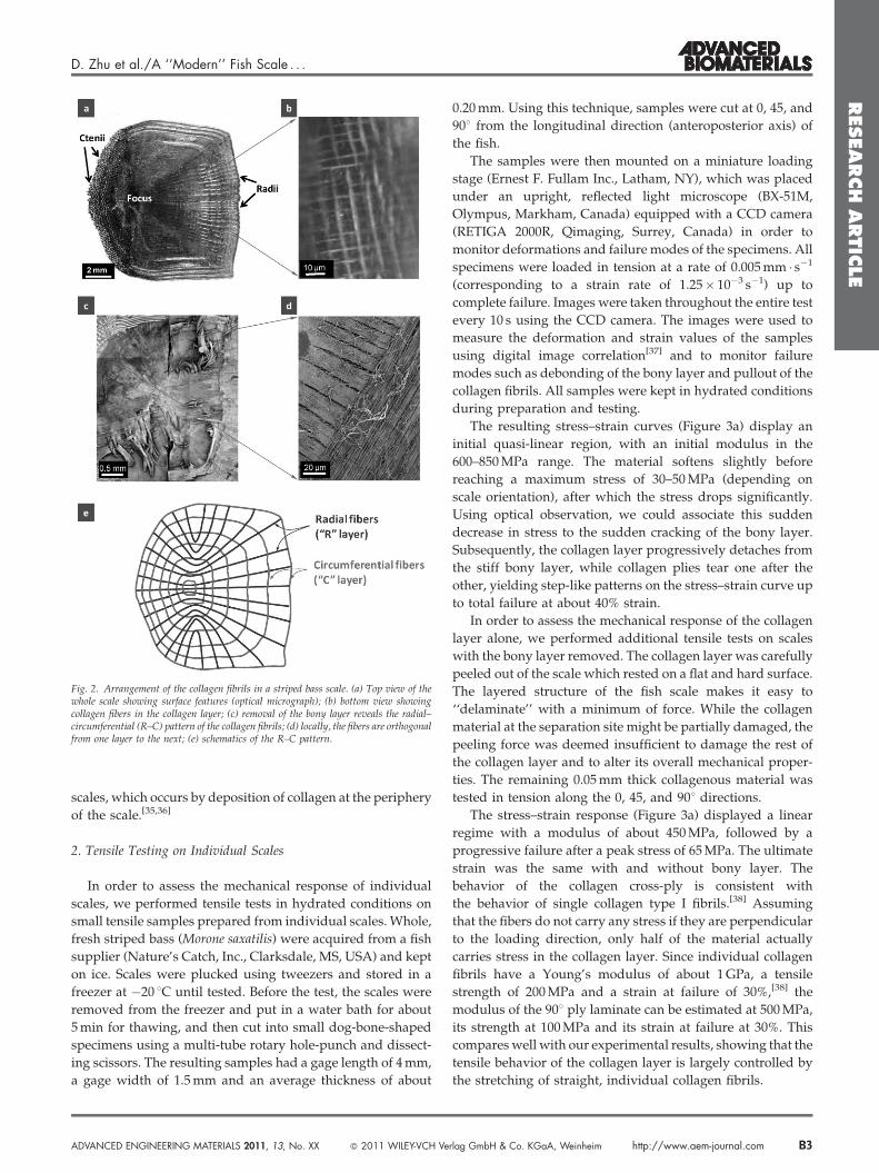
layers being organized around the focus of the scale (Figure 2).
This arrangement is consistent with the growth of individual
. KGaA, Weinheim ADVANCED ENGINEERING MATERIALS 2011, 13, No. XX
RESEARCH
ARTIC
LE
D. Zhu et al./A ‘‘Modern’’ Fish Scale . . .
Fig. 2. Arrangement of the collagen fibrils in a striped bass scale. (a) Top view of thewhole scale showing surface features (optical micrograph); (b) bottom view showingcollagen fibers in the collagen layer; (c) removal of the bony layer reveals the radial–circumferential (R–C) pattern of the collagen fibrils; (d) locally, the fibers are orthogonalfrom one layer to the next; (e) schematics of the R–C pattern.
scales, which occurs by deposition of collagen at the periphery
of the scale.[35,36]
2. Tensile Testing on Individual Scales
In order to assess the mechanical response of individual
scales, we performed tensile tests in hydrated conditions on
small tensile samples prepared from individual scales. Whole,
fresh striped bass (Morone saxatilis) were acquired from a fish
supplier (Nature’s Catch, Inc., Clarksdale, MS, USA) and kept
on ice. Scales were plucked using tweezers and stored in a
freezer at �20 8C until tested. Before the test, the scales were
removed from the freezer and put in a water bath for about
5 min for thawing, and then cut into small dog-bone-shaped
specimens using a multi-tube rotary hole-punch and dissect-
ing scissors. The resulting samples had a gage length of 4 mm,
a gage width of 1.5 mm and an average thickness of about
ADVANCED ENGINEERING MATERIALS 2011, 13, No. XX � 2011 WILEY-VCH Ve
0.20 mm. Using this technique, samples were cut at 0, 45, and
908 from the longitudinal direction (anteroposterior axis) of
the fish.
The samples were then mounted on a miniature loading
stage (Ernest F. Fullam Inc., Latham, NY), which was placed
under an upright, reflected light microscope (BX-51M,
Olympus, Markham, Canada) equipped with a CCD camera
(RETIGA 2000R, Qimaging, Surrey, Canada) in order to
monitor deformations and failure modes of the specimens. All
specimens were loaded in tension at a rate of 0.005 mm � s�1
(corresponding to a strain rate of 1.25� 10�3 s�1) up to
complete failure. Images were taken throughout the entire test
every 10 s using the CCD camera. The images were used to
measure the deformation and strain values of the samples
using digital image correlation[37] and to monitor failure
modes such as debonding of the bony layer and pullout of the
collagen fibrils. All samples were kept in hydrated conditions
during preparation and testing.
The resulting stress–strain curves (Figure 3a) display an
initial quasi-linear region, with an initial modulus in the
600–850 MPa range. The material softens slightly before
reaching a maximum stress of 30–50 MPa (depending on
scale orientation), after which the stress drops significantly.
Using optical observation, we could associate this sudden
decrease in stress to the sudden cracking of the bony layer.
Subsequently, the collagen layer progressively detaches from
the stiff bony layer, while collagen plies tear one after the
other, yielding step-like patterns on the stress–strain curve up
to total failure at about 40% strain.
In order to assess the mechanical response of the collagen
layer alone, we performed additional tensile tests on scales
with the bony layer removed. The collagen layer was carefully
peeled out of the scale which rested on a flat and hard surface.
The layered structure of the fish scale makes it easy to
‘‘delaminate’’ with a minimum of force. While the collagen
material at the separation site might be partially damaged, the
peeling force was deemed insufficient to damage the rest of
the collagen layer and to alter its overall mechanical proper-
ties. The remaining 0.05 mm thick collagenous material was
tested in tension along the 0, 45, and 908 directions.
The stress–strain response (Figure 3a) displayed a linear
regime with a modulus of about 450 MPa, followed by a
progressive failure after a peak stress of 65 MPa. The ultimate
strain was the same with and without bony layer. The
behavior of the collagen cross-ply is consistent with
the behavior of single collagen type I fibrils.[38] Assuming
that the fibers do not carry any stress if they are perpendicular
to the loading direction, only half of the material actually
carries stress in the collagen layer. Since individual collagen
fibrils have a Young’s modulus of about 1 GPa, a tensile
strength of 200 MPa and a strain at failure of 30%,[38] the
modulus of the 908 ply laminate can be estimated at 500 MPa,
its strength at 100 MPa and its strain at failure at 30%. This
compares well with our experimental results, showing that the
tensile behavior of the collagen layer is largely controlled by
the stretching of straight, individual collagen fibrils.
rlag GmbH & Co. KGaA, Weinheim http://www.aem-journal.com B3
RESEARCH
ARTIC
LE
D. Zhu et al./A ‘‘Modern’’ Fish Scale . . .
Fig. 3. (a) Tensile stress–strain curves for fish scales along 0, 45, and 908 from the longitudinal axis of the fish. Summary of results for (b) Young’s modulus; (c) strength. Theerror bars indicate standard deviations.
While it was not possible to isolate the bony layer for
testing, its properties were inferred from the whole scale
and collagen only tensile test results. In the elastic regime, the
whole scale behaves like a two-layer, constant strain
composite. Since the thickness of the bony and
collagen layers is similar, the modulus of the scale is
B4 http://www.aem-journal.com � 2011 WILEY-VCH Verlag GmbH & Co
given by:
ES ¼1
2EC þ EBð Þ (1)
where EC and EB are the Young’s moduli of collagen and bony
layers, respectively. The modulus of bony layer can then be
. KGaA, Weinheim ADVANCED ENGINEERING MATERIALS 2011, 13, No. XX
RESEARCH
ARTIC
LE
D. Zhu et al./A ‘‘Modern’’ Fish Scale . . .
estimated using:
EB ¼ 2ES�EC (2)
The strength of bony layer can be evaluated with a similar
approach. In the linear regime, with the uniform strain
assumption, the stresses in the bony and collagen layers are
proportional to their stiffnesses. From the whole scale test, the
stress sS at which the bony layer fails is known. Just prior to
failure the stress in the bony layer is then given by:
sB ¼EB
ESsS (3)
This model assumes that both materials are in the linear
elastic range up to the failure of the bony layer. In reality
Figure 3a shows that the scale softens slightly when loaded in
tension, probably due to damage accumulation in the bony layer
(collagen behaves linearly over this range of strain). Equation 3
therefore slightly overestimates the actual strength of the bony
layer. We used Equations (2) and (3) to estimate the modulus
and strength of the bony layer. The results show that the bony
layer is about twice as stiff as the collagen layer, with about the
same tensile strength (Figure 3b,c). The bony layer is, however,
more brittle, failing at about 10% strain while the collagen layer
fails at strains in excess of 40%. Interestingly, we also found that
the whole scale displays in-plane anisotropic properties, but
only because of the bony layer; the collagen layer is isotropic in
plane in terms of both modulus and strength. This set of
experiments highlights the main traits of the scale’s compo-
nents: the bony layer is stiff, hard and brittle because of its high
mineral content, while the underlying collagen cross-ply is
softer and more deformable, with larger strains at failure.
3. Resistance to Sharp Penetration: Puncture of IndividualScales
Adult striped bass have a few natural predators, including
various aquatic birds, marine mammals, and potentially large
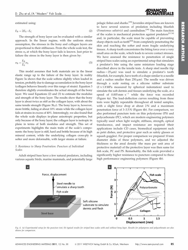
Fig. 4. (a) Experimental setup for the puncture test; (b) typical results for striped bass sshown for comparison.
ADVANCED ENGINEERING MATERIALS 2011, 13, No. XX � 2011 WILEY-VCH Ve
pelagic fishes and sharks.[39] Juveniles striped bass are known
to have several sources of predation including bluefish
(Pomatomus saltatrix) and cannibalism.[39] The main function
of the scales is mechanical protection against predators[3,7,8]
and, in particular, the scale must be capable of preventing
sharp objects such as teeth[40] from completely penetrating the
skin and reaching the softer and more fragile underlying
tissues. A sharp tooth concentrates the biting force over a very
small area on the scale, which leads to severe contact stresses.
We have assessed the resistance to penetration of single
striped bass scales using an experimental setup that simulates
a predator’s bite using the same miniature loading stage
described above in the tensile tests. A sharp steel needle (tip
radius¼ 25 mm) was used to simulate a predator’s tooth
(bluefish, for example, have teeth of a shape similar to a needle
and a radius smaller than 250 mm). The needle was driven
through a scale resting on a silicone rubber substrate
(E� 1.8 MPa measured by spherical indentation) used to
simulate the soft dermis and tissues underlying the scale, at a
speed of 0.005 mm � s�1 while the force was recorded
(Figure 4a). The load-deflection curves resulting from these
tests were highly repeatable throughout all tested samples,
with a slight force drop at about 2 N and a maximum
penetration force of 3–3.5 N (Figure 4b). For comparison, we
also performed puncture tests on thin polystyrene (PS) and
polycarbonate (PC), which are modern engineering polymers
typically used when light weight, stiffness, strength, optical
translucence, and impact resistance are required (their
applications include CD cases, biomedical equipment such
as petri dishes, and protective gear such as safety glasses or
squash goggles). For proper comparison we prepared 10 mm
diameter disks of these polymers, and we adjusted the
thickness so the areal density (the mass per unit area of
protective material) of the protective layer was then same for
fish scale, PC and PS. Remarkably, the fish scale provided a
significantly higher resistance to puncture compared to these
high performance engineering polymers (Figure 4b).
cales with and without bony layer. Results for polystyrene and polycarbonate are also
rlag GmbH & Co. KGaA, Weinheim http://www.aem-journal.com B5

RESEARCH
ARTIC
LE
D. Zhu et al./A ‘‘Modern’’ Fish Scale . . .
Finally, we performed additional puncture tests on scales
with the bony layer removed. The isolated collagen layer was
easily penetrated, providing only half of puncture resistance
in terms of force. The bony layer is therefore an important
component of the system, operating in synergy with the
collagen layer to increase the performance of individual
scales. The fish scale followed a sequence of mechanisms that
was highly repeatable from scale to scale as well as across
locations on a given scale. The penetration curves consist of
three distinct stages that we investigated in detail by imaging
of the puncture site at different points on the penetration curve
(Figure 5). Stage I is the initial linear region, which is
dominated by flexion of the entire scale and by damage and
indentation of the surface of the bony layer. At a force of about
2 N, the force drops slightly, which we associated to the
sudden cracking of the bony layer. Bony and collagen layers
Fig. 5. Detailed sequence of a puncture test. (a) Load–displacement curve showing three distthe last one of the collagen side were obtained by scanning electron microscopy (SEM), w
B6 http://www.aem-journal.com � 2011 WILEY-VCH Verlag GmbH & Co
have the same thickness, but since the bony layer is stiffer, the
neutral plane of the scale lies within the bony layer. As a
result, flexural deformations generate tensile stresses in the
lower side of the bony layer. Once these stresses reach
the tensile strength of the bony layer, cracks initiate at the
collagen/bone interface and rapidly propagate towards
the surface of the bony layer. Interestingly, the patterns of
the flexural cracks always followed a cross pattern, whose
orientation invariably followed the orientation of the local
radii and circuli (Figure 5c) and underlying collagen fibrils.
The microstructure of the bony layer therefore induces the
failure of the bony layer along specific directions. Upon
cracking of the bony layer, four ‘‘flaps’’ of bony material
immediately deflect downwards, generating circumferential
cracks. At this point, the underlying collagen layer, while
remaining intact, detaches from the bony layer over a ring-like
inct stages; (b) associated mechanisms; and (c) imaging. The images of the bony side andhile the first four images of the collagen side were taken with an optical microscope.
. KGaA, Weinheim ADVANCED ENGINEERING MATERIALS 2011, 13, No. XX
RESEARCH
ARTIC
LE
D. Zhu et al./A ‘‘Modern’’ Fish Scale . . .
area observable with an optical microscope (the scale, while
opaque to electrons, is transparent to visible light). The
cracking of the bony layer marks the beginning of stage II,
dominated by further flexion of the scale, opening of the cross
cracks as the four ‘‘flaps’’ of bony material are bent towards
the collagen layer, radial propagation of the cross cracks, and
further delamination between collagen and bony layers.
Eventually the deflection and opening of the flaps are
sufficient to let the needle reach the collagen layer and
completely puncture it (stage III). The initial failure of the
collagen layer indicates the beginning of stage III, and the
sharp drop in force at this point suggests that the failure is
rapid, possibly because the collagen layer is stretched.
Throughout the rest of stage III, the scale is deflected by
the needle, the delamination between collagen and bony
layers propagates more extensively, and the radial cracks
continue to grow.
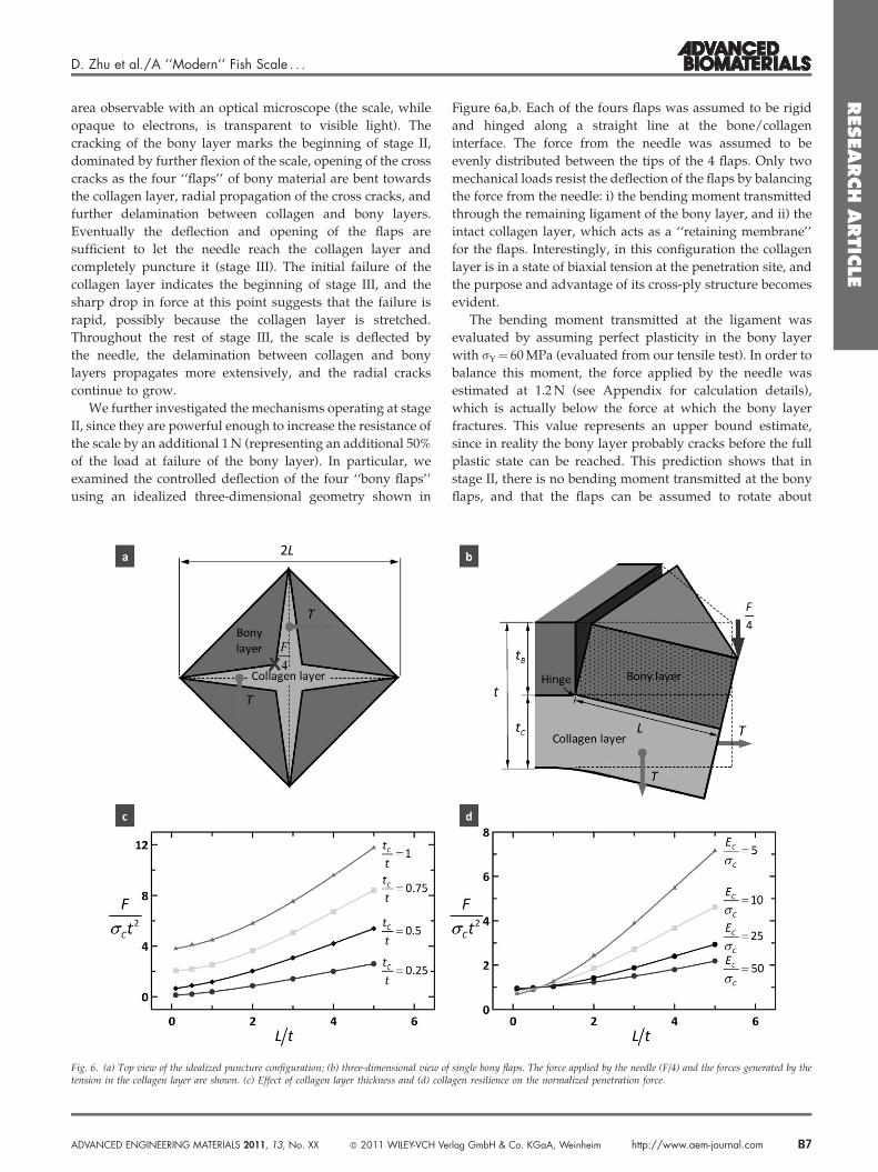
We further investigated the mechanisms operating at stage
II, since they are powerful enough to increase the resistance of
the scale by an additional 1 N (representing an additional 50%
of the load at failure of the bony layer). In particular, we
examined the controlled deflection of the four ‘‘bony flaps’’
using an idealized three-dimensional geometry shown in
Fig. 6. (a) Top view of the idealized puncture configuration; (b) three-dimensional view oftension in the collagen layer are shown. (c) Effect of collagen layer thickness and (d) coll
ADVANCED ENGINEERING MATERIALS 2011, 13, No. XX � 2011 WILEY-VCH Ve
Figure 6a,b. Each of the fours flaps was assumed to be rigid
and hinged along a straight line at the bone/collagen
interface. The force from the needle was assumed to be
evenly distributed between the tips of the 4 flaps. Only two
mechanical loads resist the deflection of the flaps by balancing
the force from the needle: i) the bending moment transmitted
through the remaining ligament of the bony layer, and ii) the
intact collagen layer, which acts as a ‘‘retaining membrane’’
for the flaps. Interestingly, in this configuration the collagen
layer is in a state of biaxial tension at the penetration site, and
the purpose and advantage of its cross-ply structure becomes
evident.
The bending moment transmitted at the ligament was
evaluated by assuming perfect plasticity in the bony layer
with sY¼ 60 MPa (evaluated from our tensile test). In order to
balance this moment, the force applied by the needle was
estimated at 1.2 N (see Appendix for calculation details),
which is actually below the force at which the bony layer
fractures. This value represents an upper bound estimate,
since in reality the bony layer probably cracks before the full
plastic state can be reached. This prediction shows that in
stage II, there is no bending moment transmitted at the bony
flaps, and that the flaps can be assumed to rotate about
single bony flaps. The force applied by the needle (F/4) and the forces generated by theagen resilience on the normalized penetration force.
rlag GmbH & Co. KGaA, Weinheim http://www.aem-journal.com B7
RESEARCH
ARTIC
LE
D. Zhu et al./A ‘‘Modern’’ Fish Scale . . .
frictionless hinges. The experiments show circumferential
cracks in the region of the hinges, confirming that little or no
bending moment can be transmitted though the bony hinge.
The second mechanism examined was associated with the
collagen, which acts as a retaining membrane for the flaps.
In the model, the collagen layer was assumed to have
completely delaminated from the overlying surface of the
bony layer, which is consistent with experimental observa-
tions towards the end of stage II. The collagen layer then acts
as a retaining membrane, with biaxial tension as the dominant
stress. Based on the idealized geometry, the moment balance
of a single bony flap about the hinge led to a simple expression
for the penetration force as a function of bony and collagen
layer thickness (tB, tC), length of the flaps (L), and stiffness (EC)
and strength (sC) of the collagen layer. Full penetration at the
end of stage II was assumed to be reached when the collagen
failed in tension (at a stress of 65 MPa according to our tensile
tests). Our model predicted a penetration force of F¼ 3 N
based on the properties of the collagen layer (EC¼ 500 MPa,
sC¼ 65 MPa) and optical observation (tB¼ tC¼ 100 mm,
L¼ 200 mm). This prediction is remarkably close to penetra-
tion force we measured experimentally, which demonstrates
that the retaining membrane effect dominates stage II and
controls the ultimate penetration resistance of the scale.
In terms of design, the model reveals that longer flaps are
desirable, and since L is larger for larger ‘‘teeth’’ and higher
forces, the scale provides a greater resistance to penetration for
larger teeth and stronger bites (Figure 6c,d). A thick collagen
layer is also beneficial, although a minimum of bony material
is required to form stiff flaps. This finding substantiates
previous discussions on mechanical benefits of thicker
collagen fibrils and plywood organization in the collagen
layer.[8,32,33,41] Finally, a soft and strong material increases
resistance to penetration, although a too soft backing layer
may lead to excessive deflection that may damage the
underlying tissues even before needle penetration. A cross-ply
of collagen is therefore ideal for this function, and the function
of the harder bony layer is to protect the collagen layer from
direct contact with the tip of the needle, and to mitigate the
stresses transmitted onto the softer collagen layer by
redistributing them over a large area.
4. Conclusions
Individual teleost fish scales are therefore high perfor-
mance natural protective systems, offering resistance to
puncture superior to modern engineering polymers typically
used for protective applications. Remarkably, fish scales are
made of a set of materials that are both softer and weaker than
these engineering polymers, which highlights the important
role of the structure and architecture of the scale in
‘‘amplifying’’ the properties (as seen in other class of
biological materials[42–44]). The high performance of the scales
is the result of a fine balance of structure and material
properties, and in particular the hardness and stiffness of the
outer layer, the softness and strength of the inner layer, and an
B8 http://www.aem-journal.com � 2011 WILEY-VCH Verlag GmbH & Co
interface weak enough to delaminate and allow the collagen
layer to stretch under the bony flaps. While this first study on
the puncture mechanics of fish scale does not consider viscous
effects, it is likely that viscoelastic and viscoplastic effects also
contribute to the energy dissipation capability of the scale. The
actual skin of the fish is of course covered with a large number
of overlapping scales and, for striped bass, we counted that
any given point on the surface of the body is covered with 3 or
4 layers of scales. The resulting multilayer system alternates
hard and soft layers in an arrangement reminiscent of the
design of bulletproof glass. In addition, overlapping scales
ensures compliance and breathability, two highly desirable
properties for personal armors. A biomimetic design at the
individual scale level could therefore be combined with a
clever arrangement of the scales at the macroscale to yield a
hierarchical protective system with attractive properties.
Appendix: Analytical ‘‘Four Flaps’’ Model
A simplified analytical model was derived to capture the
progressive deflection of the four flaps and the retaining
membrane effect provided by the underlying collagen layer
(Figure A.1).
A.1. Strain
The strains in the collagen were determined by tracking the
displacement of the point in the centroid of the section of
collagen (point D). Upon deflection of the flap point D moves
to D’, so that its displacement vector can be written, in the
Cartesian system xyz (Figure A.1):
D~D0 ¼
L
2ffiffiffi2p cosu�1ð Þ� tC
2sinu
0
� L
2ffiffiffi2p sinu� tC
2cosu�1ð Þ
���������(A.1)
Any fiber crossing the cracks will extend by a distance
(using scalar product to project D~D0
on the unit vector
collinear to T):
L
2ffiffiffi2p cosu�1ð Þ� tC
2sinu
0
� L
2ffiffiffi2p sinu� tC
2cosu�1ð Þ
266664
377775:� 1ffiffiffi
2p
1ffiffiffi2p
0
2666664
3777775¼ L
41�cosuð Þ þ tC
2ffiffiffi2p sinu
(A.2)
The collagen is detached from the bony layer in the
puncture area, which leads to the assumption that the tensile
strain in the collagen is uniform and equal to:
"C ¼1
21�cosuð Þ þ tC
Lffiffiffi2p sinu (A.3)
Interestingly, the geometry and kinematics for the system
is such that the strain in the collagen layer is equi-biaxial and
uniform in the puncture site. Knowledge of strain in the
collagen layer leads to stress, using the modulus found from
the tensile tests (E¼ 500 MPa).
. KGaA, Weinheim ADVANCED ENGINEERING MATERIALS 2011, 13, No. XX

RESEARCH
ARTIC
LE
D. Zhu et al./A ‘‘Modern’’ Fish Scale . . .
Fig. A.1. Schematic diagram of the deformation and force in the four flaps model.
A.2. Deflection of the Flaps
Similarly, point C at the upper tip of the flaps moves to C’
upon deflection, and
C~C0 ¼
Lffiffiffi2p cosu�1ð Þ þ tBsinu
0
� Lffiffiffi2p sinu þ tB cosu�1ð Þ
���������(A.4)
The deflection at the loading point is then the z component
of C~C0:
d ¼ Lffiffiffi2p sinu�tB cosu�1ð Þ (A.5)
A.3. Force
Under the concentrated force F/4, the rigid bony flap
rotates along the y axis and the loading location changes from
C to C’. The force vectors acting on the bony and collagen
layers are:
~F ¼0
0
�F=4
264
375 ~T ¼
Tffiffiffi2p
Tffiffiffi2p
0
2666666664
3777777775
(A.6)
As the cross product of the vectors O~D0and ~T is equivalent
to half of the cross product of the vectors O~C0
and ~F, then we
ADVANCED ENGINEERING MATERIALS 2011, 13, No. XX � 2011 WILEY-VCH Ve
get the force retained by the collagen layer as follows:
F ¼ 2LtCsC
ffiffiffi2p
tanu þ 2tC
L
1þffiffiffi2p tB
Ltanu
0B@
1CA (A.7)
Normalized by the square of the scale thickness t and the
tensile strength of collagen sC:
F
sCt2¼ 2
L
t
tC
t
ffiffiffi2p
tanu þ 2tC
L
1þffiffiffi2p tB
Ltanu
0B@
1CA (A.8)
A.4. Force Generated by Flexural Stresses at the Hinges
Assuming that the entire thickness of the bony layer
undergoes plasticity, the bending moment transmitted is
calculated as follows:
M ¼ffiffiffi2p
LtB
2
� �2
sB (A.9)
The force applied by the needle is balanced by this
moment:
Lffiffiffi2p F
4¼M (A.10)
Combining Equations (A.9) and (A.10), we get the force
generated by flexural stresses at the hinges:
F ¼ 8tB
2
� �2
sB (A.11)
rlag GmbH & Co. KGaA, Weinheim http://www.aem-journal.com B9
RESEARCH
ARTIC
LE
D. Zhu et al./A ‘‘Modern’’ Fish Scale . . .
Received: June 21, 2011
Final Version: August 10, 2011
[1] L. F. Wang, J. H. Song, C. Ortiz, M. C. Boyce, J. Mater. Res.
2009, 24, 3477.
[2] F. G. Torres, O. P. Troncoso, J. Nakamatsu, C. J. Grande,
C. M. Gomez, Mat. Sci. Eng. C Bio. S 2008, 28, 1276.
[3] B. J. F. Bruet, J. H. Song, M. C. Boyce, C. Ortiz, Nat. Mater.
2008, 7, 748.
[4] T. Ikoma, H. Kobayashi, J. Tanaka, D. Walsh, S. Mann,
Int. J. Biol. Macromol. 2003, 32, 199.
[5] T. Ikoma, H. Kobayashi, J. Tanaka, D. Walsh, S. Mann, J.
Struct. Biol. 2003, 142, 327.
[6] K. V. Kardong, Vertebrates: Comparative Anatomy, Func-
tion, Evolution, McGraw-Hill, New York 2008.
[7] L. Zylberberg, J. Geraudie, F. Meunier, J. Sire, Bone 1992,
4, 171.
[8] J. Bereiter-Hahn, L. Zylberberg, Comp. Biochem. Phys. A
1993, 105, 625.
[9] S. Sudo, K. Tsuyuki, Y. Ito, T. Ikohagi, JSME Int. J. C
Mech. Sy. 2002, 45, 1100.
[10] J. Y. Sire, J. Fish Biol. 1986, 28, 713.
[11] J. D. Currey, J. Exp. Biol. 1999, 202, 3285.
[12] J. H. Long, B. Adcock, R. G. Root, Comp. Biochem. Physiol.
A Mol. Integr. Physiol. 2002, 133, 911.
[13] J. H. Long, M. E. Hale, M. J. McHenry, F M. W. Westneat,
J. Exp. Biol. 1996, 199, 2139.
[14] J. H. Long, T. J. Koob, K. Irving, K. Combie, V. Engel,
N. Livingston, A. Lammert, J. Schumacher, J. Exp. Biol.
2006, 209, 4732.
[15] M. R. Hebrank, J. H. Hebrank, Biol. Bull. 1986, 171, 236.
[16] M. A. Meyers, A. Y. M. Lin, Y. Seki, P. Y. Chen, B. K. Kad,
S. Bodde, JOM J. Miner. Metals Mater. Soc. 2006, 58, 35.
[17] P. Fratzl, R. Weinkamer, Prog. Mater. Sci. 2007, 52, 1263.
[18] L. J. Szewciw, D. G. de Kerckhove, G. W. Grime,
D. S. Fudge, P. Roy. Soc. B Biol. Sci. 2010, 277, 2597.
[19] F. J. Vernerey, F. Barthelat, Int. J. Solids Struct. 2010, 47,
2268.
B10 http://www.aem-journal.com � 2011 WILEY-VCH Verlag GmbH & Co
[20] M. R. Hebrank, Biol. Bull. 1980, 158, 58.
[21] L. A. Jawad, J. Nat. Hist. 2005, 39, 2643.
[22] W. T. Liu, Y. Zhang, G. Y. Li, Y. Q. Miao, X. H. Wu, J. Fish
Biol. 2008, 72, 1055.
[23] A. A. Schonborner, G. Boivin, C. A. Baud, Cell Tissue Res.
1979, 202, 203.
[24] R. V. Seshaiya, P. Ambujabay, M. Kalyani, Amino acid
composition of Icthylepedin from fish scales, in: Aspects
of Protein Structure (Ed: G. N. Ramachandran), Academic
Press, New York p. 343.
[25] F. J. Meunier, Am. Zool. 1984, 24, 953.
[26] A. Bigi, M. Burghammer, R. Falconi, M. H. J. Koch,
S. Panzavolta, C. Riekel, J. Struct. Biol. 2001, 136, 137.
[27] L. Zylberberg, J. Bereiterhahn, J. Y. Sire, Cell Tissue Res.
1988, 253, 597.
[28] F. J. Meunier, J. Castanet, Zool. Scr. 1982, 11, 141.
[29] J. Meunier, Tissue Cell 1981, 13, 165.
[30] T. M. Scheyer, P. M. Sander, W. G. Joyce, W. Boehme,
U. Witzel, Org. Divers Evol. 2007, 7, 136.
[31] E. C. Bass, F. A. Ashford, M. R. Segal, J. C. Lotz, Ann.
Biomed. Eng. 2004, 32, 1231.
[32] L. Zylberberg, J. Bonaventure, L. Cohensolal,
D. J. Hartmann, J. Bereiterhahn, J. Cell Sci. 1992, 103,
273.
[33] H. S. Youn, T. J. Shin, J. Struct. Biol. 2009, 168, 332.
[34] F. J. Meunier, P. M. Brito, Cybium 2004, 28, 225.
[35] C. Liu, S. Shen, J. Taiwan Museum 1991, 44, 321.
[36] G. K. Ostrander, The Laboratory Fish, Academic Press,
Oxford 2000.
[37] F. Barthelat, J. Poissant, Exp. Mech. 2010, 50, 353.
[38] Z. L. Shen, M. R. Dodge, H. Kahn, R. Ballarini,
S. J. Eppell, Biophys. J. 2008, 95, 3956.
[39] K. J. Hartman, Proc. Atlantic Striped Bass Workshop and
Roundtable Discussion, 1999, p. 6.
[40] W. E. Bemis, A. Giuliano, B. McGuire, Zoology 2005, 108,
317.
[41] H. Onozato, N. Watabe, Cell Tissue Res. 1979, 201, 409.
[42] F. Barthelat, R. Rabiei, J. Mech. Phys. Solids 2011, 59, 829.
[43] M. J. Buehler, Nano Today 2010, 5, 379.
[44] S. Keten, Z. P. Xu, B. Ihle, M. J. Buehler, Nat. Mater. 2010,
9, 359.
. KGaA, Weinheim ADVANCED ENGINEERING MATERIALS 2011, 13, No. XX