Structure and Function of the Hip - Mercer County ...behrensb/documents/Week10A-FINAL.pdfStructure...
35
Structure and Function of the Hip
Transcript of Structure and Function of the Hip - Mercer County ...behrensb/documents/Week10A-FINAL.pdfStructure...

Structure and Function
of the Hip

Objectives
• Identify the supporting structures of the hip joint
• Discuss the actions of the hip musculature
through understanding of the origin and insertion
• Identify the force couples involved with an
anterior and posterior pelvic tilt
• Explain the function of the hip abductor muscles
while walking
• Identify the one joint and two joint muscles of the
hip joint

Ligaments in the Pelvis
• Iliofemoral (Y), Ischiofemoral, pubofemoral

Relevance of Anterior Hip
Ligaments
•Where was the plumb
line to describe optimal
posture? Ant or post to
hip joint

The Ilioinguinal Ligament
• No function at hip joint; Separates abd wall and thigh.
One border of the femoral triangle
The Iliotibial Band (ITB)
•A very long, tendinous
portion of the tensor
fascia latae muscle
•Both the gluteus
maximus and tensor
fascia latae attach to it

The Hip Joint
• Three degrees of Freedom– Flexion/Extension (Sagittal plane)
• Medial/Lateral axis
– Anterior to axis will ____ the hip
– Posterior to axis will ____ the hip
– Abduction/Adduction (Frontal plane)• Anterior/Posterior axis
– Medial to axis will ______ the hip
– Lateral to axis will ______ the hip
– Internal/External Rotation (Transverse plane)• Vertical axis
– Anterior and/or medial will _____ the hip
– Posterior and/or lateral will _____ the hip

Which line of pull will flex/ext, and
which will int/ext rotate?
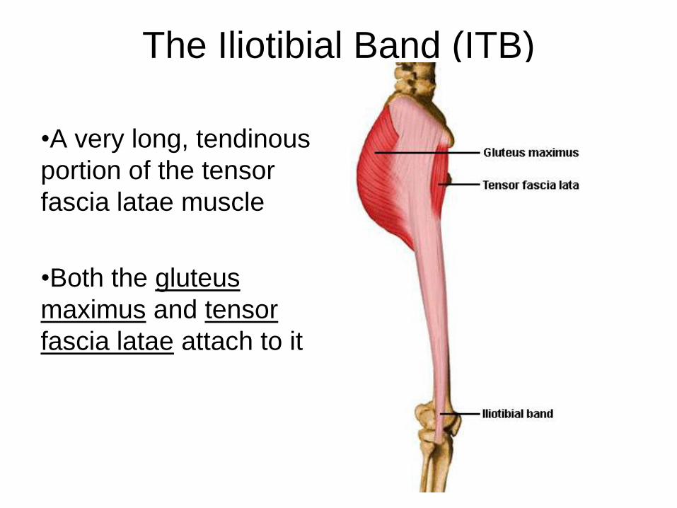
Innervation of Hip
• Femoral nerve innervates most of the hip flexors
• Obturator nerve innervates primarily the hip adductors
• Sciatic nerve (tibial portion): hamstrings and extensor head of adductor magnus
• Gluteal nerve (superior and inferior) will innervate the rest
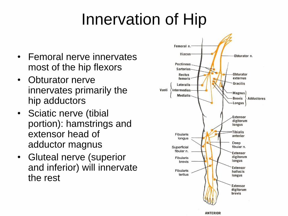
Iliopsoas
• Origin:
– Iliacus:
• Iliac fossa
– Psoas Major:
• transverse processes of T12-
L5
• Insertion:
– Lesser trochanter
• Action:
– Hip flexion
– Trunk flexion
– Anterior pelvic tilt
• Innervation: femoral nerve

Rectus Femoris
• Origin:
– Anterior inferior iliac spine
• Insertion:
– Tibial tuberosity via the
quadricep tendon
• Action:
– Hip flexion
– Knee extension
• Innervation: femoral nerve
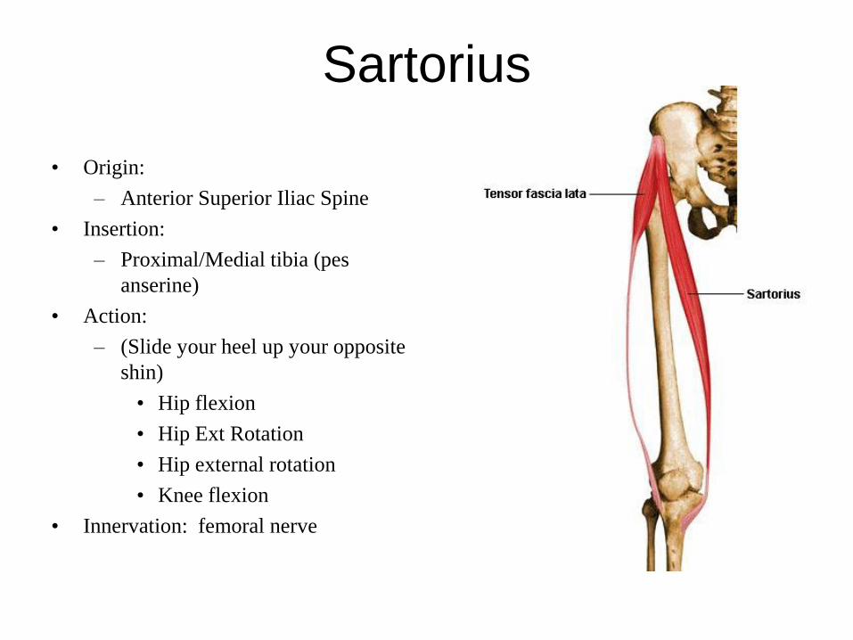
Sartorius
• Origin:
– Anterior Superior Iliac Spine
• Insertion:
– Proximal/Medial tibia (pes
anserine)
• Action:
– (Slide your heel up your opposite
shin)
• Hip flexion
• Hip Ext Rotation
• Hip external rotation
• Knee flexion
• Innervation: femoral nerve

Tensor Fascia Latae
• Origin:
– Outer surface of the iliac crest
posterior to the ASIS
• Insertion:
– Proximal 1/3 of the ITB
• Action:
– Hip flexion
– Hip ABDuction
– Hip Internal rotation
• Innervation: Superior gluteal n.

Primary Hip Flexors
• ILiopsoas
• Rectus Femoris
• Sartorius
• Tensor Fascia Latae

Biceps Femoris
(Part of the hamstrings)
• Origin:
– Ischial Tuberosity
• Insertion:
– Head of the fibula
• Action:
– Hip extension
– Knee flexion
• Innervation: sciatic nerve (tibial
portion)

Semimembranosus
(Part of the hamstrings)• Origin:
– Ischial tuberosity
• Insertion:
– Medial condyle of the tibia
(posterior)
• Action:
– Hip extension
– Knee flexion
• Innervation: sciatic nerve (tibial
portion)

Semitendinosus
(Part of the hamstrings)• Origin:
– Ischial tuberosity
• Insertion:
– Prox/medial surface of the tibia
(pes anserine)
• Action:
– Hip extension
– Knee flexion
• Innervation: sciatic nerve (tibial
portion)


Gluteus Maximus
• Origin:
– Posterior Ilium
– Sacrum and Coccyx
• Insertion:
– ITB
– Gluteal Tuberosity of the femur
• Action:
– Hip extension
– Hip external rotation
• Innervation: Superior gluteal nerve

Primary Hip Extensors
• Gluteus Maximus
• Hamstrings
– Semitendinosus
– Semimembranosus
– Biceps Femoris
• Adductor Magnus
(extensor head)

Gluteus Medius
• Origin:
– Outer surface of the ilium
• Insertion:
– Greater trochanter of the femur
• Action:
– Hip ABDuction
• Innervation:
– Superior gluteal nerve

Gluteus Minimus
• Origin:
– Outer surface of the ilium,
inferior to the gluteus medius
• Insertion:
– Greater trochanter
• Action:
– Hip ABDuction
– Hip Int rotation
• Innervation: Superior gluteal nerve

Trendelenburg Sign
• During single leg stance of
walking, the hip abductor muscles
need to work to keep pelvis level;
– If weak, u see the pelvis tilt
inferiorly on the non-affected
side.
http://www.youtube.com/watch?v=IhsP
0YhDUu8

Primary Hip Abductors
• Gluteus Medius
• Gluteus Minimus
• TFL

Pectinius
• Origin:
– pectineal line on the superior
pubic ramus
• Insertion:
– pectineal line on the posterior
femur
• Action:
– Hip ADDuct
– Hip flexion
• Innervation: Obturator nerve

Adductor Magnus
• Extensor Head
• Origin:
– Ischial tuberosity
• Insertion:
– Adductor tubercle (distal femur)
• Action:
– Hip ADDuct, hip extension
• Innervation: sciatic n. (tibial portion)
• Adductor Head
• Origin:
– Ischial ramus
• Insertion:
– Entire linea aspera of femur
• Action:
– Hip ADDuct, hip flexion
• Innervation: Obturator n.

Adductor Longus
• Origin:
– Anterior surface of the body of
the pubis
• Insertion:
– Middle 1/3 of the linea aspera
of the femur
• Action:
– Hip Adduct
– Hip flexion
• Innervation: Obturator n.

Adductor Brevis
• Origin:
– Inferior pubic ramus
• Insertion:
– Prox 1/3 of linea aspera
• Action:
– Hip ADDuct
– Hip flexion
• Innervation: Obturator n.

Gracilis
• Origin:
– Inferior ramus and body of pubis
• Insertion:
– Prox/medial aspect of tibia (pes
anserine)
• Action:
– Hip ADDuct
– Hip flexion
– Knee flexion
• Innervation: Obturator n.

Primary Hip Adductors
• Pectinius
• Adductor Magnus -
both
• Adductor Longus
• Adductor Brevis
• Gracilis

Primary Hip External Rotators
•Intrinsic Hip ER: (6
muscles)– Piriformis
– Obturator Internus
– Obturator Exterunus
– Gemelus Superior
– Gemelus Inferior
– Quadratus Femoris
•Piriformis Syndrome:– The sciatic nerve passes deep
to the piriformis in most cases
(approximately 85% of people) but
can in fact pierce the piriformis
itself, predisposing to piriformis
syndrome and subsequent
sciatica.
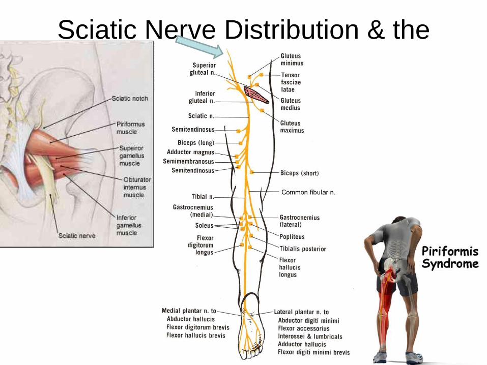
Sciatic Nerve Distribution & the
Piriformis

Primary Hip Internal Rotators
• Gluteus Medius
• Gluteus Minimus
• Tensor Fasica Latae

Pelvic Tilting – Force Couple

Optional Project
Up to 5 points on next exam
• One page paper about why you should
hold and use a cane in the opposite hand
of the injured or weak leg