Structure and dynamics of condensed multivalent ions ...
14
Structure and dynamics of condensed multivalent ions within polyelectrolyte bundles: a combined x-ray diffraction and solid-state NMR study This article has been downloaded from IOPscience. Please scroll down to see the full text article. 2005 J. Phys.: Condens. Matter 17 S1123 (http://iopscience.iop.org/0953-8984/17/14/001) Download details: IP Address: 164.67.190.90 The article was downloaded on 02/11/2010 at 00:31 Please note that terms and conditions apply. View the table of contents for this issue, or go to the journal homepage for more Home Search Collections Journals About Contact us My IOPscience
Transcript of Structure and dynamics of condensed multivalent ions ...

Structure and dynamics of condensed multivalent ions within polyelectrolyte bundles: a
combined x-ray diffraction and solid-state NMR study
This article has been downloaded from IOPscience. Please scroll down to see the full text article.
2005 J. Phys.: Condens. Matter 17 S1123
(http://iopscience.iop.org/0953-8984/17/14/001)
Download details:
IP Address: 164.67.190.90
The article was downloaded on 02/11/2010 at 00:31
Please note that terms and conditions apply.
View the table of contents for this issue, or go to the journal homepage for more
Home Search Collections Journals About Contact us My IOPscience
INSTITUTE OF PHYSICS PUBLISHING JOURNAL OF PHYSICS: CONDENSED MATTER
J. Phys.: Condens. Matter 17 (2005) S1123–S1135 doi:10.1088/0953-8984/17/14/001
Structure and dynamics of condensed multivalent ionswithin polyelectrolyte bundles: a combined x-raydiffraction and solid-state NMR study
Thomas E Angelini1, Lori K Sanders2, Hongjun Liang2, Willy Wriggers3,Jay X Tang4 and Gerard C L Wong1,2
1 Department of Physics, University of Illinois at Urbana-Champaign, IL 61801, USA2 Department of Materials Science and Engineering, University of Illinois at Urbana-Champaign,IL 61801, USA3 School of Health Information Sciences, University of Texas Health Science Center at Houston,7000 Fannin Street Suite 600, Houston, TX 77030, USA4 Department of Physics, Brown University, Providence, RI 02912, USA
E-mail: [email protected]
Received 25 August 2004, in final form 25 November 2004Published 24 March 2005Online at stacks.iop.org/JPhysCM/17/S1123
AbstractLike-charged polyelectrolytes can attract and condense into compact orderedstates via counterion-mediated interactions (Gelbart et al 2000 Phys. Today 5338–44). Recent examples include DNA toroids and F-actin bundles. We haveinvestigated the structure and dynamics of condensed divalent ions withinF-actin polyelectrolyte bundles. Using synchrotron x-ray diffraction, thestructural organization of Ba2+ ions on F-actin has been directly observed.The Ba2+ ions organize into counterion charge density waves (CDWs) parallelto the actin filaments. Moreover, these 1D counterion charge density wavescouple to twist deformations of the oppositely charged actin filaments, andmediate attractions by effecting a ‘zipper-like’ charge alignment between thecounterions and the polyelectrolyte charge distribution. We have also examinedcondensed divalent 25Mg ions within F-actin bundles using solid-state NMR.Preliminary measurements indicate that the longitudinal relaxation time T1 ofMg2+ ions decreases by approximately an order of magnitude as they organizeinto the CDW state within condensed F-actin bundles. The measured value ofT1 for Mg2+ ions in the CDW is intermediate between typical liquid-like andsolid-like values.
(Some figures in this article are in colour only in the electronic version)
1. Introduction
Electrostatics in aqueous media is often understood in terms of mean field theories such as thePoisson–Boltzmann formalism [2–4], in which like-charged objects such as polyelectrolytes
0953-8984/05/141123+13$30.00 © 2005 IOP Publishing Ltd Printed in the UK S1123
S1124 T E Angelini et al
always repel. Indeed, DNA chains in water containing monovalent ions always repel oneanother. Linearized versions of mean field theory provide the theoretical underpinning forwidely employed tools such as Debye–Huckel theory and DLVO (Derjaguin–Landau–Verwey–Overbeek) theory [5, 6], which constitute the usual starting point for understanding chargedpolyelectrolyte or colloidal systems.
In systems with strong electrostatic interactions (containing, for example, high surfacecharge densities, multivalent ions, etc), however, the physics is qualitatively different.Interactions between polyelectrolytes can be controlled by the structure and dynamics of thecondensed ions surrounding the polyelectrolyte. It has been recognized for some time thatthe mean field Poisson–Boltzmann approach cannot produce attractions unless some formof correlation between counterions is introduced [7–9]. As Kirkwood [8] suggested andOosawa [9] showed using an approximate analytical model, like-charged attractions betweenpolyelectrolytes may be possible due to the correlated fluctuations of the condensed ion layersaround strongly charged polyelectrolytes. A large number of theoretical investigations havefocused on the existence and form of multivalent ion induced like-charge attraction betweencylindrical polyelectrolytes [10, 11].
Recently, a number of anionic biopolymers (with persistence lengths of 1 µm or more)have been used as experimental systems for the study of like-charge polyelectrolyte attraction,such as the filamentous bacteriophages, microtubules, and F-actin [12, 13]. Due to their largepersistence lengths, they can be thought of as idealized rod-like polyelectrolytes, and a numberof interesting new effects have been found. For example, in the presence of divalent ions, F-actin progressively condenses into close-packed bundles via an intermediate state comprisedof 1D lamellar stacks of liquid crystalline actin networks [14–16]. The dependence of DNAand actin condensation on ion valence, size, and structure has been systematically studiedexperimentally [12, 17] and an empirically motivated criterion for the valence dependence hasbeen proposed based on experimental results for ‘dumbbell’ divalent ions [18].
Polyelectrolytes in aqueous solution are coated by a condensed ‘Manning’ layer of mobileoppositely charged counterions [19]. All proposed theoretical explanations for like-chargeattraction introduce some form of positional counterion correlations within this layer. Forexample, dynamic ‘van der Waals’-like correlations of long-wavelength ion fluctuations havebeen suggested [8, 9, 20–23]. Static mechanisms consisting of ion correlations along the axisof the polyelectrolyte rods in the form of a Wigner lattice have also been considered [24–27]. Recently, theoretical models based on discretized distributions of condensed ions onpolyelectrolytes have been solved analytically and numerically [28–30].
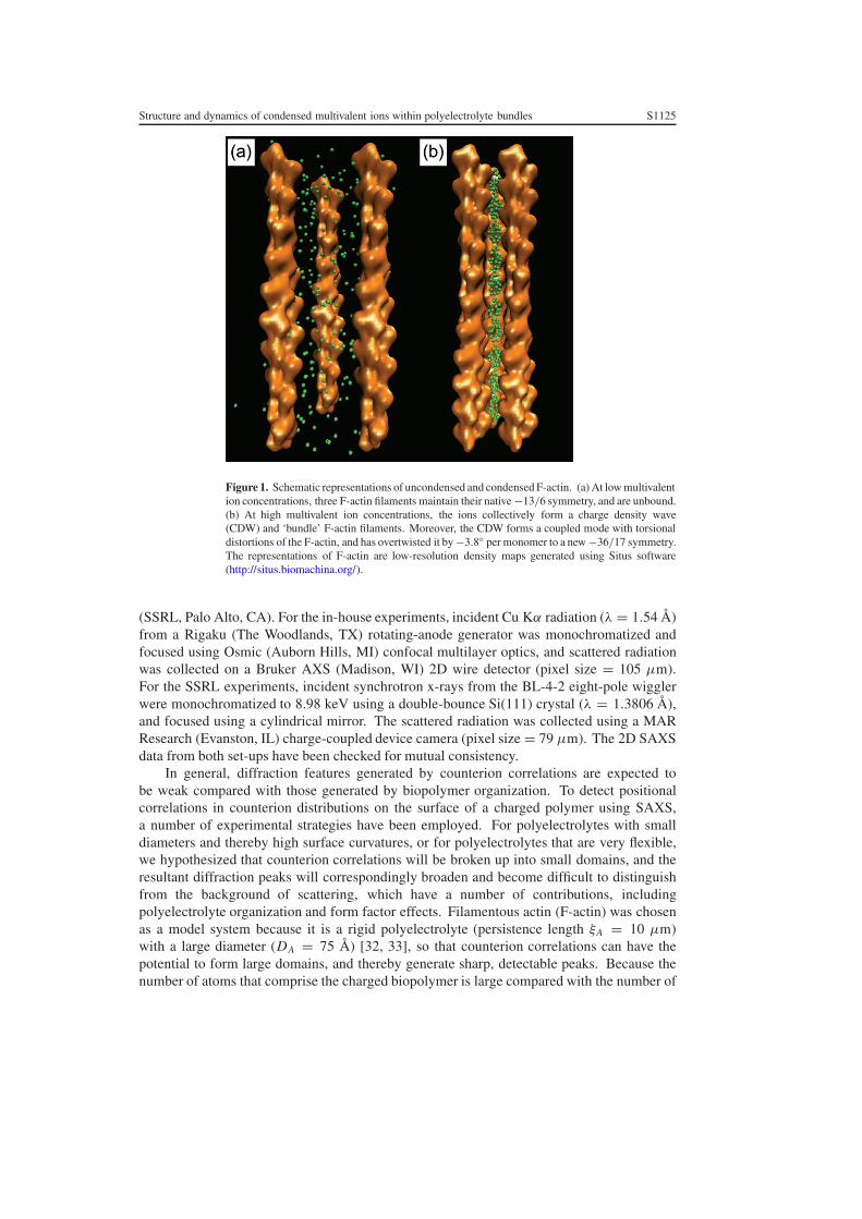
We have investigated the structure and dynamics of condensed divalent ions within F-actin polyelectrolyte bundles. The organization of multivalent ions on actin filaments hasbeen directly measured using synchrotron x-ray diffraction [31]. The counterions do notform a lattice that simply follows actin’s helical symmetry; rather, they organize into one-dimensional (1D) charge density waves parallel to the actin filaments (figure 1). Moreover,this 1D counterion charge density wave forms a coupled mode with torsional distortions of theoppositely charged polyelectrolyte. We have also performed solid-state NMR experiments oncondensed divalent ions within F-actin bundles. Preliminary measurements of the longitudinalrelaxation time T1 show that Mg2+ ions condensed in F-actin bundles are much less mobilethat those in solution.
2. Experimental details
Small angle x-ray scattering (SAXS) measurements were performed using both an in-housex-ray source as well as beam line 4-2 at the Stanford Synchrotron Radiation Laboratory
Structure and dynamics of condensed multivalent ions within polyelectrolyte bundles S1125
Figure 1. Schematic representations of uncondensed and condensed F-actin. (a) At low multivalention concentrations, three F-actin filaments maintain their native −13/6 symmetry, and are unbound.(b) At high multivalent ion concentrations, the ions collectively form a charge density wave(CDW) and ‘bundle’ F-actin filaments. Moreover, the CDW forms a coupled mode with torsionaldistortions of the F-actin, and has overtwisted it by −3.8◦ per monomer to a new −36/17 symmetry.The representations of F-actin are low-resolution density maps generated using Situs software(http://situs.biomachina.org/).
(SSRL, Palo Alto, CA). For the in-house experiments, incident Cu Kα radiation (λ = 1.54 Å)from a Rigaku (The Woodlands, TX) rotating-anode generator was monochromatized andfocused using Osmic (Auborn Hills, MI) confocal multilayer optics, and scattered radiationwas collected on a Bruker AXS (Madison, WI) 2D wire detector (pixel size = 105 µm).For the SSRL experiments, incident synchrotron x-rays from the BL-4-2 eight-pole wigglerwere monochromatized to 8.98 keV using a double-bounce Si(111) crystal (λ = 1.3806 Å),and focused using a cylindrical mirror. The scattered radiation was collected using a MARResearch (Evanston, IL) charge-coupled device camera (pixel size = 79 µm). The 2D SAXSdata from both set-ups have been checked for mutual consistency.
In general, diffraction features generated by counterion correlations are expected tobe weak compared with those generated by biopolymer organization. To detect positionalcorrelations in counterion distributions on the surface of a charged polymer using SAXS,a number of experimental strategies have been employed. For polyelectrolytes with smalldiameters and thereby high surface curvatures, or for polyelectrolytes that are very flexible,we hypothesized that counterion correlations will be broken up into small domains, and theresultant diffraction peaks will correspondingly broaden and become difficult to distinguishfrom the background of scattering, which have a number of contributions, includingpolyelectrolyte organization and form factor effects. Filamentous actin (F-actin) was chosenas a model system because it is a rigid polyelectrolyte (persistence length ξA = 10 µm)with a large diameter (DA = 75 Å) [32, 33], so that counterion correlations can have thepotential to form large domains, and thereby generate sharp, detectable peaks. Because thenumber of atoms that comprise the charged biopolymer is large compared with the number of
S1126 T E Angelini et al
counterions, high molecular weight counterions (Ba2+) were used to improve the Z-contrastbetween counterion scattering and biopolymer scattering. Finally, to further highlight thediffraction features that are generated by the counterions, we compared the SAXS patternsfrom F-actin that has been concentrated non-electrostatically (with no added multivalent ions)with F-actin that has been condensed electrostatically by using multivalent ions.
In order to do this experiment, it was necessary to obtain at least partially aligned samples,so that correlations along different directions can be discerned. The best alignment of the F-actin biopolymer is usually achieved in samples with monovalent ion based buffers such asthose typically employed in typical structural biology experiments, and in non-electrostaticallyconcentrated samples, such as those used in our control experiments. Unlike those samples,however, multivalent ion condensed biopolymer samples typically exhibit poor alignmentbecause of the existence of ion mediated strong attractions that cause the precipitation of theF-actin. Typically, the precipitated F-actin is compacted into a dense pellet during mixing.These pellets consist of many coexisting domains of actin bundles locally oriented alongdifferent random directions, which leads to ‘powder averaging’ of the diffraction pattern andthe associated loss of orientational information. In order to minimize these effects, we use asmall (300 × 300 µm2) x-ray beam to get diffraction information on locally aligned domainswithin a pellet.
The most abundant intracellular protein in eukaryotic cells is actin, one of the principalcomponents of the cytoskeleton. Actin associates to form polymeric actin filaments, whichcan in turn form bundles and networks in a highly coordinated way by interacting with actinbinding proteins and salts. The actin cytoskeleton determines cell shape, and plays a centralrole in cellular locomotion [32]. Monomeric actin (G-actin) (MW 43 000) was prepared froma lyophilized powder of rabbit skeletal muscle purchased from Cytoskeleton, Inc. (Denver,CO). The non-polymerizing G-actin solution contained a 5 mM Tris buffer at pH 8.0, with0.2 mM CaCl2, 0.5 mM ATP, 0.2 mM DTT, and 0.01% NaN3. G-actin (2 mg ml−1) waspolymerized into F-actin (linear charge density λA ≈ −1e/2.5 Å at pH 7) with the additionof salt (100 mM KCl final concentration). F-actin rods are comprised of G-actin monomerspacked into a helix with a −13/6 symmetry, or 13 monomers in six left handed helical turns.Each G-actin monomer has dimensions of approximately 55 Å×55 Å×35 Å, and is comprisedof four subdomains. For the purpose of modelling x-ray diffraction from actin filaments, thesefour subdomains can be approximated as four spheres with appropriate radii and molecularweights [34]. Human plasma gelsolin, an actin severing and capping protein (Cytoskeleton,Inc.), was used to regulate the F-actin length. The average length of F-actin is controlledby the gelsolin concentration, which has been independently calibrated [35]. The filamentswere treated with phalloidin (MW 789.2; Sigma Aldrich, St. Louis, MO) to prevent F-actindepolymerization. F-actin gels were ultracentrifuged at 100000 g for 1 h to pellet the filaments.After the removal of the supernatant buffer solution, the F-actin was resuspended to a finalconcentration of 10 mg ml−1 by using Millipore H2O (18.2 M�). Electrostatically condensedsamples were prepared by mixing with MgCl2 or BaCl2 salt solutions. Non-electrostaticallyconcentrated samples were prepared by ultracentrifugation (100 000 × g) of F-actin solutions.The supernatant buffer was subsequently removed, and the actin concentration set by theaddition of 5 mM pH 7.0 PIPES buffer. All samples were sealed in 1.5 mm diameter quartzcapillaries.
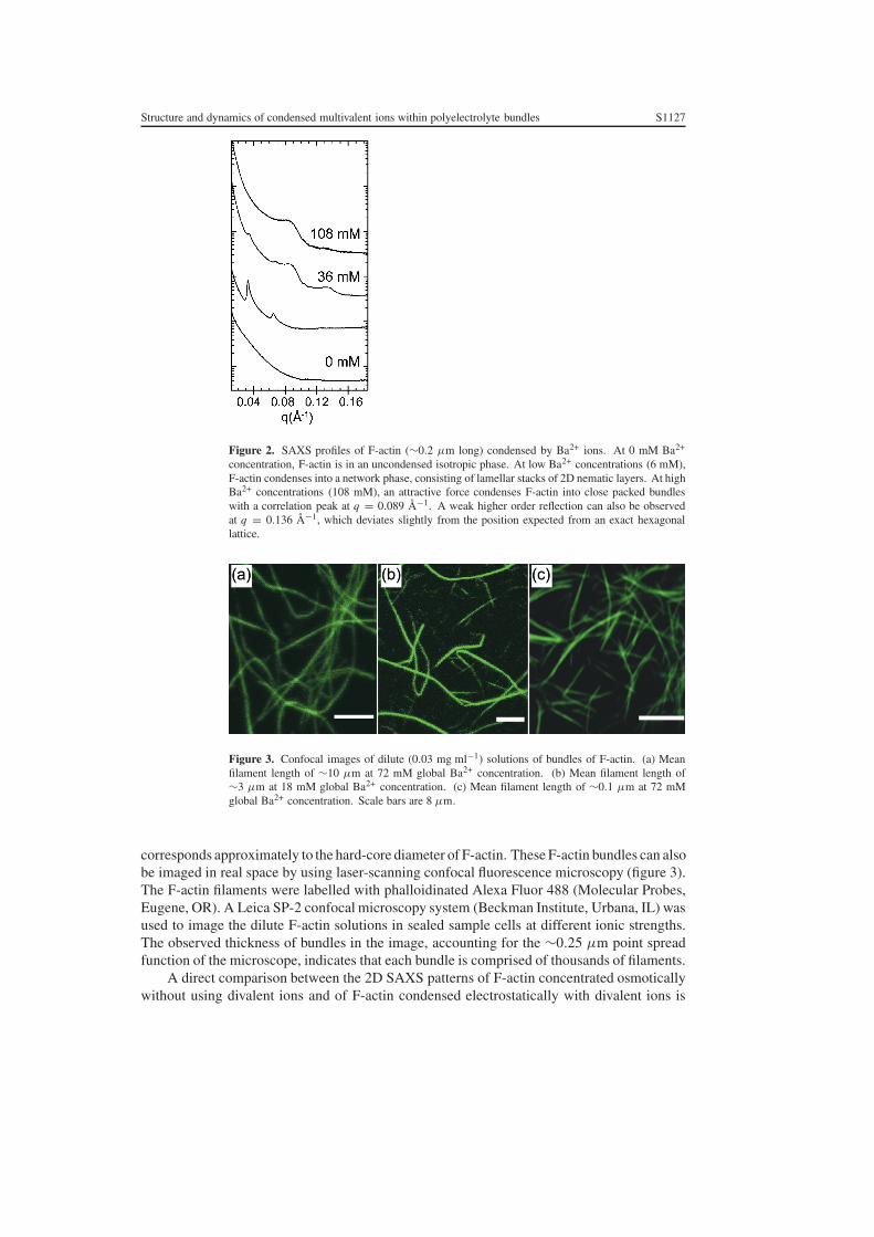
The progressive condensation of F-actin bundles with increasing multivalent ionconcentrations can be observed in figure 2. F-actin condenses into intermediate state comprisedof 1D lamellar stacks of liquid crystalline actin networks before condensing into close-packedbundles [14–16]. SAXS measurements of F-actin bundles condensed with Ba2+ exhibit anactin–actin close-packing peak characteristic of the bundled phase at q = 0.089 Å−1, which
Structure and dynamics of condensed multivalent ions within polyelectrolyte bundles S1127
Figure 2. SAXS profiles of F-actin (∼0.2 µm long) condensed by Ba2+ ions. At 0 mM Ba2+
concentration, F-actin is in an uncondensed isotropic phase. At low Ba2+ concentrations (6 mM),F-actin condenses into a network phase, consisting of lamellar stacks of 2D nematic layers. At highBa2+ concentrations (108 mM), an attractive force condenses F-actin into close packed bundleswith a correlation peak at q = 0.089 Å−1. A weak higher order reflection can also be observedat q = 0.136 Å−1, which deviates slightly from the position expected from an exact hexagonallattice.
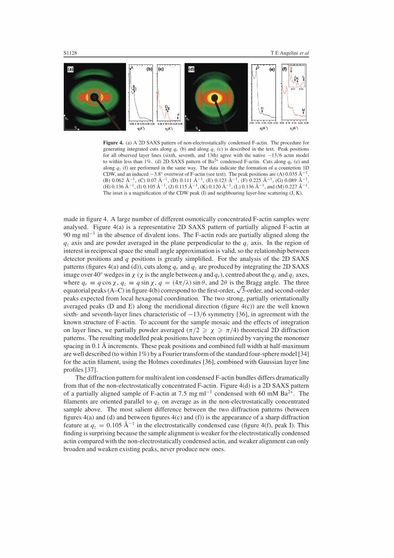
Figure 3. Confocal images of dilute (0.03 mg ml−1) solutions of bundles of F-actin. (a) Meanfilament length of ∼10 µm at 72 mM global Ba2+ concentration. (b) Mean filament length of∼3 µm at 18 mM global Ba2+ concentration. (c) Mean filament length of ∼0.1 µm at 72 mMglobal Ba2+ concentration. Scale bars are 8 µm.
corresponds approximately to the hard-core diameter of F-actin. These F-actin bundles can alsobe imaged in real space by using laser-scanning confocal fluorescence microscopy (figure 3).The F-actin filaments were labelled with phalloidinated Alexa Fluor 488 (Molecular Probes,Eugene, OR). A Leica SP-2 confocal microscopy system (Beckman Institute, Urbana, IL) wasused to image the dilute F-actin solutions in sealed sample cells at different ionic strengths.The observed thickness of bundles in the image, accounting for the ∼0.25 µm point spreadfunction of the microscope, indicates that each bundle is comprised of thousands of filaments.
A direct comparison between the 2D SAXS patterns of F-actin concentrated osmoticallywithout using divalent ions and of F-actin condensed electrostatically with divalent ions is
S1128 T E Angelini et al
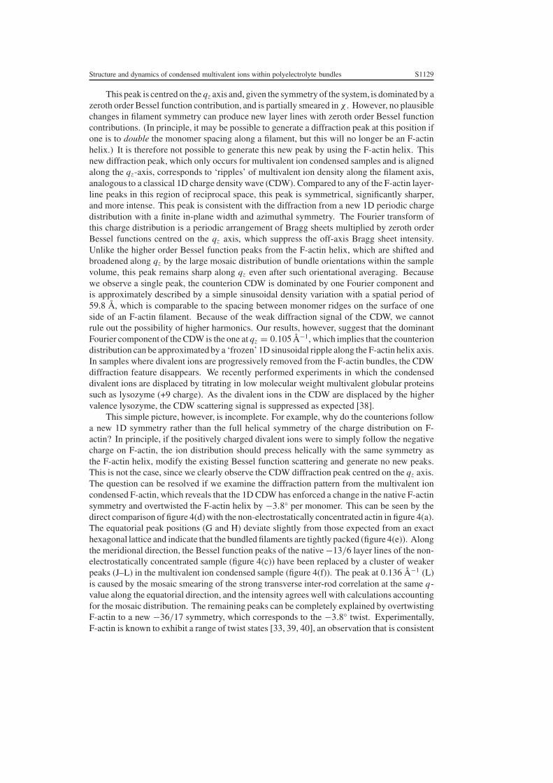
Figure 4. (a) A 2D SAXS pattern of non-electrostatically condensed F-actin. The procedure forgenerating integrated cuts along qr (b) and along qz (c) is described in the text. Peak positionsfor all observed layer lines (sixth, seventh, and 13th) agree with the native −13/6 actin modelto within less than 1%. (d) 2D SAXS pattern of Ba2+ condensed F-actin. Cuts along qr (e) andalong qz (f) are performed in the same way. The data indicate the formation of a counterion 1DCDW, and an induced −3.8◦ overtwist of F-actin (see text). The peak positions are (A) 0.035 Å−1,(B) 0.062 Å−1, (C) 0.07 Å−1, (D) 0.111 Å−1, (E) 0.123 Å−1, (F) 0.225 Å−1, (G) 0.089 Å−1,(H) 0.136 Å−1, (I) 0.105 Å−1, (J) 0.115 Å−1, (K) 0.120 Å−1, (L) 0.136 Å−1, and (M) 0.227 Å−1.The inset is a magnification of the CDW peak (I) and neighbouring layer-line scattering (J, K).
made in figure 4. A large number of different osmotically concentrated F-actin samples wereanalysed. Figure 4(a) is a representative 2D SAXS pattern of partially aligned F-actin at90 mg ml−1 in the absence of divalent ions. The F-actin rods are partially aligned along theqz axis and are powder averaged in the plane perpendicular to the qz axis. In the region ofinterest in reciprocal space the small angle approximation is valid, so the relationship betweendetector positions and q positions is greatly simplified. For the analysis of the 2D SAXSpatterns (figures 4(a) and (d)), cuts along qr and qz are produced by integrating the 2D SAXSimage over 40◦ wedges in χ (χ is the angle between q and qr ), centred about the qr and qz axes,where qr ≡ q cos χ , qz ≡ q sin χ , q = (4π/λ) sin θ , and 2θ is the Bragg angle. The threeequatorial peaks (A–C) in figure 4(b) correspond to the first-order,
√3-order, and second-order
peaks expected from local hexagonal coordination. The two strong, partially orientationallyaveraged peaks (D and E) along the meridional direction (figure 4(c)) are the well knownsixth- and seventh-layer lines characteristic of −13/6 symmetry [36], in agreement with theknown structure of F-actin. To account for the sample mosaic and the effects of integrationon layer lines, we partially powder averaged (π/2 � χ � π/4) theoretical 2D diffractionpatterns. The resulting modelled peak positions have been optimized by varying the monomerspacing in 0.1 Å increments. These peak positions and combined full width at half-maximumare well described (to within 1%) by a Fourier transform of the standard four-sphere model [34]for the actin filament, using the Holmes coordinates [36], combined with Gaussian layer lineprofiles [37].
The diffraction pattern for multivalent ion condensed F-actin bundles differs dramaticallyfrom that of the non-electrostatically concentrated F-actin. Figure 4(d) is a 2D SAXS patternof a partially aligned sample of F-actin at 7.5 mg ml−1 condensed with 60 mM Ba2+. Thefilaments are oriented parallel to qz on average as in the non-electrostatically concentratedsample above. The most salient difference between the two diffraction patterns (betweenfigures 4(a) and (d) and between figures 4(c) and (f)) is the appearance of a sharp diffractionfeature at qz = 0.105 Å−1 in the electrostatically condensed case (figure 4(f), peak I). Thisfinding is surprising because the sample alignment is weaker for the electrostatically condensedactin compared with the non-electrostatically condensed actin, and weaker alignment can onlybroaden and weaken existing peaks, never produce new ones.
Structure and dynamics of condensed multivalent ions within polyelectrolyte bundles S1129
This peak is centred on the qz axis and, given the symmetry of the system, is dominated by azeroth order Bessel function contribution, and is partially smeared in χ . However, no plausiblechanges in filament symmetry can produce new layer lines with zeroth order Bessel functioncontributions. (In principle, it may be possible to generate a diffraction peak at this position ifone is to double the monomer spacing along a filament, but this will no longer be an F-actinhelix.) It is therefore not possible to generate this new peak by using the F-actin helix. Thisnew diffraction peak, which only occurs for multivalent ion condensed samples and is alignedalong the qz-axis, corresponds to ‘ripples’ of multivalent ion density along the filament axis,analogous to a classical 1D charge density wave (CDW). Compared to any of the F-actin layer-line peaks in this region of reciprocal space, this peak is symmetrical, significantly sharper,and more intense. This peak is consistent with the diffraction from a new 1D periodic chargedistribution with a finite in-plane width and azimuthal symmetry. The Fourier transform ofthis charge distribution is a periodic arrangement of Bragg sheets multiplied by zeroth orderBessel functions centred on the qz axis, which suppress the off-axis Bragg sheet intensity.Unlike the higher order Bessel function peaks from the F-actin helix, which are shifted andbroadened along qz by the large mosaic distribution of bundle orientations within the samplevolume, this peak remains sharp along qz even after such orientational averaging. Becausewe observe a single peak, the counterion CDW is dominated by one Fourier component andis approximately described by a simple sinusoidal density variation with a spatial period of59.8 Å, which is comparable to the spacing between monomer ridges on the surface of oneside of an F-actin filament. Because of the weak diffraction signal of the CDW, we cannotrule out the possibility of higher harmonics. Our results, however, suggest that the dominantFourier component of the CDW is the one at qz = 0.105 Å−1, which implies that the counteriondistribution can be approximated by a ‘frozen’ 1D sinusoidal ripple along the F-actin helix axis.In samples where divalent ions are progressively removed from the F-actin bundles, the CDWdiffraction feature disappears. We recently performed experiments in which the condenseddivalent ions are displaced by titrating in low molecular weight multivalent globular proteinssuch as lysozyme (+9 charge). As the divalent ions in the CDW are displaced by the highervalence lysozyme, the CDW scattering signal is suppressed as expected [38].
This simple picture, however, is incomplete. For example, why do the counterions followa new 1D symmetry rather than the full helical symmetry of the charge distribution on F-actin? In principle, if the positively charged divalent ions were to simply follow the negativecharge on F-actin, the ion distribution should precess helically with the same symmetry asthe F-actin helix, modify the existing Bessel function scattering and generate no new peaks.This is not the case, since we clearly observe the CDW diffraction peak centred on the qz axis.The question can be resolved if we examine the diffraction pattern from the multivalent ioncondensed F-actin, which reveals that the 1D CDW has enforced a change in the native F-actinsymmetry and overtwisted the F-actin helix by −3.8◦ per monomer. This can be seen by thedirect comparison of figure 4(d) with the non-electrostatically concentrated actin in figure 4(a).The equatorial peak positions (G and H) deviate slightly from those expected from an exacthexagonal lattice and indicate that the bundled filaments are tightly packed (figure 4(e)). Alongthe meridional direction, the Bessel function peaks of the native −13/6 layer lines of the non-electrostatically concentrated sample (figure 4(c)) have been replaced by a cluster of weakerpeaks (J–L) in the multivalent ion condensed sample (figure 4(f)). The peak at 0.136 Å−1 (L)is caused by the mosaic smearing of the strong transverse inter-rod correlation at the same q-value along the equatorial direction, and the intensity agrees well with calculations accountingfor the mosaic distribution. The remaining peaks can be completely explained by overtwistingF-actin to a new −36/17 symmetry, which corresponds to the −3.8◦ twist. Experimentally,F-actin is known to exhibit a range of twist states [33, 39, 40], an observation that is consistent

S1130 T E Angelini et al
with its low torsional rigidity, as measured with single-molecule techniques [41]. We considerchanges in the F-actin helical twist away from its native symmetry by starting with the standardfour-sphere model in the −13/6 helix and monomer spacing of 28.7 Å (the value extractedfrom the measurement of actin in figure 4(a)) and twisting the filament over a range of ±10◦per monomer in increments of 0.05◦. Long repeat helical symmetries up to the 108/51 rangehave been checked for the appearance of new intense layer lines, and none were found. Themeasured peaks at q = 0.115 and 0.120 Å−1 agree well with the l = 17 and 19 layer lines ofthe overtwisted −36/17 symmetry (calculated peaks at q = 0.113 and 0.120 Å−1 after addingmosaic smearing). Even the higher-order feature at q = 0.227 Å−1 is reproduced.
3. Discussion
It is interesting to examine why the composite polyelectrolyte–counterion system chooses theovertwisted −36/17 symmetry. The angle between adjacent monomers on a −13/6 helixis given by −6 × 2π/13, or −166.2◦. By contrast, the angle per monomer on a −36/17helix is −170◦, which indicates the overtwist of −3.8◦ per monomer. An examination of thelocal environment around a 1D CDW reveals why the −36/17 symmetry is favoured. Thepacking of helical f-actin rods into a simple columnar hexagonal array will result in interstitial‘channels’ through which the CDW can thread. The arrangement of charge within thesechannels, however, does not match perfectly with a spatially periodic linear CDW and a seriesof defects is necessarily generated by subdomain 1 of the actin monomer precessing in and outof a given interstitial channel. In twisting from the −13/6 to −36/17, it can be shown that thedensity of these defects is greatly reduced [31]. The −36/17 symmetry produces evenly spacednegative-charge patches (when projected along z) with a period equal to twice the monomerspacing on a single filament, d = 57.4 Å, which is quite close to 59.8 Å, our measured periodfor the 1D CDW of multivalent ions. In fact, this observed organization of multivalent ions intoa classical CDW may be related to the occurrence of these defects. Furthermore, although sd-1seems to be the most relevant choice as a reference point for making these observations, thesame arguments hold true for the entire actin monomer and the entire actin charge distribution.
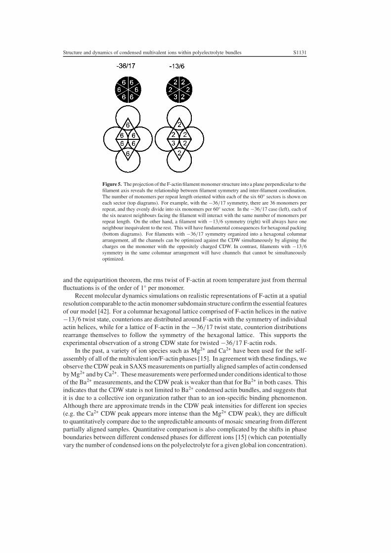
Optimizing the electrostatic interactions in a single interstitial channel will not necessarilyoptimize all of the interstitial channels within a hexagonal array of filaments. F-actin is ahelix, and the charged patches will precess with a spatial frequency determined by its helicalsymmetry. As a result, optimizing the charge matching for a CDW threading one channelwill not in general optimize charge matching in adjacent channels, since different aspects ofthe helices will be presented in adjacent tunnels. An examination of this question reveals oneother important advantage in changing the actin twist state from −13/6 to −36/17. There are36 monomers in the crystallographic repeat of twisted F-actin in the −36/17 configuration.The 36 monomers are evenly divisible by six, the number of nearest neighbours in hexagonalcoordination, whereas 13, the number of monomers in the native crystallographic repeat ofF-actin, is not (figure 5). For F-actin rods with −36/17 symmetry organized into a sixfoldcolumnar arrangement, all of the interstitial channels can be optimized simultaneously. Aunit cell can be generated with two principal channel arrangements that can be simultaneouslyoptimized, and the entire 2D plane can be tiled with a unit cell without frustration effects. Thisis clearly not true for −13/6 F-actin. Since 13 is not divisible by six nearest neighbours, thepattern of charges presented by helices to adjacent channels depends sensitively on the phaseof the actin helices, and it is not possible to tile the 2D surface without frustration.
It is important to assess whether the −3.8◦ twist which is necessary for the formationof the −36/17 helix is forbiddingly large. The torsional rigidity of F-actin has beenmeasured [41]. By using the measured value for the torsional rigidity (κ ≈ 8 × 10⁻²⁶ N m2)
Structure and dynamics of condensed multivalent ions within polyelectrolyte bundles S1131
Figure 5. The projection of the F-actin filament monomer structure into a plane perpendicular to thefilament axis reveals the relationship between filament symmetry and inter-filament coordination.The number of monomers per repeat length oriented within each of the six 60◦ sectors is shown oneach sector (top diagrams). For example, with the −36/17 symmetry, there are 36 monomers perrepeat, and they evenly divide into six monomers per 60◦ sector. In the −36/17 case (left), each ofthe six nearest neighbours facing the filament will interact with the same number of monomers perrepeat length. On the other hand, a filament with −13/6 symmetry (right) will always have oneneighbour inequivalent to the rest. This will have fundamental consequences for hexagonal packing(bottom diagrams). For filaments with −36/17 symmetry organized into a hexagonal columnararrangement, all the channels can be optimized against the CDW simultaneously by aligning thecharges on the monomer with the oppositely charged CDW. In contrast, filaments with −13/6symmetry in the same columnar arrangement will have channels that cannot be simultaneouslyoptimized.
and the equipartition theorem, the rms twist of F-actin at room temperature just from thermalfluctuations is of the order of 1◦ per monomer.
Recent molecular dynamics simulations on realistic representations of F-actin at a spatialresolution comparable to the actin monomer subdomain structure confirm the essential featuresof our model [42]. For a columnar hexagonal lattice comprised of F-actin helices in the native−13/6 twist state, counterions are distributed around F-actin with the symmetry of individualactin helices, while for a lattice of F-actin in the −36/17 twist state, counterion distributionsrearrange themselves to follow the symmetry of the hexagonal lattice. This supports theexperimental observation of a strong CDW state for twisted −36/17 F-actin rods.
In the past, a variety of ion species such as Mg2+ and Ca2+ have been used for the self-assembly of all of the multivalent ion/F-actin phases [15]. In agreement with these findings, weobserve the CDW peak in SAXS measurements on partially aligned samples of actin condensedby Mg2+ and by Ca2+. These measurements were performed under conditions identical to thoseof the Ba2+ measurements, and the CDW peak is weaker than that for Ba2+ in both cases. Thisindicates that the CDW state is not limited to Ba2+ condensed actin bundles, and suggests thatit is due to a collective ion organization rather than to an ion-specific binding phenomenon.Although there are approximate trends in the CDW peak intensities for different ion species(e.g. the Ca2+ CDW peak appears more intense than the Mg2+ CDW peak), they are difficultto quantitatively compare due to the unpredictable amounts of mosaic smearing from differentpartially aligned samples. Quantitative comparison is also complicated by the shifts in phaseboundaries between different condensed phases for different ions [15] (which can potentiallyvary the number of condensed ions on the polyelectrolyte for a given global ion concentration).
S1132 T E Angelini et al
We have performed solid-state NMR experiments on divalent 25Mg ions that comprisethe counterion density wave organized within condensed F-actin bundles. Nuclear magneticresonance (NMR) spectroscopy is a valuable technique for studying ion binding in biologicalsystems. For example 43Ca, 25Mg, 113Cd, 23Na, and 39K NMR have been used to investigateion binding in proteins such as calmodulin, troponin, and parvalbumin [43, 44]. NMR hasalso been used to study the interactions of counterions with polyelectrolytes by monitoringquadrupole relaxation of ions with spin I > 1/2. 25Mg has a spin-5/2 nucleus with a nuclearquadrupole moment that can relax via interactions with electric field gradients. Measurementsof the lineshapes can give insight into the dynamics of Mg2+ ions, as well as interactionsbetween Mg2+ and F-actin filaments.
Xian et al used solution NMR to monitor 25Mg2+ counterion dynamics on uncondensedactin filaments [45]. Their results suggest that 25Mg2+ rotational correlation times areindependent of the rotational dynamics of the actin filaments. Moreover, the competitivebinding between 25Mg2+ and trivalent cobalt hexamine was directly monitored. However, dueto the rapid quadrupole relaxation of the 25Mg2+ (I = 5/2) and the solid nature of condensedF-actin, solution NMR is not feasible to study the dynamics of 25Mg2+ condensed into theCDW state within F-actin bundles, which form at high Mg2+ concentrations. In order to gaininsight into the dynamics of the counterions within condensed F-actin bundles, we have usedsolid-state 25Mg NMR techniques, where an accurate determination of longitudinal relaxationtimes of 25Mg2+ in condensed F-actin can be related to correlation times of the counterionsthrough the spectral density.
25Mg isotope enriched MgO was purchased from Oak Ridge National Laboratory (OakRidge, TN) and dissolved in HCl to obtain a 25MgCl2 stock solution. Monomeric actin waspurified following the method of Spudich and Watt [46] and polymerized using 25MgCl2 to afinal concentration of 2 mM. Gelsolin was added at a molar ratio of 1:370 (gelsolin:actin molarratio) to restrict the length of the F-actin polymers to ∼1 µm, for comparison with the x-rayresults. Phalloidin (Sigma Aldrich, St Louis, MO) was added to prevent depolymerization ofactin.
All 25Mg NMR experiments were carried out at 45.898 MHz on a 17.5 T OxfordInstruments (Witney, UK) 750 MHz magnet controlled by a Varian (Palo Alto, CA) UnityInova operating system. Solution NMR experiments employed a Varian 10 mm 15N-31P BBsolution probe while the solid-state NMR experiments used a Doty Scientific (Columbia, SC)XC5 15N-31P CPMAS solid probe. A 90◦ pulse width of 22 µs was used along with a 100 µspost-acquisition delay time. Longitudinal relaxation times were measured by both inversionrecovery and saturation recovery methods. Magic angle spinning (MAS) experiments wereperformed at a rotation rate of 2 kHz. Solid-state NMR samples were prepared by centrifugingF-actin bundles into Doty Scientific 5 mm XC5 sealing cells and removing excess supernatant,in order to maximize the signal from the 25Mg in the bundles.
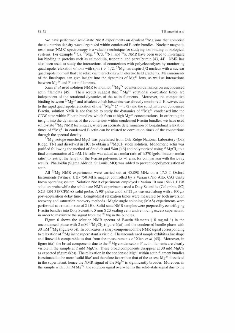
Figure 6 shows the solution NMR spectra of F-actin filaments (10 mg ml−1) in theuncondensed phase with 2 mM 25MgCl2 (figure 6(a)) and the condensed bundle phase with30 mM 25Mg (figure 6(b)). In both cases, a sharp component of the NMR signal correspondingto relaxation of 25Mg in the supernatant is visible. The uncondensed sample exhibits a lineshapeand linewidth comparable to that from the measurements of Xian et al [45]. Moreover, infigure 6(a), the broad components due to the 25Mg condensed on F-actin filaments are clearlyvisible in the sample at 2 mM MgCl2. These broad components disappear at 30 mM MgCl2as expected (figure 6(b)). The relaxation in the condensed Mg2+ within actin filament bundlesis estimated to be more ‘solid like’ and therefore faster than that of the excess Mg2+ dissolvedin the supernatant, hence the NMR signal of the Mg2+ is significantly broader. Moreover, inthe sample with 30 mM Mg2+, the solution signal overwhelms the solid-state signal due to the
Structure and dynamics of condensed multivalent ions within polyelectrolyte bundles S1133
Figure 6. (a) 25Mg NMR spectrum of 2 mM Mg2+ in 10 mg ml−1 F-actin solution. The F-actinis uncondensed. (b) 25Mg NMR spectrum of 30 mM Mg2+ in condensed 10 mg ml−1 F-actinsolution. F-actin is condensed into a bundled phase by the Mg2+ ions. The solution Mg signal islarge compared to the solid-state Mg signal.
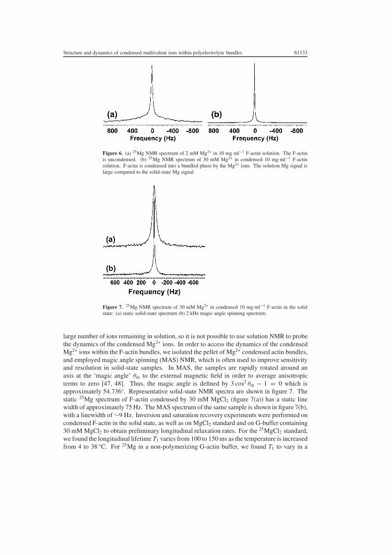
Figure 7. 25Mg NMR spectrum of 30 mM Mg2+ in condensed 10 mg ml−1 F-actin in the solidstate: (a) static solid-state spectrum (b) 2 kHz magic angle spinning spectrum.
large number of ions remaining in solution, so it is not possible to use solution NMR to probethe dynamics of the condensed Mg2+ ions. In order to access the dynamics of the condensedMg2+ ions within the F-actin bundles, we isolated the pellet of Mg2+ condensed actin bundles,and employed magic angle spinning (MAS) NMR, which is often used to improve sensitivityand resolution in solid-state samples. In MAS, the samples are rapidly rotated around anaxis at the ‘magic angle’ θm to the external magnetic field in order to average anisotropicterms to zero [47, 48]. Thus, the magic angle is defined by 3 cos2 θm − 1 = 0 which isapproximately 54.736◦. Representative solid-state NMR spectra are shown in figure 7. Thestatic 25Mg spectrum of F-actin condensed by 30 mM MgCl2 (figure 7(a)) has a static linewidth of approximately 75 Hz. The MAS spectrum of the same sample is shown in figure 7(b),with a linewidth of ∼9 Hz. Inversion and saturation recovery experiments were performed oncondensed F-actin in the solid state, as well as on MgCl2 standard and on G-buffer containing30 mM MgCl2 to obtain preliminary longitudinal relaxation rates. For the 25MgCl2 standard,we found the longitudinal lifetime T1 varies from 100 to 150 ms as the temperature is increasedfrom 4 to 38 ◦C. For 25Mg in a non-polymerizing G-actin buffer, we found T1 to vary in a
S1134 T E Angelini et al
similar manner, from 62 to 96 ms as a function of temperature ranging from 4 to 38 ◦C. This isexpected since Mg can be chelated by ATP in the solution. For condensed F-actin, however, wefind that T1 decreases to a range from 11 to 15 ms over the same temperature range. From ourx-ray measurements presented above, we see that divalent ions are organized into CDWs withinF-actin bundles. The preliminary NMR results presented here give a quantitative measure forthe decreased mobility of Mg2+ ions condensed within F-actin bundles, compared to Mg2+
ions dissolved in the supernatant. In this context, it is interesting to note that the value of T1
for Mg2+ ions in the CDW is intermediate between typical liquid-like and solid-like values.
4. Conclusion
We have experimentally examined the mechanism for like-charge attraction in cytoskeletal F-actin and found evidence for counterion organization. The microscopic mechanism involves acoupled mode between a counterion CDW and the polyelectrolyte twist. (The counterion CDWcannot exist without the polyelectrolyte twist and vice versa.) This implies that within a bundle,it is possible for many actin filaments to collectively twist by the same amount in concert, inresponse to binding by crosslinking agents (divalent ions in the present case). This couplingbetween filament twist and linker binding may have biological implications. For example, ithas been observed that the actin binding protein cofilin can change the twist state of individualactin filaments [49]. Similar compromises between F-actin twist and F-actin crosslinking byactin binding proteins within bundles can influence the hierarchy of existing interactions bylocal modifications of binding sites and impinge on cytoskeletal regulation. We have alsodirectly measured the dynamics of the ions condensed within actin bundles by using magicangle spinning solid-state NMR. We find that their longitudinal relaxation time decreases byapproximately an order of magnitude relative to ions in free solution. It is known that differentpolyelectrolytes require ions of different valences to condense, and an experimentally motivatedcriterion for the degree of ion multivalence required for generating attractions has been recentlyproposed [18]. We plan to extend this preliminary NMR investigation by obtaining relaxationdata at several field strengths and for ions of different valences.
Acknowledgments
We are grateful to W Xian and P Janmey for their generous gift of gelsolin and helpfuldiscussions on the NMR experiments. We thank P Molitor and E Oldfield for assistancewith the solid-state NMR experiments. We also thank K Schweizer and K Schulten for theirinsights at the early stages of this work. This material is based upon work supported bythe US Department of Energy, Division of Materials Sciences, under award No DEFG02-91ER45439, through the Frederick Seitz Materials Research Laboratory at the Universityof Illinois at Urbana-Champaign, NSF under DMR-0409769 and CAMPWS, and by NIH1R21DK68431-01.
References
[1] Gelbart W M, Bruinsma R F, Pincus P A and Parsegian V A 2000 Phys. Today 53 38–44[2] Israelachvili J 1992 Intermolecular and Surface Forces 2 edn (London: Academic)[3] Evans D R and Wennerstrom H 1999 The Colloidal Domain 2 edn (New York: Wiley)[4] Nelson P 2004 Biological Physics (New York: Freeman)[5] Derjaguin B V and Landau L 1941 Acta Physicochimica (URSS) 14 633[6] Verwey E J and Overbeek J T G 1948 Theory of the Stability of Lyophobic Colloids (Amsterdam: Elsevier)
Structure and dynamics of condensed multivalent ions within polyelectrolyte bundles S1135
[7] LeBret M and Zimm B H 1984 Biopolymers 23 287–312[8] Kirkwood J G and Shumaker J B 1952 Proc. Natl Acad. Sci. USA 38 863–71[9] Oosawa F 1968 Biopolymers 6 1688
[10] Ray J and Manning G S 1994 Langmuir 10 2450–61[11] Deserno M, Arnold A and Holm C 2003 Macromolecules 36 249–59[12] Tang J X and Janmey P A 1996 J. Biol. Chem. 271 8556–63[13] Tang J X, Wong S, Tran P T and Janmey P A 1996 Ber. Bunsenges. Phys. Chem. 100 796–806[14] Borukhov I, Bruinsma R F, Gelbart W M and Liu A J 2001 Phys. Rev. Lett. 86 2182–5[15] Wong G C L, Lin A, Tang J X, Li Y, Janmey P A and Safinya C R 2003 Phys. Rev. Lett. 91 018103[16] Borukhov I and Bruinsma R 2001 Phys. Rev. Lett. 87 158101[17] Deng H and Bloomfield V A 1999 Biophys. J. 77 1556–61[18] Butler J C, Angelini T E, Tang J X and Wong G C L 2003 Phys. Rev. Lett. 91 028301[19] Borukhov I, Lee K C, Bruinsma R F, Gelbart W M, Liu A J and Stevens M J 2002 J. Chem. Phys. 117 462–80[20] Ha B-Y and Liu A J 1997 Phys. Rev. Lett. 79 1289–92[21] Ha B-Y and Liu A J 1998 Phys. Rev. Lett. 81 1011–4[22] Podgornik R and Parsegian V A 1998 Phys. Rev. Lett. 80 1560–3[23] Golestanian R and Liverpool T B 2002 Phys. Rev. E 66 051802[24] Rouzina I and Bloomfield V A 1996 J. Phys. Chem. 100 9977–89[25] Grønbech-Jensen N, Mashl R J, Bruinsma R F and Gelbart W M 1997 Phys. Rev. Lett. 78 2477–80[26] Shklovskii B I 1999 Phys. Rev. Lett. 82 3268–871[27] Kornyshev A A and Leikin S 1999 Phys. Rev. Lett. 82 4138–41[28] Solis F J and Olvera de la Cruz M 1999 Phys. Rev. E 60 4496[29] Arenzon J J, Stilck J F and Levin Y 1999 Eur. Phys. J. B 12 79[30] Diehl A, Carmona H A and Levin Y 2001 Phys. Rev. E 6401 1804 (1 part 1)[31] Angelini T E, Liang H, Wriggers W and Wong G C L 2003 Proc. Natl Acad. Sci. USA 100 8634–7[32] Lodish H, Berk A, Zipursky S L, Matsudaira P, Baltimore D and Darnell J 2000 Molecular Cell Biology 4th edn
(New York: Freeman)[33] Sheterline P, Clayton J and Sparrow J C 1998 Actin (New York: Oxford University Press)[34] Al-Khayat H A, Yagi N and Squire J M 1995 J. Mol. Biol. 252 611–32[35] Janmey P A, Peetermans J, Zaner K S, Stossel T P and Tanaka T 1986 J. Biol. Chem. 261 8357–62[36] Holmes K C, Popp D, Gebhard W and Kabsch W 1990 Nature 347 44–9[37] Tibbits T T and Caspar D L D 1993 Acta Crystallogr. A 49 532–45[38] Coridan R H, Angelini T E, Sanders L K, Xian W and Wong G C L 2004 unpublished results[39] Egelman E H, Francis N and DeRosier D J 1982 Nature 298 131–5[40] Aebi U, Millonig R, Salvo H and Engel A 1986 Ann. New York Acad. Sci. 483 100–19[41] Tsuda Y, Yasutake H, Ishijima A and Yanagida T 1996 Proc. Natl Acad. Sci. USA 93 12937–42[42] Lee J and Luijten E 2004 unpublished results[43] Forsen S and Lindman B 1981 Annual Reports on NMR Spectroscopy ed G Webb (New York: Academic)
pp 183–226[44] Forsen S and Vogel H J 1987 Biological Magnetic Resonance ed L Berliner and J Reubin (New York: Plenum)
pp 249–309[45] Xian W, Tang J X, Janmey P A and Braunlin W H 1999 Biochemistry 38 7219–26[46] Spudich J and Watt S 1971 J. Biol. Chem. 246 4866–71[47] Andrew E R 1959 Arch. Sci. 12 103–8[48] Lowe I J 1959 Phys. Rev. Lett. 2 285–7[49] McGough A, Pope B, Chiu W and Weeds A 1997 J. Cell. Biol. 138 771–81