
Structural studies of the primary donor cation radical P"0 in reaction
5
Proc. Nati. Acad. Sci. USA Vol. 81, pp. 1401-1405, March 1984 Biophysics Structural studies of the primary donor cation radical P"0 in reaction centers of Rhodospirillum rubrum by electron-nuclear double resonance in solution (photosynthesis/primary process/bacteriochlorophyll/special pair geometry) W. LUBITZ*, F. LENDZIANt, H. SCHEERt, J. GOTTSTEINt, M. PLATOt, AND K. MOBIUSt *Institut fur Organische Chemie, Freie Universitat Berlin, Takustrasse 3, D-1000 Berlin 33; tInstitut fur Molekuilphysik, Freie Universitat Berlin, Arnimallee 14, D-1000 Berlin 33; and fBotanisches Institut, Universitat Munchen, Menzinger Strasse 67, D-8000 Munchen 19, Federal Republic of Germany Communicated by Joseph J. Katz, September 1, 1983 ABSTRACT The light-induced cation radical of the pri- mary electron donor, Ps70, in photosynthetic reaction centers from Rhodospirillum rubrum G-9, has been investigated by electron-nuclear double resonance (ENDOR) in liquid aque- ous solution. The measured hyperfine coupling constants are assigned to specific molecular positions by partial deuteration. Comparison with the bacteriochlorophyll a cation radical shows different reduction factors of the individual coupling constants deviating from the value 2.0 reported in earlier in- vestigations in frozen solutions. The average of the coupling constants is, however, reduced by a factor very close to 2.0. EPR simulations using the ENDOR coupling constants support a dimer model for PsO with C2 symmetry, where the two mac- rocycles are close enough to form a supermolecular orbital re- sulting in a different distribution of the unpaired electron, compared with the monomeric bacteriochlorophyll a cation radical. Molecular orbital calculations were used to obtain structural information about this dimer. In bacterial photosynthesis, the light-induced charge separa- tion starts with the fast donation of an electron from an excit- ed singlet primary donor P to an electron-transport chain in the reaction center protein (RC) (1-5). RCs offer a conve- nient system for the investigation of the cation and anion radicals formed in this process. Much of our present knowledge about the various species and their interactions in RCs has evolved from the applica- tion of paramagnetic resonance methods (3, 6). The dimeric nature of the primary donor cation radical P8t,0 in some bac- teria was originally proposed by Norris et al. (7) to explain the observed narrowing of the EPR line by a reduction factor (RF) of \/2 compared with the monomeric bacteriochloro- phyll a cation radical (BChl-a+ ) (Fig. 1). Further support for this model came from electron-nuclear double resonance (ENDOR) at low temperatures in which a reduction of the hyperfine coupling constants (hfcs) by a factor of two was deduced when going from BChl-a+ to P8+70 (8-14). The de- tailed structure of the suggested dimer is still controversial (3, 4, 14), but its geometry is of prime importance for basic understanding of the primary act of light-induced charge sep- aration in photosynthesis. ENDOR in solution has been used to elucidate the elec- tronic structure of the various isolated pigment radicals and has provided an almost complete set of isotropic hfcs (15- 20). Liquid-state ENDOR is superior to frozen-solution EN- DOR (10, 21, 22) because the linewidths are smaller due to the absence of anisotropic broadening so that hfcs from all magnetic nuclei in the radical can often be obtained (23, 24). However, most of the ENDOR studies of photosynthesis thus far were carried out in frozen matrices (8-13). An EN- DOR in solution study on RC [Rhodopseudomonas (Rp.) sphaeriodes R-26] in water at room temperature has been reported only recently (25). In those experiments, the addi- tional application of electron-nuclear-nuclear triple reso- nance (26) proved to be useful for increasing the ENDOR intensity and resolution and for determining the signs of the hfcs. This study is now extended to another bacterium-i.e., Rhodospirillum (Rs.) rubrum G-9. MATERIALS AND METHODS Rs. rubrum G-9 was grown anaerobically in Hutner medium (27) at 25-32°C. After freezing and thawing, the cells were incubated for 2 hr at 25°C with lysozyme [2.5 mg/g (wet weight) of bacteria; Boehringer, Darmstadt] and EDTA (10 mM; Merck, Darmstadt), sonicated (three 1-min periods; Branson) and centrifuged for 30 min at 12,000 x g. The su- pernatant was then centrifuged for 1.5 hr at 55,000 x g, and the chromatophore pellet without the hard core was sus- pended in Tris buffer to A870 = 50. One method for the isola- tion of RCs followed essentially the procedure of Snozzi and Bachofen (28). In other preparations, the crude RCs were purified on DEAE-cellulose (DE 52, Whatman) with Tris buffer containing 0.02% lauryldimethylamine oxide instead of Triton X-100. Lauryldimethylamine oxide exchange against Triton X-100 was done after chemical reduction of the detergent to the amine (29) on a DEAE-cellulose column. The RCs were dialyzed against the same buffer after elution with 0.3 M NaCl. Ubiquinone removal was carried out by the method of Okamura et al. (30). RC solutions with A870 < 3 (c < 25 AM) were first concen- trated on DEAE-cellulose and then further by membrane fil- tration (Millipore, type PSED 25,000, 13-mm diameter) to c - 250 ,uM. This solution was centrifuged for 20 min at 12,000 x g and decanted from an eventually appearing greyish pel- let, and 50 ul of the resulting solution was placed under ar- gon in the ENDOR tube (-1 mm i.d.). BChl-a esterified with geranylgeraniol (BChl-agg) was isolated according to Strain and Svec (31). The cation radical was prepared by iodine oxidation (5 molar excess) in CH2CI2/CH3OH, 6:1 (vol/vol) under high vacuum conditions (32). The starting concentra- tion of BChl-a was 0.1 mM; for the ENDOR measurements the radical solution was usually concentrated by a factor of ca. 3. The stability of samples prepared in this manner was up to 1 month. ENDOR and triple resonance measurements were carried out on a laboratory-built spectrometer (23, 26, 32). In situ illumination of the sample in the spectral range 450-900 nm Abbreviations: BChl-a, bacteriochlorophyll a; BChl-agg and BChl- ap, geranylgeranyl and phytyl esters, respectively, of BChl-a; RC, reaction center; hfc, hyperfine coupling constant; hfs, hyperfine structure; ENDOR, electron-nuclear double resonance; RF, reduc- tion factor. 1401 The publication costs of this article were defrayed in part by page charge payment. This article must therefore be hereby marked "advertisement" in accordance with 18 U.S.C. §1734 solely to indicate this fact.
Transcript of Structural studies of the primary donor cation radical P"0 in reaction
Proc. Nati. Acad. Sci. USAVol. 81, pp. 1401-1405, March 1984Biophysics
Structural studies of the primary donor cation radical P"0 inreaction centers of Rhodospirillum rubrum by electron-nucleardouble resonance in solution
(photosynthesis/primary process/bacteriochlorophyll/special pair geometry)
W. LUBITZ*, F. LENDZIANt, H. SCHEERt, J. GOTTSTEINt, M. PLATOt, AND K. MOBIUSt*Institut fur Organische Chemie, Freie Universitat Berlin, Takustrasse 3, D-1000 Berlin 33; tInstitut fur Molekuilphysik, Freie Universitat Berlin, Arnimallee 14,D-1000 Berlin 33; and fBotanisches Institut, Universitat Munchen, Menzinger Strasse 67, D-8000 Munchen 19, Federal Republic of Germany
Communicated by Joseph J. Katz, September 1, 1983
ABSTRACT The light-induced cation radical of the pri-mary electron donor, Ps70, in photosynthetic reaction centersfrom Rhodospirillum rubrum G-9, has been investigated byelectron-nuclear double resonance (ENDOR) in liquid aque-ous solution. The measured hyperfine coupling constants areassigned to specific molecular positions by partial deuteration.Comparison with the bacteriochlorophyll a cation radicalshows different reduction factors of the individual couplingconstants deviating from the value 2.0 reported in earlier in-vestigations in frozen solutions. The average of the couplingconstants is, however, reduced by a factor very close to 2.0.EPR simulations using the ENDOR coupling constants supporta dimer model for PsO with C2 symmetry, where the two mac-rocycles are close enough to form a supermolecular orbital re-sulting in a different distribution of the unpaired electron,compared with the monomeric bacteriochlorophyll a cationradical. Molecular orbital calculations were used to obtainstructural information about this dimer.
In bacterial photosynthesis, the light-induced charge separa-tion starts with the fast donation ofan electron from an excit-ed singlet primary donor P to an electron-transport chain inthe reaction center protein (RC) (1-5). RCs offer a conve-nient system for the investigation of the cation and anionradicals formed in this process.Much of our present knowledge about the various species
and their interactions in RCs has evolved from the applica-tion of paramagnetic resonance methods (3, 6). The dimericnature of the primary donor cation radical P8t,0 in some bac-teria was originally proposed by Norris et al. (7) to explainthe observed narrowing of the EPR line by a reduction factor(RF) of \/2 compared with the monomeric bacteriochloro-phyll a cation radical (BChl-a+ ) (Fig. 1). Further support forthis model came from electron-nuclear double resonance(ENDOR) at low temperatures in which a reduction of thehyperfine coupling constants (hfcs) by a factor of two wasdeduced when going from BChl-a+ to P8+70 (8-14). The de-tailed structure of the suggested dimer is still controversial(3, 4, 14), but its geometry is of prime importance for basicunderstanding of the primary act of light-induced charge sep-aration in photosynthesis.ENDOR in solution has been used to elucidate the elec-
tronic structure of the various isolated pigment radicals andhas provided an almost complete set of isotropic hfcs (15-20). Liquid-state ENDOR is superior to frozen-solution EN-DOR (10, 21, 22) because the linewidths are smaller due tothe absence of anisotropic broadening so that hfcs from allmagnetic nuclei in the radical can often be obtained (23, 24).However, most of the ENDOR studies of photosynthesisthus far were carried out in frozen matrices (8-13). An EN-
DOR in solution study on RC [Rhodopseudomonas (Rp.)sphaeriodes R-26] in water at room temperature has beenreported only recently (25). In those experiments, the addi-tional application of electron-nuclear-nuclear triple reso-nance (26) proved to be useful for increasing the ENDORintensity and resolution and for determining the signs of thehfcs. This study is now extended to another bacterium-i.e.,Rhodospirillum (Rs.) rubrum G-9.
MATERIALS AND METHODS
Rs. rubrum G-9 was grown anaerobically in Hutner medium(27) at 25-32°C. After freezing and thawing, the cells wereincubated for 2 hr at 25°C with lysozyme [2.5 mg/g (wetweight) of bacteria; Boehringer, Darmstadt] and EDTA (10mM; Merck, Darmstadt), sonicated (three 1-min periods;Branson) and centrifuged for 30 min at 12,000 x g. The su-pernatant was then centrifuged for 1.5 hr at 55,000 x g, andthe chromatophore pellet without the hard core was sus-pended in Tris buffer to A870 = 50. One method for the isola-tion of RCs followed essentially the procedure of Snozzi andBachofen (28). In other preparations, the crude RCs werepurified on DEAE-cellulose (DE 52, Whatman) with Trisbuffer containing 0.02% lauryldimethylamine oxide insteadof Triton X-100. Lauryldimethylamine oxide exchangeagainst Triton X-100 was done after chemical reduction ofthe detergent to the amine (29) on a DEAE-cellulose column.The RCs were dialyzed against the same buffer after elutionwith 0.3 M NaCl. Ubiquinone removal was carried out bythe method of Okamura et al. (30).RC solutions with A870 < 3 (c < 25 AM) were first concen-
trated on DEAE-cellulose and then further by membrane fil-tration (Millipore, type PSED 25,000, 13-mm diameter) to c- 250 ,uM. This solution was centrifuged for 20 min at 12,000x g and decanted from an eventually appearing greyish pel-let, and 50 ul of the resulting solution was placed under ar-gon in the ENDOR tube (-1 mm i.d.). BChl-a esterified withgeranylgeraniol (BChl-agg) was isolated according to Strainand Svec (31). The cation radical was prepared by iodineoxidation (5 molar excess) in CH2CI2/CH3OH, 6:1 (vol/vol)under high vacuum conditions (32). The starting concentra-tion of BChl-a was 0.1 mM; for the ENDOR measurementsthe radical solution was usually concentrated by a factor ofca. 3. The stability of samples prepared in this manner wasup to 1 month.ENDOR and triple resonance measurements were carried
out on a laboratory-built spectrometer (23, 26, 32). In situillumination of the sample in the spectral range 450-900 nm
Abbreviations: BChl-a, bacteriochlorophyll a; BChl-agg and BChl-ap, geranylgeranyl and phytyl esters, respectively, of BChl-a; RC,reaction center; hfc, hyperfine coupling constant; hfs, hyperfinestructure; ENDOR, electron-nuclear double resonance; RF, reduc-tion factor.
1401
The publication costs of this article were defrayed in part by page chargepayment. This article must therefore be hereby marked "advertisement"in accordance with 18 U.S.C. §1734 solely to indicate this fact.
Proc. Natl. Acad. Sci USA 81 (1984)
H 4bCH3
H3C 1c12 2liwihn
H ~ M\ H
Rp.sphaeroides.6
H3C 17 CH3
H
H2C H2 H
CH3R lob
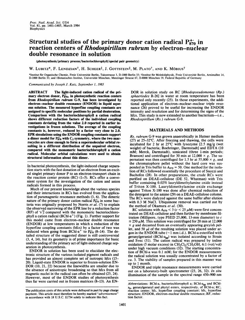
FIG. 1. Molecular structure of BChl-a with numbering scheme;
R = geranylgeranyl (BChl-a,,) in Rs. rubrum and phytyl (BWhl-ap~) in
Rp. sphaeroides.
was by a tungsten/halogen lamp (250 W). The temperaturestability of the sample was within ±10C.
RESULTS AND DISCUSSIONENDOR/Triple Resonance Study of BChl-a". From BChl-
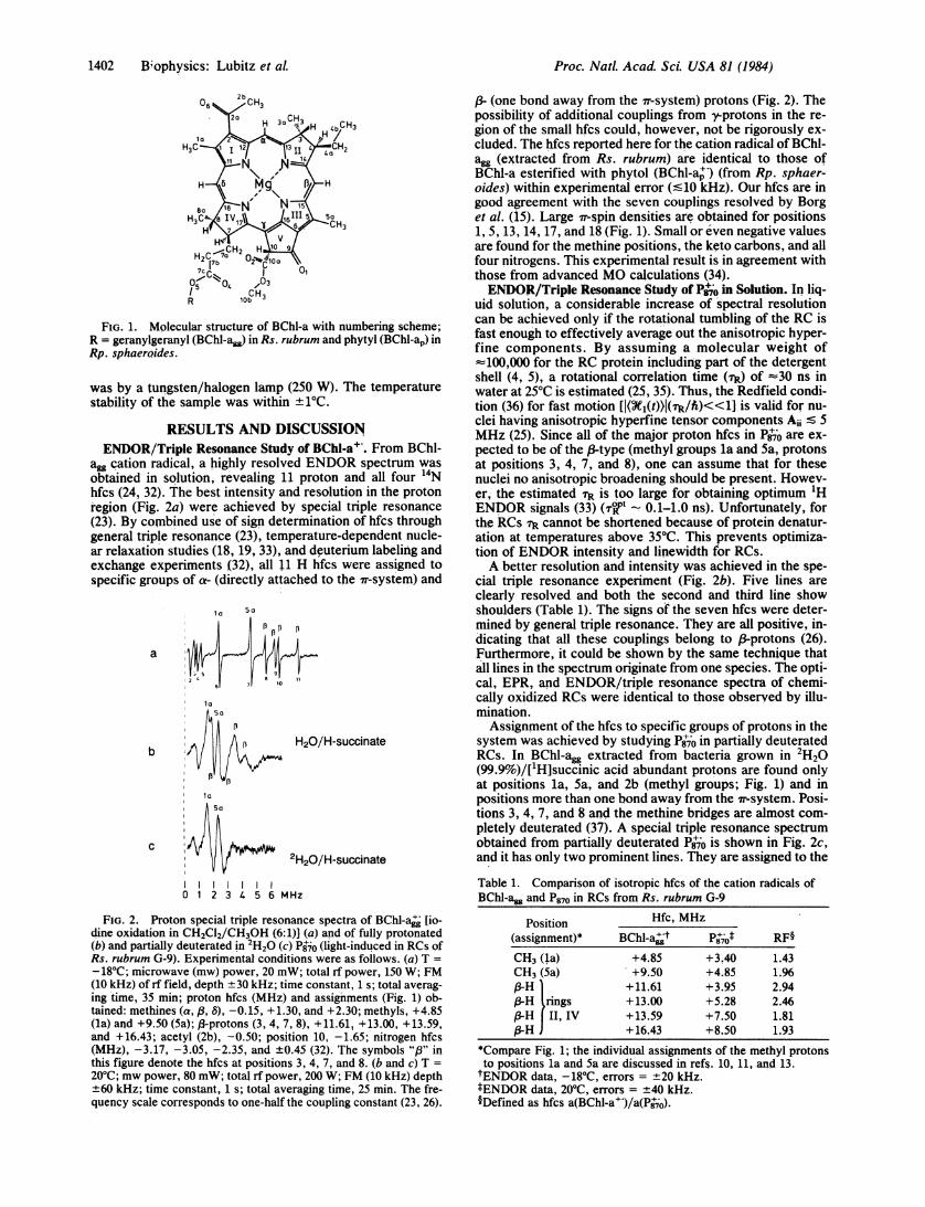
agg cation radical, a highly resolved ENDOR spectrum wasobtained in solution, revealing 11 proton and all four '4Nhfcs (24, 32). The best intensity and resolution in the protonregion (Fig. 2a) were achieved by special triple resonance(23). By combined use of sign determination of hfcs throughgeneral triple resonance (23), temperature-dependent nucle-ar relaxation studies (18, 19, 33), and deuterium labeling andexchange experiments (32), all 11 H hfcs were assigned tospecific groups of' a- (directly attached to the ir-system) and
aa5a
a
1a
bA _ , H20/H-succinate
la
Sa
C I2H20/H-succinate
0 1 2 3 4 5 6 MHz
FIG. 2. Proton special triple resonance spectra of BChl-aj [io-dine oxidation in CH2CI2/CH3OH (6:1)1 (a) and of fully protonated(b) and partially deuterated in 2H20 (c) P0jo (light-induced in RCs ofRs. rubrum G-9). Experimental conditions were as follows. (a) T =
-18'C; microwave (mw) power, 20 mW; total rf power, 150 W; FM(10 kHz) of rf field, depth ±30 kHz; time constant, 1 s; total averag-ing time, 35 min; proton hfcs (MHz) and assignments (Fig. 1) ob-tained: methines (a, /, 8), -0.15, +1.30, and +2.30; methyls, +4.85(la) and +9.50 (5a); ,3-protons (3, 4, 7, 8), +11.61, +13.00, +13.59,and +16.43; acetyl (2b), -0.50; position 10, -1.65; nitrogen hfcs(MHz), -3.17, -3.05, -2.35, and ±0.45 (32). The symbols "/3" inthis figure denote the hfcs at positions 3, 4, 7, and 8. (b and c) T =
20'C; mw power, 80 mW; total rf power, 200 W; FM (10 kHz) depth±60 kHz; time constant, 1 s; total averaging time, 25 min. The fre-quency scale corresponds to one-half the coupling constant (23, 26).
P- (one bond away from the 7r-system) protons (Fig. 2). Thepossibility of additional couplings from -t-protons in the re-gion of the small hfcs could, however, not be rigorously ex-cluded. The hfcs reported here for the cation radical of BChl-agg (extracted from Rs. rubrum) are identical to those ofBChl-a esterified with phytol (BChl-a") (from Rp. sphaer-oides) within experimental error ('10 kHz). Our hfcs are ingood agreement with the seven couplings resolved by Borget al. (15). Large fr-spin densities are obtained for positions1, 5, 13, 14, 17, and 18 (Fig. 1). Small or even negative valuesare found for the methine positions, the keto carbons, and allfour nitrogens. This experimental result is in agreement withthose from advanced MO calculations (34).ENDOR/Triple Resonance Study of Pj70 in Solution. In liq-
uid solution, a considerable increase of spectral resolutioncan be achieved only if the rotational tumbling of the RC isfast enough to effectively average out the anisotropic hyper-fine components. By assuming a molecular weight of=100,000 for the RC protein including part of the detergentshell (4, 5), a rotational correlation time (TR) of =30 ns inwater at 250C is estimated (25, 35). Thus, the Redfield condi-tion (36) for fast motion [I(W1(t))I(r/h)<<«1 is valid for nu-clei having anisotropic hyperfine tensor components Ai Z 5MHz (25). Since all of the major proton hfcs in Pg7o are ex-pected to be of the 3-type (methyl groups la and 5a, protonsat positions 3, 4, 7, and 8), one can assume that for thesenuclei no anisotropic broadening should be present. Howev-er, the estimated TR is too large for obtaining optimum 1HENDOR signals (33) (°rTt - 0.1-1.0 ns). Unfortunately, forthe RCs TR cannot be shortened because of protein denatur-ation at temperatures above 35TC. This prevents optimiza-tion of ENDOR intensity and linewidth for RCs.A better resolution and intensity was achieved in the spe-
cial triple resonance experiment (Fig. 2b). Five lines areclearly resolved and both the second and third line showshoulders (Table 1). The signs of the seven hfcs were deter-mined by general triple resonance. They are all positive, in-dicating that all these couplings belong to (-protons (26).Furthermore, it could be shown by the same technique thatall lines in the spectrum originate from one species. The opti-cal, EPR, and ENDOR/triple resonance spectra of chemi-cally oxidized RCs were identical to those observed by illu-mination.Assignment of the hfcs to specific groups of protons in the
system was achieved by studying P8'0 in partially deuteratedRCs. In BChl-agg extracted from bacteria grown in 2H20(99.9%)/['H]succinic acid abundant protons are found onlyat positions la, 5a, and 2b (methyl groups; Fig. 1) and inpositions more than one bond away from the ir-system. Posi-tions 3, 4, 7, and 8 and the methine bridges are almost com-pletely deuterated (37). A special triple resonance spectrumobtained from partially deuterated P8tjo is shown in Fig. 2c,and it has only two prominent lines. They are assigned to the
Table 1. Comparison of isotropic hfcs of the cation radicals ofBChl-a~g and P8,0 in RCs from Rs. rubrum G-9
Position Hfc, MHz(assignment)* BChl a;;t P8iot RF§CH3 (la) +4.85 +3.40 1.43CH3 (5a) +9.50 +4.85 1.96j3-H +11.61 +3.95 2.943-H rings +13.00 +5.28 2.46(-H II, IV +13.59 +7.50 1.81/-H +16.43 +8.50 1.93
*Compare Fig. 1; the individual assignments of the methyl protonsto positions la and 5a are discussed in refs. 10, 11, and 13.tENDOR data, -18TC, errors = +20 kHz.tENDOR data, 20TC, errors = ±40 kHz.1Defined as hfcs a(BChl-a")/a(P8'jO).
1402 B.ophysics: Lubitz et aL
Proc. Natl. Acad. Sci. USA 81 (1984) 1403
methyl groups in positions la and 5a. There are only twohfcs for these methyl groups: the triple resonance lines showno tendency to split. If we assume a dimer for P8'0, this find-ing suggests a symmetric spin distribution over both halves-i.e., protons at corresponding positions in both halves ofthe dimer exhibit the same hfcs. Four lines are missing ascompared with Fig. 2b. They should therefore belong to thefour 83-protons (Fig. 2b) in the hydrated rings II and IV (posi-tions 3, 4, 7, and 8; Fig. 1). Assignment of the smallest hfc inFig. 2b and c is not so straightforward and will be discussedbelow.Dimeric Nature of Ps+O. Because there are independent as-
signments of the measured hfcs to molecular positions forboth BChl-a" and P8t10, we can compare the spectra of thosetwo radicals by assuming that the ordering within one groupof hfcs with regard to magnitude is retained. In P8+7O all hfcsare reduced in magnitude as compared with BChl-a" but notby a constant RF of 2 (Table 1), as expected in the simplest"special pair" model (7-13). The average RF factor is none-theless very close to 2 (2.06), still indicating a dimeric struc-ture of P870. The procedure of comparing hfcs is, however,questionable because of the uncertain relationship betweenhfcs and spin densities [dihedral angles of the 13protons (38),shifting of spin density to "blind" positions].An independent approach to confirm the dimeric nature of
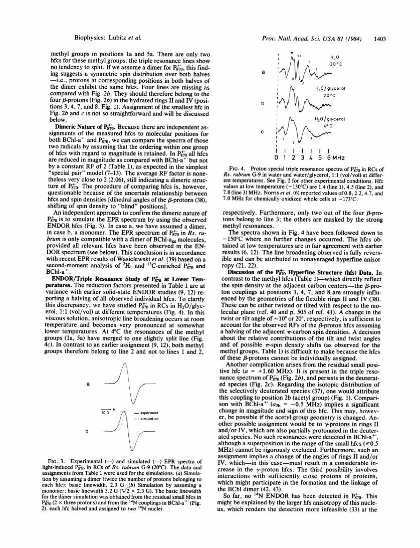
P8+io is to simulate the EPR spectrum by using the observedENDOR hfcs (Fig. 3). In case a, we have assumed a dimer,in case b, a monomer. The EPR spectrum of P8jO in Rs. ru-brum is only compatible with a dimer of BChl-agg molecules,provided all relevant hfcs have been observed in the EN-DOR spectrum (see below). This conclusion is in accordancewith recent EPR results of Wasielewski et al. (39) based on asecond-moment analysis of 2H1- and 13C-enriched P8+70 andBChl-a+.ENDOR/Triple Resonance Study of Ps+,o at Lower Tem-
peratures. The reduction factors presented in Table 1 are atvariance with earlier solid-state ENDOR studies (9, 12) re-porting a halving of all observed individual hfcs. To clarifythis discrepancy, we have studied P870 in RCs in H20/glyc-erol, 1:1 (vol/vol) at different temperatures (Fig. 4). In thisviscous solution, anisotropic line broadening occurs at roomtemperature and becomes very pronounced at somewhatlower temperatures. At 4°C the resonances of the methylgroups (la, 5a) have merged to one slightly split line (Fig.4c). In contrast to an earlier assignment (9, 12), both methylgroups therefore belong to line 2 and not to lines 1 and 2,
a
H10 G - experiment
8 simulation
b
FIG. 3. Experimental (-) and simulated (---) EPR spectra oflight-induced P8jO in RCs of Rs. rubrum G-9 (20°C). The data andassignments from Table 1 were used for the simulations. (a) Simula-tion by assuming a dimer (twice the number of protons belonging toeach hfc); basic linewidth, 2.3 G. (b) Simulation by assuming amonomer; basic linewidth 3.2 G (\/2 x 2.3 G). The basic linewidthfor the dimer simulation was obtained from the residual small hfcs inP8jO (2 x three protons) and from the '4N couplings in BChl-a+ (Fig.2), each hfc halved and assigned to two 14N nuclei.
Ia5a
a 1.4
b I?
/\, H20/glycerol
C
I 2
0 1 2 3 4 5 6 MHz
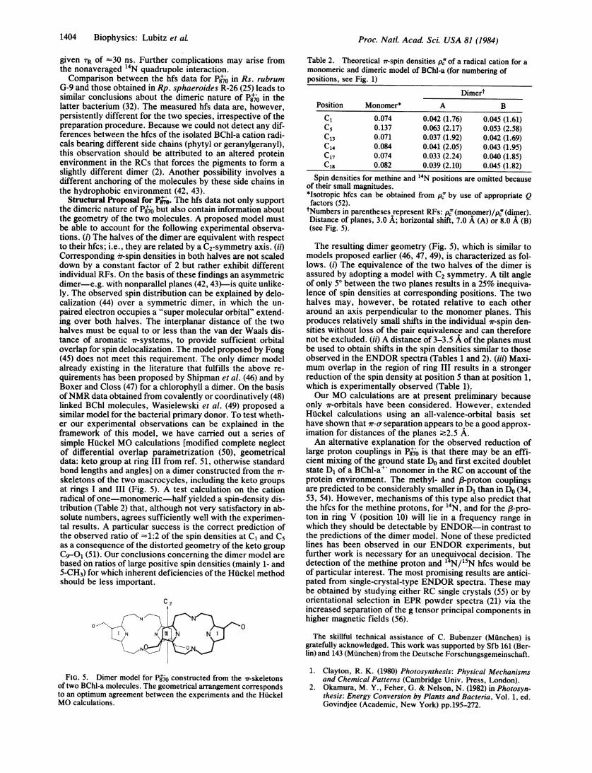
FIG. 4. Proton special triple resonance spectra of P8jo in RCs ofRs. rubrum G-9 in water and water/glycerol, 1:1 (vol/vol) at differ-ent temperatures. See Fig. 2 for other experimental conditions. Hfcvalues at low temperature (-130°C) are 1.4 (line 1), 4.5 (line 2), and7.8 (line 3) MHz. Norris et al. (6) reported values of 0.8, 2.2, 4.7, and7.0 MHz for chemically oxidized whole cells at -173°C.
respectively. Furthermore, only two out of the four /3pro-tons belong to line 3; the others are masked by the strongmethyl resonances.The spectra shown in Fig. 4 have been followed down to
-150°C where no further changes occurred. The hfcs ob-tained at low temperatures are in fair agreement with earlierresults (6, 12). The line broadening observed is fully revers-ible and can be attributed to nonaveraged hyperfine anisot-ropy (21, 22).
Discussion of the P87j Hyperfine Structure (hfs) Data. Incontrast to the methyl hfcs (Table 1)-which directly reflectthe spin density at the adjacent carbon centers-the P-pro-ton couplings at positions 3, 4, 7, and 8 are strongly influ-enced by the geometries of the flexible rings II and IV (38).These can be either twisted or tilted with respect to the mo-lecular plane (ref. 40 and p. 505 of ref. 41). A change in thetwist or tilt angle of -10° or 20°, respectively, is sufficient toaccount for the observed RFs of the p-proton hfcs assuminga halving of the adjacent ir-carbon spin densities. A decisionabout the relative contributions of the tilt and twist anglesand of possible ir-spin density shifts (as observed for themethyl groups, Table 1) is difficult to make because the hfcsof these p-protons cannot be individually assigned.Another complication arises from the residual small posi-
tive hfc (a = +1.60 MHz). It is present in the triple reso-nance spectrum of P8j0 (Fig. 2b), and persists in the deuterat-ed species (Fig. 2c). Regarding the isotopic distribution ofthe selectively deuterated species (37), one would attributethis coupling to position 2b (acetyl group) (Fig. 1). Compari-son with BChl-a+ (a2b = -0.5 MHz) implies a significantchange in magnitude and sign of this hfc. This may, howev-er, be possible if the acetyl group geometry is changed. An-other possible assignment would be to y-protons in rings IIand/or IV, which are also partially protonated in the deuter-ated species. No such resonances were detected in BChl-a+,although a superposition in the range of the small hfcs (<0.5MHz) cannot be rigorously excluded. Furthermore, such anassignment implies a change of the angles of rings II and/orIV, which-in this case-must result in a considerable in-crease in the y-proton hfcs. The third possibility involvesinteractions with sufficiently close protons of proteins,which might participate in the formation and the linkage ofthe BChl dimer (42, 43).So far, no 14N ENDOR has been detected in P8Y0. This
might be explained by the larger hfs anisotropy of this nucle-us, which renders the detection more infeasible (33) at the
Biophysics: Lubitz et aL
1404 Biophysics: Lubitz et aL
given TR of -30 ns. Further complications may arise fromthe nonaveraged 14N quadrupole interaction.Comparison between the hfs data for P870 in Rs. rubrum
G-9 and those obtained in Rp. sphaeroides R-26 (25) leads tosimilar conclusions about the dimeric nature of P870 in thelatter bacterium (32). The measured hfs data are, however,persistently different for the two species, irrespective of thepreparation procedure. Because we could not detect any dif-ferences between the hfcs of the isolated BChl-a cation radi-cals bearing different side chains (phytyl or geranylgeranyl),this observation should be attributed to an altered proteinenvironment in the RCs that forces the pigments to form aslightly different dimer (2). Another possibility involves adifferent anchoring of the molecules by these side chains inthe hydrophobic environment (42, 43).
Structural Proposal for Psj0. The hfs data not only supportthe dimeric nature of P8'jo but also contain information aboutthe geometry of the two molecules. A proposed model mustbe able to account for the following experimental observa-tions. (t) The halves of the dimer are equivalent with respectto their hfcs; i.e., they are related by a C2-symmetry axis. (ii)Corresponding 7'r-spin densities in both halves are not scaleddown by a constant factor of 2 but rather exhibit differentindividual RFs. On the basis of these findings an asymmetricdimer-e.g. with nonparallel planes (42, 43)-is quite unlike-ly. The observed spin distribution can be explained by delo-calization (44) over a symmetric dimer, in which the un-paired electron occupies a "super molecular orbital" extend-ing over both halves. The interplanar distance of the twohalves must be equal to or less than the van der Waals dis-tance of aromatic ir-systems, to provide sufficient orbitaloverlap for spin delocalization. The model proposed by Fong(45) does not meet this requirement. The only dimer modelalready existing in the literature that fulfills the above re-quirements has been proposed by Shipman et al. (46) and byBoxer and Closs (47) for a chlorophyll a dimer. On the basisofNMR data obtained from covalently or coordinatively (48)linked BChl molecules, Wasielewski et al. (49) proposed asimilar model for the bacterial primary donor. To test wheth-er our experimental observations can be explained in theframework of this model, we have carried out a series ofsimple Huckel MO calculations [modified complete neglectof differential overlap parametrization (50), geometricaldata: keto group at ring III from ref. 51, otherwise standardbond lengths and angles] on a dimer constructed from the ir-
skeletons of the two macrocycles, including the keto groupsat rings I and III (Fig. 5). A test calculation on the cationradical of one-monomeric-half yielded a spin-density dis-tribution (Table 2) that, although not very satisfactory in ab-solute numbers, agrees sufficiently well with the experimen-tal results. A particular success is the correct prediction ofthe observed ratio of -1:2 of the spin densities at C, and C5as a consequence of the distorted geometry of the keto group
C9--O (51). Our conclusions concerning the dimer model arebased on ratios of large positive spin densities (mainly 1- and5-CH3) for which inherent deficiencies of the Huckel methodshould be less important.
C 2
N N
0
IN N N
N
FIG. 5. Dimer model for P8jo constructed from the ir-skeletonsof two BChl-a molecules. The geometrical arrangement correspondsto an optimum agreement between the experiments and the HuckelMO calculations.
Table 2. Theoretical ir-spin densities pj of a radical cation for amonomeric and dimeric model of BChl-a (for numbering ofpositions, see Fig. 1)
DimertPosition Monomer* A B
C1 0.074 0.042 (1.76) 0.045 (1.61)C5 0.137 0.063 (2.17) 0.053 (2.58)C13 0.071 0.037 (1.92) 0.042 (1.69)C14 0.084 0.041 (2.05) 0.043 (1.95)C17 0.074 0.033 (2.24) 0.040 (1.85)C18 0.082 0.039 (2.10) 0.045 (1.82)
Spin densities for methine and 14N positions are omitted becauseof their small magnitudes.*Isotropic hfcs can be obtained from pc' by use of appropriate Qfactors (52).tNumbers in parentheses represent RFs: pj (monomer)/pc (dimer).Distance of planes, 3.0 A; horizontal shift, 7.0 A (A) or 8.0 A (B)(see Fig. 5).
The resulting dimer geometry (Fig. 5), which is similar tomodels proposed earlier (46, 47, 49), is characterized as fol-lows. (i) The equivalence of the two halves of the dimer isassured by adopting a model with C2 symmetry. A tilt angleof only 5° between the two planes results in a 25% inequiva-lence of spin densities at corresponding positions. The twohalves may, however, be rotated relative to each otheraround an axis perpendicular to the monomer planes. Thisproduces relatively small shifts in the individual ff-spin den-sities without loss of the pair equivalence and can thereforenot be excluded. (ii) A distance of 3-3.5 A of the planes mustbe used to obtain shifts in the spin densities similar to thoseobserved in the ENDOR spectra (Tables 1 and 2). (iii) Maxi-mum overlap in the region of ring III results in a strongerreduction of the spin density at position 5 than at position 1,which is experimentally observed (Table 1).Our MO calculations are at present preliminary because
only ir-orbitals have been considered. However, extendedHuckel calculations using an all-valence-orbital basis sethave shown that ir-o, separation appears to be a good approx-imation for distances of the planes -2.5 A.An alternative explanation for the observed reduction of
large proton couplings in P8+jo is that there may be an effi-cient mixing of the ground state Do and first excited doubletstate D1 of a BChl-a" monomer in the RC on account of theprotein environment. The methyl- and 13-proton couplingsare predicted to be considerably smaller in D1 than in Do (34,53, 54). However, mechanisms of this type also predict thatthe hfcs for the methine protons, for "'N, and for the 13-pro-ton in ring V (position 10) will lie in a frequency range inwhich they should be detectable by ENDOR-in contrast tothe predictions of the dimer model. None of these predictedlines has been observed in our ENDOR experiments, butfurther work is necessary for an unequivocal decision. Thedetection of the methine proton and 14N/15N hfcs would beof particular interest. The most promising results are antici-pated from single-crystal-type ENDOR spectra. These maybe obtained by studying either RC single crystals (55) or byorientational selection in EPR powder spectra (21) via theincreased separation of the g tensor principal components inhigher magnetic fields (56).
The skillful technical assistance of C. Bubenzer (Munchen) isgratefully acknowledged. This work was supported by Sfb 161 (Ber-lin) and 143 (Munchen) from the Deutsche Forschungsgemeinschaft.
1. Clayton, R. K. (1980) Photosynthesis: Physical Mechanismsand Chemical Patterns (Cambridge Univ. Press, London).
2. Okamura, M. Y., Feher, G. & Nelson, N. (1982) in Photosyn-thesis: Energy Conversion by Plants and Bacteria, Vol. 1, ed.Govindjee (Academic, New York) pp. 195-272.
Proc. NatL Acad Sci. USA 81 (1984)
Proc. Natl. Acad. Sci. USA 81 (1984) 1405
3. Hoff, A. J. (1982) in Light Reaction Path of Photosynthesis,ed. Fong, F. K. (Springer, Berlin), pp. 80-151; and 322-326.
4. Feher, G. & Okamura, M. Y. (1978) in The PhotosyntheticBacteria, eds. Clayton, R. K. & Sistrom, W. R. (Plenum, NewYork), pp. 349-386.
5. Gingras, G. in The Photosynthetic Bacteria, eds. Clayton,R. K. & Sistrom, W. R. (Plenum, New York), pp. 119-132.
6. Norris, J. R., Scheer, H. & Katz, J. J. (1979) in The Porphy-rins, ed. Dolphin, D. (Academic, New York), Vol. 4, pp. 159-195.
7. Norris, J. R., Uphaus, R. A., Crespi, H. L. & Katz, J. J.(1971) Proc. Natl. Acad. Sci. USA 68, 625-629.
8. Norris, J. R., Druyan, M. E. & Katz, J. J. (1973) J. Am.Chem. Soc. 95, 1680-1682.
9. Feher, G., Hoff, A. J., Isaacson, R. A. & McElroy, J. D.(1973) Biophys. J. 13, 61 (abstr.).
10. Feher, G., Hoff, A. J., Isaacson, R. A. & Ackerson, L. C.(1975) Ann. N. Y. Acad. Sci. 244, 239-259.
11. Norris, J. R., Scheer, H. & Katz, J. J. (1975) Ann. N. Y. Acad.Sci. 244, 260-280.
12. Norris, J. R., Scheer, H., Druyan, M. E. & Katz, J. J. (1974)Proc. Natl. Acad. Sci. USA 71, 4897-4900.
13. Norris, J. R. & Katz, J. J. (1978) in The Photosynthetic Bacte-ria, eds. Clayton, R. K. & Sistrom, W. R. (Plenum, NewYork), pp. 397-418.
14. Parson, W. W. (1982) Annu. Rev. Biophys. Bioeng. 11, 57-80.15. Borg, C. D., Forman, A. & Fajer, J. (1976) J. Am. Chem. Soc.
98, 6889-6893.16. Fajer, J., Forman, A., Davis, M. S., Spaulding, L. D., Brune,
D. C. & Felton, R. H. (1977) J. Am. Chem. Soc. 9, 4134-4140.
17. Hoff, A. J. & Mobius, K. (1978) Proc. Natl. Acad. Sci. USA75, 2296-2300.
18. Lubitz, W., Lendzian, F. & Mobius, K. (1981) Chem. Phys.Lett. 81, 235-241.
19. Lubitz, W., Lendzian, F. & Mobius, K. (1981) Chem. Phys.Lett. 84, 33-38.
20. Lendzian, F., Lubitz, W. & Mobius, K. (1982) Chem. Phys.Lett. 90, 375-381.
21. Kevan, L. & Kispert, L. D. (1976) Electron Spin Double Reso-nance Spectroscopy (Wiley, New York), pp. 233-253.
22. Hyde, J. S., Rist, G. H. & Eriksson, L. E. G. (1968) J. Phys.Chem. 72, 4269-4275.
23. Mobius, K., Plato, M. & Lubitz, W. (1982) Phys. Rep. 82, 171-208.
24. M6bius, K., Frohling, W., Lendzian, F., Lubitz, W., Plato, M.& Winscom, C. J. (1982) J. Phys. Chem. 86, 4491-4507.
25. Lendzian, F., Lubitz, W., Scheer, H., Bubenzer, C. & Mobi-us, K. (1981) J. Am. Chem. Soc. 103, 4635-4637.
26. M6bius, K. & Biehl, R. (1979) in Multiple Electron ResonanceSpectroscopy, eds. Dorio, M. M. & Freed, J. H. (Plenum,New York), pp. 475-507.
27. Cohen-Bazire, G., Sistrom, W. R. & Stanier, J. (1957) J. Cell.Comp. Physiol. 49, 25-68.
28. Snozzi, M. & Bachofen, R. (1979) Biochim. Biophys. Acta 546,236- 247.
29. Pachence, J., Dutton, P. L. & Blasie, J. K. (1979) Biochim.Biophys. Acta 548, 348-373.
30. Okamura, M. Y., Isaacson, R. A. & Feher, G. (1975) Proc.Natl. Acad. Sci. USA 72, 3491-3495.
31. Strain, H. H. & Svec, W. A. (1966) in The Chlorophylls, eds.Vernon, L. P. & Seely, G. R. (Academic, New York).
32. Lendzian, F. (1982) Dissertation (Freie Universitat, Berlin,Federal Republic of Germany).
33. Plato, M., Lubitz, W. & Mobius, K. (1981) J. Phys. Chem. 85,1202-1219.
34. Petke, J. D., Maggiora, G. M., Shipman, L. L. & Christoffer-son, R. E. (1980) Photochem. Photobiol. 31, 243-257.
35. Chasteen, N. D. & Francavilla, J. (1976) J. Phys. Chem. 80,867-871.
36. Redfield, A. G. (1965) Adv. Magn. Reson. 1, 1-32.37. Katz, J. J., Dougherty, R. C., Crespi, H. L. & Strain, H. H.
(1966) J. Am. Chem. Soc. 88, 2856-2857.38. Davis, M. S., Forman, A., Hanson, L. K., Thornber, J. P. &
Fajer, J. (1979) J. Phys. Chem. 83, 3325-3332.39. Wasielewski, M. R., Norris, J. R., Crespi, H. L. & Harper, J.
(1981) J. Am. Chem. Soc. 103, 7664-7665.40. Chow, H. C., Serlin, R. & Strouse, C. E. (1975) J. Am. Chem.
Soc. 97, 7230-7237.41. Scheer, H. & Katz, J. J. (1975) in Porphyrins and Metallopor-
phyrins, ed. Smith, K. M. (Elsevier, New York), pp. 399-524.42. Schaafsma, T. J. (1982) in Triplet State ODMR Spectroscopy,
ed. Clarke, R. H. (Wiley, New York), pp. 291-365.43. Hoff, A. J. (1982) in Triplet State ODMR Spectroscopy, ed.
Clarke, R. H. (Wiley, New York), pp. 367-425.44. Bowman, M. K. & Norris, J. R. (1982) J. Am. Chem. Soc. 104,
1512-1515.45. Fong, F. K. (1974) Proc. Natl. Acad. Sci. USA 71, 3692-3695.46. Shipman, L. L., Cotton, T. M., Norris, J. R. & Katz, J. J.
(1976) Proc. Natl. Acad. Sci. USA 73, 1791-1794.47. Boxer, S. G. & Closs, G. L. (1976) J. Am. Chem. Soc. 98,
5406-5408.48. Cotton, T. M., Loach, P. A., Katz, J. J. & Ballschmiter,
K. H. (1978) Photochem. Photobiol. 27, 735-749.49. Wasielewski, M. R., Smith, U. H., Cope, B. T. & Katz, J. J.
(1977) J. Am. Chem. Soc. 99, 4172-4173.50. Pople, J. A. & Beveridge, D. L. (1970) Approximate Molecu-
lar Orbital Theory (McGraw-Hill, New York), pp. 75-79.51. Barkigia, K. M., Fajer, J., Smith, K. M. & Williams, G. J. B.
(1981) J. Am. Chem. Soc. 103, 5890-5893.52. Sanders, J. K. M., Newton, C. G. & Waterton, J. C. (1978) J.
Magn. Reson. 31, 49-53.53. Fajer, J., Borg, D. C., Forman, A., Felton, R. H., Vegh, L. &
Dolphin, D. (1973) Ann. N. Y. Acad. Sci. 206, 349-364.54. O'Malley, P. M. & Babcock, G. T. (1984) in Proc. 6th Int.
Congr. Photosynthesis, Brussels, ed. Sybesma, C., in press[(1983) Proc. 6th Int. Congr. Photosynthesis Abstr. 1, abstr.104.1].
55. Michel, H. (1982) J. Mol. Biol. 158, 567-572.56. Grinberg, 0. Ya., Dadali, A. A., Dubinskii, A. A., Vasser-
man, A. M., Buchachenko, A. L. & Lebedev, Ya. S. (1979)Teoreticheskaya i Eksperimental'naya Khimiya 15, 583-588.
Biophysics: Lubitz et aL


![Radical Cation π‐Dimers of Conjugated Oligomers as ... › contents › ... · transport through molecular wires has been pointed out.[43-47] This intimate relationship was deduced](https://static.fdocuments.net/doc/165x107/5f0c70957e708231d43568ca/radical-cation-adimers-of-conjugated-oligomers-as-a-contents-a-.jpg)


![Polythiophene and oligothiophene systems modified by TTF ...€¦ · cation radical [23], and the metallic behaviour of the TTF- TCNQ charge transfer complex [24], great attention](https://static.fdocuments.net/doc/165x107/60836cc308f72c0ddd6a56f6/polythiophene-and-oligothiophene-systems-modified-by-ttf-cation-radical-23.jpg)