Structural mechanism of ergosterol regulation by fungal...
20
ARTICLE Received 17 Jun 2014 | Accepted 15 Dec 2014 | Published 6 Feb 2015 Structural mechanism of ergosterol regulation by fungal sterol transcription factor Upc2 Huiseon Yang 1 , Junsen Tong 1 , Chul Won Lee 2 , Subin Ha 1 , Soo Hyun Eom 3 & Young Jun Im 1 Transcriptional regulation of ergosterol biosynthesis in fungi is crucial for sterol homeostasis and for resistance to azole drugs. In Saccharomyces cerevisiae, the Upc2 transcription factor activates the expression of related genes in response to sterol depletion by poorly understood mechanisms. We have determined the structure of the C-terminal domain (CTD) of Upc2, which displays a novel a-helical fold with a deep hydrophobic pocket. We discovered that the conserved CTD is a ligand-binding domain and senses the ergosterol level in the cell. Ergosterol binding represses its transcription activity, while dissociation of the ligand leads to relocalization of Upc2 from cytosol to nucleus for transcriptional activation. The C-terminal activation loop is essential for ligand binding and for transcriptional regulation. Our findings highlight that Upc2 represents a novel class of fungal zinc cluster transcription factors, which can serve as a target for the developments of antifungal therapeutics. DOI: 10.1038/ncomms7129 1 College of Pharmacy, Chonnam National University, Gwangju 500-757, South Korea. 2 Department of Chemistry, Chonnam National University, Gwangju 500-757, South Korea. 3 School of Life Science, Steitz Center for Structural Biology, and Department of Chemistry, Gwangju Institute of Science and Technology, Gwangju 500-712, South Korea. Correspondence and requests for materials should be addressed to Y.J.I. (email: [email protected]). NATURE COMMUNICATIONS | 6:6129 | DOI: 10.1038/ncomms7129 | www.nature.com/naturecommunications 1 & 2015 Macmillan Publishers Limited. All rights reserved.
Transcript of Structural mechanism of ergosterol regulation by fungal...

ARTICLE
Received 17 Jun 2014 | Accepted 15 Dec 2014 | Published 6 Feb 2015
Structural mechanism of ergosterol regulation byfungal sterol transcription factor Upc2Huiseon Yang1, Junsen Tong1, Chul Won Lee2, Subin Ha1, Soo Hyun Eom3 & Young Jun Im1
Transcriptional regulation of ergosterol biosynthesis in fungi is crucial for sterol homeostasis
and for resistance to azole drugs. In Saccharomyces cerevisiae, the Upc2 transcription factor
activates the expression of related genes in response to sterol depletion by poorly understood
mechanisms. We have determined the structure of the C-terminal domain (CTD) of Upc2,
which displays a novel a-helical fold with a deep hydrophobic pocket. We discovered that the
conserved CTD is a ligand-binding domain and senses the ergosterol level in the cell.
Ergosterol binding represses its transcription activity, while dissociation of the ligand leads to
relocalization of Upc2 from cytosol to nucleus for transcriptional activation. The C-terminal
activation loop is essential for ligand binding and for transcriptional regulation. Our findings
highlight that Upc2 represents a novel class of fungal zinc cluster transcription factors, which
can serve as a target for the developments of antifungal therapeutics.
DOI: 10.1038/ncomms7129
1 College of Pharmacy, Chonnam National University, Gwangju 500-757, South Korea. 2 Department of Chemistry, Chonnam National University, Gwangju500-757, South Korea. 3 School of Life Science, Steitz Center for Structural Biology, and Department of Chemistry, Gwangju Institute of Science andTechnology, Gwangju 500-712, South Korea. Correspondence and requests for materials should be addressed to Y.J.I. (email: [email protected]).
NATURE COMMUNICATIONS | 6:6129 | DOI: 10.1038/ncomms7129 | www.nature.com/naturecommunications 1
& 2015 Macmillan Publishers Limited. All rights reserved.
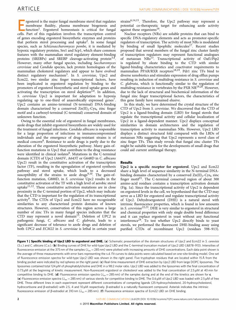
Ergosterol is the major fungal membrane sterol that regulatesmembrane fluidity, plasma membrane biogenesis andfunction1. Ergosterol homeostasis is critical for fungal
cells. Part of this regulation involves the transcription controlof genes encoding ergosterol biosynthetic enzymes and proteinsthat perform sterol processing and uptake2. In some fungalspecies, such as Schizosaccharomyces pombe, it is mediated byhypoxic regulatory proteins, Sre1 and Scp1, which share commonfeatures with the mammalian sterol regulatory element-bindingproteins (SREBPs) and SREBP cleavage-activating protein3,4.However, many other fungal species, including Saccharomycescerevisiae and Candida species, lack protein homologues of themammalian cholesterol regulators and appear to have evolveddistinct regulatory mechanism5. In S. cerevisiae, Upc2 andEcm22, two similar zinc finger transcriptional factors, havebeen implicated in ergosterol regulation by binding to thepromoters of ergosterol biosynthetic and sterol uptake genes andactivating the transcription on sterol depletion2,6. In addition,S. cerevisiae Upc2 is important for adaptation to hypoxiaregulating up to one-third of anaerobically expressed genes7.Upc2 contains an amino-terminal (N-terminal) DNA-bindingdomain characterized by a conserved Zn(II)2-Cys6 zinc fingermotif and a carboxyl-terminal (C-terminal) conserved domain ofunknown function.
Owing to the essential role of ergosterol in fungal membranes,azole drugs that inhibit ergosterol biosynthesis are widely used forthe treatment of fungal infections. Candida albicans is responsiblefor a large proportion of infections in immunocompromisedindividuals and the emergence of drug-resistant strains is ofmedical concern8. Resistance to azole drugs is partly due to thealteration of the ergosterol biosynthetic pathway. Many gain-of-function mutations in Upc2 that contribute to the drug resistancewere identified in clinical isolates8. Mutations in the C-terminaldomain (CTD) of Upc2 (A643V, A643T or G648D in C. albicansUpc2) result in the constitutive activation of the transcriptionfactor (TF), resulting in the upregulation of ergosterol synthesispathway and sterol uptake, which leads to a decreasedsusceptibility of the strains to azole drugs9,10. The gain-of-function mutation, G888D, in S. cerevisiae Upc2 results in theconstitutive activation of the TF with a high level of aerobic steroluptake11,12. These constitutive activation mutations are in closeproximity in the C-terminal portion of Upc2, which may indicatethat the CTD is important for the regulation of its transcriptionalactivity9. The CTDs of Upc2 and Ecm22 have no recognizablesimilarities to any characterized protein domains of knownstructures. However, conservation of this region across a largenumber of zinc TFs in many fungal species indicates that theCTD may represent a novel domain13. Deletion of UPC2 inpathogenic fungi, C. albicans and C. glabrata, leads to asignificant decrease of tolerance to azole drugs and deletion ofboth UPC2 and ECM22 in S. cerevisiae is lethal in certain yeast
strains6,14,15. Therefore, the Upc2 pathway may represent apotential co-therapeutic target for enhancing azole activityagainst these organisms16.
Nuclear receptors (NRs) are soluble proteins that can bind tospecific DNA-regulatory elements and acts as promotor-specificregulators of transcription. The activity of most NRs is modulatedby binding of small lipophilic molecules17. Recent studiesproposed that several members of the fungal zinc cluster familyof transcription regulators may represent functional analoguesof metazoan NRs13. Transcriptional activity of Oaf1/Pip2is regulated by oleate binding to the CTD with similarligand-binding characteristics and coactivator requirements tomammalian PPAR/RXR receptors18,19. Pdr1 zinc TFs bind todiverse xenobiotics and stimulate expression of drug efflux pumpsresulting in induction of multidrug resistance in S. cerevisiae andC. glabrata, which is functionally similar to the regulation ofmultidrug resistance in vertebrates by the PXR NR19,20. However,due to the lack of structural and biochemical information of thefungal zinc finger transcriptional factors, the characteristics ofthis gene family have remained elusive.
In this study, we have determined the crystal structure of theCTD of Upc2 from S. cerevisiae. We discovered that the CTD ofUpc2 is a ligand-binding domain (LBD) for fungal sterols thatregulate the transcriptional activity and cellular localization ofUpc2 in a ligand-dependent manner. Upc2 displays conceptualsimilarities in domain architectures and the regulation oftranscription activity to mammalian NRs. However, Upc2 LBDdisplays a distinct structural fold compared with the LBDs ofmetazoan NRs suggesting that Upc2 represents a novel class ofzinc finger TFs. This study reveals that fungal zinc cluster TFsmight be suitable targets for the developments of small drugs thatcould aid current antifungal therapy.
ResultsUpc2 is a specific receptor for ergosterol. Upc2 and Ecm22share a high level of sequence similarity in the N-terminal DNA-binding domains characterized by a conserved Zn(II)2-Cys6 zincfinger motif21. The C-terminal conserved region of about 300amino-acid residues contains a transcription activation domain(Fig. 1a). Since the transcriptional activity of Upc2 is dependenton ergosterol levels in the cell, we hypothesized that the CTD mayserve as a LBD for ergosterol and regulate transcriptional activityof Upc2. Dehydroergosterol (DHE) is a natural sterol withintrinsic fluorescence properties, which is found in low amountsin S. cerevisiae22,23. DHE is very similar to ergosterol in structuraland chemical properties with only single double bond differenceand it can replace ergosterol in yeast without any functionalinterference24. To test whether Upc2 directly binds to yeaststerols, we performed the fluorescent DHE-binding assay usingpurified CTDs of recombinant Upc2 (residues 598–913).
Figure 1 | Specific binding of Upc2 LBD to ergosterol and DHE. (a) Schematic presentation of the domain structures of Upc2 and Ecm22 in S. cerevisia
(S.c.) and C. albicans (C.a.). (b) Binding curves of DHE for wild-type Upc2 LBD and the C-terminal truncation mutant of Upc2 LBD (D878–913). Intensities of
fluorescence emission at the 375 nm of the samples (lex¼ 285 nm) were plotted with increasing amounts of DHE concentrations. Each data point shown is
the average of three measurements with error bars representing the s.d. Fit curves to data points were calculated based on one-site-binding model. One set
of fluorescence emission spectra for wild-type Upc2 LBD was shown in the right panel. Five tryptophan residues that are located within 15 Å from the
binding pocket were indicated by red spheres on the right panel. (c) Real-time measurement of DHE extraction by Upc2 LBD from large DOPC liposomes. The
liposomes contained total 124mM of phosphatidylcholine and DHE in a 98:2 molar ratio. Upc2 LBD was added to the liposomes with the final concentration of
0.73mM at the beginning of kinetic measurement. Non-fluorescent ergosterol or cholesterol was added to the final concentration of 2.5mM at 40 min for
competitive binding to DHE. (d) Fluorescence emission spectra (lex¼ 285 nm) of the samples during and at the end of the kinetics are shown for c.
(e) Fluorescence emission spectra (lex¼ 285 nm) of various sterols for competitive binding to DHE. The 0.6mM of Upc2 LBD was loaded with 2.5mM of free
DHE. Three different lines in each experiment represent different concentrations of competing ligands (25-hydroxycholesterol, 20-hydroxycholesterol,
hydrocortisone and b-estradiol) with 2.5, 4 and 10mM respectively. b-estradiol is a naturally fluorescent compound. Asterisk indicates the intrinsic
fluorescence emission of b-estradiol at 310 nm (lex¼ 285 nm). (f) Fluorescence emission spectra of Ecm22 LBD on DHE binding.
ARTICLE NATURE COMMUNICATIONS | DOI: 10.1038/ncomms7129
2 NATURE COMMUNICATIONS | 6:6129 | DOI: 10.1038/ncomms7129 | www.nature.com/naturecommunications
& 2015 Macmillan Publishers Limited. All rights reserved.

0 min
+ Erg, 0 min
0 min
+ Erg, 7 min
13 min
+ Erg, 71 min
+ Erg, 97 min20 min
40 min+ Erg, 18 min
70 min
150 min
S.c. Upc21
1
Nuclear localization signal
Linker
DNA-binding domain
Zn2Cys6 cluster 289Coiled-coils
ActivationloopLigand-binding domain
Crystal structure (598–878)
600 880
801500
712350
140
120
100
80
300
200
Ex = 285 nm
Upc2 LBD
80
60
40
0
100
80
60
40
20
0300 350 400 450
nm
Upc2 + DHE +25-hydroxycholesterol
Upc2 + DHE +20-hydroxycholesterol
Upc2 + DHE +hydrocortisone
Upc2 + DHE +β-estradiol
300 350 400 450
nm
300 350 400 450
nm
300 350 400 450
nm
300 350 400 450
nm
300 350 400 450
nm
30010
50
100
120
10
50
100
120
10
50
100
120 230200
100
10
*
350 400 450
nm
20 40 60 80
Dehydroergosterol (DHE)
Ergosterol
Cholesterol
HO
HO
HO
H
HH
H
H
HMin.
DOPC/DHE liposomes
DOPC/DHE + Upc2 DOPC/DHE:Upc2 + ergosterol Ecm22 LBD + DHE
Cholesterol (2.5 μM)
Ergosterol (2.5 μM)
100
0300 350 400 450
nm
60
40
20
Upc2
Upc2 Δ879–913
Kd = 2.5 nM ± 0.4
No binding
00.0 0.1 0.2 0.3 0.4 0.5
[DHE]t μM
Em
375
nm
(a.
u.)
Em
375
nm
(a.
u.)
Inte
nsity
(a.
u.)
Inte
nsity
(a.
u.)
100
80
60
40
20
0
Inte
nsity
(a.
u.)
7060
40
20
0
Inte
nsity
(a.
u.)
Inte
nsity
913
1
S.c. Ecm22
C.a. Upc2
NATURE COMMUNICATIONS | DOI: 10.1038/ncomms7129 ARTICLE
NATURE COMMUNICATIONS | 6:6129 | DOI: 10.1038/ncomms7129 | www.nature.com/naturecommunications 3
& 2015 Macmillan Publishers Limited. All rights reserved.
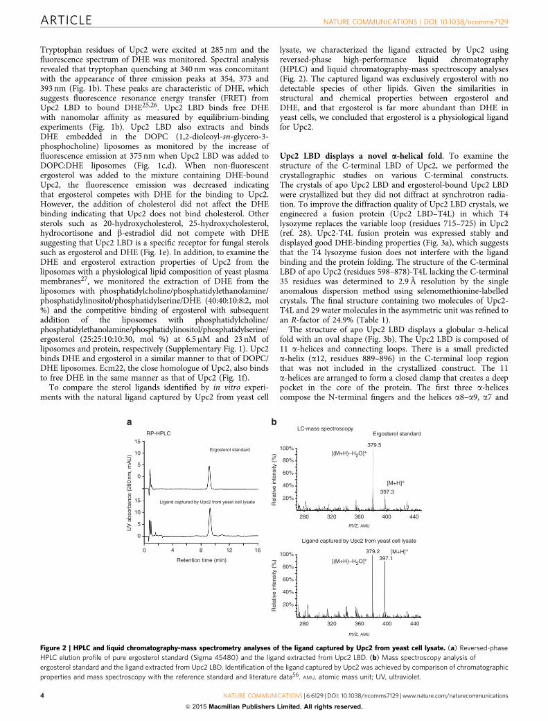
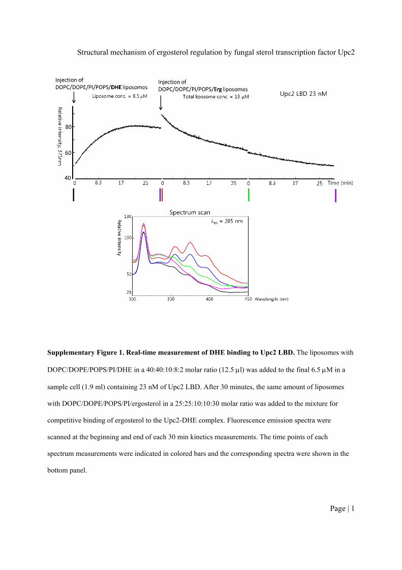
Tryptophan residues of Upc2 were excited at 285 nm and thefluorescence spectrum of DHE was monitored. Spectral analysisrevealed that tryptophan quenching at 340 nm was concomitantwith the appearance of three emission peaks at 354, 373 and393 nm (Fig. 1b). These peaks are characteristic of DHE, whichsuggests fluorescence resonance energy transfer (FRET) fromUpc2 LBD to bound DHE25,26. Upc2 LBD binds free DHEwith nanomolar affinity as measured by equilibrium-bindingexperiments (Fig. 1b). Upc2 LBD also extracts and bindsDHE embedded in the DOPC (1,2-dioleoyl-sn-glycero-3-phosphocholine) liposomes as monitored by the increase offluorescence emission at 375 nm when Upc2 LBD was added toDOPC:DHE liposomes (Fig. 1c,d). When non-fluorescentergosterol was added to the mixture containing DHE-boundUpc2, the fluorescence emission was decreased indicatingthat ergosterol competes with DHE for the binding to Upc2.However, the addition of cholesterol did not affect the DHEbinding indicating that Upc2 does not bind cholesterol. Othersterols such as 20-hydroxycholesterol, 25-hydroxycholesterol,hydrocortisone and b-estradiol did not compete with DHEsuggesting that Upc2 LBD is a specific receptor for fungal sterolssuch as ergosterol and DHE (Fig. 1e). In addition, to examine theDHE and ergosterol extraction properties of Upc2 from theliposomes with a physiological lipid composition of yeast plasmamembranes27, we monitored the extraction of DHE from theliposomes with phosphatidylcholine/phosphatidylethanolamine/phosphatidylinositol/phosphatidylserine/DHE (40:40:10:8:2, mol%) and the competitive binding of ergosterol with subsequentaddition of the liposomes with phosphatidylcholine/phosphatidylethanolamine/phosphatidylinositol/phosphatidylserine/ergosterol (25:25:10:10:30, mol %) at 6.5 mM and 23 nM ofliposomes and protein, respectively (Supplementary Fig. 1). Upc2binds DHE and ergosterol in a similar manner to that of DOPC/DHE liposomes. Ecm22, the close homologue of Upc2, also bindsto free DHE in the same manner as that of Upc2 (Fig. 1f).
To compare the sterol ligands identified by in vitro experi-ments with the natural ligand captured by Upc2 from yeast cell
lysate, we characterized the ligand extracted by Upc2 usingreversed-phase high-performance liquid chromatography(HPLC) and liquid chromatography-mass spectroscopy analyses(Fig. 2). The captured ligand was exclusively ergosterol with nodetectable species of other lipids. Given the similarities instructural and chemical properties between ergosterol andDHE, and that ergosterol is far more abundant than DHE inyeast cells, we concluded that ergosterol is a physiological ligandfor Upc2.
Upc2 LBD displays a novel a-helical fold. To examine thestructure of the C-terminal LBD of Upc2, we performed thecrystallographic studies on various C-terminal constructs.The crystals of apo Upc2 LBD and ergosterol-bound Upc2 LBDwere crystallized but they did not diffract at synchrotron radia-tion. To improve the diffraction quality of Upc2 LBD crystals, weengineered a fusion protein (Upc2 LBD–T4L) in which T4lysozyme replaces the variable loop (residues 715–725) in Upc2(ref. 28). Upc2-T4L fusion protein was expressed stably anddisplayed good DHE-binding properties (Fig. 3a), which suggeststhat the T4 lysozyme fusion does not interfere with the ligandbinding and the protein folding. The structure of the C-terminalLBD of apo Upc2 (residues 598–878)-T4L lacking the C-terminal35 residues was determined to 2.9 Å resolution by the singleanomalous dispersion method using selenomethionine-labelledcrystals. The final structure containing two molecules of Upc2-T4L and 29 water molecules in the asymmetric unit was refined toan R-factor of 24.9% (Table 1).
The structure of apo Upc2 LBD displays a globular a-helicalfold with an oval shape (Fig. 3b). The Upc2 LBD is composed of11 a-helices and connecting loops. There is a small predicteda-helix (a12, residues 889–896) in the C-terminal loop regionthat was not included in the crystallized construct. The 11a-helices are arranged to form a closed clamp that creates a deeppocket in the core of the protein. The first three a-helicescompose the N-terminal fingers and the helices a8–a9, a7 and
15
15
10
5
0
UV
abs
orba
nce
(280
nm, m
AU
)
Rel
ativ
e in
tens
ity (
%)
Rel
ativ
e in
tens
ity (
%)
10
5
0
0 4 8
Retention time (min)
Ergosterol standard 100%
LC-mass spectroscopy
Ligand captured by Upc2 from yeast cell lysate
Ergosterol standard
379.5
[(M+H)–H2O]+
[M+H]+
[M+H]+
[(M+H)–H2O]+
397.3
379.2397.1
80%
60%
40%
20%
100%
80%
60%
40%
20%
280 320 360 400 440
280 320 360 400 440
m/z, AMU
m/z, AMU
Ligand captured by Upc2 from yeast cell lysate
RP-HPLC
12 16
Figure 2 | HPLC and liquid chromatography-mass spectrometry analyses of the ligand captured by Upc2 from yeast cell lysate. (a) Reversed-phase
HPLC elution profile of pure ergosterol standard (Sigma 45480) and the ligand extracted from Upc2 LBD. (b) Mass spectroscopy analysis of
ergosterol standard and the ligand extracted from Upc2 LBD. Identification of the ligand captured by Upc2 was achieved by comparison of chromatographic
properties and mass spectroscopy with the reference standard and literature data56. AMU, atomic mass unit; UV, ultraviolet.
ARTICLE NATURE COMMUNICATIONS | DOI: 10.1038/ncomms7129
4 NATURE COMMUNICATIONS | 6:6129 | DOI: 10.1038/ncomms7129 | www.nature.com/naturecommunications
& 2015 Macmillan Publishers Limited. All rights reserved.

a11 compose the other fingers of the clamp. The four helicesa3–a6 compose the central palm as a helical bundle. The a7–a8loop (residues 760–799) surround the helices a8–a9 as a longestrandom coil in the structure. The T4 lysozyme replacing nineresidues in the a5–a6 loop is closely located on the surface ofUpc2 LBD (Fig. 3c).
The structure of Upc2 LBD reveals a central hydrophobicpocket surrounded by a helices (Fig. 3d). The deep hydrophobicpocket has a volume of 287 Å3. The helices a5 and a6 composethe bottom of the pocket and the helices a1–a2, a7, a9 and a11compose the wall of the hydrophobic binding pocket. The pocketis accessible to a solvent with a small pore on the surface. Thepresence of a hydrophobic pocket suggested to us that it mayserve as a binding site for small lipophilic molecules. Structuralcomparison of Upc2 with known structures in the PDB using theDALI server did not find related structures with Z scores 47suggesting that the Upc2 LBD represents a novel fold of a LBD.
Upc2 LBD forms a constitutive homodimer. Size-exclusionchromatography (SEC) analysis demonstrated that the recombi-nant Upc2 LBD or Upc2 LBD–T4L is a homogeneous dimerwithout a detectable monomeric species, suggesting that dimericUpc2 is a biologically relevant form. The dimer of Upc2 LBDcontains two sterol-binding sites and the ligand binding did notaffect the oligomeric state of Upc2 as analysed by SEC (Fig. 4a).There were two molecules of Upc2 LBD–T4L related by a non-crystallographic two-fold axis in the asymmetric unit of thecrystals. Upc2 LBD dimerizes by helices a1 and a2 forming afour-helix bundle in the dimer interface (Fig. 4b). The N terminiof Upc2 LBD dimer are exposed to the same side near the centerof dimer interface. The dimer interface is mainly composed of
hydrophobic residues burying 1,562 Å2 of surface area at theinterface (Fig. 4c). The Met610 is located in the center of dimerinterface. M610R mutant is a monomer in solution analysed bySEC suggesting that the dimer observed in the crystal structure isconsistent with that observed in solution (Fig. 4a). The LBDs ofC. albicans Upc2 and S. cerevisiae Ecm22 are also dimers insolution and the M506R mutation of C. albicans Upc2 led to themonomerization of the LBD. The conservation of the residuescomposing the dimer interface in Upc2 homologues suggests thatthe homodimerization of LBDs is a general feature of theseproteins (Supplementary Fig. 2).
The C-terminal loop is essential for ligand binding. Themajority of the constitutive activation mutations in S. cerevisiaeUpc2 and C. albicans Upc2 are clustered in the C-terminal loopbetween helix a11 and the predicted a12 helix21,29. The crystalstructure does not include this C-terminal activation loop of35 residues. To examine the functional importance of thisC-terminal loop, we performed the ligand-binding assays on theC-terminal deletion mutants of Upc2 LBD (Fig. 5a). Truncationof the C-terminal 14 residues (D900–913) did not affectligand binding. However, deletion of the C-terminal 35 residues(D879–913), including the predicted a12, abolished DHE binding,suggesting that the C-terminal glycine rich loop and helix a12 areessential for ligand binding (Fig. 5b). In addition, G888D, aconstitutive activation mutation in the a11–a12 loop inhibitedDHE binding significantly.
The crystal structure of Upc2 LBD shows a deep hydrophobicpocket that might accommodate a single sterol molecule.However, the size of the binding pocket in apo Upc2 LBDlacking the C-terminal activation loop is slightly smaller than the
60
40
20
0
300 350 400 450
C
I758L820
V629
F823L703V699
I690
V687
L749
I759
α4
α5
α6
α11α1′
T4 Lysozyme(residue 2–161)
T4 Lysozyme(residue 2–161)
N
N C
N-term604
C-term878
Residue 715–725lysozme fusion
α1
α1′α2
α3
α4α5
α11α10
α9 α8
α7
α6
Buffer
Buffer + DHE
apo Upc2-T4L
Upc2-T4L + DHE 1 min
Upc2-T4L + DHE 5 minUpc2-T4L + DHE 30 min
Protein conc. = 0.20 μMDHE conc. = 2.5 μM
�ex = 285 nm
Wavelength (nm)
A chain
B chain
Flu
ores
cenc
e in
tens
ity (
a.u.
)
Figure 3 | Structure of Upc2 LBD. (a) Fluorescence emission spectra of Upc2 LBD–T4L fusion construct (residues 598–913) on DHE binding.
(b) Overall structure of Upc2 LBD. The structure was coloured with red to blue based on the secondary structure succession. The T4 lysozyme fused
in the a5–a6 loop was not shown for clarity. (c) Overall structure of Upc2 LBD–T4L dimer with cylindrical representation. The location of deep hydrophobic
pocket in one of the Upc2 protomers is indicated by a black circle. (d) Surface representation of Upc2 LBD monomer. The residues composing the
wall of the hydrophobic pocket are shown in sticks. The hydrophobic pocket is shown in grey surface representation. Conc., concentration; C-term,
C-terminal; N-term, N-terminal.
NATURE COMMUNICATIONS | DOI: 10.1038/ncomms7129 ARTICLE
NATURE COMMUNICATIONS | 6:6129 | DOI: 10.1038/ncomms7129 | www.nature.com/naturecommunications 5
& 2015 Macmillan Publishers Limited. All rights reserved.
dimensions of ergosterol implying that Upc2 LBD must undergoa conformational change on ligand binding. To identify thestructural determinants for sterol binding of Upc2, we tried to co-crystallize Upc2 LBD with ergosterol. However, due to the poordiffraction of sterol-bound Upc2 crystals, the structural determi-nation of the ligand complex was not possible. Alternatively, wetested the effect of several mutations in the hydrophobic pocketon sterol binding. The mutation of residues distal to the pocket,V699Y and R752A, did not affect ligand binding. Mutation ofresidues in the hydrophobic binding pocket to bulky residues(L703F and L820W) significantly reduced DHE binding con-firming that the hydrophobic pocket accommodates sterol(Fig. 5b). In addition, the M610R mutation completely abolishedthe sterol binding, implying that the dimerization of Upc2 LBD isrequired for sterol binding. The LBDs of all Upc2 homologues infungal species display a high degree of sequence conservation(Supplementary Fig. 2). Specifically, the residues located in thedimerization interface, ligand-binding pocket and the C-terminalactivation loop are strictly conserved.
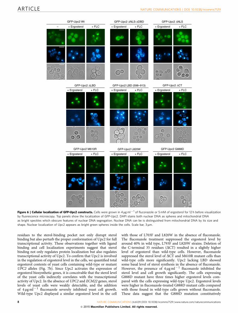
Cytosolic Upc2 relocalizes into the nucleus on activation. Theunique structural and ligand-binding features of Upc2 suggestedto us that Upc2 may relocalize to nucleus on ligand binding in thecytosol. We examined the cellular localization of full-length Upc2and its mutants under ergosterol-rich or ergosterol-deficientconditions. Previously, there were several conflicting reports onthe localization of Upc2 in the nucleus, cytoplasm and peri-nuclear foci using the C-terminal green fluorescent protein (GFP)fusion30–32. However, the key determinants of Upc2 localizationhave not been experimentally defined. In this study, to avoid thefunctional interference of GFP with the C-terminal activationloop and ligand binding, GFP was tagged to the N terminus of
Upc2. Wild-type Upc2 is present mostly in the cytosol with mildnuclear localization in 30% of the cell population. When the yeastcells were grown in the presence of 5 mM ergosterol, Upc2 waslocalized almost exclusively to the cytoplasm. However, additionof 4 mg ml� 1 of fluconazole to the culture media, which inhibitsthe biosynthesis of ergosterol, shifted the localization of Upc2 tonucleus in an entire cell population (Fig. 6). Upc2 contains abipartite nuclear localization signal (NLS) upstream of itsZn2-Cys6 cluster sequence that confers an intrinsic nuclearlocalization property. The deletion of NLS (residues 2–43) madeUpc2 completely cytosolic. In addition, the isolated LBD (residues598–913) or the construct lacking NLS-DBD (construct residues283–913) was completely cytosolic regardless of sterol levels. Incontrast, deletion of the C-terminal LBD (DLBD, constructresidues 1–559) showed strong nuclear localization independentof sterol levels. These data suggest that the ergosterol-boundC-terminal LBD suppresses the nuclear transport of Upc2 byinhibiting the NLS function. The dissociation of sterol ligandfrom Upc2 may lead to exposure of the NLS or release Upc2 fromthe capture by cytosolic proteins for nuclear transport. Thedeletion of the C-terminal loop (D879–913) that disruptsergosterol binding led to modest nuclear localization of Upc2.G888D or mutations in the sterol-binding pocket (L820W orL703F) led to strong constitutive nuclear localization of Upc2.The M610R mutation that disrupts dimerization of LBD andligand binding shows mostly nuclear localization regardless ofsterol level, suggesting that dimerization of Upc2 is essential forits regulatory function. In conclusion, these observations togetherwith ligand-binding assays suggest that sterol binding is thekey mechanism in regulating the localization of the TF. In asterol-rich condition, Upc2 is bound to ergosterol and is presentin the cytosol as a repressed form. On depletion of ergosterol,unliganded Upc2 is activated and moves to the nucleus fortranscriptional activation of related genes.
Upc2 regulates ergosterol level in yeast cells. Upc2 and Ecm22bind to a conserved 7-bp sterol regulatory element within pro-motors of most ERG genes including ERG2, ERG3 and ERG11(refs 2,21). To investigate the mechanism of transcriptionalregulation of Upc2, we check the transcriptional activity ofvarious Upc2 constructs using ERG2-LacZ as a reporter (Fig. 7a).The wild-type Upc2 displayed a moderate transcriptional activityin a normal growth condition. Fluconazole treatment increasesthe transcription activity more than two times. However, emptyplasmid or deletion of the C-terminal LBD showed very weaktranscription activity suggesting that LBD is essential for thetranscriptional activity of Upc2. G888D mutation, regardless offluconazole treatment, showed a full constitutive activation ofUpc2, which is 1.2 times higher than the induced wild type. Thisobservation is consistent with the previous report that the CTD isessential for the transcription activation of Upc2 (ref. 21). TheM610R showed no increase of transcriptional activity on steroldepletion suggesting that dimerization is essential for theregulatory function. Deletion of the C-terminal activation loop(DCT) that shows constitutive nuclear localization, however,displayed at least 30% lower transcriptional activity thanuninduced wild type. In addition, the transcription activity ofDCT did not increase on fluconazole treatment. These datasuggest that the C-terminal activation loop is essential not onlyfor the regulation of protein localization but for the fulltranscription activity of Upc2. Disruption of sterol binding byL703F or L820W showed constitutive nuclear localization.However, the transcription activities of these mutants were 40%less than the wild type and were not enhanced significantly onfluconazole induction. It seems that introduction of bulky
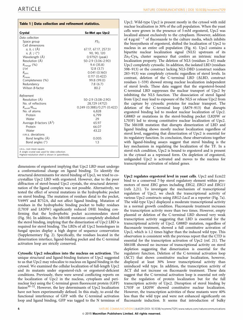
Table 1 | Data collection and refinement statistics.
Crystal Se-Met apo Upc2
Data collectionSpace group P32
Cell dimensionsa, b, c (Å) 67.17, 67.17, 257.51a, b, g (�) 90, 90, 120
Wavelength (Å) 0.97921 (peak)Resolution (Å) 50–2.9 (3.06–2.90)Rmerge (%) 9.4 (35.8)I/sI 12.8 (3.7)Rpim 0.041 (0.160)Rmeas 0.117 (0.422)Completeness (%) 99.8 (99.0)Redundancy 7.8 (6.7)Wilson B-factor 80.2
RefinementResolution (Å) 50–2.9 (3.08–2.90)No. of reflections 28,729 (4712)Rwork/Rfree 0.249 (0.388)/0.271 (0.422)No. of atoms 6,828
Protein 6,799Water 29
Average B-factors (Å2) 65.97Protein 66.07Water 43.22
r.m.s. deviationsBond lengths (Å) 0.005Bond angles (�) 1.3
r.m.s., root mean square.A single crystal was used for data collection.Highest-resolution shell is shown in parenthesis.
ARTICLE NATURE COMMUNICATIONS | DOI: 10.1038/ncomms7129
6 NATURE COMMUNICATIONS | 6:6129 | DOI: 10.1038/ncomms7129 | www.nature.com/naturecommunications
& 2015 Macmillan Publishers Limited. All rights reserved.
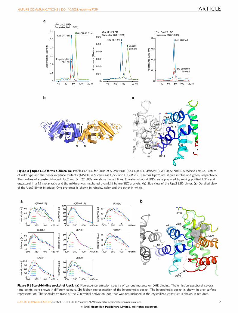
S.c. Upc2 LBDSuperdex 200 (16/60)
0.6
0.5
0.4
0.3
0.2
0.1
040 60 80 100 120 ml 40 60 80 100 ml 120 ml100806040
0.1
0
0.2
0.3
0.4
0.04
0.06
0.05
0.03
0.02
0.01
0
C.a. Upc2 LBDSuperdex 200 (16/60)
S.c. Ecm22 LBDSuperdex 200 (16/60)
Erg complex74.9 ml
Apo 74.7 mlM610R 86.5 ml
Apo 75.1 ml
L506R88.5 ml
Apo 76.2 ml
Erg complex75.9 ml
Abs
orba
nce
(280
nm
)
Abs
orba
nce
(280
nm
)
Abs
orba
nce
(280
nm
)
C
N
C
N
M610
α1
α1′α2
α5
α3
α2
α1′
α1
α1
α2
F644
F644
H614
K611
L607
C617
E641
H614
M610
F613
F613
Figure 4 | Upc2 LBD forms a dimer. (a) Profiles of SEC for LBDs of S. cerevisiae (S.c.) Upc2, C. albicans (C.a.) Upc2 and S. cerevisiae Ecm22. Profiles
of wild type and the dimer interface mutants (M610R in S. cerevisiae Upc2 and L506R in C. albicans Upc2) are shown in blue and green, respectively.
The profiles of ergosterol-bound Upc2 and Ecm22 LBDs are shown in red lines. Ergosterol-bound LBDs were prepared by mixing purified LBDs and
ergosterol in a 1:5 molar ratio and the mixture was incubated overnight before SEC analysis. (b) Side view of the Upc2 LBD dimer. (c) Detailed view
of the Upc2 dimer interface. One protomer is shown in rainbow color and the other in white.
0 min11 min61 min
110 min
0 min20 min80 min
140 min
0 min30 min63 min
120 min
0 min20 min80 min
0 min60 min
110 min150 min
0 min26 min76 min
0 min15 min70 min
140 min
0 min17 min70 min97 min
L703F
V699YG888D M610R
L820W
R752AΔ(879–913)Δ(900–913)
Inte
nsity
(a.
u.)
Inte
nsity
(a.
u.)
Inte
nsity
(a.
u.)
Inte
nsity
(a.
u.)
Inte
nsity
(a.
u.)
Inte
nsity
(a.
u.)
Inte
nsity
(a.
u.)
Inte
nsity
(a.
u.)
R752
M610
V699
L703
L820
F823
Q878
α1
α1
α2
α3
α5
α2α11
60
40
20
0300 350 400 450 nm 300 350 400 450nm 300 350 400 450nm
300 350 400 450 nm 300 350 400 450nm
300 350 400 450 nm 300 350 400 450nm
300 350 400 450nm
60
40
20
0
60
40
20
0
60
80
100
40
20
0
0
30
60
90
120
60
40
20
0
0
30
60
90
–10
10
30
50
Figure 5 | Sterol-binding pocket of Upc2. (a) Fluorescence emission spectra of various mutants on DHE binding. The emission spectra at several
time points were shown in different colours. (b) Ribbon representation of the hydrophobic pocket. The hydrophobic pocket is shown in grey surface
representation. The speculative trace of the C-terminal activation loop that was not included in the crystallized construct is shown in red dots.
NATURE COMMUNICATIONS | DOI: 10.1038/ncomms7129 ARTICLE
NATURE COMMUNICATIONS | 6:6129 | DOI: 10.1038/ncomms7129 | www.nature.com/naturecommunications 7
& 2015 Macmillan Publishers Limited. All rights reserved.
residues to the sterol-binding pocket not only disrupt sterolbinding but also perturb the proper conformation of Upc2 for fulltranscriptional activity. These observations together with ligandbinding and cell localization experiments suggest that sterolbinding not only regulates protein localization but also regulatestranscriptional activity of Upc2. To confirm that Upc2 is involvedin the regulation of ergosterol level in the cells, we quantified totalergosterol contents of yeast cells containing wild-type or mutantUPC2 alleles (Fig. 7b). Since Upc2 activates the expression ofergosterol biosynthetic genes, it is conceivable that the sterol levelof the yeast cells indirectly correlates with the transcriptionalactivity of Upc2. In the absence of UPC2 and ECM22 genes, sterollevels of yeast cells were weakly detectable, and the additionof 4mg ml� 1 fluconazole severely inhibited yeast cell growth.Wild-type Upc2 displayed a similar ergosterol level in the cell
with those of L703F and L820W in the absence of fluconazole.The fluconazole treatment suppressed the ergosterol level byaround 40% in wild type, L793F and L820W strains. Deletion ofthe C-terminal 35 residues (DCT) resulted in a slightly higherlevel of ergosterol than wild-type cells. However, fluconazolesuppressed the sterol level of DCT and M610R mutant cells thanwild-type cells more significantly. Upc2 lacking LBD showedsome basal level of sterol synthesis in the absence of fluconazole.However, the presence of 4mg ml� 1 fluconazole inhibited thesterol level and cell growth significantly. The cells expressingG888D mutant have three times higher ergosterol levels com-pared with the cells expressing wild-type Upc2. Ergosterol levelswere higher in fluconazole-treated G888D mutant cells comparedwith those found in wild-type cells grown without fluconazole.These data suggest that the G888D mutation constitutively
GFP-Upc2 Wt
GFP
DAPI
BF
GFP-Upc2 ΔNLS-ΔDBD
GFP-Upc2 LBD (598–913)GFP-Upc2 ΔLBD
GFP-Upc2 M610R GFP-Upc2 L820W GFP-Upc2 G888D
GFP-Upc2 ΔCT
GFP-Upc2 ΔNLS
+ Ergosterol
+ Ergosterol
+ Ergosterol
+ Ergosterol
+ Ergosterol
+ Ergosterol+ Ergosterol
+ Ergosterol
+ Ergosterol + FLC
+ FLC
+ FLC+ FLC
+ FLC
+ FLC
+ FLC
+ FLC
+ FLC–
Figure 6 | Cellular localization of GFP-Upc2 constructs. Cells were grown in 4mg ml� 1 of fluconazole or 5 mM of ergosterol for 12 h before visualization
by fluorescence microscopy. Top panels show the localization of GFP-Upc2. DAPI stains both nuclear DNA as spheres and mitochondrial DNA
as bright speckles which obscure features of nuclear DNA segregation. Nuclear DNA can be is distinguished from mitochondrial DNA by its size and
shape. Nuclear localization of Upc2 appears as bright green spheres inside the cells. Scale bar, 3mm.
ARTICLE NATURE COMMUNICATIONS | DOI: 10.1038/ncomms7129
8 NATURE COMMUNICATIONS | 6:6129 | DOI: 10.1038/ncomms7129 | www.nature.com/naturecommunications
& 2015 Macmillan Publishers Limited. All rights reserved.

activates Upc2 for the biosynthesis of ergosterol, which leads toan increased resistance to azole antifungals. To confirm that Upc2is involved in the regulation of gene expression of ergosterolbiosynthetic enzymes, we performed quantitative gene expressionanalysis of ERG11 by real-time PCR using the upc2/ecm22-knockout strains with wild-type and mutant UPC2 alleles. Wemeasured the mRNA levels of ERG11 genes that were normalizedby TAF10 gene as an internal control33. The data indicate thatthe mRNA expression levels of ERG11 correlate with thepromotor activities of ERG2 and ergosterol levels as expected(Supplementary Fig. 3).
Activation of Upc2 confers resistance to antifungal agent. Toinvestigate the relationship between the activation of Upc2 and
the resistance to antifungal agents, we checked the fluconazolesusceptibilities of upc2D ecm22D or ecm22D strains expressingwild-type or mutant UPC2 alleles (Fig. 7c). Since fluconazoleinterferes with yeast cell growth by inhibiting ergosterol bio-synthesis, the overall patterns of cell growths displayed a corre-lation with the sterol levels of the cells treated with fluconazole.Double knockout cells of upc2D ecm22D did not grow in thepresence of fluconazole. Introduction of wild-type UPC2permitted weak cell growth at 8 mg ml� 1 of fluconazole. The yeastcells expressing a G888D mutant showed a significant increase ofresistance to fluconazole at 8 mg ml� 1. However, G888D mutantgrew more weakly than wild type in the absence of fluconazolesuggesting that full constitutive activation of Upc2 is dis-advantageous under non-selective condition by imposing meta-bolic burden34. DLBD mutant lacking transcription activity did
Empty YCplac33
24
*
*
*
* *
**
#
#
##
#
#
#
#
1.6
1.2
0.8
0.4
0.0
Rel
ativ
e er
gost
erol
leve
l (A
bs. 2
80 n
m)
20
β-ga
lact
osid
ase
activ
ity (
mili
uni
ts)
15
10
5
0
WT
M61
0R
L703
F
L820
W
G888D
YCplac33-UPC2
ΔCTΔLB
D
upc2� ecm22�
upc2� ecm22�pERG2::lacZ expression upc2� ecm22�
No fluconazoleFluconazole 4 μg ml–1 No fluconazole
Fluconzaole 4 μg ml–1
upc2�
Fluconazole 0 μg ml–1 4 μg ml–1 6 μg ml–1 8 μg ml–1
Fluconazole 0 μg ml–1 4 μg ml–1 6 μg ml–1 8 μg ml–1
Upc2 WT
Upc2 M610R
Upc2 L703F
Upc2 L820W
Upc2 G888D
Upc2 ΔCT
Upc2 ΔLBD
Empty YCplac33
Upc2 WT
Upc2 M610R
Upc2 L703F
Upc2 L820W
Upc2 G888D
Upc2 ΔCT
Upc2 ΔLBD
EmptyplasmidW
T
M61
0R
L703
F
L820
W
G888D
YCplac33-UPC2
ΔCTΔLB
DEmptyplasmid
Figure 7 | Ergosterol level and antibiotic resistance. (a) Transcriptional activity of Upc2. ERG2-lacZ reporters were used to determine the contribution
of each Upc2 constructs to the regulation of ERG2 promotor. b-Galactosidase assays were performed on each transformants after 5 ml cultures were
grown for 16 h either in minimal medium (uninduced) or in the minimal medium containing 4mg ml� 1 of fluconazole (induced). The assay values represent
the average of three independent experiments. All error bars indicate s.d. P values were assessed by two-tailed Student’s t-test. *Po0.05 versus wild
type, #Po0.05 versus each mutant without fluconazole. (b) Quantification of total ergosterol in yeast cells. Yeast upc2D ecm22D strains containing
various UPC2 alleles were analysed for their ergosterol contents. Each data point is the average of three measurements. All error bars indicate s.d.
P values were assessed by two-tailed Student’s t-test. *Po0.05 versus wild type, #Po0.05 versus fluconazole-treated wild-type. (c) Susceptibilities to
fluconazole of yeast strains expressing wild-type or mutant UPC2 allele. The strains were initially grown without fluconazole and the dilution series
were spotted on the agar plates containing fluconazole and incubated for 36 h.
NATURE COMMUNICATIONS | DOI: 10.1038/ncomms7129 ARTICLE
NATURE COMMUNICATIONS | 6:6129 | DOI: 10.1038/ncomms7129 | www.nature.com/naturecommunications 9
& 2015 Macmillan Publishers Limited. All rights reserved.
not grow at the lowest fluconazole concentration. L820W, L703F,M610R and DCT mutants that interfere with sterol binding weremore susceptible to fluconazole than the wild type. Single upc2D-knockout strain harbouring ECM22 allele permitted weak growthin a low concentration of fluconazole suggesting that ECM22 hasa partially overlapping role in activating genes involved in azoleresistance. However, in the fluconazole concentrations higherthan 6 mg ml� 1, upc2D and upc2D ecm22D strains did not displaysignificant differences in the susceptibilities of all Upc2constructs, suggesting that Upc2 plays a dominant role in thesterol regulation. This is consistent with the previous observationsthat deletion of UPC2 results in ketoconazole sensitivity, whereasno effect was observed with ECM22 deletion suggesting thatEcm22 and Upc2 have specific targets with some essentialoverlapping functions35,36.
Upc2 is a novel class of zinc finger TFs. Recent studies proposedthat several members of the fungal zinc cluster family of tran-scription regulators may represent functional analogues ofmetazoan NRs13,18–20.
While fungal zinc cluster TFs have no apparent sequence orstructural similarity to metazoan NRs, Upc2 displays conceptualsimilarities with steroid NRs in general domain architectures,sterol ligand binding, homodimerization, ligand-dependenttranscriptional regulation and nuclear translocation.
Upc2 and metazoan NRs both contain zinc finger DNA-binding domains in their N-terminal regions. Zinc fingerscoordinating two zinc ions are composed of six cysteines forUpc2 and four cysteines in each of the two fingers for NRs37. TheDNA-binding domains of NRs and zinc cluster TFs arestructurally similar and they bind to DNA as homo- orheterodimers13. Zinc finger TFs have a C-terminal negativeregulatory domain, which for a number of family membersmediates ligand binding or response to small molecules such ascellular metabolites and lipophilic ligands13. Transcriptionalactivity of Upc2 is regulated by ergosterol, which is functionallysimilar to Type I steroid NRs in terms of ligand specificities.Upc2 and steroid receptors both form homodimers. Upc2 formsconstitutive homodimers regardless of ligand binding. Oestrogenreceptor a has been reported to exist as a dimer even in the apostate and the ligand binding further stabilize the homodimer38.
Structural comparison of Upc2 with mammalian steroid NRsreveals that Upc2 has a distinctive a-helical fold of LBD frommammalian NRs (Supplementary Fig. 4). Both gene regulators aremainly composed of around 12 a-helical structures with similardimensions of LBDs. LBDs of Upc2, oestrogen and progesteronereceptors form dimers by helix bundle interactions. However,except the overall configuration of dimers with ligand-bindingsites closely located to the dimer interface, there is no clearstructural homology between Upc2 and the NRs. On ligandbinding, steroid receptors dissociate from heat shock proteinHsp90 in the cytosol and translocate to the nucleus, binding totheir target gene promotors as homodimers17. Consistently, thetranslocation of Upc2 from the cytosol to the nucleus is regulatedin a ligand-dependent manner.
However, Upc2 LBD displays a completely different structuralfold compared with the LBDs of metazoan NRs. The lack ofsequence and structural similarities of fungal zinc cluster TFs andmetazoan NRs suggests that the architectural and functionalsimilarities are the product of convergent evolution with commonmechanistic strategy. Phylogenetic analysis shows that theorthologues of Upc2 are only identifiable within the Sacchar-omycotina and Upc2/Ecm22 proteins form monophyletic cladethat is not closely related to any other Zn2-Cys6 proteins ofSaccharomycotina5. The budding yeast, S. cerevisiae, containsaround 50 Zn2-Cys6 TFs39. Even though sequence homology ofUpc2 to other types of zinc finger TFs is low, they harbour aconserved fungal-specific sequence within the C-terminal LBDsthat was previously known as the MHR (middle homologyregion)40. The C-terminal region, including the MHR, in zinc TFscommonly contains 13–15 a-helices of secondary structureelements, which implies that the presence of a LBD is a generalfeature of this protein family. In conclusion, Upc2 represent anovel class of zinc finger TFs displaying a functional analogy withmetazoan steroid receptors.
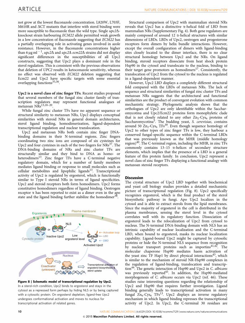
DiscussionThe crystal structure of Upc2 LBD together with biochemicaland yeast cell biology studies provides a detailed mechanisticpicture of transcriptional regulation (Fig. 8). Upc2 specificallyrecognizes ergosterol, which is the final product of the sterolbiosynthetic pathway in fungi. Apo Upc2 localizes in thecytosol and is able to extract sterols from the lipid membranes.Since the majority of ergosterol in the cell is distributed in theplasma membranes, sensing the sterol level in the cytosolcorrelates well with its regulatory function. Dissociation ofergosterol leads to the relocalization of Upc2 from cytosol tonucleus. The N-terminal DNA-binding domain with NLS has anintrinsic capability of nuclear localization and the C-terminalLBD, when bound to ergosterol, masks its nuclear localizationcapability. Ligand-bound Upc2 might be captured by cytosolicproteins or hide the N-terminal NLS sequence from recognitionby nuclear transport proteins such as importins41,42. Themolecular chaperone Hsp90 mediates haem activation ofthe yeast zinc TF Hap1 by direct physical interactions43, whichis similar to the mechanism of steroid NR-Hsp90 complexes inthe regulation of ligand-binding, translocation and transcrip-tion44. The genetic interaction of Hsp90 and Upc2 in C. albicanswas previously reported45. In addition, the Hsp90-mediatedmorphogenesis of C. albicans occurs via Upc2 (ref. 46). Thesestudies raise interesting questions regarding the relationship ofUpc2 and Hsp90 that requires further investigation. Ligandbinding generally leads to transcriptional activation in manyfungal Zn6-Cys6 TFs13. Upc2 displays an inverse regulatorymechanism in which ligand binding represses the transcriptionalactivity of Upc2. In Upc2, the C-terminal 30 residues are
ErgosterolsPlasma membrane
or
Cytosolic proteinInactive Upc2 Low
ergosterol level
NLS
Activated Upc2
Nucleus
Coactivator(unknown)
Transcriptional activation ofergosterol biosynthesis &sterol uptake
DBD
LBDActivation loop
Figure 8 | Schematic model of transcriptional regulation by Upc2.
In a sterol-rich condition, Upc2 binds to ergosterol and stays in the
cytosol as a repressed form perhaps by hiding NLS or by being captured
with a cytosolic protein. On ergosterol depletion, ligand-free Upc2
undergoes conformational activation and moves to nucleus for
transcriptional activation of related genes.
ARTICLE NATURE COMMUNICATIONS | DOI: 10.1038/ncomms7129
10 NATURE COMMUNICATIONS | 6:6129 | DOI: 10.1038/ncomms7129 | www.nature.com/naturecommunications
& 2015 Macmillan Publishers Limited. All rights reserved.
defined as an ‘activation loop’. It contains a highly conservedglycine residues connected to the predicted two-turn amphipathica-helix (a12). The activation loop in Upc2 is not only involvedin sterol binding and regulation of protein localization, butalso involved in transcriptional activation. The conformationalflexibility of the C-terminal activation loop seems to be essentialfor the ligand binding and for the transcriptional regulation.Any substitution larger than alanine at G888 results in an increaseof gene expression similar to that of the original G888Dmutation21. Upc2 DCT does not bind DHE and it showsconstitutive nuclear localization. G888D mutation in theactivation loop also abolishes DHE binding, which indicatesubtle changes in the activation loop have a significant influenceon ligand binding properties. This suggests that the activatedform of apo Upc2 LBD has a different conformation from theligand-bound state. The structure of Upc2 LBD DCT shows adeep hydrophobic pocket (287 Å3) that has a volume smallerthan the value of ergosterol (427 Å). We suppose that the DCTor G888D mutations lead to an irreversible change to theactivated conformation of LBD. Dissociation of ergosterol fromthe Upc2 LBD might result in conformational changes thatliberate the C-terminal activation loop from intramolecularinhibitory contacts, allowing recruitment of transcriptionalcoactivators. In yeast, the mediator Gal1p/MED15 is known toplay a critical role as a subunit of coactivators for Oaf1 and Pdr1TFs18,20. Currently, the downstream coactivator of Upc2 isunknown. Discovery of the coactivator for Upc2 and themechanistic characterization of coactivator regulation wouldprovide important clues for the relationship between fungalzinc cluster TFs and metazoan NRs.
Many zinc cluster TFs bind small-molecule intermediates,substrates or end products of the metabolic pathways that areunder their transcriptional controls13. In contrast, manymammalian NRs bind small molecules of extracellular origin.Therefore, fungal zinc cluster TFs seem to have evolved tomonitor and regulate the metabolic pathways by sensing their keycompounds. Some fungal species that contains SREBPhomologues do not harbour the UPC2 gene and vice versa,implying that they might have developed separate regulatorymechanism of sterol homeostasis during evolution5. Depletion ofsterol levels by azole antifungals stimulates the activation of steroluptake genes by Upc2 (ref. 2). Although there are severalstructural differences between cholesterol and ergosterol, theyboth efficiently support the growth of S. cerevisiae and C. glabrataincapable of ergosterol biosynthesis47. Since the human bodyunder azole treatment provides conditions favouring thestimulation of sterol import from the fungal environment, theability of C. glabrata to replace ergosterol with a host sterol maybe responsible for its elevated azole resistance in vivo6,47. Thedeletion of UPC2 leads to anaerobic inviability and highsusceptibility to azole antifungals in S. cerevisiae and inC. albicans6,14. Therefore, the inhibition of Upc2, whichsubsequently suppresses the adaptive responses of fungal cellsto azoles, could be a novel strategy to improve the combinedtherapy with antifungal agents16,48.
In this study, we uncovered the mechanism of how Upc2senses sterol levels and activates the ergosterol biosyntheticpathway. Upc2 LBD displays a novel fold of LBD and a deephydrophobic pocket that could serve as an excellent pharmaco-pore for the design of small-molecule inhibitors. In addition,Upc2 is specific to fungal sterols and has no affinities to othertypes of sterols providing key information for ligand-based drugdesign. Thus, the discovery of Upc2 as a novel class of ligand-dependent TFs opens the way for the development of newantimicrobial agents.
MethodsPreparation of recombinant proteins. Details of construct design, proteinexpression and crystallographic procedures were described elsewhere28. In brief,DNA encoding various portions of Upc2 (UniProt id: Q12151) was amplified byPCR using S. cerevisiae genomic DNA as a template. The PCR products weresubcloned into the NdeI and XhoI sites of a pET28b vector. The variable region(residue 715–725) in Upc2 was replaced by a dipeptide sequence (Val-Asp) of theSalI restriction enzyme recognition sequence by PCR-based mutagenesis. Toimprove the diffraction quality of Upc2, a DNA encoding T4 lysozyme (residues2–161) with SalI restriction enzyme sites in 50 and 30 overhangs was insertedinto the SalI site of pET28b-UPC2. The pET28b-UPC2-T4L (residues 598–878,715–725D::T4L) was transferred into an Escherichia coli strain BL21(DE3)cells and the protein was expressed by addition of 0.5 mM isopropyl b-D-1-thiogalactopyranoside. Cells expressing His-Upc2-T4L were resuspended in2� PBS, 20 mM imidazole and were lysed by sonication. The supernatantcontaining His-Upc2-T4L was applied to a Ni-NTA affinity column. The elutedsample in 0.1 M Tris-HCl pH 7.5, 0.3 M imidazole, 0.3 M NaCl was concentrated to10 mg ml� 1 and the His-tag was removed by cleavage with thrombin protease.Upc2-T4L was subjected to SEC on a Superdex 200 column (GE Healthcare)equilibrated with 20 mM Tris-HCl pH 8.0, 0.3 M NaCl. The peak fractionscontaining Upc2-T4L were concentrated to 10 mg ml� 1 for crystallization.Purification of selenomethionyl Upc2-T4L constructs essentially followed the sameprocedure using a methionine pathway inhibition technique using a heterotrophicE. coli strain49. For the ligand-binding assay, various C-terminal constructs of Upc2and their mutants were cloned into pET28b and the proteins expressed andpurified using the procedure described above.
Crystallization and crystallographic analysis of Upc2-T4L. Upc2 LBD–T4Lcrystallized in space group P32 with two copies in the asymmetric unit and asolvent content of 0.64. Se-SAD data sets were collected to 2.9 Å resolution at thepeak wavelength of 0.97921 Å at the Photon Factory AR-NW12A beamline.SAD phasing was carried out at 3.0 Å resolution using the programme SOLVE.Twenty-two Se sites out of a total of 24 selenomethionines in the Upc-T4L dimerwere found by automated heavy atom search with a figure of merit of 0.44. Thephases were further improved by density modification using the programmeRESOLVE50. The resulting electron density maps with a figure of merit of 0.70were readily interpretable. Two molecules of Upc2 and one molecule of T4L wereclearly visible in the electron density maps. The N-terminal subdomain (residues15–56) of the second T4L molecule in the asymmetric unit was poorly definedin the electron density maps due to the disorder in the lattice. The structure ofUpc2-T4L was built manually using the programme Coot51. The structure ofUpc2-T4L was refined to Rwork/Rfree of 24.9%/27.1% using CNS52. The final modelconsisted of two copies of Upc2 (residues 604–878), two copies of T4 lysozyme(residues 2–161) and 29 water molecules. There are 95.4% of the residues in themost favoured and additional allowed regions of the Ramachandran plot. Sevenresidues have conformations in disallowed regions, and all of these are locatedin regions of high mobility at the extreme termini of chains or in the flexibleloop regions. A stereo image of selected electron density is presented inSupplementary Fig. 5.
Liposomes. DOPC, DOPE (1,2-dioleoyl-sn-glycero-3-phosphoethanolamine),POPS (1-palmitoyl-2-oleoyl-sn-glycero-3-phospho-L-serine), were obtained fromAvanti Polar Lipids Inc. Phosphatidylinositol (soybean), ergosterol and DHE wereobtained from Sigma-Aldrich. The liposomes were prepared as described withslight modifications26. Lipids in stock solutions in chloroform or in ethanol weremixed at the desired molar ratio, incubated at 37 �C for 5 min and the solvent wasevaporated by nitrogen stream. The dried lipids were resuspended in 50 mMHEPES, pH7.2 and 120 mM potassium acetate (HK buffer) by vortexing. Theliposomes were prepared at a total lipid concentration of 1.3 mM. The hydratedlipid mixture was frozen and thawed five times using water bath and cooled ethanolat � 70 �C. The lipid mixture was extruded 10 times through a 0.1-mm (pore size)polycarbonate filter. Liposomes were stored at 4 �C in the dark and used within3 days.
Kinetic measurement of ligand binding using FRET. Fluorescent measurementsof DHE binding to Upc2 LBD were based on FRET between Trp and bound DHEand were carried out using methods as previously reported26,53. For kineticsmeasurements, Trp fluorescence was measured at 340 nm (bandwidth 5 nm) onexcitation at 285 nm (bandwith 5 nm) in a spectrofluorometer (FP-6200; JASCO).One ml of sample initially containing the liposomes (1.3 mM) was diluted twotimes with HK buffer. The liposomes were diluted with the buffer to desiredconcentrations right before the experiments. The sample (volume 2 ml) was placedin a square quartz cell and continuously stirred with a small magnetic bar atambient temperature. Upc2 proteins were injected into the sample cell with a finalprotein concentration of 0.6 mM and the kinetics measurement was startedinstantly. 2.5 mM of free ergosterol or cholesterol was added to the mixture at40 min and the measurement was continued for another 40 min. Emission spectrawere recorded at several time points on excitation at 285 nm.
NATURE COMMUNICATIONS | DOI: 10.1038/ncomms7129 ARTICLE
NATURE COMMUNICATIONS | 6:6129 | DOI: 10.1038/ncomms7129 | www.nature.com/naturecommunications 11
& 2015 Macmillan Publishers Limited. All rights reserved.
Free DHE-binding and competition assays. To measure the binding affinity ofUpc2 to DHE, a serial dilution series of free DHE (0, 2.5, 5, 25, 50, 100, 250 and500 nM) were mixed with 47 nM of Upc2 LBD and incubated for 14 h. On the basisof the amounts of protein and the ligand bound, free ligand concentrations in eachpoint were calculated and used for one-site-binding model analysis. Emission at375 nm was recorded with maximum sensitivity of fluorometer and the bindingcurve was fitted with a one-site-binding model using the programme SigmaPlot. Tomonitor the binding of other sterol ligands, a competition assay was performed.Upc2 LBD (0.6 mM) was preloaded with DHE by incubating Upc2 and 2.5 mM offree DHE for 6 h in ice. Increasing amounts of second sterol ligands (2.5, 5.0 and10mM) were added and incubated for 2 h at room temperature before the emissionspectra of the mixtures were recorded.
Reversed-phase HPLC and MS analyses. The purified His-tagged Upc2 LBD(5 mg) from E. coli in apo-form was incubated with yeast cell lysate (50 ml culture)for 30 min at room temperature. Upc2 LBD was isolated by Ni-NTA affinitychromatography. The sterol ligand captured by Upc2 was extracted to n-heptane asdescribed54. Three ml of 25% alcoholic potassium hydroxide solution was added toUpc2 LBD sample and vortex mixed for 1 min and the mixture was incubated at85 �C for 1 h. Sterols were then extracted by addition of a mixture of 1 ml ofdistilled water and 3 ml of n-heptane followed by vortex mixing for 3 min.The heptane layer containing extracted sterol was collected for subsequentchromatographic analysis. Heptane was vacuum dried out and the sterol pellet wassolubilized in methanol. The samples dissolved in methanol were analysed byRP-HPLC (Shimadzu 10Avp HPLC system) with a Thermo Hypersil BDS (C8)50� 3 mm, 3 mm column and coupled with a triple-quadrupole equipped massspectrometer (API2000, AB Sciex) with an ESI source. The mobile phasecomponents were 0.05% trifluoroacetic acid in water and 0.05% trifluoroacetic acidin acetonitrile. We used a method based on an acetonitrile isocratic condition thatallowed the detection of compounds. The products were eluted at a flow rate of0.4 ml min� 1 with 75% acetonitrile, and monitored by determining absorbance at280 nm. Compounds were identified based on their retention times and the massesdetected in the liquid chromatography-mass spectrometry system were comparedwith the pure ergosterol standard (Sigma 45480).
Plasmid construction and yeast strains. The complete expression cassette andthe open reading frame of Upc2 were amplified from yeast genomic DNA andcloned into YCplac33 and pRS413MET-GFP. Mutant constructs were generated bysite-directed mutagenesis and confirmed by DNA sequencing. The upc2D ecm22Dstrain was prepared by replacing the ECM22 gene with LEU2 gene from anupc2D strain (BY4741, MATa his3D1 leu2D0 met15D0 ura3D0) by homologousrecombination. Plasmids encoding the UPC2 gene were transformed to the upc2Decm22D strain (BY4741, MATa his3D1 leu2D0 met15D0 ura3D0 ecm22D::LEU2).
Fluconazole susceptibility test. Fluconazole susceptibility testing was performedby introducing YCplac33 plasmids containing the full expression cassette of wild-type or mutant UPC2 into the upc2D ecm22D strain. The transformed cells weregrown at 30 �C until the cell density reached to OD600 of 1. The serial dilutions ofcells were spotted on agar plates containing fluconazole and incubated for 36 h.
Microscopy. Yeast strains expressing the appropriate GFP-UPC2 alleles weregrown to an OD600 of 0.4–0.6 in selection media. Cells were grown with 4 mg ml� 1
of fluconazole or 5 mM ergosterol. Yeast cells were labelled by adding DAPI(40 ,6-diamidino-2-phenylindole) at a final concentration of 5 mg ml� 1 formitochondrial and nuclear DNA staining. The cells were further incubated for1 h before harvesting. The cells were washed two times with 1� PBS and resus-pended in selection media for observation under the microscope. Visualization ofcells was performed on an Eclipse Ti fluorescence microscope (Nikon) equippedwith fluorescein isothiocyanate and DAPI filters and the images were captured withan Andor ixon EMCCD camera.
Measurement of transcription activity. pRS413-ERG2-lacZ was an ERG2-lacZreporter made by subcloning an ERG2 promoter, lacZ DNA, into the multiplecloning sites of the pRS413MET vector. ERG2 promotor from � 751 to � 1 wasamplified by PCR from yeast genomic DNA. lacZ was amplified by PCR fromE. coli genomic DNA. The 30 end of ERG2 promotor was fused to the 50 end of lacZopen reading frame by PCR amplification. ERG2-lacZ DNA was subcloned toBamHI/XhoI sites of pRS413 vector. pRS413-ERG2-lacZ and the YCplac33-UPC2construct were co-transformed to the upc2D ecm22D strain. Five ml of yeast cellswere grown to an OD600 of 1 and the activity of b-galactosidase was measured asdescribed55.
Sterol quantification of yeast cells. Total intracellular sterols were extracted asdescribed54 with slight modifications. In brief, 5 ml overnight culture from a singleS. cerevisiae colony was used to inoculate 50 ml of a selection medium containing 0,or 4 mg ml� 1 of fluconazole. The cultures were incubated for 16 h at 30 �C. Thestationary-phase cells were harvested by centrifugation at 2,700 r.p.m. for 5 min
and washed once with distilled water. Cell densities were measured based on OD600
of the cultures and equal amounts of cells were processed for sterol extraction.Three ml of 25% alcoholic potassium hydroxide solution (25 g of KOH and 35 mlof sterile distilled water, brought to 100 ml with 100% ethanol) was added to eachpellet and vortex mixed for 1 min. Cell suspensions were transferred to glass tubesand were incubated in an 85 �C water bath for 1 h. Following incubation, the tubeswere allowed to cool to room temperature. Sterols were then extracted by additionof a mixture of 1 ml of distilled water and 3 ml of n-heptane followed by vortexmixing for 3 min. The heptane layer was transferred to a clean tube and stored at� 20 �C. Before analysis, a 20-ml aliquot of sterol extract was diluted fivefold in100% ethanol and scanned spectrophotometrically between 240 and 300 nm with aNanoDrop 1000 Spectrophotometer (Thermo Fisher Scientific). The presence ofergosterol and DHE in the extracted sample resulted in a characteristic four-peakcurve.
References1. Zhang, Y. Q. & Rao, R. Beyond ergosterol: linking pH to antifungal
mechanisms. Virulence 1, 551–554 (2010).2. Vik, A. & Rine, J. Upc2p and Ecm22p, dual regulators of sterol biosynthesis in
Saccharomyces cerevisiae. Mol. Cell. Biol. 21, 6395–6405 (2001).3. Porter, J. R., Burg, J. S., Espenshade, P. J. & Iglesias, P. A. Ergosterol regulates
sterol regulatory element binding protein (SREBP) cleavage in fission yeast.J. Biol. Chem. 285, 41051–41061 (2010).
4. Hughes, A. L., Todd, B. L. & Espenshade, P. J. SREBP pathway responds tosterols and functions as an oxygen sensor in fission yeast. Cell 120, 831–842(2005).
5. Maguire, S. L. et al. Zinc finger transcription factors displaced SREBP proteinsas the major sterol regulators during Saccharomycotina evolution. PLoS Genet.10, e1004076 (2014).
6. Zavrel, M., Hoot, S. J. & White, T. C. Comparison of sterol import underaerobic and anaerobic conditions in three fungal species, Candida albicans,Candida glabrata, and Saccharomyces cerevisiae. Eukaryot. Cell 12, 725–738(2013).
7. Kwast, K. E. et al. Genomic analyses of anaerobically induced genes inSaccharomyces cerevisiae: functional roles of Rox1 and other factors inmediating the anoxic response. J. Bacteriol. 184, 250–265 (2002).
8. Dunkel, N. et al. A gain-of-function mutation in the transcription factor Upc2pcauses upregulation of ergosterol biosynthesis genes and increased fluconazoleresistance in a clinical Candida albicans isolate. Eukaryot. Cell 7, 1180–1190(2008).
9. Hoot, S. J., Smith, A. R., Brown, R. P. & White, T. C. An A643V amino acidsubstitution in Upc2p contributes to azole resistance in well-characterizedclinical isolates of Candida albicans. Antimicrob. Agents Chemother. 55,940–942 (2011).
10. Heilmann, C. J., Schneider, S., Barker, K. S., Rogers, P. D. & Morschhauser, J.An A643T mutation in the transcription factor Upc2p causes constitutiveERG11 upregulation and increased fluconazole resistance in Candida albicans.Antimicrob. Agents Chemother. 54, 353–359 (2010).
11. Lewis, T. L., Keesler, G. A., Fenner, G. P. & Parks, L. W. Pleiotropic mutationsin Saccharomyces cerevisiae affecting sterol uptake and metabolism. Yeast 4,93–106 (1988).
12. Valachovic, M. et al. Cumulative mutations affecting sterol biosynthesis in theyeast Saccharomyces cerevisiae result in synthetic lethality that is suppressed byalterations in sphingolipid profiles. Genetics 173, 1893–1908 (2006).
13. Naar, A. M. & Thakur, J. K. Nuclear receptor-like transcription factors in fungi.Genes Dev. 23, 419–432 (2009).
14. MacPherson, S. et al. Candida albicans zinc cluster protein Upc2p confersresistance to antifungal drugs and is an activator of ergosterol biosyntheticgenes. Antimicrob. Agents Chemother. 49, 1745–1752 (2005).
15. Whaley, S. G. et al. UPC2A is required for high-level azole antifungal resistancein Candida glabrata. Antimicrob. Agents Chemother. 58, 4543–4554 (2014).
16. Gallo-Ebert, C. et al. Novel antifungal drug discovery based on targetingpathways regulating the fungus-conserved Upc2 transcription factor.Antimicrob. Agents Chemother. 58, 258–266 (2014).
17. Gronemeyer, H., Gustafsson, J. A. & Laudet, V. Principles for modulation of thenuclear receptor superfamily. Nat. Rev. Drug Discov. 3, 950–964 (2004).
18. Thakur, J. K. et al. Mediator subunit Gal11p/MED15 is required for fattyacid-dependent gene activation by yeast transcription factor Oaf1p. J. Biol.Chem. 284, 4422–4428 (2009).
19. Phelps, C. et al. Fungi and animals may share a common ancestor to nuclearreceptors. Proc. Natl Acad. Sci. USA 103, 7077–7081 (2006).
20. Thakur, J. K. et al. A nuclear receptor-like pathway regulating multidrugresistance in fungi. Nature 452, 604–609 (2008).
21. Davies, B. S., Wang, H. S. & Rine, J. Dual activators of the sterol biosyntheticpathway of Saccharomyces cerevisiae: similar activation/regulatory domains butdifferent response mechanisms. Mol. Cell. Biol. 25, 7375–7385 (2005).
22. Schroeder, F. et al. Membrane cholesterol dynamics: cholesterol domains andkinetic pools. Proc. Soc. Exp. Biol. Med. 196, 235–252 (1991).
ARTICLE NATURE COMMUNICATIONS | DOI: 10.1038/ncomms7129
12 NATURE COMMUNICATIONS | 6:6129 | DOI: 10.1038/ncomms7129 | www.nature.com/naturecommunications
& 2015 Macmillan Publishers Limited. All rights reserved.

23. Le Fur, Y., Maume, G., Feuillat, M. & Maume, B. F. Characterization by gaschromatography/mass spectrometry of sterols in Saccharomyces cerevisiaeduring autolysis. J. Agric. Food Chem. 47, 2860–2864 (1999).
24. Mukherjee, S., Zha, X., Tabas, I. & Maxfield, F. R. Cholesterol distribution inliving cells: fluorescence imaging using dehydroergosterol as a fluorescentcholesterol analog. Biophys. J. 75, 1915–1925 (1998).
25. Schroeder, F., Butko, P., Nemecz, G. & Scallen, T. J. Interaction of fluorescentdelta 5,7,9(11),22-ergostatetraen-3 beta-ol with sterol carrier protein-2. J. Biol.Chem. 265, 151–157 (1990).
26. de Saint-Jean, M. et al. Osh4p exchanges sterols for phosphatidylinositol4-phosphate between lipid bilayers. J. Cell Biol. 195, 965–978 (2011).
27. Blagovic, B., Rupcic, J., Mesaric, M. & Maric, V. Lipid analysis of the plasmamembrane and mitochondria of brewer’s yeast. Folia Microbiol. 50, 24–30(2005).
28. Ha, S. et al. Crystallization and preliminary X-ray crystallographic analysis ofsterol transcription factor Upc2 from Saccharomyces cerevisiae. ActaCrystallogr. Sect. F Struct. Biol. Cryst. Commun. 69, 147–152 (2013).
29. Flowers, S. A. et al. Gain-of-function mutations in UPC2 are a frequent cause ofERG11 upregulation in azole-resistant clinical isolates of Candida albicans.Eukaryot. Cell 11, 1289–1299 (2012).
30. Huh, W. K. et al. Global analysis of protein localization in budding yeast.Nature 425, 686–691 (2003).
31. Habeler, G. et al. YPL.db: the yeast protein localization database. Nucleic AcidsRes. 30, 80–83 (2002).
32. Marie, C., Leyde, S. & White, T. C. Cytoplasmic localization of steroltranscription factors Upc2p and Ecm22p in S. cerevisiae. Fungal Genet. Biol. 45,1430–1438 (2008).
33. Teste, M. A., Duquenne, M., Francois, J. M. & Parrou, J. L. Validation ofreference genes for quantitative expression analysis by real-time RT-PCR inSaccharomyces cerevisiae. BMC Mol. Biol. 10, 99 (2009).
34. Sasse, C. et al. The stepwise acquisition of fluconazole resistance mutationscauses a gradual loss of fitness in Candida albicans. Mol. Microbiol. 86, 539–556(2012).
35. Akache, B. & Turcotte, B. New regulators of drug sensitivity in the family ofyeast zinc cluster proteins. J. Biol. Chem. 277, 21254–21260 (2002).
36. Shianna, K. V., Dotson, W. D., Tove, S. & Parks, L. W. Identification of a UPC2homolog in Saccharomyces cerevisiae and its involvement in aerobic steroluptake. J. Bacteriol. 183, 830–834 (2001).
37. Krishna, S. S., Majumdar, I. & Grishin, N. V. Structural classification of zincfingers: survey and summary. Nucleic Acids Res. 31, 532–550 (2003).
38. Tamrazi, A., Carlson, K. E., Daniels, J. R., Hurth, K. M. & Katzenellenbogen, J.A. Estrogen receptor dimerization: ligand binding regulates dimer affinity anddimer dissociation rate. Mol. Endocrinol. 16, 2706–2719 (2002).
39. MacPherson, S., Larochelle, M. & Turcotte, B. A fungal family of transcriptionalregulators: the zinc cluster proteins. Microbiol. Mol. Biol. Rev. 70, 583–604(2006).
40. Schjerling, P. & Holmberg, S. Comparative amino acid sequence analysis ofthe C6 zinc cluster family of transcriptional regulators. Nucleic Acids Res. 24,4599–4607 (1996).
41. Fernandez-Cid, A., Vega, M., Herrero, P. & Moreno, F. Yeast importin-beta isrequired for nuclear import of the Mig2 repressor. BMC Cell Biol. 13, 31 (2012).
42. Echeverria, P. C. & Picard, D. Molecular chaperones, essential partners ofsteroid hormone receptors for activity and mobility. Biochim. Biophys. Acta1803, 641–649 (2010).
43. Lee, H. C., Hon, T. & Zhang, L. The molecular chaperone Hsp90 mediatesheme activation of the yeast transcriptional activator Hap1. J. Biol. Chem. 277,7430–7437 (2002).
44. Picard, D. Chaperoning steroid hormone action. Trends Endocrinol. Metab. 17,229–235 (2006).
45. Diezmann, S., Michaut, M., Shapiro, R. S., Bader, G. D. & Cowen, L. E.Mapping the Hsp90 genetic interaction network in Candida albicans reveals
environmental contingency and rewired circuitry. PLoS Genet. 8, e1002562(2012).
46. Van Hauwenhuyse, F., Fiori, A. & Van Dijck, P. Ascorbic acid inhibition ofCandida albicans Hsp90-mediated morphogenesis occurs via thetranscriptional regulator Upc2. Eukaryot. Cell 13, 1278–1289 (2014).
47. Bard, M. et al. Sterol uptake in Candida glabrata: rescue of sterol auxotrophicstrains. Diagn. Microbiol. Infect. Dis. 52, 285–293 (2005).
48. Vasicek, E. M., Berkow, E. L., Flowers, S. A., Barker, K. S. & Rogers, P. D. UPC2is universally essential for azole antifungal resistance in Candida albicans.Eukaryot. Cell 13, 933–946 (2014).
49. Doublie, S. Preparation of selenomethionyl proteins for phase determination.Methods Enzymol. 276, 523–530 (1997).
50. Terwilliger, T. C. & Berendzen, J. Evaluation of macromolecular electron-density map quality using the correlation of local r.m.s. density. Acta.Crystallogr. D Biol. Crystallogr. 55, 1872–1877 (1999).
51. Emsley, P., Lohkamp, B., Scott, W. G. & Cowtan, K. Features and developmentof Coot. Acta. Crystallogr. D Biol. Crystallogr. 66, 486–501 (2010).
52. Brunger, A. T. et al. Crystallography & NMR system: A new software suite formacromolecular structure determination. Acta. Crystallogr. D Biol. Crystallogr.54, 905–921 (1998).
53. Liu, R., Lu, P., Chu, J. W. & Sharom, F. J. Characterization of fluorescent sterolbinding to purified human NPC1. J. Biol. Chem. 284, 1840–1852 (2009).
54. Arthington-Skaggs, B. A., Jradi, H., Desai, T. & Morrison, C. J. Quantitation ofergosterol content: novel method for determination of fluconazole susceptibilityof Candida albicans. J. Clin. Microbiol. 37, 3332–3337 (1999).
55. Guarente, L. Yeast promoters and lacZ fusions designed to study expression ofcloned genes in yeast. Methods Enzymol. 101, 181–191 (1983).
56. McIntosh, A. L. et al. Fluorescence techniques using dehydroergosterol to studycholesterol trafficking. Lipids 43, 1185–1208 (2008).
AcknowledgementsWe are grateful to T.A. Leonard for discussions and for the editing of this manuscript.This project was supported by the National Research Foundation of Korea (NRF)grant funded by the Ministry of Education, Science and Technology (grant nosNRF-2010-0013448 and NRF-2014R1A1A1003060).
Author contributionsY.J.I. and H.Y. designed the project; H.Y. and S.H. cloned the genes, purified andcrystallized the recombinant proteins; H.Y., J.T. and S.H. performed the ligand-bindingassays and yeast cell biology experiments; C.W.L. performed the HPLC and massspectroscopy analyses; H.Y., S.H.E. and Y.J.I. collected and analysed the X-ray data; Y.J.I.carried out structure refinements; Y.J.I. wrote the manuscript. All authors discussed theresults and approved the manuscript.
Additional informationAccession codes: Coordinates and structure factors for the reported crystal structurehave been deposited in the Protein Data Bank with the accession code, 4N9N.
Supplementary Information accompanies this paper at http://www.nature.com/naturecommunications
Competing financial interests: The authors declare no competing financial interests.
Reprints and permission information is available online at http://npg.nature.com/reprintsandpermissions/
How to cite this article: Yang, H. et al. Structural mechanism of ergosterol regulation byfungal sterol transcription factor Upc2. Nat. Commun. 6:6129 doi: 10.1038/ncomms7129(2015).
NATURE COMMUNICATIONS | DOI: 10.1038/ncomms7129 ARTICLE
NATURE COMMUNICATIONS | 6:6129 | DOI: 10.1038/ncomms7129 | www.nature.com/naturecommunications 13
& 2015 Macmillan Publishers Limited. All rights reserved.
Structural mechanism of ergosterol regulation by fungal sterol transcription factor Upc2
Page | 1
Supplementary Figure 1. Real-time measurement of DHE binding to Upc2 LBD. The liposomes with
DOPC/DOPE/POPS/PI/DHE in a 40:40:10:8:2 molar ratio (12.5 l was added to the final 6.5 M in a
sample cell (1.9 ml) containing 23 nM of Upc2 LBD. After 30 minutes, the same amount of liposomes
with DOPC/DOPE/POPS/PI/ergosterol in a 25:25:10:10:30 molar ratio was added to the mixture for
competitive binding of ergosterol to the Upc2-DHE complex. Fluorescence emission spectra were
scanned at the beginning and end of each 30 min kinetics measurements. The time points of each
spectrum measurements were indicated in colored bars and the corresponding spectra were shown in the
bottom panel.

Structural mechanism of ergosterol regulation by fungal sterol transcription factor Upc2
Page | 2
Supplementary Figure 2. Sequence alignment of LBDs from fungal Upc2 homologs. Top two
sequences of Upc2 and Ecm22 LBDs are from Saccharomyces cerevisia and their orthologs in other

Structural mechanism of ergosterol regulation by fungal sterol transcription factor Upc2
Page | 3
species are shown with abbreviations as follow: C.a. (Candida albicans); D.h. (Debaryomyces hansenii);
K.p. (Komagataella pastoris); S.s. (Scheffersomyces stipitis); C.g. (Candida glabrata); L.t. (Lachancea
thermotolerans); Z.r. (Zygosaccharomyces rouxii); K.l. (Kluyveromyces lactis); A.g. (Ashbya gossypii).
Secondary structure elements of -helices for S.c. Upc2 are indicated by orange rectangles. The gain of
function mutation site, Gly888 is indicated by a red arrow. The residues in the hydrophobic pockets are
marked with red dots. The residues in the dimer interface are shown in green dots. The C-terminal
activation loop (879-913) which was not included in the crystallized constructs is indicated in red dotted
lines.

Structural mechanism of ergosterol regulation by fungal sterol transcription factor Upc2
Page | 4
Supplementary Figure 3. Gene expression analysis of ERG11 using real-time PCR
For RNA extraction, 5 ml of yeast cell culture was used for each sample. Frozen yeast cells were mechanically
disrupted using Zirconia Beads and total RNA was extracted using the RiboPureTM-Yeast Kit (lifetechnologies).
Subsequently, to eliminate genomic DNA contamination, DNaseI was treated according to the RiboPure kit
instruction. To isolate pure mRNA, the extracted total RNA was applied with Dynabeads mRNA DIRECT Kit
(lifetechnologies). As a quality control, the absence of contaminant genomic DNA in RNA preparations was verified
using RNA as a template in PCR amplification assays. The iosolated mRNA was converted to cDNA by reverse
transcription in a 20 μl reaction mixture using AccuPower RocketScript Cycle RT PreMix (BIONEER) containing
20-mer oligo(dT) primers. For Quantitative RT-PCR, oligonucleotides for the amplification of ERG11 and TAF10
(ERG11: 5’- TTGGTTTCTCCAGGTTACACTC-3’ (sense), 5’- CGGAATAAGAGGAGGCAGAATC-3’
(antisense), TAF10: 5’- CTGTAGTGGATGATGGGAGTG-3’ (sense), 5’-ACTGCATCGGGAATGATAGG-3’
(antisense) were designed using PrimerQuest tool (Integrated DNA Technologies, Inc). The designed primers were
confirmed for specificity confidence by a BLAST analysis against S. cerevisiae genome sequence and were
analyzed using the GeneRunner software to avoid formation of risky secondary structures. The cDNA levels of
ERG11 and TAF10 were then analyzed using MiniOpticon real-time PCR system (Bio-Rad). Each sample was tested
in triplet using AccuPower GreenStar qPCR PreMix (BIONEER). The thermocycling program consisted of one hold
at 95°C for 4 min, followed by 50 cycles of 10 s at 95°C and 45 s at 58°C. After completion of these cycles,
melting-curve data were then collected to verify PCR specificity, contamination and the absence of primer dimers.
The threshold cycle (Ct) was set to 0.12 relative fluorescence unit of the machine. Relative expression levels of each
sample were determined by ΔΔCt method of comparative quantification algorithm, using a TAF10 gene as an
internal control1. mRNA levels of empty plasmid and ΔLBD could not be measured due to poor cell growth under
funconazole treatment. The values represent the average of three independent experiments. All error bars indicate
s.d.

Structural mechanism of ergosterol regulation by fungal sterol transcription factor Upc2
Page | 5
Supplementary Figure
4. Structural
comparison of LBDs
between Upc2 and
human steroid
receptors. The ligand
binding domains of
human steroid receptors
(PDB IDs are
ER3UUD, GR:1M2Z,
PR:1A28, AR:1T5Z,
MR:2AA2) and S.c.
Upc2 (this study) are
shown in a cartoon
representation with
bound estradiol in sphere
model. One protomer of
the dimer is colored in
red to blue by secondary
structure succession and
the other protomer is
shown in gray.

Structural mechanism of ergosterol regulation by fungal sterol transcription factor Upc2
Page | 6
Supplementary Figure 5. 2Fo-Fc electron density maps. A stereo image (side by side) of the 2.9 Å
2Fo-Fc composite annealed omit maps of S.c. Upc2 LBD contoured at 0.8 were shown with the final
model superimposed.
Structural mechanism of ergosterol regulation by fungal sterol transcription factor Upc2
Page | 7
SUPPLEMENTARY REFERENCES
1 Teste, M. A., Duquenne, M., Francois, J. M. & Parrou, J. L. Validation of reference genes
for quantitative expression analysis by real-time RT-PCR in Saccharomyces cerevisiae.
BMC molecular biology 10, 99, doi:10.1186/1471-2199-10-99 (2009).


![Structural basis of sterol recognition and nonvesicular ...myweb.chonnam.ac.kr/~stbiochm/Publications/PDF/[2018 PNAS] LAM.pdf · Structural basis of sterol recognition and nonvesicular](https://static.fdocuments.net/doc/165x107/5c95af6109d3f2de7d8d04e3/structural-basis-of-sterol-recognition-and-nonvesicular-myweb-stbiochmpublicationspdf2018.jpg)