Structural insights into human heme oxygenase-1...
14
rsif.royalsocietypublishing.org Review Cite this article: Rahman MN, Vukomanovic D, Vlahakis JZ, Szarek WA, Nakatsu K, Jia Z. 2012 Structural insights into human heme oxygenase-1 inhibition by potent and selective azole-based compounds. J R Soc Interface 20120697. http://dx.doi.org/10.1098/rsif.2012.0697 Received: 28 August 2012 Accepted: 3 October 2012 Subject Areas: biochemistry Keywords: heme oxygenase, inhibitor, X-ray crystallography, structure, HO-1, azole Author for correspondence: Zongchao Jia e-mail: [email protected] Structural insights into human heme oxygenase-1 inhibition by potent and selective azole-based compounds Mona N. Rahman 1 , Dragic Vukomanovic 1 , Jason Z. Vlahakis 2 , Walter A. Szarek 2 , Kanji Nakatsu 1 and Zongchao Jia 1 1 Department of Biomedical and Molecular Sciences, and 2 Department of Chemistry, Queen’s University, Kingston, Ontario, Canada K7L 3N6 The development of heme oxygenase (HO) inhibitors, especially those that are isozyme-selective, promises powerful pharmacological tools to elucidate the regulatory characteristics of the HO system. It is already known that HO has cytoprotective properties and may play a role in several disease states, making it an enticing therapeutic target. Traditionally, the metalloporphyrins have been used as competitive HO inhibitors owing to their structural similarity with the substrate, heme. However, given heme’s important role in several other proteins (e.g. cytochromes P450, nitric oxide synthase), non- selectivity is an unfortunate side-effect. Reports that azalanstat and other non-porphyrin molecules inhibited HO led to a multi-faceted effort to develop novel compounds as potent, selective inhibitors of HO. This resulted in the creation of non-competitive inhibitors with selectivity for HO, including a subset with isozyme selectivity for HO-1. Using X-ray crystallography, the structures of several complexes of HO-1 with novel inhibitors have been eluci- dated, which provided insightful information regarding the salient features required for inhibitor binding. This included the structural basis for non- competitive inhibition, flexibility and adaptability of the inhibitor binding pocket, and multiple, potential interaction subsites, all of which can be exploited in future drug-design strategies. 1. Introduction The heme oxygenase (HO) system comprises two active isozymes, HO-1 and HO- 2, and is responsible for the regioselective, oxidative cleavage of heme at the a- meso carbon. The degradation of heme produces equimolar amounts of carbon monoxide (CO), ferrous iron (Fe 2þ ) and biliverdin, which is subsequently con- verted to bilirubin by biliverdin reductase (figure 1) [1–4]. While these products were originally viewed as ‘waste’ products, increasing evidence has shown that all three are biologically active, and have contributing as well as complementary roles to provide significant cytoprotection (reviewed in [5]). Studies in knock-out mice have shown that HO-1 deficiency is characterized by intrauterine mortality and chronic inflammation; over 95 per cent of HO-1 2 / 2 knock-out mice die in utero [6,7]. In the only two human cases of HO-1 deficiency reported to date [8,9], numerous anomalies were observed, including hemolysis, inflammation, nephritis, asplenia and early death [10]. Thus, HO-1 appears to play a critical role in normal cellular function in both laboratory animals and humans, largely due to conversion of a toxic molecule, heme, to cytoprotective molecules. The pro-oxidative, pro-inflammatory effects of excess free heme, which lead to fibrotic events, can be countered by its degradation by the HO system as well as the cyto- protective and anti-inflammatory effects of its by-products—namely CO, biliverdin (bilirubin) and Fe 2þ —making them novel targets to alleviate tissue inflammation, oxidative stress and fibrosis (reviewed in [11]). Endogenously formed CO, of which the HO system produces approxi- mately 85 per cent, has been shown to be an important gasotransmitter, with a regulatory role in a variety of cellular functions, including anti-inflammatory, & 2012 The Author(s) Published by the Royal Society. All rights reserved. on July 10, 2018 http://rsif.royalsocietypublishing.org/ Downloaded from
Transcript of Structural insights into human heme oxygenase-1...

on July 10, 2018http://rsif.royalsocietypublishing.org/Downloaded from
rsif.royalsocietypublishing.org
ReviewCite this article: Rahman MN, Vukomanovic
D, Vlahakis JZ, Szarek WA, Nakatsu K, Jia Z.
2012 Structural insights into human heme
oxygenase-1 inhibition by potent and selective
azole-based compounds. J R Soc Interface
20120697.
http://dx.doi.org/10.1098/rsif.2012.0697
Received: 28 August 2012
Accepted: 3 October 2012
Subject Areas:biochemistry
Keywords:heme oxygenase, inhibitor, X-ray
crystallography, structure, HO-1, azole
Author for correspondence:Zongchao Jia
e-mail: [email protected]
& 2012 The Author(s) Published by the Royal Society. All rights reserved.
Structural insights into human hemeoxygenase-1 inhibition by potent andselective azole-based compounds
Mona N. Rahman1, Dragic Vukomanovic1, Jason Z. Vlahakis2,Walter A. Szarek2, Kanji Nakatsu1 and Zongchao Jia1
1Department of Biomedical and Molecular Sciences, and 2Department of Chemistry, Queen’s University,Kingston, Ontario, Canada K7L 3N6
The development of heme oxygenase (HO) inhibitors, especially those that are
isozyme-selective, promises powerful pharmacological tools to elucidate the
regulatory characteristics of the HO system. It is already known that HO has
cytoprotective properties and may play a role in several disease states,
making it an enticing therapeutic target. Traditionally, the metalloporphyrins
have been used as competitive HO inhibitors owing to their structural
similarity with the substrate, heme. However, given heme’s important role
in several other proteins (e.g. cytochromes P450, nitric oxide synthase), non-
selectivity is an unfortunate side-effect. Reports that azalanstat and other
non-porphyrin molecules inhibited HO led to a multi-faceted effort to develop
novel compounds as potent, selective inhibitors of HO. This resulted in the
creation of non-competitive inhibitors with selectivity for HO, including a
subset with isozyme selectivity for HO-1. Using X-ray crystallography, the
structures of several complexes of HO-1 with novel inhibitors have been eluci-
dated, which provided insightful information regarding the salient features
required for inhibitor binding. This included the structural basis for non-
competitive inhibition, flexibility and adaptability of the inhibitor binding
pocket, and multiple, potential interaction subsites, all of which can be
exploited in future drug-design strategies.
1. IntroductionThe heme oxygenase (HO) system comprises two active isozymes, HO-1 and HO-
2, and is responsible for the regioselective, oxidative cleavage of heme at the a-
meso carbon. The degradation of heme produces equimolar amounts of carbon
monoxide (CO), ferrous iron (Fe2þ) and biliverdin, which is subsequently con-
verted to bilirubin by biliverdin reductase (figure 1) [1–4]. While these products
were originally viewed as ‘waste’ products, increasing evidence has shown that
all three are biologically active, and have contributing as well as complementary
roles to provide significant cytoprotection (reviewed in [5]). Studies in knock-out
mice have shown that HO-1 deficiency is characterized by intrauterine mortality
and chronic inflammation; over 95 per cent of HO-1 2/2 knock-out mice die inutero [6,7]. In the only two human cases of HO-1 deficiency reported to date
[8,9], numerous anomalies were observed, including hemolysis, inflammation,
nephritis, asplenia and early death [10]. Thus, HO-1 appears to play a critical
role in normal cellular function in both laboratory animals and humans, largely
due to conversion of a toxic molecule, heme, to cytoprotective molecules. The
pro-oxidative, pro-inflammatory effects of excess free heme, which lead to fibrotic
events, can be countered by its degradation by the HO system as well as the cyto-
protective and anti-inflammatory effects of its by-products—namely CO, biliverdin
(bilirubin) and Fe2þ—making them novel targets to alleviate tissue inflammation,
oxidative stress and fibrosis (reviewed in [11]).
Endogenously formed CO, of which the HO system produces approxi-
mately 85 per cent, has been shown to be an important gasotransmitter, with
a regulatory role in a variety of cellular functions, including anti-inflammatory,

oxygen
NADP+ + H2ONADPH
hemeoxygenase
heme
NADPHreductase
biliverdinCO + Fe2+
Figure 1. The oxidative degradation of heme in the heme oxygenase/carbonmonoxide (HO/CO) pathway results in the release of equimolar amounts ofcarbon monoxide, ferrous iron and biliverdin, the latter of which is convertedto bilirubin by biliverdin reductase.
rsif.royalsocietypublishing.orgJ
RSoc
Interface20120697
2
on July 10, 2018http://rsif.royalsocietypublishing.org/Downloaded from
antiapoptotic, antiproliferative, as well as vasodilatory effects
[12–15]. Many of these activities contribute to the cytoprotec-
tive characteristics of HO. In many cases, the mechanisms
underlying these effects involve an increase in the activity of a
pathway such as: synthesis of cyclic guanosine monophosphate
via activation of soluble guanylyl cyclase (sGC) [16,17], stimu-
lation of calcium-dependent potassium channels [18] and
activation of mitogen-activated protein kinase signalling path-
ways [19–22]. In other instances, CO may be inhibitory
through its interaction with a heme moiety, as has been
reported for haemoglobin, myoglobin, prostaglandin endoper-
oxide synthase, nitric oxide synthase (NOS), catalase,
peroxidases, respiratory burst oxidase, pyrrolases, cytochrome
c oxidase, cytochrome P450 and tryptophan dioxygenase. This
is further complicated by cross-talk between the NOS and HO
systems via a common interaction of nitric oxide (NO) and CO
with sGC [22].
In keeping with the cytoprotective role of HO, both bili-
verdin and its proximal product, bilirubin, have antioxidant
properties, and are important scavengers for free radicals,
such as superoxide, peroxides, hydroxides, hypochlorous
acid, singlet oxygen, nitroxides and peroxynitrite [23–27].
Although seemingly counterintuitive, free iron, which pro-
motes production of intracellular reactive oxygen species
(ROS) [28], ultimately triggers the activation of redox-sensi-
tive signalling pathways to result in cytoprotective benefits
with respect to inflammation, mitochondrial biogenesis,
apoptosis and cell survival [29–31]. Moreover, the increase
in free intracellular iron via heme degradation results in an
augmentation of synthesis of ferritin, a protein involved in
iron sequestration [32,33]. Indeed, the binding of free iron
to the cytoplasmic ‘iron-sensing’ RNA-binding proteins,
iron-regulatory protein-1 and -2 (IRP1 and IRP2), causes the
coordination of events to modify mRNA stability, through
binding to iron-regulatory elements of proteins such as
H- and L-ferritin, transferrin receptor 1, and ferroportin1, all
of which are critical for iron processing and trafficking [34,35].
1.1. Heme oxygenase in disease: important, yetambiguous and conflicting, roles
The protective role of the HO/CO system has been reported
in several disease conditions, including diabetes, heart
disease, hypertension, neurological disorders (Alzheimer’s
disease) and endotoxemia as well as organ transplantation,
fibrosis and inflammation [11,36,37]. There have also been
some studies that support a protective role of HO-1 against
the development of some types of cancers, i.e. breast and
lymph node, which may also involve its role in protection
against oxidative stress [38,39]. By contrast, the observation
of substantial HO-1 activity in a variety cancers, including
pancreatic and prostate, is consistent with the idea that the
cytoprotective properties of HO can be disadvantageous for
the host through conferring an advantage to the tumour by
contributing to cellular resistance to standard cancer treat-
ments. In this type of situation, HO activity becomes a
potential therapeutic target for selective HO inhibitors.
Indeed, HO-1 activity has been found to be upregulated in
response to several therapeutic treatments and implicated
in promoting tumour growth; its inhibition has been found
to increase responsiveness of pancreatic cancer cells to
chemo- and radiotherapy [40–43]. Further implications can
be made regarding tumour angiogenesis and subsequent
tumour growth as HO-1-derived CO has also been associated
with angiogenesis, inducing vascular endothelial growth
factor (VEGF) synthesis [44,45], and stimulating the prolifer-
ation of endothelial cells [46,47], as well as being implicated
in the promotion of angiogenesis by stromal-cell-derived
factor 1 [48]. There are also conflicting reports regarding the
role HO-1 may have in neuronal cells; its overexpression
has been implicated in protection from oxidative injury,
while lack of or inhibition of HO-1 activity may attenuate
neuronal death (reviewed in [37]). Furthermore, studies
have shown that gene deletion of HO-2, which predominates
in the brain, is cytoprotective. Thus, the precise role of the
HO/CO system in neuronal complications, particularly that
of HO-1, though important, is still not clear.
1.2. Heme oxygenase inhibitorsThe development of isozyme-selective HO inhibitors would
serve as powerful pharmacological tools to dissect the HO/
CO system, especially in clarifying situations in which
conflicting observations have been reported such as in neur-
onal and cancer cells, as well as the mechanisms underlying
its physiological effects in conjunction with specific roles
for each isozyme. Moreover, this avenue may provide novel
therapeutic strategies in dealing with various disease states.
For example, in the treatment of hemolytic disease such
as hyperbilirubinemia, in which the Hmox1 gene is signifi-
cantly induced, it may be beneficial to inhibit the inducible
HO-1 isozyme selectively without any deleterious effect
to the ‘housekeeping’ isozyme, HO-2 [49]. Inhibition of
HO-1, using pegylated zinc protoporphyrin, has already
been associated with anti-tumour activity [50] and has been
shown to enhance the effects of contemporary chemo-
therapeutic agents, particularly those that generate ROS
[51]. Traditionally, the metalloporphyrins (e.g. tin/zinc/
chromium protoporphyrin) have been used as inhibitors
in this field, on the basis of their structure being similar to
the heme moiety, which makes for useful competitive inhibi-
tors for HOs. Interestingly, a recent comprehensive study
looked at the ability of a variety of metalloporphyrins
(i.e. iron/zinc/tin/chromium bis glycol/deuteron/meso/
protoporphyrin) to inhibit the HO isozymes [49]. Tin meso-
porphyrin was found to be the most potent inhibitor for
both isozymes, with HO-2 selectivity being greatest for tin
protoporphyrin while zinc compounds were the least inhibi-
tory towards HO-2. However, none of the metalloporphyrins
tested were selective for HO-1.
(a)
(b) (c)
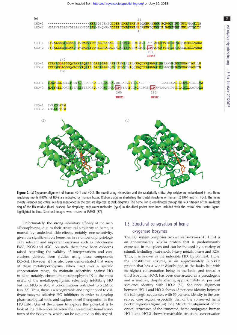
Figure 2. (a) Sequence alignment of human HO-1 and HO-2. The coordinating His residue and the catalytically critical Asp residue are emboldened in red. Hemeregulatory motifs (HRMs) of HO-2 are indicated by maroon boxes. Ribbon diagrams illustrating the crystal structures of human (b) HO-1 and (c) HO-2. The hememoiety (orange) and critical residues mentioned in the text are depicted as stick diagrams. The heme iron is coordinated through the N-3 nitrogen of the imidazolering of the His residue (black dashes). For simplicity, only water molecules (cyan) in the distal pocket have been included with the critical distal water ligandhighlighted in blue. Structural images were created in PYMOL [57].
rsif.royalsocietypublishing.orgJ
RSoc
Interface20120697
3
on July 10, 2018http://rsif.royalsocietypublishing.org/Downloaded from
Unfortunately, the strong inhibitory efficacy of the met-
alloporphyrins, due to their structural similarity to heme, is
marred by undesired side-effects, notably non-selectivity,
given the significant role heme has in a number of physiologi-
cally relevant and important enzymes such as cytochrome
P450, NOS and sGC. As such, there have been concerns
raised regarding the validity of interpretations and con-
clusions derived from studies using these compounds
[52–54]. However, it has also been demonstrated that some
of these metalloporphyrins, when used over a specific
concentration range, do maintain selectivity against HO
in vitro; notably, chromium mesoporphyrin IX is the most
useful of the metalloporphyrins, selectively inhibiting HO
but not NOS or sGC at concentrations restricted to 5 mM or
less [55]. Thus, there is a recognizable and urgent need to cul-
tivate isozyme-selective HO inhibitors in order to develop
pharmacological tools and explore novel therapeutics in the
HO field. One of the means to explore this potential is to
look at the differences between the three-dimensional struc-
tures of the isozymes, which can be exploited in this regard.
1.3. Structural conservation of hemeoxygenase isozymes
The HO system comprises two active isozymes [4]. HO-1 is
an approximately 32 kDa protein that is predominantly
expressed in the spleen and can be induced by a variety of
stimuli, including heat-shock, heavy metals, heme and ROS.
Thus, it is known as the inducible HO. By contrast, HO-2,
the constitutive enzyme, is an approximately 36.5 kDa
protein that has a wider distribution in the body, but with
its highest concentration being in the brain and testes. A
third isozyme, HO-3, has been demarcated as a pseudogene
and is inactive, despite sharing approximately 90 per cent
sequence identity with HO-2 [56]. Sequence alignment
between HO-1 and HO-2 shows 45 per cent identity between
the full-length sequences, with 55 per cent identity in the con-
served core region, especially that of the conserved heme
pocket regions (figure 2a) [58]. Structural alignment of the
crystal structures of the truncated, heme-conjugated human
HO-1 and HO-2 shows remarkable structural conservation

rsif.royalsocietypublishing.orgJ
RSoc
Interface20120697
4
on July 10, 2018http://rsif.royalsocietypublishing.org/Downloaded from
with r.m.s.d. of the a-carbons ranging from 0.697 to 0.862 A,
for the various conformations determined thus far
(i.e. open and closed conformations of the human HO-1
holoenzyme) [58].
The three-dimensional structures of both HOs are predo-
minantly a-helical with the heme sandwiched between the
distal and proximal helices (figure 2b,c) [58–61]. A number
of conserved glycines in the distal helix provide flexibility
to accommodate substrate binding and product release. The
active site of the apoprotein is generally more open than
the holoenzyme. In the holoenzyme, the heme iron is coordi-
nated by His25 in HO-1 (His45 in HO-2) on the proximal side
with a water molecule serving as the sixth ligand on the distal
side, and the distal and proximal helices are positioned closer
together to form a more ‘closed’ conformation. As elucidated
by the crystal structure of the HO-1 holoenzyme, there is still
an inherent flexibility of the distal helix, which allowed it to be
crystallized in both ‘open’ and ‘closed’ conformations. Both
apo- and holoenzymes contain a hydrogen-bond network
involving Asn210, Arg136, as well as a second level of residues,
which includes Tyr58 and Tyr114 to stabilize the position of the
catalytically critical Asp140 residue (Asp160 in HO-2). In the
high-resolution structure of the HO-1 holoenzyme [61], it
was revealed that the distal and proximal helices ‘clamp
down’ to tighten the structure and allow coordination of the
heme moiety with His25 as well as interaction of its propionate
groups with basic side chains without perturbation of the
Asp140 side chain. The tightened structure due to heme conju-
gation also traps critical water molecules in the distal cavity,
which form part of a well-ordered hydrogen-bond network
involving Asp140. This network may serve as a proton shuttle
to anchor the catalytically critical distal water ligand of heme
required for oxygen activation [61]. Although the analogous
network was not fully resolved in the heme-conjugated HO-2
crystal structure, the conformation of Asp160 is similar to
that of Asp140 in HO-1; thus, it is presumed to follow the
same underlying mechanism of catalysis [58].
The major point of sequence divergence between HO-1 and
HO-2 is in the C-terminal region (figure 2a), as well as the pres-
ence of three heme regulatory motifs (HRMs) in HO-2 with a
characteristic Cys-Pro signature; while HRM3 is centred
around Cys127, HRM1 and HRM2 reside in the C-terminus
[58]. There is some controversy with respect to the precise role
of these HRMs. Early studies indicated that these HRMs may
bind heme directly [62,63]. However, recent reports have indi-
cated that the HRMs affect catalytic efficiency without
affecting heme affinity and do not result in additional heme-
binding sites [64,65]. Rather, HRM1 and HRM2 form a thiol/
redox switch that modulates HO-2 affinity, on the basis of the
redox environment. Reducing conditions release the Cys265
thiol group from a disulphide bond with Cys282, allowing it
to form an alternative axial heme ligand with lower affinity
than His45 [66]. Thus, only one heme moiety conjugates with
HO-2, coordinated by either His45 or the lower affinity Cys265.
2. Development of isozyme-selective hemeoxygenase inhibitors: a structure – activityrelationship approach
Our research group is involved in a multi-disciplinary pro-
gramme comprising investigators specializing in medicinal
chemistry, pharmacology and toxicology, and biochemistry/
structural biology, and focuses upon the design and deve-
lopment of novel and potent, isozyme-selective inhibitors
of HO. Our initial investigations were stimulated by reports
of some non-porphyrin-based compounds that were found
to inhibit HO. Novel synthetic peptides were identified as
potent inhibitors of HO-1 activity and shown to have the
ability to prolong heterotopic heart graft survival in rats
[67]. The most notable HO-1 inhibitors included
peptide RESLRNLRGY, having an IC50 of 200 mM, and pep-
tide RNleNleNleRNleNleNleGY-CONH2 (RDP1258), with an
IC50 of 20 mM, against rat spleen HO-1 in vitro. However, this
effect of HO-1 inhibitory activity was compromised by a
secondary effect of inducing HO-1 expression. A series of
imidazopyridines and substituted prolines were also reported
to inhibit both the HO from the parasite Plasmodium yoeliiand the HO of the host mice [68]. Several compounds
were identified in this study, most notably 2-[3-(2-piperidin-
1-yl)ethoxyphenyl]imidazo[1,2-a]pyridine (100% inhibition
of mouse HO at 100 mM) and one stereoisomer of 5-(2-
hydroxyphenyl)-2-methylpyrrolidine-2,3,4-tricarboxylic acid
3,4-diethyl ester (100% inhibition of mouse HO at 100 mM).
Initial work by our group on these compounds did not give
favourable results as potential lead compounds. However, an
imidazole-based compound, namely (2S,4S)-2-[2-(4-chloro-
phenyl)ethyl]-2-[(1H-imidazol-1-yl)methyl]-4-[((4-aminophe-
nyl)thio)methyl]-1,3-dioxolane (azalanstat, QC-1, figure 3),
originally designed by Syntex as an inhibitor of mammalian
lanosterol 14a-demethylase (14-DM), gave promising results
in its ability to inhibit HO [3]. The non-porphyrin-based
structure of azalanstat was a key initial criterion because
it would minimize cross-reactivity with other heme-binding
proteins. A medicinal chemistry programme was initiated
based on the lead compound, QC-1, and the topological
analysis shown in figure 3. The resultant analogues were
screened by an in vitro CO formation assay, using micro-
somal preparations from rat spleen and brain as
physiological sources of HO-1 and HO-2, respectively [69].
The in vivo efficacies of some select compounds were also
evaluated in mice and rats, as well as in cultured renal
proximal tubule epithelial cells [70]. Initial studies identified
a number of compounds that were selective for the induci-
ble HO-1 isozyme [69]. Indeed, kinetic analyses of QC-1
and its analogues confirmed a non-competitive mode of
inhibition, unlike the metalloporphyrins that compete for
the heme-binding site, thus putatively minimizing
the chances of cross-reactivity with other heme-binding pro-
teins such as NOS and sGC, and increasing selectivity for
the HO isozymes.
The initial structure–activity relationship (SAR) study [69]
investigated the ortho-, meta- and para-amino derivatives of
each of the four diastereomeric analogues of QC-1. Although
no pattern in potency/selectivity for HO-1 was observed
with respect to the position of the amino functionality, it was
found that the (2S,4S) and the (2R,4S) diastereomeric configur-
ations produced analogues with greater potency and
selectivity for HO-1. In a further study, candidate compounds
lacking the aminothiophenol moiety of azalanstat were syn-
thesized and evaluated; the specific compounds synthesized
were the four diastereomeric analogues having a methyl
group in place of the methylene(aminothiophenol) moiety
attached to the 1,3-dioxolane ring, as well as the compound
(QC-15) in which this moiety was replaced by a hydrogen

O O
O O
N
western region
N
N
NH2
Scentral region
Clazalanstat (QC-1)
N
Cl
NN
O
O
NN
N
N
N
O
4
4
32
15
321
5
O ON
N
CF3
S
N
northeastern region
eastern region
ClQC-15 QC-80
QC-86QC-82
QC-308
Figure 3. Structures of representative QC-inhibitors used for X-ray crystallographic studies, including azalanstat (QC-1) depicting regions of interest for SAR studies.
rsif.royalsocietypublishing.orgJ
RSoc
Interface20120697
5
on July 10, 2018http://rsif.royalsocietypublishing.org/Downloaded from
atom. The most potent and selective compound towards HO-1
inhibition was (2R,4R)-2-[2-(4-chlorophenyl)ethyl]-2-[(1H-imi-
dazol-1-yl)methyl]-4-methyl-1,3-dioxolane hydrochloride. QC-
15 exhibited similar potency/selectivity for HO-1, albeit with-
out containing any stereogenic centres, an attractive feature for
synthesis. The results of the study thus far would suggest that
structural differences between HO-1 and HO-2 exist, differ-
ences that may be exploited to introduce selectivity. The
compounds resulting from the second study were also found
to be selective for HO, with little or no effect on neuronal
NOS and sGC, although they still showed potent inhibitory
activity towards cytochromes P450 (i.e. CYP3A1/3A2,
CYP2E1) [71,72].
The structures of the novel imidazole–dioxolane-based
HO inhibitors are similar to the known azole-based anti-
fungal compounds, particularly ketoconazole (KTZ), which
has been shown also to have anti-tumour effects in prostate
cancer [73]. Given the increase in HO-1 protein expression
in tumours, such as hyperplastic and undifferentiated malig-
nant prostate tissue [40], as well as the requirement of HO-1
for many solid tumours [43], it was hypothesized that
perhaps the anti-tumour effect of KTZ may be mediated
by HO-1 inhibition [74]. Testing of a series of antifungal
agents demonstrated that the structures containing diazoles
or triazoles are potent inhibitors of both HO isozymes.
It was hypothesized that the azole moiety of these
compounds may interfere with HO activity by binding the
heme iron and forming a complex that is inaccessible to the
catalytic site. However, it was found that KTZ did not
affect the heme absorbance spectrum at the low concen-
trations resulting in inhibition. Indeed, analyses showed
that KTZ inhibition was non-competitive and that it was
also not due to interference with cytochrome P450 reductase
or destruction of the HO protein. Moreover, KTZ showed
weak inhibition towards NOS, yet no inhibition towards
NADPH cytochrome P450 reductase.
In continuation of our medicinal chemistry programme
based on the azalanstat lead, a series of 2-oxy-substituted
1-(1H-imidazol-1-yl)-4-phenylbutanes were synthesized hav-
ing halogens in the phenyl ring and replacement of the
dioxolane moiety by a hydroxyl or carbonyl functional
group [75]. The entire library of compounds was found to
be highly active, with the bromine- and iodine-substituted
derivatives being the most potent. The imidazole–dioxolanes
were all selective for HO-1, and exhibited substantially lower
activity towards HO-2. The corresponding imidazole–
ketones and imidazole–alcohols showed selectivity towards
HO-1 to a lesser degree than the similarly substituted
imidazole–dioxolanes.
The SAR study has involved the design and synthesis of
many analogues in addition to the types described earlier,
for example: imidazole–dioxolane compounds having a

rsif.royalsocietypublishing.orgJ
RSoc
Interface20120697
6
on July 10, 2018http://rsif.royalsocietypublishing.org/Downloaded from
variety of substituents at the 4-position of the dioxolane ring,
2-oxy-substituted 1-azolyl-4-phenylbutanes to study the
effect of the variation of the azole moiety, a series of 1-aryl-2-
(1H-imidazol-1-yl/1H-1,2,4-triazol-1-yl)ethanones and their
derivatives, and a series of a-(1H-imidazol-1-yl)-v-phenylalk-
anes to study the effect of introduction of heteroatoms
in the alkyl linker. Reference to these analogues is made
later in the discussions of the X-ray crystallographic
structural analyses.
2.1. Enhancing structural knowledge of inhibition withX-ray crystallography
As the number of HO inhibitors increased, functional analyses
identified a number of potent inhibitors, including several
deemed isozyme-selective for HO-1. With the knowledge
obtained from the three-dimensional structure of a truncated
human HO-1 derivative based on previous X-ray crystallo-
graphy studies (figure 2b) [59–61], a structural study of
complexes formed between recombinant human HO-1 in com-
plex with some of our potent and representative inhibitors was
initiated. While the previous approach gave valuable infor-
mation regarding SARs, and the iterative and systematic
methodology was successful in identifying salient features
for inhibition, what was missing was the ‘why’—i.e. the
underlying mechanism leading to inhibition as well as iso-
zyme selectivity. The inhibitors for which crystal structures
were determined in complex with HO-1 were: 2-[2-(4-chloro-
phenyl)ethyl]-2-[(1H-imidazol-1-yl) methyl]-1,3-dioxolane
(QC-15), 1-(adamantan-1-yl)-2-(1H-imidazol-1-yl)ethanone
(QC-82), 4-phenyl-1-(1,2,4-1H-triazol-1-yl)butan-2-one (QC-
86), (2R,4S)-2-[2-(4-chlorophenyl)ethyl]-2-[(1H-imidazol-1-
yl)methyl]-4-[((5-trifluoromethylpyridin-2-yl)thio)methyl]-1,3-
dioxolane (QC-80), and 1-(1H-imidazol-1-yl)-4,4-diphenyl-2-
butanone (QC-308) (figure 3). Structural analyses and com-
parisons of the various complexes enabled us to definitively
identify structure–functional relationships to understand the
mode of binding to HO-1 and the mechanism underlying
the inhibition of heme degradation. Moreover, identification
of essential structural features critical for inhibition, both
within the inhibitor as well as structural changes within the
protein, provided valuable insights that enhanced the design
of more effective HO-1 inhibitors. During the course of our
study, the X-ray crystal structure of a truncated, human HO-
2 derivative containing a Cys127Ala mutation was also deter-
mined [58], which provided more insights regarding potential
structural differences that may be used in inhibitor design to
enhance isozyme selectivity (figure 2c).
2.2. A common binding mode for imidazole-basedheme oxygenase-1 inhibitors
One of the early, potent inhibitors found to be isozyme-
selective for HO-1 was QC-15, with IC50’s of 4 + 2 mM for
HO-1 and greater than 100 mM for HO-2 as determined with
CO-formation assays using rat spleen and brain microsomal
fractions, respectively [72]. The potency of this compound
was very similar to that of QC-1 (6 + 1 mM) for HO-1, but
removal of the bulky 4-aminophenylthiol groups in the north-
eastern region resulted in decreased activity towards HO-2 in
comparison with QC-1 whose IC50 was 28 + 18 mM. This com-
pound was also the first to be crystallized, as reported by
Sugishima et al. [76], in complex with rat HO-1 (rHO-1,
2.70 A). Soon thereafter, we successfully crystallized QC-82,
also an isozyme-selective and potent inhibitor of HO-1 with
an IC50 of 7 + 1 mM, in complex with human HO-1 (hHO-1)
at a resolution of 1.54 A [77]. The similarity in inhibitory
effect was quite interesting, given the structural differences
between these two compounds. While QC-15 is similar to
QC-1 except in the absence of the bulky northeastern sub-
stituent, the structure of QC-82 diverges even further.
The central dioxolane group was replaced by a ketone, the
4-chlorophenyl group in the western region was replaced by
a bulky adamantanyl group and the central linker region
between the two had been shortened by two carbon atoms.
The only commonality was the eastern imidazole moiety. Inter-
estingly, spectral analyses of both inhibitor–enzyme
complexes also showed a red-shift in the Soret peak implying
that, while the heme was still conjugated to the enzyme, its
environment had been modified [76,77].
Comparison of the structures of the two inhibitor–HO-1
complexes revealed a common binding mode for these see-
mingly structurally distinct, imidazole-based compounds
(figure 4a,b) [76,77]. While it appears that the inhibitors
bind within the heme-binding pocket, they do not displace
and rather require the heme moiety for binding, which
explains the non-competitive nature of the inhibition as
determined through kinetic analyses. This observation has
been confirmed recently by Sorrenti et al. [78], who reported
that the substrate was required for binding to membrane-free
full-length hHO-1. The inhibitor binds to the distal side of
heme, with the imidazolyl group serving as an anchor with
the nitrogen at position 3 coordinating with the heme iron
to form a true hexacoordinated heme, replacing the critical
distal water molecule as the sixth coordinating ligand. The
western region extends back into the distal side of the
heme-binding pocket where it fits into a hydrophobic
pocket. Comparison of the structures of inhibitor–HO-1 com-
plexes with the respective holoenzymes revealed that there is
very little change in the overall structure to accommodate the
inhibitors. Alignment of the hHO-1–QC-82 complex with the
‘closed’, active conformation of the native holoenzyme gave a
Ca r.m.s.d. of only 0.74 A. Both the heme and proximal helix
shift back to expand the distal pocket to some extent. In the
structure of rHO-1 with QC-15, the heme and proximal
helix are shifted approximately 0.8 A towards the a-meso
carbon along the a–g axis of heme; in that of hHO-1 with
QC-82, the coordinating nitrogen of His25 and the Fe
atom are shifted by 0.91 and 0.85 A, respectively. However,
it is the inherent flexibility of the distal helix that allows the
heme-binding pocket to open up to allow the inhibitor to
slip in and also expands the distal pocket to accommodate
the inhibitor. Moreover, the extent of expansion is dependent
upon the size of the inhibitor with the helix being shifted to a
greater extent to accommodate the bulkier adamantanyl
group of QC-82, resulting in a distal pocket which is more
open relative to either the native holoenzyme or rHO-1 in
complex with QC-15. Indeed, accommodation of the bulky
adamantanyl group of QC-82 resulted from a significant con-
formational change in the distal helix (Ser142 to Ile150) with a
maximal displacement of 3.92 A corresponding to Gly144
(figure 5a). In the complex of rHO-1 with QC-15, the main-
chain conformation of Ser142 and Gly143 changed along
with the bending angle so as to open up the distal helix to
such an extent that the helical structure at the bending
point (Gly143–Gln143) was disrupted. By contrast, the

(c) (d )
(a)
(c)
(e)
(b)
Figure 4. Ribbon diagrams depicting the structures of HO-1 in complex with the respective QC-inhibitors. Heme (orange) and inhibitors ( purple) are depicted asstick diagrams. Black dashes illustrate metal coordination via the nitrogen atom of the azole group of either His25 or the QC compound. Hydrogen bonds aredepicted as yellow dashed lines. Selected residues mentioned in the text are depicted as stick diagrams and labelled for clarity. For simplicity, only water moleculesin the distal pocket are shown. Electrostatic surface potentials, as calculated using PYMOL [57], depict positively (blue) and negatively (red) charged areas, and revealhydrophobic pockets (white) into which the western and northeastern regions of the QC-inhibitors fit. All images were prepared using PYMOL [57]. (a) rHO-1 incomplex with QC-15 (PDB #2DY5), (b) hHO-1 in complex with QC-82 (PDB #3CZY—Chain A), (c) hHO-1 in complex with QC-86 (PDB #3K4F), (d ) hHO-1 in complexwith QC-80 (PDB #3HOK—Chain B), (e) hHO-1 in complex with QC-308 (PDB #3TGM—Chain B). The two distal hydrophobic pockets (18 HP and 28 HP) thatstabilize the two phenyl moieties of QC-308 allow a ‘double-clamp’ mode of binding.
rsif.royalsocietypublishing.orgJ
RSoc
Interface20120697
7
on July 10, 2018http://rsif.royalsocietypublishing.org/Downloaded from
greater shift to accommodate the adamantanyl group of
QC-82 in the hHO-1 complex resulted in an apparent helical
break from Ser142 to Gly144. Thus, the adamantanyl group
may represent the upper limit of what can be accommodated
by shifts in the distal helix.
While the imidazolyl group serves as an anchor by coor-
dinating with the heme iron moiety, the binding of the
western region is stabilized through hydrophobic interactions
involving residues lining the distal hydrophobic pocket, i.e.
Phe33, Met34, Phe37, Val50, Leu54, Leu147, Phe167 and
Phe214. The extent of stabilization of the western region by
the hydrophobic pocket depends on the nature of the western
region as a whole. For example, QC-15 was found to be a
more potent inhibitor than QC-82 when spectral analyses
were used to assay inhibitor binding to the purified, recombi-
nant, truncated hHO-1 used for crystallization, despite the
greater hydrophobicity of the latter’s adamantanyl group
relative to the former’s chlorophenyl moiety in the western

(a) (b)
Figure 5. Structural alignments of hHO-1 complexes with QC-inhibitor (green) with the native holoenzyme (PDB #1N45—Chain A) (cyan), both depicted as ribbondiagrams. QC-inhibitors are coloured purple and shown in stick form. (a) QC-82 complex (PDB #3CZY—Chain A). The distal helix is shifted with a maximaldisplacement of Gly144 (3.92 A) as highlighted by comparison of the complex (black label) and native protein (italicized blue label). The resultant helical break isindicated (black arrow). (b) QC-80 complex (PDB #3HOK—Chain B). The proximal helix is shifted with a rearrangement of residues, highlighted as wheat-colouredstick diagrams (black labels) in the complex in comparison with their relative positions in the native protein, highlighted as teal stick diagrams (italicized bluelabels). Structural alignments were performed using SUPERPOSE in CCP4 [79] and the images prepared using PYMOL [57].
rsif.royalsocietypublishing.orgJ
RSoc
Interface20120697
8
on July 10, 2018http://rsif.royalsocietypublishing.org/Downloaded from
region. Further observation revealed that the shorter, central
linker of QC-82 resulted in this group not extending as far
into the hydrophobic pocket, hence making fewer contacts.
Moreover, in addition to hydrophobic contacts, the chloro-
phenyl group of QC-15 may also be stabilized by p–p
stacking interactions attributable to the prevalence of aro-
matic residues in this region. More information regarding
the structural basis of inhibitor binding was gleaned by
using the known structure data to interpret previous func-
tional data. Previously, a negative correlation was found
between potency and electronegativity of the halogen substi-
tuent in the western region within a series of alcohol
derivatives of 2-oxy-substituted 1-(1H-imidazol-1-yl-4-phe-
nylbutanes) [75,77]. For example, the potency of the alcohol
derivative of QC-15 (IC50 ¼ 0.5 + 0.1 mM) increased upon
substitution of the chlorophenyl group by bromophenyl
(IC50 ¼ 0.14 + 0.06 mM) or the more electronegative iodo-
phenyl group (IC50 ¼ 0.06 + 0.03 mM), and decreased
when replaced by the less electronegative fluorophenyl
(IC50 ¼ 1.4 + 1.1 mM) or a non-substituted phenyl group
(IC50 ¼ 6.2 + 0.8 mM). A close inspection of the hydrophobic
pocket revealed the presence of a polar thiol group (Met34),
which may explain why electronegative moieties in the wes-
tern region still give rise to potent inhibitors. Alternatively,
the larger halogens may provide more points and better
contact with the distal hydrophobic pocket.
One of the key differences between QC-15 and QC-82 is
the central linker region, which contains a dioxolane and
ketone group, respectively. Analysis of the differences in
interaction with HO-1 provided insights into potential fea-
tures, which could be exploited in future drug design. The
dioxolane group of QC-15 is involved in a hydrogen bond
network involving a water molecule as well as the carbonyl
group of Thr135 [76]. A subsequent structure of hHO-1 in
complex with QC-80 (IC50 ¼ 2.1 + 0.6 mM), which also con-
tains a central dioxolane component, revealed a similar
stabilizing interaction [80]. However, this network appears
not to be essential for binding the central region of these
imidazole-based compounds, as there is no analogous
water molecule available for a similar hydrogen-bond inter-
action with the ketone group of QC-82 in one of the
molecules in the asymmetric unit. By contrast, in the crystal
structure of hHO-1 in complex with QC-86 (IC50 ¼ 2.5 +0.4 mM), another azole-based compound with a central
ketone moiety, it appears that the carbonyl group is stabilized
by a hydrogen bond involving an active site water molecule
that is a part of a potential hydrogen-bond network involving
another water molecule and the Thr135 carbonyl group [81];
the water molecule may also be involved in the Asp140
hydrogen-bond network. Given the more open conformation
of the distal pocket to accommodate the bulky adamantanyl
group of QC-82 relative to the other compounds for which
structures have been determined, it is likely that this struc-
tural expansion acts to impede its ability to trap water
molecules, resulting in greater fluidity of the solvent struc-
ture. Further insight into this central region was gained by
looking at previous functional data involving the series
of 2-oxy-substituted 1-(1H-imidazol-1-yl-4-phenylbutanes),
which showed an interesting trend. Comparison of IC50
values showed that, generally, compounds with a central
hydroxyl group tended to be more potent than their dioxo-
lane or ketone counterparts. Substitution of the central
dioxolane group of QC-15 with a ketone group did not
affect its IC50 significantly (IC50 ¼ 4.7 + 0.5 mM), whereas
substitution to a central hydroxyl group increased its potency
by almost 10-fold (IC50 ¼ 0.5 + 0.1 mM). This may be due,
in part, to a greater potential to form hydrogen bonds
with trapped water molecules or neighbouring residues.
The increased rotational flexibility of the hydroxyl group,
relative to a carbonyl or dioxolane, may also allow interac-
tion with Asp140 to stabilize binding further and/or
disrupt the critical Asp140 hydrogen-bond network further.
Unfortunately, the increased potency observed for the
alcohol derivative of QC-15 is marred by a loss of selecti-
vity (IC50 ¼ 4.0 + 0.6 mM for HO-2). Thus, culmination of
all the structural and functional data reveals that, while

rsif.royalsocietypublishing.orgJ
RSoc
Interface20120697
9
on July 10, 2018http://rsif.royalsocietypublishing.org/Downloaded from
hydrogen-bond networks involving trapped water
molecules and neighbouring residues may contribute to the
stabilization of the central region of an inhibitor, they are not
critical for its binding to HO-1.
Recently, to gain more insights into the structural require-
ments for the central region of the QC compounds, a series of
a-(1H-imidazol-1-yl)-v-phenylalkanes was synthesized to
examine the effect of introducing heteroatoms into the central
alkyl linker [82]. Moreover, linkers of various lengths were
also investigated, which provided information regarding
the size limits required for effective binding. Interestingly,
introduction of an oxygen atom decreased inhibitory potency
against HO-1, while derivatives containing a sulphur atom
had increased potency relative to those with a simple alkyl
linker. The presence of polar groups within the distal hydro-
phobic pocket, e.g. Met34 and Met51, may be playing a role
in these stabilizing/destabilizing interactions. Increasing
linker length from one to five carbon atoms resulted in a pro-
gressive increase in inhibitory potency against HO-1, from
44 + 6 to 3.5 + 0.7 mM, respectively; the absence of a
linker resulted in no activity. Presumably, this trend is due
to greater contacts with the distal hydrophobic pocket as
the linker is increased, resulting in greater stabilization of
the western phenyl moiety. A minimal linker is required
for activity, which suggests that stabilization by the distal
hydrophobic pocket is in fact crucial for binding of this
class of compounds. While a five-carbon linker resulted in
the most potent compounds, the most HO-1 selective com-
pounds contained a four-atom linker between the phenyl
and imidazolyl moieties. Alignment of the crystal structures
of the hHO-1 complex with QC-82 (PDB code 3CZY, i.e. our
largest inhibitor binding site) with that of hHO-2 (PDB code
2QPP) shows that a potential difference in the size of the
western hydrophobic pocket may account for this selectivity.
The HO-2 pocket is larger partly due to substitutions of
shorter amino acids; for example, the side chains of
Met51 and Val50 in HO-1 extend further in towards the
western region of the QC compounds than their Thr71 and
Ala70 counterparts in HO-2. Thus, a longer linker may be
required to allow the proper hydrophobic contacts for
stabilization in HO-2.
The common binding mode of the imidazole-based HO-1
inhibitors involves coordination of the heme iron through an
eastern imidazolyl anchor and stabilization of the western
region by a distal, hydrophobic pocket through hydrophobic
interactions and potentially p–p stacking interactions.
Movement of the distal helix allows expansion of the distal
pocket to accommodate bulky groups on the western end.
The central region is not critical for binding although
water-mediated hydrogen-bond networks may contribute to
its stabilization and an optimal linker length of four carbons
may allow isozyme selectivity towards HO-1 without bind-
ing HO-2. It is possible that this central region’s main
contribution is to the proper spacing between the eastern
and western regions, i.e. the ‘anchor’ and the hydrophobic
moiety, so that they may make optimal contacts of heme
coordination and within the distal hydrophobic pocket,
respectively. Interestingly, binding of inhibitor does not per-
turb the critical Asp140 residue, nor any of the surrounding
resides involved in its critical hydrogen-bond network.
Rather, the underlying mechanism by which the imidazole-
based compounds inhibit HO is by disrupting the ordered
solvent structure and, thus, the hydrogen-bonded network,
and ultimately displacing the catalytically critical distal
water ligand to inhibit heme oxidation.
2.3. Anchoring inhibitionThe elegance of the structure and mechanism underlying heme
oxidation has allowed its inhibition by this diverse class of non-
porphyrin-based compounds. The pentacoordinate nature of
the HO-1 heme, with His25 serving as a proximal coordinating
ligand, as well as the immutable positioning of the critical
Asp140 and its hydrogen-bond network, allows the precise
positioning of the catalytically critical water molecule to
serve as a sixth ligand on the distal side upon heme conjugation
[60,61,83,84]. It also makes the distal side of heme a prime
target in designing inhibitors with a coordinating functional
group to serve as an anchor for binding to displace the critical
water ligand. Indeed, the main feature for binding of our QC
compounds is coordination of the heme iron through the N-3
nitrogen of an unsubstituted imidazolyl moiety. Thus, a poten-
tial strategy for improving inhibitor potency is to build a
stronger ‘anchor’ by modifying this imidazolyl group.
A series of compounds was designed in which different
azole groups were substituted for imidazole in the eastern
region [85]. Functional analyses determined that 1,2,4-tria-
zole- and 1H-tetrazole-based inhibitors are potent inhibitors
of HO-1, with an improved selectivity over HO-2. A crystal
structure for hHO-1 in complex with a compound containing
a 1,2,4-triazolyl substituent, namely 4-phenyl-1-(1,2,4-1H-
triazol-1-yl)butan-2-one (QC-86, IC50 ¼ 2.5 + 0.4 mM), was
determined to a resolution of 2.20 A. Analysis of the
enzyme–inhibitor interaction revealed that the triazolyl
moiety is able to provide an additional point of anchor stabil-
ization as the presence and positioning of the N-2 nitrogen
atom is close enough to form a potential hydrogen bond
with the amide group of Gly143 (figure 4c). This additional
hydrogen bond may explain the slightly increased potency
of this compound relative to its imidazolyl counterpart
(cf. IC50 ¼ 4 + 2 mM). Interestingly, shifting the position of
the additional nitrogen atom to form the 1,2,3-triazolyl
variant significantly decreases potency (IC50 ¼ 89 + 1 mM).
Because there was no obvious structural basis for the
decrease, this phenomenon was investigated further by calcu-
lating the Mulliken charges for the coordinating nitrogens of
each of these azole variants. Similar values were determined
for both the 1,2,4-triazole ring (20.464) and the imidazole
ring (20.446), while the resultant delocalized electron
system of the 1,2,3-triazole ring decreased the electronegativ-
ity of the ‘coordinating’ nitrogen (20.281) to potentially
weaken its coordination with the heme iron. Further destabi-
lization of the anchor may be a result of repulsive forces
contributed by the ketone group of Gly139 as the N-2 nitro-
gen of the 1,2,3-triazole would be located 3.05 A away from
this functional group.
Recent studies have shown that replacing the imidazole
with either a 1,2,4-triazole or a tetrazole significantly dimini-
shes the inhibitory effects on the cytochrome P450 family of
drug metabolizing enzymes [86]. Thus, strengthening the
QC-anchor in this manner would be an efficient strategy to
design potent and selective HO-1 inhibitors. It may also be
worthwhile to explore other strategies to strengthen the
anchor by replacing the coordinating atom itself. It was puz-
zling, however, to observe that the azole–dioxolanes and the
azole–alcohols derived from the active 1,2,4-triazole- and

r
10
on July 10, 2018http://rsif.royalsocietypublishing.org/Downloaded from
1H-tetrazole-based ketones were generally less active than
their imidazole-based derivatives [81].
sif.royalsocietypublishing.orgJ
RSoc
Interface20120697
2.4. A novel, inducible binding modeWhile initial work had concluded that the aminothiophenol
moiety in the northeastern region of azalanstat is not essential
for HO inhibition, it had been noticed that general HO
potency was dependent upon the position of the nitrogen
atom and that, more importantly, its removal altogether
enhanced HO-1 selectivity [69,71,72]. Thus, further investi-
gation of this region was postulated to be important in
determining selectivity for HO-2 using thiophenol and sub-
stituted phenol derivatives as well as small functionalized
derivatives at the 4-position of the dioxolane ring, i.e. the
northeastern region [87]. Unfortunately, most of the com-
pounds synthesized were highly potent inhibitors of both
HOs with only moderate selectivity for HO-1. The addition
of a second aromatic ring system was well tolerated by
HO-1 and enhanced selectivity, although an upper limit to
this region was found based on the loss of inhibitory activity
when this was extended to a bulky adamantanyl group. Inter-
estingly, within the thiophenol/phenol series, four of the six
most selective compounds contained hydrocarbon moieties
in the northeastern region, suggesting the importance of
non-polar functionality in this region. Replacement of the
4-aminothiophenol moiety with smaller, polar, functional
groups also resulted in potent inhibitors but with no to
modest selectivity for HO-1. Thus, it was concluded that modi-
fications solely in this northeastern region would not result in
HO-2 selectivity and, moreover, may not be an efficient
avenue in the development of highly selective HO-1 inhibitors.
From a structural point of view, however, it was not
directly obvious as to how these bulky northeastern groups
would be accommodated to allow binding to HO-1. None
of the previous structures indicated much flexibility within
the proximal region, with most of the movement occurring
distally. A putative proximal hydrophobic pocket had been
observed in the structure of hHO-1 in complex with the
bulky adamantanyl derivative, QC-82, which had not been
apparent in previous structures (figure 4b). Computational
analyses were successful in virtually docking both QC-1
[77] as well as (2R, 4S)-2-[2-(4-chlorophenyl)ethyl]-2-[(1H-
imidazol-1-yl)methyl]-4-[(phenylsulphanyl)methyl]-1,3-dioxo-
lane [87] such that the bulky northeastern groups fit into this
potential pocket. However, the crystal structure of QC-80
(IC50 ¼ 2.1 + 0.6 mM) in complex with hHO-1 revealed that
the binding of this class of compounds is, in fact, a result of
a novel, inducible binding mode, which could not be antici-
pated from any of the structural data obtained previously
[80]. The bulky northeastern region is accommodated by
shifting residues in the proximal helix to induce the creation
of a secondary hydrophobic pocket distinct from that
suggested from previous structures (figure 4d ).
Despite the bulky northeastern region, QC-80 follows the
typical binding mode, interacting with hHO-1 in a manner
analogous to the azole-based compounds that lack substitu-
ents in this region, with one exception. Previous structures
of hHO-1, including the apo- and holoenzymes, as well as
in complex with the other QC-inhibitors lacking a north-
eastern region showed a sharp bend of approximately 608in the proximal helix (Asn30-Ala31) that precedes another
helical segment (Glu32-Lys39). Interaction of the bulky
5-trifluoromethylpyridin-2-yl moiety of QC-80 appeared to
push back residues in this region to ‘unkink’ and extend
the proximal helix up to Gln38 and induce the formation of
a secondary peripheral hydrophobic pocket to accommodate
the northeastern substituent. Residues linking this newly
formed pocket that may contribute hydrophobic interactions
to stabilize the 5-trifluoromethylpyridin-2-yl group include
Ala28, Ala31, Arg35, Phe214 and Glu215. This conformation-
al change expands the binding pocket to a greater extent than
observed with any of the other inhibitors, shifting Gln38 as
much as 11 A from its original position in the native holoen-
zyme. Moreover, there is a rearrangement of residues
between the interior and exterior of the pocket. Binding of
the northeastern region of the inhibitor displaces the
thiol group of Met34, which shifts to the exterior of the
newly formed hydrophobic pocket along with the functio-
nal groups of Phe33, Phe37 and Gln38, while the functional
groups of both Arg35 and Glu32 shift into the interior
where they may be stabilized by electrostatic interactions
between the two side chains (figure 5b).
Thus, the crystal structure of the QC-80 complex revealed
that the proximal helix of hHO-1 also contains a degree of
intrinsic flexibility, although not to the same extent as the
distal helix, which allows binding of inhibitors with a bulky
northeastern region, such as QC-1, by inducing the formation
of a secondary hydrophobic pocket to stabilize this region.
While binding by an ‘induced fit’ mechanism would be more
energetically costly than binding to a preformed binding site,
it has provided further insights into the structural properties
of hHO-1 that were not apparent previously, as well as a
rationale underlying the potency of inhibitors such as azalan-
stat which contain bulky northeastern regions. This new
information may be helpful in future inhibitor design.
2.5. Structure-aided drug design: a novel,‘double-clamp’ binding mode
The discovery of the novel, induced formation of the secondary
hydrophobic pocket to bind the northeastern region of QC-80
shed new light on the observation of the putative proximal, sec-
ondary hydrophobic pocket seen in the structure of hHO-1 with
QC-82 [77]. While this proximal pocket is not involved in bind-
ing the northeastern region as previously postulated, there was
potential in its exploitation to improve inhibitor potency by
creating compounds that could occupy both preformed hydro-
phobic pockets simultaneously. The synthesis and
characterization of 1-(1H-imidazol-1-yl)-4,4-diphenyl-2-buta-
none (QC-308, IC50¼ 0.27 + 0.07 mM), which contains an
additional phenyl moiety in the western region, in contrast to
the usual single hydrophobic group in the QC compounds,
demonstrated a approximately 15-fold increase in potency rela-
tive to its monophenyl analogue, 4-phenyl-1-(1H-imidazol-1-
yl)-2-butanone (QC-65; IC50¼ 4.06 + 1.8 mM) [75]. This was
quite remarkable given that QC-65 was already quite a potent
inhibitor. Determination of the crystal structure of the hHO-1
complex with QC-308 revealed the common elements required
for binding the majority of the QC compounds and, further,
confirmed the presence of a definitive secondary hydrophobic
pocket to accommodate and stabilize the second phenyl
moiety in the western region of QC-308 (figure 4e) [88].
Thus, each of the two phenyl moieties of the diphenyl analogue
fit into separate hydrophobic pockets, resulting in a ‘double-
clamp’ binding mode to provide additional stabilization,

rsif.royalsocietypublishing.orgJ
R
11
on July 10, 2018http://rsif.royalsocietypublishing.org/Downloaded from
which likely accounts for the increased potency relative to
the monophenyl variant. Unfortunately, neither QC-65
nor QC-308 are isozyme-selective, being potent inhibitors
of both HO isozymes (IC50¼ 11.3 + 4.7 mM [75] and 0.46 +0.15 mM [88], respectively, for HO-2 in rat brain microsomes).
Replacement of the central ketone moiety of QC-65 with a diox-
olane group had conferred isozyme selectivity (QC-57; 2-[2-
phenylethyl]-2-[(1H-imidazol-1-yl)methyl]1,3-dioxolane) with
IC50’s of 0.76 + 0.4 and greater than 100 mM for HO-1 and
HO-2, respectively [75]. Thus, using the ‘double-clamp’ strategy
to increase the potency of an HO-1 isozyme-selective derivative
such as this would be an avenue worth pursuing.
SocInterface
20120697
3. Looking forwardOver the past decade, since the discovery of azalanstat as a
specific HO inhibitor, we have amassed a large group of
azole-based derivatives that exhibit potent inhibitory activity
specific for HO, with several showing isozyme selectivity.
Through a combination of a medicinal chemistry SAR
approach and X-ray crystallographic structural analyses, we
have gained valuable insights into the common binding
mode of this class of compounds, as well as information
regarding the enzyme responsible for its inherent flexibility,
which also allows for an ‘induced’ fit to accommodate prox-
imal structural motifs. Each series of derivatives has allowed
us to amass more information, which can be used to develop
the next generation of inhibitors. As such, we have concluded
that this class of azole-based HO-1 inhibitors, originally
derived from azalanstat, inhibit heme oxidation through a
non-competitive, yet heme-dependent, mechanism by dis-
rupting the ordered solvent structure integral to the critical
hydrogen-bond network involving Asp140 and displacing
the catalytically critical distal water ligand.
Moreover, from a physiological point of view, this inhibi-
tory mechanism has already demonstrated potential
therapeutic applications as some of our imidazole-based
HO-1 inhibitors have shown the ability to attenuate the
growth of prostate [89] and breast cancer cells in vivo. The rel-
evance of HO in various experimental situations has been
explored by other laboratories through administration of
these novel imidazole-based drugs to both cultured cells
and intact animals. Accordingly, Di Francesco et al. [90]
tested the role of HO-1 in the response of human umbilical
vein endothelial cells in culture to laminar shear stress.
They observed that TNF-a biosynthesis was reduced by
shear stress, and that this reduction was reversed in the pres-
ence of the selective HO-1 inhibitor QC-15. Similarly,
Csongradi et al. [91] used selective HO-1 inhibitors to study
the effects of renal HO inhibition on blood pressure in
mice. Angiotensin II induced hypertension was exacerbated
in the presence of this (QC-13) selective HO-1 inhibitor, as
well as the first-generation HO inhibitor tin mesoporphyrin
(SnMP). SnMP also induced renal medullary HO-1 protein,
while QC-13 was without effect on HO protein levels,
which obviated the complication of increasing quantities of
the enzyme targeted for inhibition.
The salient features of this class of inhibitors include,
first and foremost, an anchor in the eastern region to coor-
dinate with the heme iron atom, usually an imidazole
although a 1,2,4-triazole moiety may strengthen the inter-
action with the added benefit of increasing specificity by
decreasing activity against the cytochrome P450 family of
enzymes. The anchor is connected by a linker region to a
hydrophobic group in the western region, which fits into a
distal hydrophobic pocket and is stabilized by both hydro-
phobic and aromatic stacking interactions. A four-carbon
linker is the optimal length to maximize potency without
compromising isozyme selectivity for HO-1. A five-carbon
linker results in a more potent compound but one that is
also inhibitory against HO-2, possibly due to the latter’s
larger hydrophobic pocket. The presence of polar residues
in the pocket (e.g. Met34) may also accommodate halogena-
tion of the phenyl group as well as the presence of
heteroatoms nearby in the linker region, although an
alternative explanation may be that the larger halogens pro-
vide more points and better contact in the pocket. The distal
helix of hHO-1 has a high inherent flexibility, which gives
the enzyme the capacity to bind inhibitors with a range of
‘bulkiness’ in the western region, while still maintaining
its heme moiety. The maximal limit may be the size of an
adamantanyl group, as its binding caused disruption in
the helical structure at one point of the distal helix. More-
over, the presence of a putative peripheral secondary
hydrophobic pocket can also be exploited to increase potency
by introducing diphenyl variants to the western region,
which can be stabilized by a ‘double-clamp’ binding mode.
A lesser degree of flexibility is associated with the proximal
helix, which results in the accommodation of compounds
with large substituents in the northeastern region. Interaction
of this series of compounds induces a conformational change,
essentially resulting in an ‘unkinking and extension’ of the
helix and rearrangement of resides from the interior and
exterior of the protein, to form a peripheral hydrophobic
pocket to accommodate these groups.
It should be noted that all of the structural analyses of
HO-1 in complex with the QC-inhibitors to date have been
conducted using soluble, truncated derivatives of the protein
[76,77,80,81,88]. By contrast, functional characterization of
inhibitor potency has been performed using the full-length
protein as found in microsomal preparations. A recent com-
parison of native and recombinant truncated proteins
demonstrated differential sensitivity of recombinant and
microsomal HO-1 towards inhibitors [92]. Also, this has led
to some controversy regarding data interpretation, especially
regarding the issue of isozyme selectivity, as some isozyme-
selective compounds were found to be equipotent against
both isozymes when tested against the recombinant, trun-
cated versions [76,92]. Given the structural and sequence
conservation of the HOs within the catalytic core [58], the
lack of selectivity found with the truncated purified protein
was not surprising. However, the recent characterization of
a recombinant full-length HO-1 had indicated that the full-
length form exhibits a 2- to 3-fold greater activity relative to
that of the truncated, soluble form, which was increased
even further in the presence of lipid. Moreover, the C-terminal
hydrophobic tail has an essential role with respect to mem-
brane incorporation as well as in formation of a high-affinity
complex with cytochrome P450 reductase, deemed essential
for maximal catalytic activity [93–95]. The C-terminal
domain is also the major point of sequence divergence between
the two isozymes, especially in the presence of the HRMs in
HO-2 [58]. While current structural information does not
imply a role for this hydrophobic tail in the binding of this
class of inhibitors, its potential role in influencing conformation

rsif.royalsocietypublishing.orgJ
RSoc
Interface20120697
12
on July 10, 2018http://rsif.royalsocietypublishing.org/Downloaded from
should not be ignored in future designs, especially in influen-
cing isozyme selectivity. Indeed, the determination of the
crystal structure of both full-length hHO-1 and hHO-2 would
provide valuable information in this regard.
The quest to build a better inhibitor involves much
imagination and constant exploring. While we have gained
experience and knowledge regarding this particular class of
azole-based non-competitive inhibitors for HO-1, we are
always searching for new lead compounds to develop new
classes of inhibitors for various applications. The crystal
structure of the truncated hHO-2 has already provided us
with valuable insights with regard to our complementary
goal of developing selective HO-2 inhibitors. Ideally, it
would also be useful to develop a series of competitive inhibi-
tors that prevent binding of heme altogether. Theoretically,
one would presume that a selective, competitive inhibitor
would have powerful therapeutic applications to greatly
minimize any HO activity if the substrate is not available
for degradation. Recently, caveolin-1 was discovered to com-
petitively inhibit rat HO-1 via the caveolin scaffolding
domain (residues 82–101), with a five amino acid sequence
(residues 97–101: YWFYR) identified as a minimum
sequence for binding. Two aromatic residues, Phe207 and
Phe214, in HO-1 are thought to be essential in the association
with caveolin-1 through p–p stacking and hydrophobic
interactions [96]. Further analysis of this peptide structure
may provide insights into new lead compounds to develop
a series of potential competitive inhibitors.
We hope that through this exciting process, using both
medicinal chemistry SAR and X-ray crystallography, we
will be able to develop powerful pharmacological tools for
delving deeper into the understanding of the mechanisms
by which the HO/CO system exerts its many effects and, in
turn, providing a foundation for the development of novel
therapeutic approaches for the myriad pathologies with
which it is involved.
This research was supported by CIHR. Dr Zongchao Jia holds aCanada Research Chair in Structural Biology and a Killam ResearchFellowship. We thank Dr Paul Ortiz de Montellano and Dr JohnEvans (both from the University of San Francisco) for the generousgift of the hHO1-t233 plasmid and advice regarding its expression.We are grateful to Dr Qilu Ye, Dr Jimin Zheng and Dr Frederick Fau-cher for technical expertise in structural determination, as well as MrBrian MacLaughlin, Ms Tracy Gifford and Mr Wallace Lee for assist-ance in the biological evaluations. Finally, we would like to expressour appreciation to the staff at the Advanced Photon Source (APS)at Argonne National Laboratory (Chicago, IL, USA) as well as atthe Cornell High Energy Synchrotron Source (Ithaca, NY, USA) fortheir support in X-ray data collection.
References
1. Tenhunen R, Marver HS, Schmid R. 1969 Microsomalheme oxygenase. Characterization of the enzyme.J. Biol. Chem. 244, 6388 – 6394.
2. Maines MD. 1997 The heme oxygenase system: aregulator of second messenger gases. Annu. Rev.Pharmacol. Toxicol. 37, 517 – 554. (doi:10.1146/annurev.pharmtox.37.1.517)
3. Vreman HJ, Wong RJ, Stevenson DK. 2002 Carbonmonoxide and cardiovascular function. Boca Raton,FL: CRC Press.
4. Maines MD. 1988 Heme oxygenase: function,multiplicity, regulatory mechanisms, and clinicalapplications. FASEB J. 2, 2557 – 2568.
5. Kinobe RT, Dercho RA, Nakatsu K. 2008 Inhibitors ofthe heme oxygenase – carbon monoxide system: onthe doorstep of the clinic? Can. J. Physiol.Pharmacol. 86, 577 – 599. (doi:10.1139/Y08-066)
6. Poss KD, Tonegawa S. 1997 Reduced stress defensein heme oxygenase 1-deficient cells. Proc. NatlAcad. Sci. USA 94, 10 925 – 10 930. (doi:10.1073/pnas.94.20.10925)
7. Poss KD, Tonegawa S. 1997 Heme oxygenase 1 isrequired for mammalian iron reutilization. Proc. NatlAcad. Sci. USA 94, 10 919 – 10 924. (doi:10.1073/pnas.94.20.10919)
8. Yachie A, Niida Y, Wada T, Igarashi N, Kaneda H, TomaT, Ohta K, Kasahara Y, Koizumi S. 1999 Oxidative stresscauses enhanced endothelial cell injury in humanheme oxygenase-1 deficiency. J. Clin. Invest. 103,129 – 135. (doi:10.1172/JCI4165)
9. Radhakrishnan N, Yadav SP, Sachdeva A, Pruthi PK,Sawhney S, Piplani T, Wada T, Yachie A. 2011Human heme oxygenase-1 deficiency presentingwith hemolysis, nephritis, and asplenia. J. Pediatr.
Hematol. Oncol. 33, 74 – 78. (doi:10.1097/MPH.0b013e3181fd2aae)
10. Radhakrishnan N, Yadav SP, Sachdeva A, Wada T,Yachie A. 2011 An interesting tetrad of asplenia,inflammation, hemolysis, and nephritis. Pediatr.Hematol. Oncol. 28, 723 – 726. (doi:10.3109/08880018.2011.613979)
11. Lundvig DM, Immenschuh S, Wagener FA.2012 Heme oxygenase, inflammation, andfibrosis: the good, the bad, and the ugly? Front.Pharmacol. 3, 81. (doi:10.3389/fphar.2012.00081)
12. Ryter SW, Alam J, Choi AM. 2006 Heme oxygenase-1/carbon monoxide: from basic science totherapeutic applications. Physiol. Rev. 86, 583 – 650.(doi:10.1152/physrev.00011.2005)
13. Lee TS, Chau LY. 2002 Heme oxygenase-1mediates the anti-inflammatory effect ofinterleukin-10 in mice. Nat. Med. 8, 240 – 246.(doi:10.1038/nm0302-240)
14. Lee TS, Tsai HL, Chau LY. 2003 Induction of hemeoxygenase-1 expression in murine macrophages isessential for the anti-inflammatory effect of lowdose 15-deoxy-D12,14-prostaglandin J2. J. Biol.Chem. 278, 19 325 – 19 330. (doi:10.1074/jbc.M300498200)
15. Imuta N, Hori O, Kitao Y, Tabata Y, Yoshimoto T,Matsuyama T, Ogawa S. 2007 Hypoxia-mediatedinduction of heme oxygenase type I and carbonmonoxide release from astrocytes protects nearbycerebral neurons from hypoxia-mediated apoptosis.Antioxid. Redox Signal. 9, 543 – 552. (doi:10.1089/ars.2006.1519)
16. Stone JR, Marletta MA. 1994 Soluble guanylatecyclase from bovine lung: activation with nitric
oxide and carbon monoxide and spectralcharacterization of the ferrous and ferric states.Biochemistry 33, 5636 – 5640. (doi:10.1021/bi00184a036)
17. Furchgott RF, Jothianandan D. 1991 Endothelium-dependent and -independent vasodilation involvingcyclic GMP: relaxation induced by nitric oxide, carbonmonoxide and light. Blood Vessels 28, 52 – 61.
18. Wang R, Wu L, Wang Z. 1997 The direct effect ofcarbon monoxide on KCa channels in vascularsmooth muscle cells. Pflugers Arch. 434, 285 – 291.(doi:10.1007/s004240050398)
19. Otterbein LE, Bach FH, Alam J, Soares M, Tao LH,Wysk M, Davis RJ, Flavell RA. 2000 Carbonmonoxide has anti-inflammatory effects involvingthe mitogen-activated protein kinase pathway. Nat.Med. 6, 422 – 428. (doi:10.1038/74680)
20. Amersi F et al. 2002 Ex vivo exposure to carbonmonoxide prevents hepatic ischemia/reperfusioninjury through p38 MAP kinase pathway. Hepatology35, 815 – 823. (doi:10.1053/jhep.2002.32467)
21. Ryter SW, Choi AM. 2005 Heme oxygenase-1: redoxregulation of a stress protein in lung and cellculture models. Antioxid. Redox Signal. 7, 80 – 91.(doi:10.1089/ars.2005.7.80)
22. Wu L, Wang R. 2005 Carbon monoxide: endogenousproduction, physiological functions, andpharmacological applications. Pharmacol. Rev. 57,585 – 630. (doi:10.1124/pr.57.4.3)
23. Farrera JA, Jauma A, Ribo JM, Peire MA, ParelladaPP, Roques-Choua S, Bienvenue E, Seta P. 1994 Theantioxidant role of bile pigments evaluated bychemical tests. Bioorg. Med. Chem. 2, 181 – 185.(doi:10.1016/S0968-0896(00)82013-1)

rsif.royalsocietypublishing.orgJ
RSoc
Interface20120697
13
on July 10, 2018http://rsif.royalsocietypublishing.org/Downloaded from
24. Stocker R, Yamamoto Y, McDonagh AF, Glazer AN,Ames BN. 1987 Bilirubin is an antioxidant ofpossible physiological importance. Science 235,1043 – 1046. (doi:10.1126/science.3029864)
25. Stocker R, Peterhans E. 1989 Antioxidant propertiesof conjugated bilirubin and biliverdin: biologicallyrelevant scavenging of hypochlorous acid. FreeRadic. Res. Commun. 6, 57 – 66. (doi:10.3109/10715768909073428)
26. Wagner JR, Motchnik PA, Stocker R, Sies H, AmesBN. 1993 The oxidation of blood plasma and lowdensity lipoprotein components by chemicallygenerated singlet oxygen. J. Biol. Chem. 268,18 502 – 18 506.
27. Kaur H, Hughes MN, Green CJ, Naughton P,Foresti R, Motterlini R. 2003 Interaction of bilirubinand biliverdin with reactive nitrogen species.FEBS Lett. 543, 113 – 119. (doi:10.1016/S0014-5793(03)00420-4)
28. Halliwell B, Gutteridge JM. 1992 Biologicallyrelevant metal ion-dependent hydroxyl radicalgeneration: an update. FEBS Lett. 307, 108 – 112.(doi:10.1016/0014-5793(92)80911-Y)
29. Balla J, Jacob HS, Balla G, Nath K, Eaton JW,Vercellotti GM. 1993 Endothelial-cell hemeuptake from heme proteins: induction ofsensitization and desensitization to oxidantdamage. Proc. Natl Acad. Sci. USA 90, 9285 – 9289.(doi:10.1073/pnas.90.20.9285)
30. Vile GF, Basu-Modak S, Waltner C, Tyrrell RM. 1994Heme oxygenase 1 mediates an adaptive responseto oxidative stress in human skin fibroblasts. Proc.Natl Acad. Sci. USA 91, 2607 – 2610. (doi:10.1073/pnas.91.7.2607)
31. Bilban M, Haschemi A, Wegiel B, Chin BY, WagnerO, Otterbein LE. 2008 Heme oxygenase and carbonmonoxide initiate homeostatic signaling. J. Mol.Med. (Berl.) 86, 267 – 279. (doi:10.1007/s00109-007-0276-0)
32. Vile GF, Tyrrell RM. 1993 Oxidative stress resultingfrom ultraviolet A irradiation of human skinfibroblasts leads to a heme oxygenase-dependent increase in ferritin. J. Biol. Chem. 268,14 678 – 14 681.
33. Vogt BA, Alam J, Croatt AJ, Vercellotti GM, Nath KA.1995 Acquired resistance to acute oxidative stress.Possible role of heme oxygenase and ferritin. Lab.Invest. 72, 474 – 483.
34. Hentze MW, Kuhn LC. 1996 Molecular control ofvertebrate iron metabolism: mRNA-based regulatorycircuits operated by iron, nitric oxide, and oxidativestress. Proc. Natl Acad. Sci. USA 93, 8175 – 8182.(doi:10.1073/pnas.93.16.8175)
35. Domachowske JB. 1997 The role of nitric oxide inthe regulation of cellular iron metabolism. Biochem.Mol. Med. 60, 1 – 7. (doi:10.1006/bmme.1996.2557)
36. Abraham NG, Kappas A. 2008 Pharmacological andclinical aspects of heme oxygenase. Pharmacol. Rev.60, 79 – 127. (doi:10.1124/pr.107.07104)
37. Loboda A, Jazwa A, Grochot-Przeczek A, RutkowskiAJ, Cisowski J, Agarwal A, Jozkowicz A, Dulak J.2008 Heme oxygenase-1 and the vascular bed: frommolecular mechanisms to therapeutic opportunities.
Antioxid. Redox Signal. 10, 1767 – 1812. (doi:10.1089/ars.2008.2043)
38. Hill M et al. 2005 Heme oxygenase-1 inhibits rat andhuman breast cancer cell proliferation: mutual crossinhibition with indoleamine 2,3-dioxygenase. FASEBJ. 19, 1957 – 1968. (doi:10.1096/fj.05-3875com)
39. Yanagawa T, Omura K, Harada H, Nakaso K, Iwasa S,Koyama Y, Onizawa K, Yusa H, Yoshida H. 2004Heme oxygenase-1 expression predicts cervicallymph node metastasis of tongue squamous cellcarcinomas. Oral Oncol. 40, 21 – 27. (doi:10.1016/S1368-8375(03)00128-3)
40. Maines MD, Abrahamsson PA. 1996 Expression ofheme oxygenase-1 (HSP32) in human prostate:normal, hyperplastic, and tumor tissue distribution.Urology 47, 727 – 733. (doi:10.1016/S0090-4295(96)00010-6)
41. Berberat PO et al. 2005 Inhibition of heme oxygenase-1 increases responsiveness of pancreatic cancer cellsto anticancer treatment. Clin. Cancer Res. 11,3790 – 3798. (doi:10.1158/1078-0432.CCR-04-2159)
42. Nowis D et al. 2006 Heme oxygenase-1 protectstumor cells against photodynamic therapy-mediatedcytotoxicity. Oncogene 25, 3365 – 3374. (doi:10.1038/sj.onc.1209378)
43. Fang J, Akaike T, Maeda H. 2004 Antiapoptotic roleof heme oxygenase (HO) and the potential of HO asa target in anticancer treatment. Apoptosis 9,27 – 35. (doi:10.1023/B:APPT.0000012119.83734.4e)
44. Dulak J et al. 2002 Heme oxygenase activitymodulates vascular endothelial growth factorsynthesis in vascular smooth muscle cells. Antioxid.Redox Signal. 4, 229 – 240. (doi:10.1089/152308602753666280)
45. Jozkowicz A, Dulak J. 2003 Effects ofprotoporphyrins on production of nitric oxide andexpression of vascular endothelial growth factor invascular smooth muscle cells and macrophages.Acta Biochim. Pol. 50, 69 – 79.
46. Jozkowicz A, Huk I, Nigisch A, Weigel G, Dietrich W,Motterlini R, Dulak J. 2003 Heme oxygenase andangiogenic activity of endothelial cells: stimulationby carbon monoxide and inhibition by tinprotoporphyrin-IX. Antioxid. Redox Signal. 5,155 – 162. (doi:10.1089/152308603764816514)
47. Li VG, Sacerdoti D, Sangras B, Vanella A, MezentsevA, Scapagnini G, Falck JR, Abraham NG. 2005Carbon monoxide signaling in promotingangiogenesis in human microvessel endothelialcells. Antioxid. Redox Signal. 7, 704 – 710.(doi:10.1089/ars.2005.7.704)
48. Deshane J et al. 2007 Stromal cell-derived factorpromotes angiogenesis via a heme oxygenase 1-dependent mechanism. J. Exp. Med. 204, 605 – 618.(doi:10.1084/jem.20061609)
49. Wong RJ, Vreman HJ, Schulz S, Kalish FS, PierceNW, Stevenson DK. 2011 In vitro inhibition of hemeoxygenase isoenzymes by metalloporphyrins.J. Perinatol. 31(Suppl. 1), S35 – S41. (doi:10.1038/jp.2010.173)
50. Fang J, Sawa T, Akaike T, Akuta T, Sahoo SK, KhaledG, Hamada A, Maeda H. 2003 In vivo antitumoractivity of pegylated zinc protoporphyrin: targeted
inhibition of heme oxygenase in solid tumor. CancerRes. 63, 3567 – 3574.
51. Fang J, Sawa T, Akaike T, Greish K, Maeda H. 2004Enhancement of chemotherapeutic response oftumor cells by a heme oxygenase inhibitor,pegylated zinc protoporphyrin. Int. J. Cancer 109,1 – 8. (doi:10.1002/ijc.11644)
52. Luo D, Vincent SR. 1994 Metalloporphyrins inhibitnitric oxide-dependent cGMP formation in vivo.Eur. J. Pharmacol. 267, 263 – 267. (doi:10.1016/0922-4106(94)90149-X)
53. Meffert MK, Haley JE, Schuman EM, Schulman H,Madison DV. 1994 Inhibition of hippocampal hemeoxygenase, nitric oxide synthase, and long-termpotentiation by metalloporphyrins. Neuron 13,1225 – 1233. (doi:10.1016/0896-6273(94)90060-4)
54. Grundemar L, Ny L. 1997 Pitfalls usingmetalloporphyrins in carbon monoxide research.Trends Pharmacol. Sci. 18, 193 – 195. (doi:10.1016/S0165-6147(97)01065-1)
55. Appleton SD, Chretien ML, McLaughlin BE, VremanHJ, Stevenson DK, Brien JF, Nakatsu K, Maurice DH,Marks GS. 1999 Selective inhibition of hemeoxygenase, without inhibition of nitric oxidesynthase or soluble guanylyl cyclase, bymetalloporphyrins at low concentrations. DrugMetab. Dispos. 27, 1214 – 1219.
56. Hayashi S, Omata Y, Sakamoto H, Higashimoto Y,Hara T, Sagara Y, Noguchi M. 2004 Characterizationof rat heme oxygenase-3 gene. Implication ofprocessed pseudogenes derived from hemeoxygenase-2 gene. Gene 336, 241 – 250.(doi:10.1016/j.gene.2004.04.002)
57. Schrodinger LLC. 2011 PyMOL molecular graphicssystem, version 1.5.0.4. See http://pymol.org/.
58. Bianchetti CM, Yi L, Ragsdale SW, Phillips Jr GN.2007 Comparison of apo- and heme-bound crystalstructures of a truncated human heme oxygenase-2.J. Biol. Chem. 282, 37 624 – 37 631. (doi:10.1074/jbc.M707396200)
59. Schuller DJ, Wilks A, Ortiz de Montellano PR, PoulosTL. 1998 Crystallization of recombinant humanheme oxygenase-1. Protein Sci. 7, 1836 – 1838.(doi:10.1002/pro.5560070820)
60. Schuller DJ, Wilks A, Ortiz de Montellano PR,Poulos TL. 1999 Crystal structure of human hemeoxygenase-1. Nat. Struct. Biol. 6, 860 – 867.(doi:10.1038/12319)
61. Lad L, Schuller DJ, Shimizu H, Friedman J, Li H, Ortizde Montellano PR, Poulos TL. 2003 Comparison of theheme-free and -bound crystal structures of humanheme oxygenase-1. J. Biol. Chem. 278, 7834 – 7843.(doi:10.1074/jbc.M211450200)
62. Qi Z, Hamza I, O’Brian MR. 1999 Heme is an effectormolecule for iron-dependent degradation of thebacterial iron response regulator (Irr) protein. Proc.Natl Acad. Sci. USA 96, 13 056 – 13 061. (doi:10.1073/pnas.96.23.13056)
63. Huang TJ, McCoubrey Jr WK, Maines MD. 2001Heme oxygenase-2 interaction withmetalloporphyrins: function of heme regulatorymotifs. Antioxid. Redox Signal. 3, 685 – 696. (doi:10.1089/15230860152543023)

rsif.royalsocietypublishing.orgJ
RSoc
Interface20120697
14
on July 10, 2018http://rsif.royalsocietypublishing.org/Downloaded from
64. Yi L, Ragsdale SW. 2007 Evidence that the hemeregulatory motifs in heme oxygenase-2 serve as athiol/disulfide redox switch regulating hemebinding. J. Biol. Chem. 282, 21 056 – 21 067.(doi:10.1074/jbc.M700664200)
65. Yi L, Jenkins PM, Leichert LI, Jakob U, Martens JR,Ragsdale SW. 2009 Heme regulatory motifs in hemeoxygenase-2 form a thiol/disulfide redox switchthat responds to the cellular redox state. J. Biol. Chem.284, 20 556 – 20 561. (doi:10.1074/jbc.M109.015651)
66. Gardner JD, Yi L, Ragsdale SW, Brunold TC. 2010Spectroscopic insights into axial ligation and active-site H-bonding in substrate-bound human hemeoxygenase-2. J. Biol. Inorg. Chem. 15, 1117 – 1127.(doi:10.1007/s00775-010-0672-8)
67. Cuturi MC, Christoph F, Woo J, Iyer S, Brouard S,Heslan JM, Pignon P, Soulillou JP, Buelow R. 1999RDP1258, a new rationally designedimmunosuppressive peptide, prolongs allograftsurvival in rats: analysis of its mechanism of action.Mol. Med. 5, 820 – 832.
68. Srivastava P, Pandey VC, Misra AP, Gupta P, Raj K,Bhaduri AP. 1998 Potential inhibitors of plasmodialheme oxygenase; an innovative approach forcombating chloroquine resistant malaria. Bioorg.Med. Chem. 6, 181 – 187. (doi:10.1016/S0968-0896(97)10013-X)
69. Vlahakis JZ, Kinobe RT, Bowers RJ, Brien JF, NakatsuK, Szarek WA. 2005 Synthesis and evaluation ofazalanstat analogues as heme oxygenase inhibitors.Bioorg. Med. Chem. Lett. 15, 1457 – 1461. (doi:10.1016/j.bmcl.2004.12.075)
70. Kinobe RT, Ji Y, Vlahakis JZ, Motterlini R, Brien JF,Szarek WA, Nakatsu K. 2007 Effectiveness of novelimidazole-dioxolane heme oxygenase inhibitors inrenal proximal tubule epithelial cells. J. Pharmacol. Exp.Ther. 323, 763 – 770. (doi:10.1124/jpet.107.119800)
71. Kinobe RT, Vlahakis JZ, Vreman HJ, Stevenson DK,Brien JF, Szarek WA, Nakatsu K. 2006 Selectivity ofimidazole – dioxolane compounds for in vitroinhibition of microsomal haem oxygenase isoforms.Br. J. Pharmacol. 147, 307 – 315. (doi:10.1038/sj.bjp.0706555)
72. Vlahakis JZ, Kinobe RT, Bowers RJ, Brien JF, NakatsuK, Szarek WA. 2006 Imidazole – dioxolanecompounds as isozyme-selective heme oxygenaseinhibitors. J. Med. Chem. 49, 4437 – 4441. (doi:10.1021/jm0511435)
73. Wilkinson S, Chodak G. 2004 An evaluation ofintermediate-dose ketoconazole in hormonerefractory prostate cancer. Eur. Urol. 45, 581 – 584.(doi:10.1016/j.eururo.2003.11.031)
74. Kinobe RT, Dercho RA, Vlahakis JZ, Brien JF, SzarekWA, Nakatsu K. 2006 Inhibition of the enzymaticactivity of heme oxygenases by azole-basedantifungal drugs. J. Pharmacol. Exp. Ther. 319,277 – 284. (doi:10.1124/jpet.106.102699)
75. Roman G, Riley JG, Vlahakis JZ, Kinobe RT, Brien JF,Nakatsu K, Szarek WA. 2007 Heme oxygenaseinhibition by 2-oxy-substituted 1-(1H-imidazol-1-
yl)-4-phenylbutanes: effect of halogen substitutionin the phenyl ring. Bioorg. Med. Chem. 15,3225 – 3234. (doi:10.1016/j.bmc.2007.02.034)
76. Sugishima M, Higashimoto Y, Oishi T, Takahashi H,Sakamoto H, Noguchi M, Fukuyama K. 2007 X-raycrystallographic and biochemical characterization ofthe inhibitory action of an imidazole – dioxolanecompound on heme oxygenase. Biochemistry 46,1860 – 1867. (doi:10.1021/bi062264p)
77. Rahman MN, Vlahakis JZ, Szarek WA, Nakatsu K, JiaZ. 2008 X-ray crystal structure of human hemeoxygenase-1 in complex with 1-(adamantan-1-yl)-2-(1H-imidazol-1-yl)ethanone: a common bindingmode for imidazole-based heme oxygenase-1inhibitors. J. Med. Chem. 51, 5943 – 5952. (doi:10.1021/jm800505m)
78. Sorrenti V, Guccione S, Di GC, Modica MN, Pittala V,Acquaviva R, Basile L, Pappalardo M, Salerno L. Inpress. Evaluation of imidazole-based compounds asheme oxygenase-1 inhibitors. Chem. Biol. Drug Des.(doi:10.1111/cbdd.12015)
79. Collaborative Computational Project N4. 1994 TheCCP4 suite: programs for protein crystallography. ActaCrystallogr. D 50, 760 – 763. (doi:10.1107/S0907444994003112)
80. Rahman MN, Vlahakis JZ, Vukomanovic D, SzarekWA, Nakatsu K, Jia Z. 2009 X-ray crystal structure ofhuman heme oxygenase-1 with (2R,4S)-2-[2-(4-chlorophenyl)ethyl]-2-[(1H-imidazol-1-yl)methyl]-4[((5-trifluoromethylpyridin-2-yl)thio)methyl]-1,3-dioxolane: a novel, inducible binding mode. J. Med.Chem. 52, 4946 – 4950. (doi:10.1021/jm900434f )
81. Roman G, Rahman MN, Vukomanovic D, Jia Z,Nakatsu K, Szarek WA. 2010 Heme oxygenaseinhibition by 2-oxy-substituted 1-azolyl-4-phenylbutanes: effect of variation of the azolemoiety. X-ray crystal structure of human hemeoxygenase-1 in complex with 4-phenyl-1-(1H-1,2,4-triazol-1-yl)-2-butanone. Chem. Biol. Drug Des. 75,68 – 90. (doi:10.1111/j.1747-0285.2009.00909.x)
82. Vlahakis JZ, Lazar C, Roman G, Vukomanovic D,Nakatsu K, Szarek WA. 2012 Heme oxygenaseinhibition by a-(1H-Imidazol-1-yl)-v-phenylalkanes: effect of introduction of heteroatomsin the alkyl linker. ChemMedChem 16, 897 – 902.(doi:10.1002/cmdc.201100602)
83. Sun J, Wilks A, Ortiz de Montellano PR, Loehr TM.1993 Resonance Raman and EPR spectroscopicstudies on heme-heme oxygenase complexes.Biochemistry 32, 14 151 – 14 157. (doi:10.1021/bi00214a012)
84. Sun J, Loehr TM, Wilks A, Ortiz de Montellano PR.1994 Identification of histidine 25 as the heme ligandin human liver heme oxygenase. Biochemistry 33,13 734 – 13 740. (doi:10.1021/bi00250a026)
85. Roman G, Vlahakis JZ, Vukomanovic D, Nakatsu K,Szarek WA. 2010 Heme oxygenase inhibition by1-aryl-2-(1H-imidazol-1-yl/1H-1,2,4-triazol-1-yl)ethanones and their derivatives. ChemMedChem5, 1541 – 1555. (doi:10.1002/cmdc.201000120)
86. Hum M, McLaughlin BE, Roman G, Vlahakis JZ,Szarek WA, Nakatsu K. 2010 The effects of azole-based heme oxygenase inhibitors on ratcytochromes P450 2E1 and 3A1/2 and humancytochromes P450 3A4 and 2D6. J. Pharmacol. Exp.Ther. 334, 981 – 987. (doi:10.1124/jpet.110.168492)
87. Vlahakis JZ, Hum M, Rahman MN, Jia Z, Nakatsu K,Szarek WA. 2009 Synthesis and evaluation ofimidazole – dioxolane compounds as selective hemeoxygenase inhibitors: effect of substituents at the 4-position of the dioxolane ring. Bioorg. Med. Chem.17, 2461 – 2475. (doi:10.1016/j.bmc.2009.01.078)
88. Rahman MN, Vlahakis JZ, Vukomanovic D, Lee W,Szarek WA, Nakatsu K, Jia Z, Uversky VN. 2012 Anovel, ‘double-clamp’ binding mode for humanheme oxygenase-1 inhibition. PLoS ONE 7, e29514.(doi:10.1371/journal.pone.0029514)
89. Alaoui-Jamali MA et al. 2009 A novel experimentalheme oxygenase-1-targeted therapy for hormone-refractory prostate cancer. Cancer Res. 69,8017 – 8024. (doi:10.1158/0008-5472.CAN-09-0419)
90. Di Francesco L et al. 2009 Induction ofprostacyclin by steady laminar shear stresssuppresses tumor necrosis factor-a biosynthesis viaheme oxygenase-1 in human endothelial cells. Circ.Res. 104, 506 – 513. (doi:10.1161/CIRCRESAHA.108.191114)
91. Csongradi E, Storm MV, Stec DE. 2012 Renalinhibition of heme oxygenase-1 increases bloodpressure in angiotensin II-dependenthypertension. Int. J. Hypertens. 2012, 497213.(doi:10.1155/2012/497213)
92. Vukomanovic D et al. 2010 Recombinant truncatedand microsomal heme oxygenase-1 and -2:differential sensitivity to inhibitors. Can. J. Physiol.Pharmacol. 88, 480 – 486. (doi:10.1139/Y10-004)
93. Huber III WJ, Marohnic CC, Peters M, Alam J, ReedJR, Masters BS, Backes WL. 2009 Measurement ofmembrane-bound human heme oxygenase-1activity using a chemically defined assay system.Drug Metab. Dispos. 37, 857 – 864. (doi:10.1124/dmd.108.025023)
94. Huber III WJ, Scruggs BA, Backes WL. 2009C-Terminal membrane spanning region of humanheme oxygenase-1 mediates a time-dependentcomplex formation with cytochrome P450reductase. Biochemistry 48, 190 – 197. (doi:10.1021/bi801912z)
95. Huber III WJ, Backes WL. 2007 Expression andcharacterization of full-length human hemeoxygenase-1: the presence of intact membrane-binding region leads to increased binding affinityfor NADPH cytochrome P450 reductase. Biochemistry46, 12 212 – 12 219. (doi:10.1021/bi701496z)
96. Taira J, Sugishima M, Kida Y, Oda E, Noguchi M,Higashimoto Y. 2011 Caveolin-1 is a competitiveinhibitor of heme oxygenase-1 (HO-1) with heme:identification of a minimum sequence in caveolin-1for binding to HO-1. Biochemistry 50, 6824 – 6831.(doi:10.1021/bi200601t)