Structural Elucidation of a Small Molecule Inhibitor of ... · PDF filerobotic formatting...
6
Structural Elucidation of a Small Molecule Inhibitor of Protein Disulfide Isomerase Anna Kaplan † and Brent R. Stockwell* ,†,‡,§ † Department of Biological Sciences, ‡ Department of Chemistry, and § Howard Hughes Medical Institute, Columbia University, 1208 Northwest Corner Building, MC4846, 550 West 120th Street, New York, New York 10027, United States * S Supporting Information ABSTRACT: Compound libraries provide a starting point for multiple biological investigations, but the structural integrity of compounds is rarely assessed experimentally until a late stage in the research process. Here, we describe the discovery of a neuroprotective small molecule that was originally incorrectly annotated with a chemical structure. We elucidated the correct structure of the active compound using analytical chemistry, revealing it to be the natural product securinine. We show that securinine is protective in a cell model of Huntington disease and identify the binding site of securinine to its target, protein disulfide isomerase using NMR chemical shift perturbation studies. We show that securinine displays favorable pharmaceutical properties, making it a promising compound for in vivo studies in neurodegenerative disease models. In addition to finding this unexpected activity of securinine, this study provides a systematic roadmap to those who encounter compounds with incorrect structural annotation in the course of screening campaigns. KEYWORDS: Structure elucidation, high-throughput screen, natural product, PDI, neuroprotection S mall molecule screening libraries have been widely used in academia to understand biological pathways, to discover novel protein targets, and to discover protein inhibitors. Screening chemically diverse small molecules in phenotypic and biochemical assays has been a starting point to identify compounds with desired biological effects from large libraries of compounds. Alternatively, focused chemical libraries, assembled around an active compound, have been used to determine structure-activity relationships. Fragment libraries are emerg- ing as a complementary strategy to high-throughput screening to identify lead compounds for drug design. These compound libraries can be synthesized or purchased, depending on the scope of the project. Regardless of the type of chemical library or its source, there is always likely to be some incorrect compound, due to errors in compound synthesis, character- ization, formatting, and annotation, as well as unavoidable compound degradation. If incorrect structure annotation involves inactive com- pounds, the errors are undetectable and less significant; although one must be wary of inferring structure-activity relationships from such negative results in high-throughput screening data. Irrespective, incorrect structure annotation becomes a significant problem when an active compound is mis-annotated. Such a situation occurred when we performed a phenotypic high-throughput screen for small molecules that prevent cell death in a cell model of Huntington’s disease (HD). 1 Here, we describe how we were able to identify an unknown active compound that had been mischaracterized in our compound libraries and describe a systematic approach for revealing the identities of unknown small molecules encoun- tered in high-throughput screens. Additionally, we identified the biophysical mechanism of action of the neuroprotective hit compound. HD is a neurodegenerative disorder, characterized by a selective loss of medium spiny neurons in the striatum, 2 due to the presence of mutant huntingtin gene (HTT). HTT normally has an average of 19 CAG repeats in its first exon, but pathogenesis develops when the CAG tract expands beyond 36 repeats. This produces an extensive polyglutamine repeat expansion near the N-terminus in the ubiquitously expressed huntingtin protein 3 causing it to aggregate and induce neuronal toxicity and neuronal death. The mechanism underlying mutant-huntingtin-induced cell death in the striatum remains obscure; hence, developing therapeutic treatments for the disease has been challenging. To address this challenge, we implemented a phenotypic high-throughput screen for small molecules that inhibit cell death in a PC12 cell culture model of HD. 1 In this model, the PC12 cells were stably transfected with an inducible mutant- HTT plasmid. 4 Induction of mutant HTT protein expression (mHTT-Q103) leads to cell death within 48 h, which was monitored using a fluorescent viability dye, Alamar blue. We assembled a diverse small molecule library totaling 68,887 compounds from numerous commercial vendors. These compounds included natural products and their derivatives, synthetic drug-like compounds prefiltered in silico to penetrate Received: January 13, 2015 Accepted: July 29, 2015 Letter pubs.acs.org/acsmedchemlett © XXXX American Chemical Society A DOI: 10.1021/acsmedchemlett.5b00014 ACS Med. Chem. Lett. XXXX, XXX, XXX-XXX
Transcript of Structural Elucidation of a Small Molecule Inhibitor of ... · PDF filerobotic formatting...
Structural Elucidation of a Small Molecule Inhibitor of ProteinDisulfide IsomeraseAnna Kaplan† and Brent R. Stockwell*,†,‡,§
†Department of Biological Sciences, ‡Department of Chemistry, and §Howard Hughes Medical Institute, Columbia University, 1208Northwest Corner Building, MC4846, 550 West 120th Street, New York, New York 10027, United States
*S Supporting Information
ABSTRACT: Compound libraries provide a starting point for multiple biologicalinvestigations, but the structural integrity of compounds is rarely assessed experimentallyuntil a late stage in the research process. Here, we describe the discovery of aneuroprotective small molecule that was originally incorrectly annotated with a chemicalstructure. We elucidated the correct structure of the active compound using analyticalchemistry, revealing it to be the natural product securinine. We show that securinine isprotective in a cell model of Huntington disease and identify the binding site ofsecurinine to its target, protein disulfide isomerase using NMR chemical shiftperturbation studies. We show that securinine displays favorable pharmaceuticalproperties, making it a promising compound for in vivo studies in neurodegenerativedisease models. In addition to finding this unexpected activity of securinine, this studyprovides a systematic roadmap to those who encounter compounds with incorrectstructural annotation in the course of screening campaigns.
KEYWORDS: Structure elucidation, high-throughput screen, natural product, PDI, neuroprotection
Small molecule screening libraries have been widely used inacademia to understand biological pathways, to discover
novel protein targets, and to discover protein inhibitors.Screening chemically diverse small molecules in phenotypicand biochemical assays has been a starting point to identifycompounds with desired biological effects from large libraries ofcompounds. Alternatively, focused chemical libraries, assembledaround an active compound, have been used to determinestructure−activity relationships. Fragment libraries are emerg-ing as a complementary strategy to high-throughput screeningto identify lead compounds for drug design. These compoundlibraries can be synthesized or purchased, depending on thescope of the project. Regardless of the type of chemical libraryor its source, there is always likely to be some incorrectcompound, due to errors in compound synthesis, character-ization, formatting, and annotation, as well as unavoidablecompound degradation.If incorrect structure annotation involves inactive com-
pounds, the errors are undetectable and less significant;although one must be wary of inferring structure−activityrelationships from such negative results in high-throughputscreening data. Irrespective, incorrect structure annotationbecomes a significant problem when an active compound ismis-annotated. Such a situation occurred when we performed aphenotypic high-throughput screen for small molecules thatprevent cell death in a cell model of Huntington’s disease(HD).1 Here, we describe how we were able to identify anunknown active compound that had been mischaracterized inour compound libraries and describe a systematic approach forrevealing the identities of unknown small molecules encoun-
tered in high-throughput screens. Additionally, we identifiedthe biophysical mechanism of action of the neuroprotective hitcompound.HD is a neurodegenerative disorder, characterized by a
selective loss of medium spiny neurons in the striatum,2 due tothe presence of mutant huntingtin gene (HTT). HTT normallyhas an average of 19 CAG repeats in its first exon, butpathogenesis develops when the CAG tract expands beyond 36repeats. This produces an extensive polyglutamine repeatexpansion near the N-terminus in the ubiquitously expressedhuntingtin protein3 causing it to aggregate and induce neuronaltoxicity and neuronal death. The mechanism underlyingmutant-huntingtin-induced cell death in the striatum remainsobscure; hence, developing therapeutic treatments for thedisease has been challenging.To address this challenge, we implemented a phenotypic
high-throughput screen for small molecules that inhibit celldeath in a PC12 cell culture model of HD.1 In this model, thePC12 cells were stably transfected with an inducible mutant-HTT plasmid.4 Induction of mutant HTT protein expression(mHTT-Q103) leads to cell death within 48 h, which wasmonitored using a fluorescent viability dye, Alamar blue.We assembled a diverse small molecule library totaling
68,887 compounds from numerous commercial vendors. Thesecompounds included natural products and their derivatives,synthetic drug-like compounds prefiltered in silico to penetrate
Received: January 13, 2015Accepted: July 29, 2015
Letter
pubs.acs.org/acsmedchemlett
© XXXX American Chemical Society A DOI: 10.1021/acsmedchemlett.5b00014ACS Med. Chem. Lett. XXXX, XXX, XXX−XXX
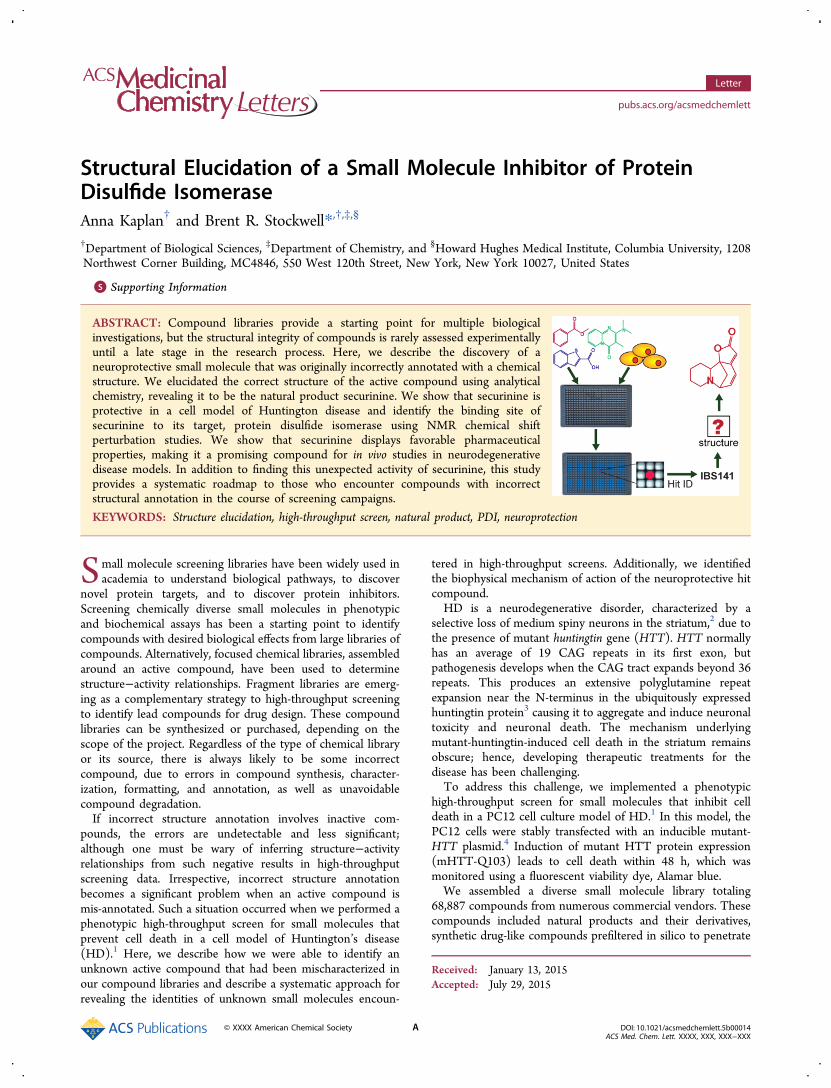
the blood−brain barrier, and compounds with publishedbiological activity.1 We screened this library in PC12 mHTT-Q103 cells to identify neuroprotective small molecules thatprevent mutant-HTT-induced apoptosis. The identified hitsfrom the primary screen were retested in an array of serialdilutions to validate the desired biological activity. One suchcompound, IBS141, was a potent small molecule hit thatshowed reproducible activity in this PC12 mHTT-Q103 cellviability assay with EC50 of 3.3 μM (Figure 1a). We pursuedthis compound in follow-up assays to probe its biologicalactivity.1
To establish the chemical stability of IBS141, we measuredthe mass and biological activity of the compound afterincubation in aqueous buffer. IBS141 was incubated at 37 °Cfor zero, one, two, or three days in PC12 growth mediumbefore aliquots were analyzed by LC−MS (for massdetermination) or added to induced PC12 mHTT-Q103 cells(for biological activity evaluation). After 48 h of treatment,Alamar blue was added to cells and viability assessed on afluorescence plate reader by quantifying the fluorescence of thedye. IBS141 showed the same neuroprotection in PC12mHTT-Q103 cells at all the time points (Figure S1a,Supporting Information), indicating that heat incubation didnot degrade its biological activity. However, by LC−MS,IBS141 showed a single mass peak of a 218.1 m/z at all thetime points (Figure S1b, Supporting Information). This wasunexpected because the predicted mass of IBS141, based on its
assigned structure in the database, was 269.38. We hypothe-sized that this 51 m/z difference between observed andpredicated mass might be due to the degradation of the originalIBS141 stock. To test this, we purchased a fresh batch ofIBS141 from the same supplier (InterBioScreen, cat. #STOCK1N-05465). The new batch of IBS141 had the samebiological activity and potency in PC12 mHTT-Q103 cells(Figure 1b), and the same mass of a 218.1 m/z as the originalbatch, but still differing from the mass assigned to its structurein the vendor database. Next, we purchased the compound withthe assigned structure of IBS141 from a different vendor(ChemBridge, cat. # 5118173). This new compound, CBG141,had the correct expected mass by LC−MS of a 269.38 m/z, butno activity in the PC12 mHTT-Q103 cell viability assay (Figure1b). Furthermore, the proton (1H) NMR of CBG141 andIBS141 showed two different NMR spectra (Figure 1b). Themajor difference is seen in the 6−7 ppm region of the protonspectra; IBS141 shows 1H resonance signals in this region, butCBG141 does not. Protons with chemical shifts observed at 6−7 ppm are due to the presence of vinylic or aromatic groups. Asthere were no protons in the putative IBS141 structure thatcould generate such peaks, this was an indication that thestructure of this hit compound was not consistent with itsassigned structure in our database.It is not uncommon to encounter mischaracterized
compounds in the course of HTS campaigns. Suchmischaracterization can occur due to compound degradation,
Figure 1. Neuroprotective hit compound, IBS141, has an incorrect structure annotation. (a) Overview of high-throughput screen used to identifyIBS141. Reported structure of IBS141 in the vendor database. Viability dose−response curve of IBS141 in PC12 mHTT-Q103 cells. (b) Viabilitydose−response curve and proton NMR spectra of IBS141 and CBG141. CBG141 is a compound with the same annotated structure as IBS141 butfrom a different chemical supplier. All viability data represent the average of triplicates ± SD (n = 3) and plotted as percent of DMSO treateduninduced cells.
ACS Medicinal Chemistry Letters Letter
DOI: 10.1021/acsmedchemlett.5b00014ACS Med. Chem. Lett. XXXX, XXX, XXX−XXX
B

robotic formatting errors, sample contamination, or supplier
errors. Frequently, when researchers encounter this situation,
they simply abandon the active compound, as structure
elucidation can be challenging. However, in this case, we
attempted to elucidate the structure of IBS141 due to its rare
and striking activity in the assay and because we sought a
systematic approach to solve such problems in the future.
To elucidate the structure of IBS141, thorough spectroscopicand analytical characterization were performed (Figure 2a).High-resolution mass spectrometry (HRMS) yielded amolecular composition of C13H15O2N, a molecular weight of217.267, and a monoisotopic mass of 217.110279 m/z for theunknown active compound. Additionally, elemental analysiswas performed to identify traces of C, H, N, B, P, S, Cl, and Felements. Results from elemental analysis confirmed the same
Figure 2. Systematic approach that helped determine the structure of unknown active IBS141 is securinine. (a) Analytical chemistry experiments andthe conclusions from the data. (b) SciFinder database search of the acquired data on the unknown IBS141 identifies securinine as the single structureto match all the criteria.
ACS Medicinal Chemistry Letters Letter
DOI: 10.1021/acsmedchemlett.5b00014ACS Med. Chem. Lett. XXXX, XXX, XXX−XXX
C

molecular composition as was determined by HRMS,C13H15O2N. From the molecular formula, an index of hydrogendeficiency (also known as degree of unsaturation) wascalculated. The unknown IBS141 had seven degrees ofunsaturation, indicating that there were a total of seven rings,double bonds, and/or triple bonds in the structure.Next, we used TLC stains to tests for the presence of specific
functional groups that would account for one nitrogen and twooxygen elements in the compound’s molecular formula. Theninhydrin stain, which tests for the presence of primary andsecondary amines, was negative, indicating that there might be atertiary amine, a nitro group, a nitrile, or an amide in thestructure. The bromocresol green stain, which tests forpresence of functional groups with a pKa of less than five,was negative, indicating that no carboxylic acids were present inthe structure. Schiff’s reagent that tests for the presence ofaldehydes and the 2,4-dinitrophenylhydrazine (DNPH) reagentthat stains for ketones and aldehydes were both negative. Thisindicated that there was an alcohol, an ether, an epoxide, or anester as an oxygen-containing functional group in thecompound.To further elucidate the presence of functional groups, an IR
spectrum was obtained (Figure 2a). The unknown activesubstance had a characteristic strong carbonyl peak atwavenumber 1738.21 cm−1 and a C−O bond vibration bandin the 1300−1000 cm−1 region (with a major peak at 1250.87cm−1). This suggested the presence of an ester functionalgroup. Furthermore, the absence of peaks at 2300−2100 cm−1
indicated no alkynes or nitriles. There were also no absorptionsbeyond 3000 cm−1, which ruled out the presence of aromatic,alcohol, or amide groups. From molecular formula we knewthere was a nitrogen in the structure. The absence of strong N−H stretch at 3500−3300 cm−1 indicated that it must be atertiary amine, which does not have an N−H bond vibration.We also observed a strong absorption at 1626.49 cm−1. Sincewe ruled out the presence of primary amines, a peak in thisposition is indicative of an alkene CC bond stretch.Next, we performed 1H NMR and 13C NMR analyses
(Figure 2a). As mentioned earlier, the proton spectrum showedresonance signals in the 7.0−5.5 ppm region, with a singlet at5.5 ppm, a doublet at 6.60 ppm, and a doublet of doublets at6.41 ppm. Since IR ruled out the presence of aromatic groups,this was indicative of vinylic conjugated protons.The standard 1D 13C NMR produced 13 peaks, each
corresponding to a carbon atom in the unknown active IBS141.To determine the carbon multiplicity, DEPT-135 13C NMRwas performed. The DEPT-135 pulse sequence shows primaryand tertiary carbon peaks with a positive phase, while secondarycarbon peaks show signals with a negative phase. To distinguishbetween primary and tertiary carbons, we also performedDEPT-90 13C NMR; this spectrum is enhanced for signals fromtertiary carbons only. With this information, we were able toidentify the multiplicity of each carbon in the standard 1D 13CNMR spectrum (Figure 2a). After assigning each carbon, wesearched 13C NMR databases for molecules that could generatethe unique peak pattern of quaternary and tertiary carbons seenfrom 180 to 85 ppm. Our search resulted in three possiblesubstructures that matched the observed pattern (Figure 2a).Figure 2a summarizes our findings at this stage. We knew the
unknown active compound had the molecular formula ofC13H15NO2, with no other atoms present. It was a multiringcompound with at least one double bond and a sum of sevenrings and/or double bonds. An amine and an ester were the
only functional groups present within a conjugated π, butnonaromatic, system. The final compound had to contain oneof the three possible substructures observed in 13C NMRspectrum. We searched the SciFinder database, which allows forfurther filtering. First, we searched for all entries with themolecular formula of C13H15NO2, which yielded 3952compounds. Then, we limited these 3952 structures to thosecontaining one of the three substructures identified from NMRstudies. Lastly, we eliminated any compounds with aromaticfunctional groups. These criteria resulted in only onecompound, the natural product securinine (Figure 2b).We purchased genuine securinine from TimTec (cat. #
ST057165) and tested it alongside IBS141 in the PC12 mHTT-Q103 cell viability assay (Figure 3a). Securinine gave an
identical activity and potency profile as unknown active IBS141in the cell viability assay. Furthermore, NMR analysis ofsecurinine showed that both 1H NMR and 13C NMR spectra ofsecurinine were identical to those of IBS141 (Figure 3b,c).From these data we concluded that IBS141 was securinine.After determining the correct structure of the neuro-
protective compound, we investigated the biophysical mecha-nism of action of securinine. We previously showed thatsecurinine can competitively inhibit compound 16F16 bindingto protein disulfide isomerase (PDI).1 16F16 is an irreversiblePDI inhibitor that covalently modifies two cysteines in theactive site of PDI.5 We hypothesized that securinine may alsobe interacting at the same site on PDI as 16F16. To betterunderstand this interaction, we used 1H−15N heteronuclearsingle quantum correlation (HSQC) NMR studies to identifythe binding site of securinine on PDI. For these NMR studieswe expressed and purified 15N-labled catalytic a domain of PDI
Figure 3. Validating the biological and chemical properties ofsecurinine as the correct annotated structure for IBS141. (a) Viabilitydose−response curve in PC12 mHTT-Q103 cells of securinine andIBS141. Data are plotted as mean percent of DMSO treateduninduced cells ± SD. Experiments were performed in triplicate.Proton and carbon NMR spectra of (b) securinine and (c) IBS141 areidentical.
ACS Medicinal Chemistry Letters Letter
DOI: 10.1021/acsmedchemlett.5b00014ACS Med. Chem. Lett. XXXX, XXX, XXX−XXX
D

(referred to as PDIa). The a domain contains the CGHC activesite, can perform the same reduction and oxidation reactions asfull length PDI,6 and is small enough (15 kDa) for NMRbinding studies. Upon securinine binding to PDIa numerouschemical shift perturbations were observed in the proteinresonance peaks (Figure 4a). The most notable changes were
seen in peaks that substantially broadened (disappeared) withcompound titration (i.e., R80, E98, W111, and A43); majority
of these were from the residues around the CGHC active site ofPDI (Figure 4b). Another significant change was observed inthe appearance of H38 peak upon compound titration. In theprotein alone spectrum H38 is undergoing a rapid hydrogenexchange with the solvent that accounts for the absence of itsHSQC peak.7 Presence of securinine prevents this exchange.Additional chemical shifts were in the region opposite theactive site (i.e., L10, A23, H24, Y26, N90, and G91). Theseshifts are indicative of the conformational change in the proteinupon securinine binding. Based on the mapped chemical shiftperturbation data, securinine may be binding adjacent to theactive site of PDI and inducing a conformation change in theprotein. Securinine interaction with H38 and R103 of PDI maylead to destabilization of active site C36 residue, thus inhibitingPDI function. Both H38 and R103 residues are needed tostabilize the negatively charged cysteine thiolate transitionstate7 of PDI. An alternative mechanism of inhibition is that theα,β-unsaturated lactone pharmacophore in securinine may beacting as a Michael acceptor for the nucleophilic attack byH38.8 This would result in a formation of an irreversiblecomplex between securinine and PDI, leading to PDIinhibition.To investigate the binding mechanism further, we monitored
the intrinsic tryptophan (Trp) fluorescence of PDIa in thepresence of securinine. As shown in Figure 5a, Trp fluorescencewas strongly quenched upon addition of securinine in aconcentration-dependent manner with a dissociation constant(Kd) of 758 ± 129 μM (Figure 5b). Next, we monitored Trpfluorescence of PDIa after performing the jump dilutionmethod to test for reversibility of binding. PDIa (1 mM) wasincubated with high concentration of securinine (4.5 mM). Thecomplex was then diluted 100-fold and Trp fluorescencerecorded. As a control, the same procedure was performed on asample containing phenylarsine oxide (PAO), a covalentirreversible modifier of vicinal thiol groups such as thosefound in the active site of PDI. After dialysis, the securinine−PDIa complex (now at 45 μM securinine and 10 μM PDIa) andcontrol PAO−PDIa samples still showed complete quenchingof intrinsic Trp fluorescence (Figure 5c). This result indicatedthat securinine, like PAO, binds irreversibly to PDI.Next, we wanted to investigate if securinine would be a good
candidate for in vivo studies by evaluating its in vitro metabolicstability. Securinine had a low intrinsic clearance value of 8.3mL/min/g in mouse liver microsomes compared to the control
Figure 4. Chemical shift perturbations upon securinine binding toPDI. (a) 15N-HSQC spectra of PDIa alone or liganded with securinine.Most perturbed residues are in blue. (b) Chemical shift changesmapped onto the surface and ribbon depiction of PDIa (PDB: 1MEK,model #35). The residue numbering is based on the mature PDIprotein sequence.
Figure 5. Intrinsic tryptophan fluorescence assay shows irreversible binding of securinine to PDI. (a) Fluorescence emission spectra and (b)normalized intensity at 330 nm of PDIa in the presence of varying concentrations of securinine. (c) Emission spectra of PDIa after ligand−proteincomplex is formed at high concentration and then diluted 100-fold. The Trp fluorescence is still quenched in securinine treated samples and in PAOcontrol after the dilution indicating irreversible binding to PDIa by the ligand. All spectra are normalized by the maximum fluorescence intensity ofthe PDIa-only sample (black). Data are plotted as mean ± SEM and fitted to a log-normal distribution.
ACS Medicinal Chemistry Letters Letter
DOI: 10.1021/acsmedchemlett.5b00014ACS Med. Chem. Lett. XXXX, XXX, XXX−XXX
E
compound 7-ethoxycoumarin (intrinsic clearance of 24.58 mL/min/g) (Table S1, Supporting Information), and it was alsorelatively stable in mouse plasma with a half-life of 1.6 h (TableS2, Supporting Information). Furthermore, only 49% ofsecurinine is bound to plasma proteins, compared to warfarin,an anticoagulant agent, with 94% plasma protein binding(Table S3, Supporting Information), indicating that in vivomore than 50% of securinine may be free to be distributed totissues to exert pharmacological effects.Securinine is a naturally occurring alkaloid that can be
extracted from the plant Securinega suf f ruticosa. It has beenshown to act as a central nervous system stimulant, at least inpart by blocking the GABA binding site on the GABAA
receptors.9,10 Securinine showed neuroprotection activities inrats with β-amyloid toxicity10 and in ameliorating the symptomsin patients with ALS.11 In our study, we found that securinine isneuroprotective in cell culture (Figure 3a) and in corticostriatalbrain slice cultures of HD.1 Since PC12 cells do not havefunctional GABAA receptors,12 we found that neuroprotectionmechanism of securinine is caused by inhibition of PDI.1 Weshow that securinine binds adjacent to the active site of PDI byforming an irreversible complex with the protein (Figure 5c).Given our experience, we emphasize the importance of
verifying the composition of active compounds at an early stageof a screening project. At a minimum, a mass spectrum shouldalways be measured on all compounds of interest. Ifmisidentification occurs, we have outlined steps that one cantake to identify an unknown active small molecule. We believethis will be useful to both chemists and biologists as these stepscan save time and resources for other researchers that are facingsimilar situations of misidentified compounds and canultimately allow further studies and development of activecompounds once their structures are identified.
■ ASSOCIATED CONTENT
*S Supporting InformationThe Supporting Information is available free of charge on theACS Publications website at DOI: 10.1021/acsmedchem-lett.5b00014.
Supplementary figures and tables, compound character-ization, and biological assays (PDF)
■ AUTHOR INFORMATION
Corresponding Author*E-mail: [email protected].
Author ContributionsA.K. performed the experiments and analyzed the data. A.K.and B.R.S. designed the experiments and wrote the manuscript.All authors have given approval to the final version of themanuscript.
FundingThis research was funded by the Howard Hughes MedicalInstitute, National Institute of Health (5R01CA097061,5R01GM085081, R01CA161061), and New York Stem CellScience (C026715) to B.R.S., and the Training Program inMolecular Biophysics (T32GM008281) to A.K.
NotesThe authors declare no competing financial interest.
■ ACKNOWLEDGMENTSWe are grateful to Dr. Yasuhiro Itagaki for HRMS data, Dr. LuisAvila for assistance with FT-IR experiments, and Dr. JohnDecatur for assistance and the use of the Columbia ChemistryNMR core facility instruments provided by NIH1S10RR25431-1A1 and NSF CHE-0840451 grant.
■ ABBREVIATIONSHD, Huntington’s disease; HTT, huntingtin gene; 2,4-DNPH,2,4-dinitrophenylhydrazine; HSQC, heteronuclear single quan-tum correlation; PDIa, a domain of protein disulfide isomerase
■ REFERENCES(1) Hoffstrom, B. G.; Kaplan, A.; Letso, R.; Schmid, R. S.; Turmel, G.J.; Lo, D. C.; Stockwell, B. R. Inhibitors of protein disulfide isomerasesuppress apoptosis induced by misfolded proteins. Nat. Chem. Biol.2010, 6, 900−6.(2) Mink, J. W. The basal ganglia: focused selection and inhibition ofcompeting motor programs. Prog. Neurobiol. 1996, 50, 381−425.(3) Kaplan, A.; Stockwell, B. R. Therapeutic approaches topreventing cell death in Huntington disease. Prog. Neurobiol. 2012,99, 262−80.(4) Aiken, C. T.; Tobin, A. J.; Schweitzer, E. S. A cell-based screen fordrugs to treat Huntington’s disease. Neurobiol. Dis. 2004, 16, 546−55.(5) Kaplan, A.; Gaschler, M. M.; Dunn, D. E.; Colligan, R.; Brown, L.M.; Palmer, A. G.; Lo, D. C.; Stockwell, B. R. Small molecule-inducedoxidation of protein disulfide isomerase is neuroprotective. Proc. Natl.Acad. Sci. U. S. A. 2015, 112, E2245−52.(6) Darby, N. J.; Creighton, T. E. Functional properties of theindividual thioredoxin-like domains of protein disulfide isomerase.Biochemistry 1995, 34, 11725−35.(7) Hernandez, G.; Anderson, J. S.; LeMaster, D. M. Electrostaticstabilization and general base catalysis in the active site of the humanprotein disulfide isomerase a domain monitored by hydrogenexchange. ChemBioChem 2008, 9, 768−78.(8) Liu, X. W.; Sok, D. E. Inactivation of protein disulfide isomeraseby alkylators including alpha, beta-unsaturated aldehydes at lowphysiological pHs. Biol. Chem. 2004, 385, 633−37.(9) Beutler, J. A.; Karbon, E. W.; Brubaker, A. N.; Malik, R.; Curtis,D. R.; Enna, S. J. Securinine alkaloids: a new class of GABA receptorantagonist. Brain Res. 1985, 330, 135−40.(10) Lin, X.; Jun-Tian, Z. Neuroprotection by D-securinine againstneurotoxicity induced by beta-amyloid (25−35). Neurol. Res. 2004, 26,792−6.(11) Copperman, R.; Copperman, G.; Marderosian, A. D. Letter:Securinine. JAMA 1974, 228, 288.(12) Hales, T. G.; Tyndale, R. F. Few cell lines with GABAA mRNAshave functional receptors. J. Neurosci. 1994, 14, 5429−36.
ACS Medicinal Chemistry Letters Letter
DOI: 10.1021/acsmedchemlett.5b00014ACS Med. Chem. Lett. XXXX, XXX, XXX−XXX
F