Metals in Medicine / Biology A/P Matthew Wilce UWA Structural Biology Computational Biology.
SPANISH NATIONAL CANCER RESEARCH CENTRE, CNIO 73ANNUAL REPORT 2015 72
Vice-Direction of Basic research structural Biology anD Biocomputing programme
STRUCTURAL BIOLOGY AND BIOCOMPUTING PROGRAMMEALFONSO VALENCIA Programme Director
The objective of the Structural Biology and Biocomputing Programme is the mechanistic understanding of key cancer-related molecular systems. The Programme was designed to combine computational and structural approaches as well as to collaborate with the CNIO Basic and Translational Research activities.
Our 3 main research goals are to :
ɗ Reconstruct the structural details of protein complexes that are active in cell adhesion and metabolism.
ɗ Predict the consequences of cancer related alterations ; we are focusing on alterations with a compensatory nature ( co-evolutionary related mutations ) as well as those affecting alternative splicing patterns.
ɗ Contribute to the analysis of cancer epigenomic and genomic information within the framework of international genome projects.
The Programme is currently composed of 3 Research Groups and 6 Core Units that provide support to the CNIO’s research activities.
Recently, 2 key groups left the CNIO in order to follow their scientific development elsewhere. Francesco Gervasio, an expert in biophysical simulations, went to the University College of London, and Guillermo Montoya, a senior crystallographer, moved to the Novo Nordisk Foundation Center for Protein Research in Copenhagen, while maintaining during 2015 a reduced research activity.
In order to maintain a reasonable level of activity and to provide the necessary support to internal projects, we established 2 new Core Units this year : a Protein Crystallography Unit, which is shared with the CNIO Experimental Therapeutics Programme ; and an Electron Microscopy Unit that works in collaboration with the laboratory of O. Llorca ( Centro de Investigaciones Biológicas, CIB-CSIC, Madrid ), where cell biology samples are prepared for further observation and imaging using our microscopes. These new Units have started their independent work by re-organising the equipment and operational aspects ; they have already commenced work with a number of CNIO groups. Moreover, we also share the Translational Bioinformatics Unit, led by Fátima Al-Sharour, with the Clinical Research Programme.
“ After a very positive initial period, the Programme is now in a phase of reorganisation, consisting of the renewal of the equipment, the replenishment of its Core Units, and most importantly, the recruitment of new group leaders in Structural and Computational Biology.”
Of particular relevance to the CNIO’s activities in Computational Biology, was the finalisation of the negotiation for membership in the European Bioinformatics Infrastructure ELIXIR. ELIXIR is a large international consortium that is now associated to a European grant ( EXCELERATE ), in which the Spanish participation is led by the CNIO. CNIO’s National Bioinformatics Institute Unit ( INB-ISCIII ) has played a key role in developing the technical aspects of the Spanish participation in ELIXIR and EXCELERATE.
During 2015, the 3-year extension of the 5+3 contracts of the 2 junior Group Leaders was evaluated by an ad-hoc expert committee and approved by the CNIO. Later in the year, the Programme was reviewed in depth by the CNIO’s External Scientific Advisory Board ( SAB ). The SAB evaluated the activity of the Programme very positively and endorsed the reorganisation of its Units, for which it made very specific recommendations in regards to the need to focus on the proper equipping of these Units. Very importantly, the SAB strongly recommended the appointment of at least one senior structural biologist. The CNIO has taken these recommendations very seriously, allocating additional funds for the renewal of the equipment of the Units, and has started the search for a senior and a junior structural biologist.
SPANISH NATIONAL CANCER RESEARCH CENTRE, CNIO 75ANNUAL REPORT 2015 74
structural Biology anD Biocomputing programme | STRUCTURAL COmPUTATIONAL BIOLOgy gROUPVice-Direction of Basic research
STRUCTURAL COMPUTATIONAL BIOLOGY GROUP
oVerVieW
The main interest of our Group is the study of the molecular bases of cancer by bringing an evolutionary perspective to the study of the interplay between genomics and epigenomics in tumour progression.
Our research is largely carried out in the context of large-scale genome projects, where we develop new computational methods for the study of genome-cancer relationships.
In this general scenario, the strategic goals of the Structural Computational Biology Group are to :
ɗ Develop software platforms for the extraction, integration and representation of cancer data, including the analysis of molecular, genomic, epigenomic and phenotypic information in collaboration with large-scale genome projects.
ɗ Analyse the function, structure and specific interactions of cancer-related proteins.
ɗ Develop methods, tools and ideas to understand and model processes relating to genome structure, organisation and evolution, with a special focus on tumour progression.
“ This year we carried out a new research effort regarding the analysis of genome/epigenome data, including chromatin 3D structure data, by combining methods for functional genome segmentation with network biology strategies. The results reveal interesting properties of chromatin organisation, including fundamental components for its organisation and dynamics.”
research highlights
The work of our Group can be described in terms of our contributions to 3 large-scale community efforts.
Our Group contributes to the organisation of the data flow and analysis of the Spanish Chronic Lymphocytic Leukaemia ( CLL ) project, which is part of the International Cancer Genome Consortium ( ICGC ) that ended in 2015 ; and to the BLUEPRINT epigenome EU flagship project, which is now entering its final phase of data production. We have participated in the main published studies of the CLL consortium, as well as in the white papers on analysis technology of the ICGC consortium. In 2016, our main efforts will be dedicated to the analysis of the massive amounts of data produced by the largest cancer genome sequencing consortia, IGCG and TCGA, in what is known as PanCancer, as well as to the completion of the integrated analysis of the BLUEPRINT data.
Organisation of chromatin and the interaction between its components
Inspired by the work of the CLL-ICGC and BLUEPRINT projects, we have developed a new framework for the analysis of large scale epigenetic and chromatin capture data. Based on concepts developed in the area of network biology, we have used the available information to reconstruct the network of interactions between a large set of more than 70 chromatin features, including DNA and histone modifications, as well as a large number of chromatin binding proteins. The analysis of the network reveals interesting properties of the components related to their specific functional activity and evolutionary history, including detection of the importance of the 5hmC modification of DNA as a network organiser.
Alfonso ValenciaGroup Leader
Staff ScientistsFederico Abascal ( until May ), Andrea Nicole Dölker, Milana Morgenstern ( until November ), Tirso Pons, Daniel Rico, Michael Tress
Post-Doctoral FellowsSimone Marsili, Vera Pancaldi, Miguel Vázquez
Graduate StudentsSimone Ecker ( until October ), Maria Rigau, Juan Rodríguez, Jon Sánchez
TechniciansDavid A. Juan ( TS )*, Martin Krallinger ( TS )*, Filipe N. Were ( since September ) ( TS )*
*Titulado Superior ( Advanced Degree )
Visiting ScientistsDimitrios Morikis ( University of California, Riverside, USA ), Evangelina Nogales ( July-September ) ( University of California, Berkeley, USA )
SPANISH NATIONAL CANCER RESEARCH CENTRE, CNIO 77ANNUAL REPORT 2015 76
structural Biology anD Biocomputing programme | STRUCTURAL COmPUTATIONAL BIOLOgy gROUPVice-Direction of Basic research
Biological text mining
Text mining is an increasingly important component of computational biology with numerous applications in biology and biomedicine.
As part of the BioCreative international effort for the evaluation of text mining systems, we organised the 2015 competition ( Seville, Sept 2015 ), with a special focus on the extraction of mentions of chemicals ( drugs and other compounds ), diseases and gene names from patient records. For the competition, we created a large, systematically annotated corpus, as well as the corresponding annotation guidelines and tools to score the results of the teams participating in the competition.
Beyond the BioCreative benchmarking effort, in the context of the IMI eTOX project, we completed the development of a
system specialised in the extraction of information related to the toxicology of biological compounds and drugs from scientific publications and toxicology reports. The relationships between administered drugs, symptoms, toxic end-points, target genes and cytochrome variants are systematically linked to the related databases and underlying text.
We have also completed the development of a text mining system to extract information related to melanoma from the literature. The system systematically explores melanoma related papers to detect mentions of genes, mutations and drugs, as well as a number of medical/pathology related terms, and exposes the information to the end-users together with facilities for navigating the information in the context of the bulk of information available from the large cancer genome projects. s
The additional incorporation of information on the three dimensional organisation of the chromatin in the nucleus ( HiC and PChiC experiments ) opens up new avenues for the study of the dynamics of the interactions between active chromatin features, such as the different isoforms of RNA polymerase II.
Alternative splicing at the protein level
This year we continued the work on alternative splicing in the context of the NIH-funded GENCODE project.
We have produced a new release of the APPRIS system for the systematic annotation of protein isoforms. APPRIS annotates binding sites, evolutionary rates, the existence of complete protein structures or models, as well as the presence of membrane regions, and determines the most likely complete isoform for each gene (“ Principal ” Isoform ). APPRIS annotations are part of the information used for the annotation of the human genome.
The Principal Isoform is the only one that has protein-like features, including presence of binding sites, coverage of known protein structures, evolutionary conservation and normal rates of evolution, and it is the genuine representative of the function of the corresponding genes. The systematic analysis of the Principal Isoforms has several interesting consequences.
Firstly, it allows a better definition of the mutations that might be relevant in cancer studies, rejecting others that map outside the boundaries of the Principal Isoforms, and therefore are expected to not have functional consequences at the protein level.
Secondly, we have been able to compare the evolutionary consequences of splicing versus gene duplication by reconstructing the history of a selected set of genes that after duplication have retained 2 very similar paralogs, or have produced 2 splice isoforms differing solely by the presence of 2 homologous exons.
Thirdly, by combining APPRIS with the systematic exploration of all the available large scale MS data sets – carried out in collaboration with the group of Jesus Vazquez ( CNIC ) – we have demonstrated that the immense majority of genes only express 1 isoform at the protein level ; at least for those genes with medium-high levels of expression. This finding is in clear contradiction with studies that have analysed expression at the level of transcription but, perhaps not surprisingly, the finding seems to fit well with the large scale analysis of gene expression in multiple tissues, carried out by the ENCODE/GTEx ( www.gtexportal.org ) consortium.
∞ PUBLICATIONS
∞ Puente XS et al. ( incl. Valencia A ) ( 2015 ). Non-coding recurrent mutations in chronic lymphocytic leukaemia. Nature 526, 519-524.
∞ Mutation Consequences and Pathway Anal-ysis working group of the International Can-cer Genome Consortium. ( 2015 ). Pathway and network analysis of cancer genomes. Nat Methods 12, 615-621.
∞ Kulis M et al. ( incl. Valencia A ) ( 2015 ). Whole-genome fingerprint of the DNA methylome during human B cell differen-tiation. Nat Genet 4, 746-756.
∞ Seguí N et al. ( incl. Valencia A ) ( 2015 ). Germline Mutations in FAN1 Cause Hered-itary Colorectal Cancer by Impairing DNA
Repair. Gastroenterology 149, 563-566. ∞ Maraver A, Fernandez-Marcos PJ, Cash TP, Mendez-Pertuz M, Dueñas M, Maietta P, Martinelli P, Muñoz-Martin M, Martín-ez-Fernández M, Cañamero M, Roncador G, Martinez-Torrecuadrada JL, Grivas D, de la Pompa JL, Valencia A, Paramio JM, Real FX, Serrano M ( 2015 ). NOTCH pathway inactiva-tion promotes bladder cancer progression. J Clin Invest 125, 824-830.
∞ Sutto L, Marsili S, Valencia A, Gervasio FL ( 2015 ). From residue coevolution to protein conformational ensembles and functional dynamics. Proc Natl Acad Sci USA 112, 13567-13572.
∞ Frenkel-Morgenstern M, Gorohovski A, Vucenovic D, Maestre L, Valencia A ( 2015 ). ChiTaRS 2.1-an improved database of the
chimeric transcripts and RNA-seq data with novel sense-antisense chimeric RNA transcripts. Nucleic Acids Res 43, D68-D75.
∞ Rodriguez JM, Carro A, Valencia A, Tress ML. ( 2015 ). APPRIS WebServer and WebSer-vices. Nucleic Acids Res 43, W455-W459.
∞ Bellido F, Pineda M, Aiza G, Valdés-Mas R, Navarro M, Puente DA, Pons T, González S, Iglesias S, Darder E, Piñol V, Soto JL, Valencia A, Blanco I, Urioste M, Brunet J, Lázaro C, Capellá G, Puente XS, Valle L, ( 2015 ). POLE and POLD1 mutations in 529 kindred with familial colorectal cancer and/or polyposis : review of reported cases and recommendations for genetic testing and surveillance. Genet Med. PMID : 26133394.
∞ Ecker S, Pancaldi V, Rico D, Valencia A ( 2015 ). Higher gene expression variability
in the more aggressive subtype of chronic lymphocytic leukemia. Genome Med 7, 8.
∞ Vazquez M, Pons T, Brunak S, Valencia A, Izarzugaza JM ( 2015 ). wKinMut-2 : Identi-fication and Interpretation of Pathogenic Variants in Human Protein Kinases. Hum Mutat. PMID : 26443060.
∞ Abascal F, Tress ML, Valencia A. ( 2015 ). Alternative splicing and co-option of trans-posable elements : the case of TMPO/LAP2a and ZNF451 in mammals. Bioinformatics 31, 2257-2261.
∞ Lees JG, Hériché JK, Morilla I, Fernández JM, Adler P, Krallinger M, Vilo J, Valencia A, Ellenberg J, Ranea JA, Orengo C ( 2015 ). FUN-L : gene prioritization for RNAi screens. Bioinformatics 31, 2052-2053.
∞ Verspoor K, Shatkay H, Hirschman L,
Blaschke C, Valencia A ( 2015 ). Summary of the BioLINK SIG 2013 meeting at ISMB/ECCB 2013. Bioinformatics 31, 297-298.
∞ Ochoa D, Juan D, Valencia A, Pazos F ( 2015 ). Detection of significant protein coevolution. Bioinformatics 31, 2166-2173.
∞ Vázquez M, Valencia A, Pons T ( 2015 ). Structure-PPi : a module for the annotation of cancer-related single-nucleotide variants at protein-protein interfaces. Bioinformatics 31, 2397-2399.
∞ Abascal F, Ezkurdia I, Rodriguez-Rivas J, Rodriguez JM, Del Pozo A, Vázquez J, Valen-cia A, Tress ML. ( 2015 ). Alternatively Spliced Homologous Exons Have Ancient Origins and Are Highly Expressed at the Protein Level. PLoS Comput Biol 11, e1004325.
∞ Krallinger M, Rabal O, Leitner F, Vazquez
M, Salgado D, Lu Z, Leaman R, Lu Y, Ji D, Lowe DM, Sayle RA, Batista-Navarro RT, Rak R, Huber T, Rocktäschel T, Matos S, Campos D, Tang B, Xu H, Munkhdalai T, Ryu KH, Ramanan SV, Nathan S, Žitnik S, Bajec M, Weber L, Irmer M, Akhondi SA, Kors JA, Xu S, An X, Sikdar UK, Ekbal A, Yoshioka M, Dieb TM, Choi M, Verspoor K, Khabsa M, Giles CL, Liu H, Ravikumar KE, Lamurias A, Couto FM, Dai HJ, Tsai RT, Ata C, Can T, Usié A, Alves R, Segura-Bedmar I, Martínez P, Oyarzabal J, Valencia A. ( 2015 ). The CHEMDNER corpus of chemicals and drugs and its annotation principles. J Chem-inform. PMID : 25810773.
∞ Krallinger M, Leitner F, Rabal O, Vazquez M, Oyarzabal J, Valencia A ( 2015 ). CHEMD-NER : The drugs and chemical names ex-
traction challenge. J Cheminform. PMID : 25810766.
∞ Ezkurdia I, Vázquez J, Valencia A, Tress M ( 2015 ). Correction to “ Analyzing the first drafts of the human proteome ”. J Proteome Res 14, 1991-0.
∞ Ezkurdia I, Rodriguez JM, Carillo-de San-ta Pau E, Vázquez J, Valencia A, Tress ML ( 2015 ). Most highly expressed protein-cod-ing genes have a single dominant isoform. J Proteome Res 14, 1880-1887.
∞ Abascal F, Tress ML, Valencia A. ( 2015 ). The evolutionary fate of alternatively spliced homologous exons after gene duplication. Genom Biol Evol 7, 1392-1403.
∞ Earl J, Rico D, Carrillo-de-Santa-Pau E, Rodríguez-Santiago B, Méndez-Pertuz M, Auer H, Gómez G, Grossman HB, Pisano
DG, Schulz WA, Pérez-Jurado LA, Carrato A, Theodorescu D, Chanock S, Valencia A, Real FX. ( 2015 ). The UBC-40 Urothelial Bladder Cancer cell line index : a genom-ic resource for functional studies. BMC Genomics 16, 403.
∞ López-Castilla A, Pons T, Pires JR ( 2015 ). NMR structure and dynamics of Q4D059, a kinetoplastid-specific and conserved protein from Trypanosoma cruzi. J Struct Biol 190, 11-20.
∞ Ezkurdia I, Calvo E, Del Pozo A, Vázquez J, Valencia A, Tress ML ( 2015 ). The potential clinical impact of the release of two drafts of the human proteome. Expert Rev Pro-teomic 12, 579-593.
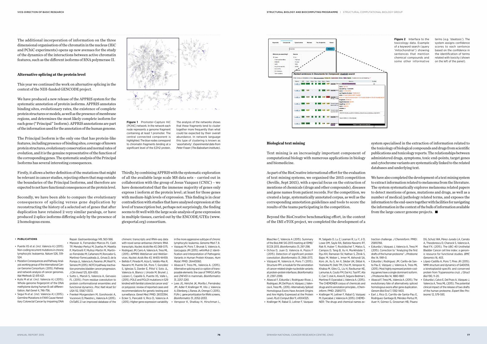
Figure 2 Interface to the toxicology data. Example of a keyword search ( query ’mitochondrial ’) showing sentences that mention chemical compounds and some other informative
terms ( e.g. ’steatosis ’). The system assigns confidence scores to each sentence based on the confidence in the identification of terms related with toxicity ( shown on the left of the panel ).
Figure 1 Promoter-Capture HiC ( PCHiC ) network. In the network each node represents a genome fragment containing at least 1 promoter. The central connected component is highlighted. The blue nodes correspond to chromatin fragments binding at a significant level of the EZH2 protein.
The analysis of the networks shows that these fragments tend to cluster together more frequently than what could be expected by their overall abundance. In network language this type of clustering is known as ‘ assortativity ’. Experimental data from Peter Fraser ( The Babraham Institute ).
SPANISH NATIONAL CANCER RESEARCH CENTRE, CNIO 79ANNUAL REPORT 2015 78
structural Biology anD Biocomputing programme | mACROmOLECULAR CRySTALLOgRAPHy gROUPVice-Direction of Basic research
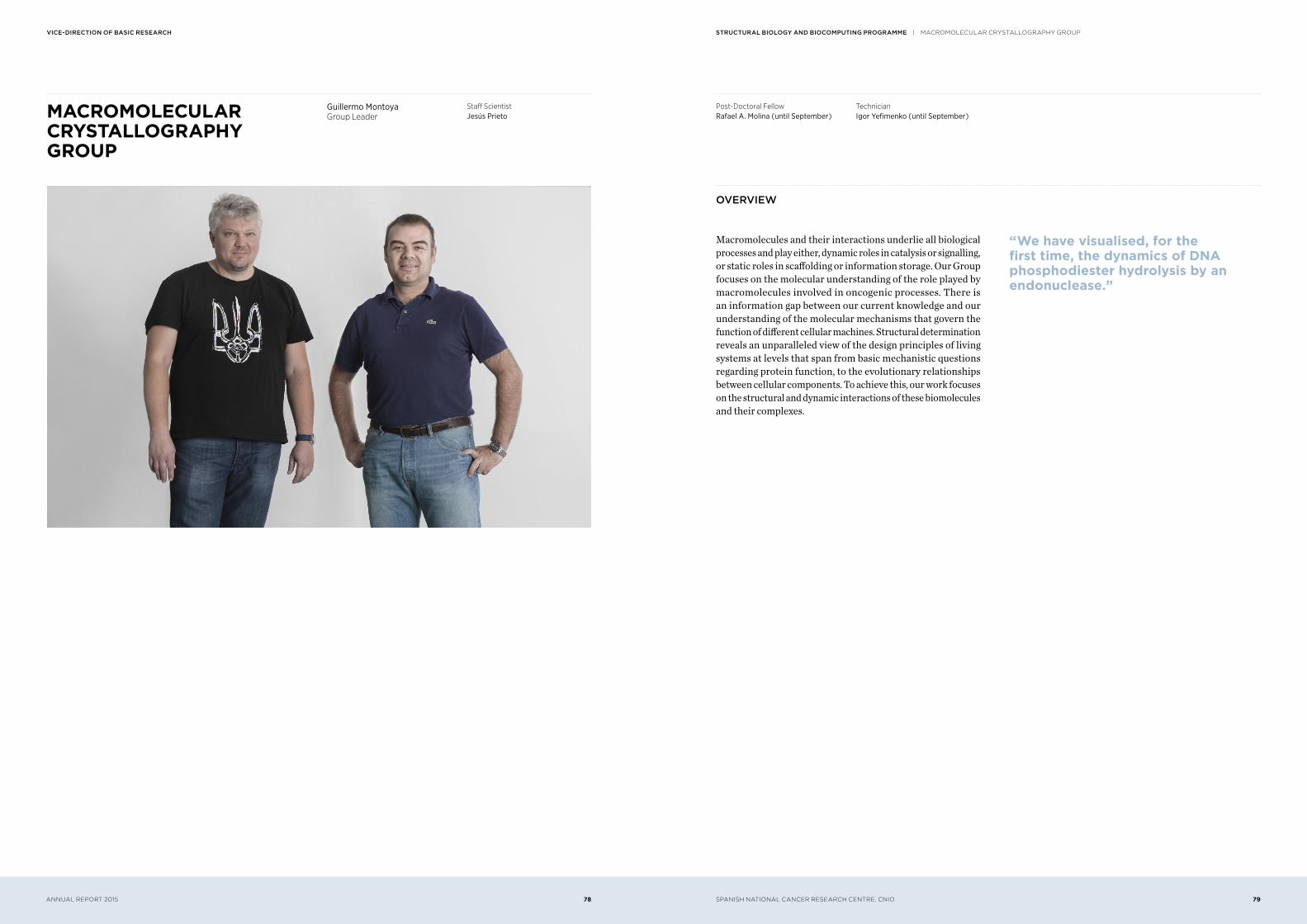
MACROMOLECULAR CRYSTALLOGRAPHY GROUP
oVerVieW
Macromolecules and their interactions underlie all biological processes and play either, dynamic roles in catalysis or signalling, or static roles in scaffolding or information storage. Our Group focuses on the molecular understanding of the role played by macromolecules involved in oncogenic processes. There is an information gap between our current knowledge and our understanding of the molecular mechanisms that govern the function of different cellular machines. Structural determination reveals an unparalleled view of the design principles of living systems at levels that span from basic mechanistic questions regarding protein function, to the evolutionary relationships between cellular components. To achieve this, our work focuses on the structural and dynamic interactions of these biomolecules and their complexes.
“ We have visualised, for the first time, the dynamics of DNA phosphodiester hydrolysis by an endonuclease.”
Guillermo MontoyaGroup Leader
Staff ScientistJesús Prieto
Post-Doctoral FellowRafael A. Molina ( until September )
TechnicianIgor Yefimenko ( until September )
SPANISH NATIONAL CANCER RESEARCH CENTRE, CNIO 81ANNUAL REPORT 2015 80
structural Biology anD Biocomputing programme | mACROmOLECULAR CRySTALLOgRAPHy gROUPVice-Direction of Basic research
Mitotic complexes
Cellular growth and division are regulated by an integrated protein network that ensures the genomic integrity of all eukaryotic cells during mitosis. Microtubules play an important role in several cellular processes, particularly in the formation of the mitotic spindle. The regulation of microtubule dynamics during mitosis is key for spindle formation. Spindle defects, arising from failures in setting up the microtubules, lead to chromosomal instability and aneuploidy, a common cause of tumour development. One of the most effective strategies for cancer treatment so far has been to interfere with the highly dynamic mitotic spindle microtubules ; tubulin remains the
most successful spindle targeted molecule in cancer. To date, novel anti-mitotic agents have demonstrated limited efficacy in clinical trials and classical anti microtubule drugs are still considered as being the best approach for cancer therapy. We are attempting to dissect the molecular working mechanism of CCT/TRiC, the molecule responsible for the folding of tubulin and actin, which are the essential building blocks of the cytoskeleton. This molecular machine is essential for sister chromatid separation through the folding of key anaphase promoting factor subunits, such as Cdc20. Using a hybrid approach, we are aiming to dissect the molecular recognition of these key substrates by the chaperonin. s
research highlights
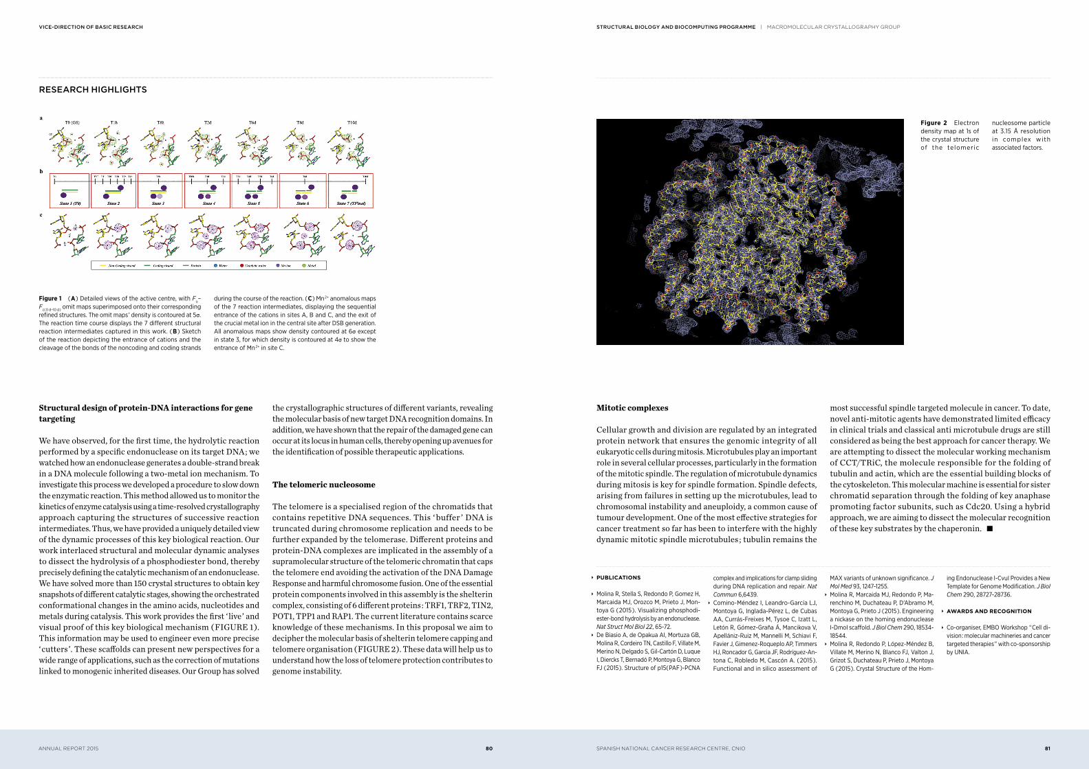
Structural design of protein-DNA interactions for gene targeting
We have observed, for the first time, the hydrolytic reaction performed by a specific endonuclease on its target DNA ; we watched how an endonuclease generates a double-strand break in a DNA molecule following a two-metal ion mechanism. To investigate this process we developed a procedure to slow down the enzymatic reaction. This method allowed us to monitor the kinetics of enzyme catalysis using a time-resolved crystallography approach capturing the structures of successive reaction intermediates. Thus, we have provided a uniquely detailed view of the dynamic processes of this key biological reaction. Our work interlaced structural and molecular dynamic analyses to dissect the hydrolysis of a phosphodiester bond, thereby precisely defining the catalytic mechanism of an endonuclease. We have solved more than 150 crystal structures to obtain key snapshots of different catalytic stages, showing the orchestrated conformational changes in the amino acids, nucleotides and metals during catalysis. This work provides the first ‘ live ’ and visual proof of this key biological mechanism ( FIGURE 1 ). This information may be used to engineer even more precise ‘ cutters ’. These scaffolds can present new perspectives for a wide range of applications, such as the correction of mutations linked to monogenic inherited diseases. Our Group has solved
the crystallographic structures of different variants, revealing the molecular basis of new target DNA recognition domains. In addition, we have shown that the repair of the damaged gene can occur at its locus in human cells, thereby opening up avenues for the identification of possible therapeutic applications.
The telomeric nucleosome
The telomere is a specialised region of the chromatids that contains repetitive DNA sequences. This ‘ buffer ’ DNA is truncated during chromosome replication and needs to be further expanded by the telomerase. Different proteins and protein-DNA complexes are implicated in the assembly of a supramolecular structure of the telomeric chromatin that caps the telomere end avoiding the activation of the DNA Damage Response and harmful chromosome fusion. One of the essential protein components involved in this assembly is the shelterin complex, consisting of 6 different proteins : TRF1, TRF2, TIN2, POT1, TPP1 and RAP1. The current literature contains scarce knowledge of these mechanisms. In this proposal we aim to decipher the molecular basis of shelterin telomere capping and telomere organisation ( FIGURE 2 ). These data will help us to understand how the loss of telomere protection contributes to genome instability.
Figure 1 ( A ) Detailed views of the active centre, with Fo– Fc( 0 d–10 d ) omit maps superimposed onto their corresponding refined structures. The omit maps ’ density is contoured at 5σ. The reaction time course displays the 7 different structural reaction intermediates captured in this work. ( B ) Sketch of the reaction depicting the entrance of cations and the cleavage of the bonds of the noncoding and coding strands
during the course of the reaction. ( C ) Mn 2+ anomalous maps of the 7 reaction intermediates, displaying the sequential entrance of the cations in sites A, B and C, and the exit of the crucial metal ion in the central site after DSB generation. All anomalous maps show density contoured at 6σ except in state 3, for which density is contoured at 4σ to show the entrance of Mn 2+ in site C.
Figure 2 Electron density map at 1s of the crystal structure of the telomeric
nucleosome particle at 3.15 Å resolution in complex with associated factors.
∞ PUBLICATIONS
∞ Molina R, Stella S, Redondo P, Gomez H, Marcaida MJ, Orozco M, Prieto J, Mon-toya G ( 2015 ). Visualizing phosphodi-ester-bond hydrolysis by an endonuclease. Nat Struct Mol Biol 22, 65-72.
∞ De Biasio A, de Opakua AI, Mortuza GB, Molina R, Cordeiro TN, Castillo F, Villate M, Merino N, Delgado S, Gil-Cartón D, Luque I, Diercks T, Bernadó P, Montoya G, Blanco FJ ( 2015 ). Structure of p15( PAF )-PCNA
complex and implications for clamp sliding during DNA replication and repair. Nat Commun 6,6439.
∞ Comino-Méndez I, Leandro-García LJ, Montoya G, Inglada-Pérez L, de Cubas AA, Currás-Freixes M, Tysoe C, Izatt L, Letón R, Gómez-Graña Á, Mancikova V, Apellániz-Ruiz M, Mannelli M, Schiavi F, Favier J, Gimenez-Roqueplo AP, Timmers HJ, Roncador G, Garcia JF, Rodríguez-An-tona C, Robledo M, Cascón A. ( 2015 ). Functional and in silico assessment of
MAX variants of unknown significance. J Mol Med 93, 1247-1255.
∞ Molina R, Marcaida MJ, Redondo P, Ma-renchino M, Duchateau P, D’Abramo M, Montoya G, Prieto J ( 2015 ). Engineering a nickase on the homing endonuclease I-DmoI scaffold. J Biol Chem 290, 18534-18544.
∞ Molina R, Redondo P, López-Méndez B, Villate M, Merino N, Blanco FJ, Valton J, Grizot S, Duchateau P, Prieto J, Montoya G ( 2015 ). Crystal Structure of the Hom-
ing Endonuclease I-CvuI Provides a New Template for Genome Modification. J Biol Chem 290, 28727-28736.
∞ AWARDS AND RECOGNITION
∞ Co-organiser, EMBO Workshop “ Cell di-vision : molecular machineries and cancer targeted therapies ” with co-sponsorship by UNIA.
SPANISH NATIONAL CANCER RESEARCH CENTRE, CNIO 83ANNUAL REPORT 2015 82
structural Biology anD Biocomputing programme | CELL SIgNALLINg ANd AdHESION JUNIOR gROUPVice-Direction of Basic research
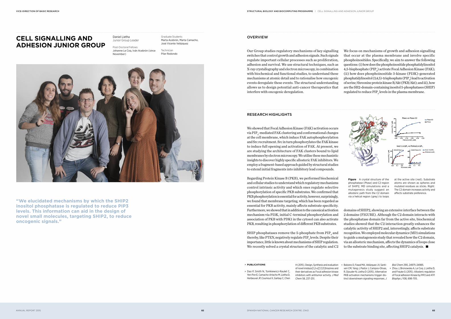
CELL SIGNALLING AND ADHESION JUNIOR GROUP
oVerVieW
Our Group studies regulatory mechanisms of key signalling switches that control growth and adhesion signals. Such signals regulate important cellular processes such as proliferation, adhesion and survival. We use structural techniques, such as X-ray crystallography and electron microscopy, in combination with biochemical and functional studies, to understand these mechanisms at atomic detail and to rationalise how oncogenic events deregulate these events. The structural understanding allows us to design potential anti-cancer therapeutics that interfere with oncogenic deregulation.
We focus on mechanisms of growth and adhesion signalling that occur at the plasma membrane and involve specific phosphoinositides. Specifically, we aim to answer the following questions : ( i ) how does the phosphoinositide phosphatidylinositol 4,5-bisphosphate ( PIP2 ) activate Focal Adhesion Kinase ( FAK ); ( ii ) how does phosphoinositide 3-kinase ( PI3K )-generated phosphatidylinositol ( 3,4,5 )-trisphosphate ( PIP3 ) lead to activation of serine/threonine protein kinase B/Akt ( PKB/Akt ); and iii ), how are the SH2-domain-containing inositol 5-phosphatases ( SHIP ) regulated to reduce PIP3 levels in the plasma membrane.
research highlights
We showed that Focal Adhesion Kinase ( FAK ) activation occurs via PIP2-mediated FAK clustering and conformational changes at the cell membrane, which induce FAK autophosphorylation and Src recruitment. Src in turn phosphorylates the FAK kinase to induce full opening and activation of FAK. At present, we are studying the architecture of FAK clusters bound to lipid membranes by electron microscopy. We utilise these mechanistic insights to discover highly specific allosteric FAK inhibitors. We employ a fragment-based approach guided by structural studies to extend initial fragments into inhibitory lead compounds.
Regarding Protein Kinase B ( PKB ), we performed biochemical and cellular studies to understand which regulatory mechanisms control intrinsic activity and which ones regulate selective phosphorylation of specific PKB substrates. We confirmed that PKB phosphorylation is essential for activity, however, surprisingly, we found that membrane targeting, which has been regarded as essential for PKB activity, mainly affects substrate specificity. Furthermore, we showed that in addition to the canonical activation mechanism via PI3K, initial C-terminal phosphorylation and association of PKB with PDK1 in the cytosol can also activate PKB, resulting in phosphorylation of different PKB substrates.
SHIP phosphatases remove the 5-phosphate from PIP3 and thereby, like PTEN, negatively regulate PIP3 levels. Despite their importance, little is known about mechanisms of SHIP regulation. We recently solved a crystal structure of the catalytic and C2
domains of SHIP2, showing an extensive interface between the 2 domains ( FIGURE ). Although the C2 domain interacts with the phosphatase domain far from the active site, biochemical studies showed that the C2 interaction greatly enhances the catalytic activity of SHIP2 and, interestingly, affects substrate recognition. We employed molecular dynamics ( MD ) simulations to guide a mutagenesis study that revealed how the C2 domain, via an allosteric mechanism, affects the dynamics of loops close to the substrate binding site, affecting SHIP2 catalysis. s
“ We elucidated mechanisms by which the SHIP2 inositol phosphatase is regulated to reduce PIP3 levels. This information can aid in the design of novel small molecules, targeting SHIP2, to reduce oncogenic signals.”
Daniel LiethaJunior Group Leader
Post-Doctoral FellowsJohanne Le Coq, Iván Acebrón ( since November )
Graduate StudentsMarta Acebrón, Marta Camacho, José Vicente Velázquez
TechnicianPilar Redondo
∞ PUBLICATIONS
∞ Dao P, Smith N, Tomkiewicz-Raulet C, Yen-Pon E, Camacho-Artacho M, Lietha D, Herbeuval JP, Coumoul X, Garbay C, Chen
H ( 2015 ). Design, Synthesis and evaluation of novel imidazo[ 1,2-a ][ 1,3,5 ]triazines and their derivatives as Focal adhesion kinase inhibitors with antitumor activity. J Med Chem 58, 237-251.
∞ Balzano D, Fawal MA, Velázquez JV, Santi-veri CM, Yang J, Pastor J, Campos-Olivas, R, Djouder N, Lietha D ( 2015 ). Alternative PKB activation mechanisms trigger dis-tinct downstream signaling responses. J
Biol Chem 290, 24975-24985. ∞ Zhou J, Bronowska A, Le Coq J, Lietha D, and Frauke G ( 2015 ). Allosteric regulation of Focal adhesion Kinase by PIP2 and ATP. Biophys J 108, 698-705.
Figure A crystal structure of the phosphatase ( Ptase ) and C2 region of SHIP2, MD simulations and a mutagenesis study suggest an allosteric path from the C2 domain via a helical region ( grey ) to loops
at the active site ( red ). Substrate atoms are shown as spheres and mutated residues as sticks. Right : The C2 domain increases activity and affects substrate preference.
SPANISH NATIONAL CANCER RESEARCH CENTRE, CNIO 85ANNUAL REPORT 2015 84
structural Biology anD Biocomputing programme | STRUCTURAL BASES Of gENOmE INTEgRITy JUNIOR gROUPVice-Direction of Basic research
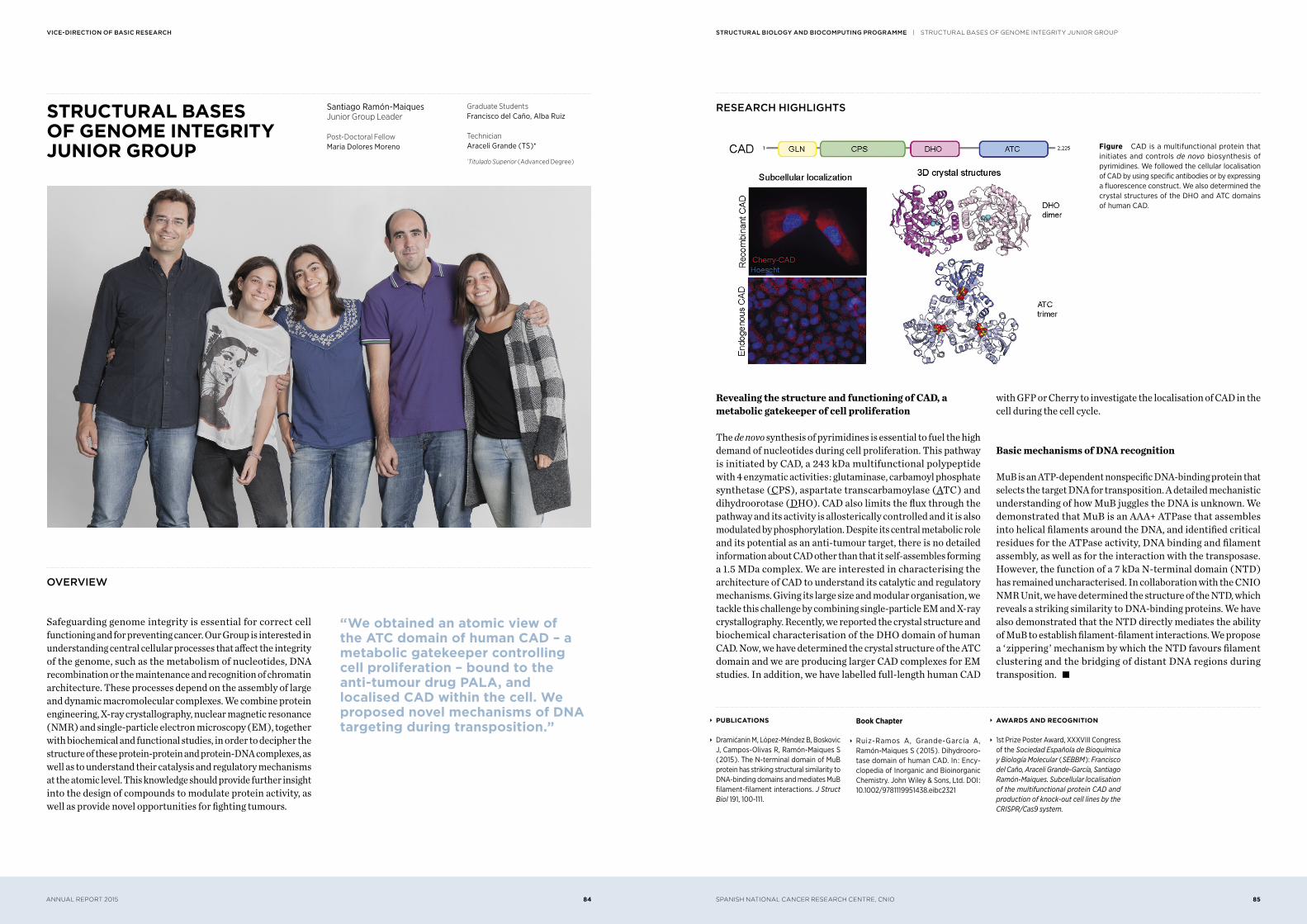
STRUCTURAL BASES OF GENOME INTEGRITY JUNIOR GROUP
research highlights
Revealing the structure and functioning of CAD, a metabolic gatekeeper of cell proliferation
The de novo synthesis of pyrimidines is essential to fuel the high demand of nucleotides during cell proliferation. This pathway is initiated by CAD, a 243 kDa multifunctional polypeptide with 4 enzymatic activities : glutaminase, carbamoyl phosphate synthetase ( CPS ), aspartate transcarbamoylase ( ATC ) and dihydroorotase ( DHO ). CAD also limits the flux through the pathway and its activity is allosterically controlled and it is also modulated by phosphorylation. Despite its central metabolic role and its potential as an anti-tumour target, there is no detailed information about CAD other than that it self-assembles forming a 1.5 MDa complex. We are interested in characterising the architecture of CAD to understand its catalytic and regulatory mechanisms. Giving its large size and modular organisation, we tackle this challenge by combining single-particle EM and X-ray crystallography. Recently, we reported the crystal structure and biochemical characterisation of the DHO domain of human CAD. Now, we have determined the crystal structure of the ATC domain and we are producing larger CAD complexes for EM studies. In addition, we have labelled full-length human CAD
with GFP or Cherry to investigate the localisation of CAD in the cell during the cell cycle.
Basic mechanisms of DNA recognition
MuB is an ATP-dependent nonspecific DNA-binding protein that selects the target DNA for transposition. A detailed mechanistic understanding of how MuB juggles the DNA is unknown. We demonstrated that MuB is an AAA+ ATPase that assembles into helical filaments around the DNA, and identified critical residues for the ATPase activity, DNA binding and filament assembly, as well as for the interaction with the transposase. However, the function of a 7 kDa N-terminal domain ( NTD ) has remained uncharacterised. In collaboration with the CNIO NMR Unit, we have determined the structure of the NTD, which reveals a striking similarity to DNA-binding proteins. We have also demonstrated that the NTD directly mediates the ability of MuB to establish filament-filament interactions. We propose a ‘ zippering ’ mechanism by which the NTD favours filament clustering and the bridging of distant DNA regions during transposition. s
oVerVieW
Safeguarding genome integrity is essential for correct cell functioning and for preventing cancer. Our Group is interested in understanding central cellular processes that affect the integrity of the genome, such as the metabolism of nucleotides, DNA recombination or the maintenance and recognition of chromatin architecture. These processes depend on the assembly of large and dynamic macromolecular complexes. We combine protein engineering, X-ray crystallography, nuclear magnetic resonance ( NMR ) and single-particle electron microscopy ( EM ), together with biochemical and functional studies, in order to decipher the structure of these protein-protein and protein-DNA complexes, as well as to understand their catalysis and regulatory mechanisms at the atomic level. This knowledge should provide further insight into the design of compounds to modulate protein activity, as well as provide novel opportunities for fighting tumours.
“ We obtained an atomic view of the ATC domain of human CAD – a metabolic gatekeeper controlling cell proliferation – bound to the anti-tumour drug PALA, and localised CAD within the cell. We proposed novel mechanisms of DNA targeting during transposition.”
Santiago Ramón-MaiquesJunior Group Leader
Post-Doctoral FellowMaria Dolores Moreno
Graduate StudentsFrancisco del Caño, Alba Ruiz
TechnicianAraceli Grande ( TS )*
*Titulado Superior ( Advanced Degree )
∞ PUBLICATIONS
∞ Dramićanin M, López-Méndez B, Boskovic J, Campos-Olivas R, Ramón-Maiques S ( 2015 ). The N-terminal domain of MuB protein has striking structural similarity to DNA-binding domains and mediates MuB filament-filament interactions. J Struct Biol 191, 100-111.
Book Chapter
∞ Ruiz-Ramos A, Grande-García A, Ramón-Maiques S ( 2015 ). Dihydrooro-tase domain of human CAD. In : Ency-clopedia of Inorganic and Bioinorganic Chemistry. John Wiley & Sons, Ltd. DOI : 10.1002/9781119951438.eibc2321
∞ AWARDS AND RECOGNITION
∞ 1st Prize Poster Award, XXXVIII Congress of the Sociedad Española de Bioquímica y Biología Molecular ( SEBBM ): Francisco del Caño, Araceli Grande-García, Santiago Ramón-Maiques. Subcellular localisation of the multifunctional protein CAD and production of knock-out cell lines by the CRISPR/Cas9 system.
Figure CAD is a multifunctional protein that initiates and controls de novo biosynthesis of pyrimidines. We followed the cellular localisation of CAD by using specific antibodies or by expressing a fluorescence construct. We also determined the crystal structures of the DHO and ATC domains of human CAD.
SPANISH NATIONAL CANCER RESEARCH CENTRE, CNIO 87ANNUAL REPORT 2015 86
structural Biology anD Biocomputing programme | SPECTROSCOPy ANd NUCLEAR mAgNETIC RESONANCE UNITVice-Direction of Basic research
SPECTROSCOPY AND NUCLEAR MAGNETIC RESONANCE UNIT
oVerVieW
The Unit consolidates the technical and scientific management of Nuclear Magnetic Resonance Spectroscopy ( NMR ) and other biophysical instrumentation made available by the Structural Biology and Biocomputing Programme. It provides CNIO researchers with instrumentation and technical support for a variety of spectroscopic and biophysical techniques. This includes the application of NMR to the in vitro characterisation of the
structure and dynamics of biomolecules ( proteins in particular ) and their interactions with other biopolymers, as well as with small molecules that could represent initial hits in the drug discovery process or research compounds for biophysical and functional studies. Furthermore, we use NMR to characterise the metabolic profiles of biofluids, cell growth media, and cell and tissue extracts from both animal models of cancer and human samples.
research highlights
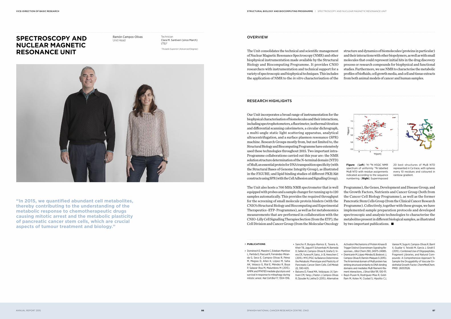
Our Unit incorporates a broad range of instrumentation for the biophysical characterisation of biomolecules and their interactions, including spectrophotometers, a fluorimeter, isothermal titration and differential scanning calorimeters, a circular dichrograph, a multi-angle static light scattering apparatus, analytical ultracentrifugation, and a surface plasmon resonance ( SPR ) machine. Research Groups mostly from, but not limited to, the Structural Biology and Biocomputing Programme have extensively used these technologies throughout 2015. Two important intra-Programme collaborations carried out this year are : the NMR solution structure determination of the N-terminal domain ( NTD ) of MuB, an essential protein for DNA transposition specificity ( with the Structural Bases of Genome Integrity Group ), as illustrated in the FIGURE ; and lipid binding studies of different PKB/Akt constructs using SPR ( with the Cell Adhesion and Signalling Group ).
The Unit also hosts a 700 MHz NMR spectrometer that is well equipped with probes and a sample changer for running up to 120 samples automatically. This provides the required throughput for the screening of small molecule protein binders ( with the CNIO’s Structural Biology and Biocomputing and Experimental Therapeutics -ETP- Programmes ), as well as for metabonomics measurements that are performed in collaboration with the CNIO-Lilly Cell Signalling Therapies Section ( from the ETP ), the Cell Division and Cancer Group ( from the Molecular Oncology
Programme ), the Genes, Development and Disease Group, and the Growth Factors, Nutrients and Cancer Group ( both from the Cancer Cell Biology Programme ), as well as the former Pancreatic Stem Cells Group ( from the Clinical Cancer Research Programme ). Collectively, together with these groups, we have implemented sample preparation protocols and developed spectroscopic and analysis technologies to characterise the metabolites present in different biological samples, as illustrated by two important publications. s
“ In 2015, we quantified abundant cell metabolites, thereby contributing to the understanding of the metabolic response to chemotherapeutic drugs causing mitotic arrest and the metabolic plasticity of pancreatic cancer stem cells, which are crucial aspects of tumour treatment and biology.”
Ramón Campos-OlivasUnit Head
TechnicianClara M. Santiveri ( since March ) ( TS )*
*Titulado Superior ( Advanced Degree )
∞ PUBLICATIONS
∞ Doménech E, Maestre C, Esteban-Martínez L, Partida D, Pascual R, Fernández-Miran-da G, Seco E, Campos-Olivas R, Pérez M, Megias D, Allen K, López M, Saha AK, Velasco G, Rial E, Méndez R, Boya P, Salazar-Roa M, Malumbres M ( 2015 ). AMPK and PFKFB3 mediate glycolysis and survival in response to mitophagy during mitotic arrest. Nat Cell Biol 17, 1304-1316.
∞ Sancho P, Burgos-Ramos E, Tavera A, Kheir TB, Jagust P, Schoenhals M, Barneda D, Sellers K, Campos-Olivas R, Graña O, Vi-era CR, Yuneva M, Sainz, Jr. B, Heeschen C ( 2015 ). MYC/PGC-1α Balance Determines the Metabolic Phenotype and Plasticity of Pancreatic Cancer Stem Cells. Cell Metab 22, 590-605.
∞ Balzano D, Fawal MA, Velázquez JV, San-tiveri CM, Yang J, Pastor J, Campos-Olivas R, Djouder N, Lietha D ( 2015 ). Alternative
Activation Mechanisms of Protein Kinase B Trigger Distinct Downstream Signaling Re-sponses. J Biol Chem 290, 24975-24985.
∞ Dramicanin M, López-Méndez B, Boskovic J, Campos-Olivas R, Ramón-Maiques S ( 2015 ). The N-terminal domain of MuB protein has striking structural similarity to DNA-binding domains and mediates MuB filament-fila-ment interactions. J Struct Biol 191, 100-111.
∞ Bayó-Puxan N, Rodríguez-Mias R, Gold-flam M, Kotev M, Ciudad S, Hipolito CJ,
Varese M, Suga H, Campos-Olivas R, Barril X, Guallar V, Teixidó M, García J, Giralt E ( 2015 ). Combined Use of Oligopeptides, Fragment Libraries, and Natural Com-pounds : A Comprehensive Approach To Sample the Druggability of Vascular En-dothelial Growth Factor. ChemMedChem. PMID : 26553526.
Figure ( Left ) 1H- 15N HSQC NMR spectrum of uniformly 15N labelled MuB NTD with residue assignments indicated according to the sequence numbering. ( Right ) Superimposed
20 best structures of MuB NTD represented in Ca trace, with spheres every 10 residues and coloured in rainbow gradient.
SPANISH NATIONAL CANCER RESEARCH CENTRE, CNIO 89ANNUAL REPORT 2015 88
structural Biology anD Biocomputing programme | BIOINfORmATICS UNITVice-Direction of Basic research
BIOINFORMATICS UNIT
In collaboration with the laboratory of C. Heeschen ( Barts Cancer Institute, London ), we helped to unveil specific metabolic features of pancreatic Cancer Stem Cells ( CSCs ) ( Sancho et al. 2015 ), and also to describe the pancreatic ductal adenocarcinoma ( PDAC ) microenvironment in order to better understand its biology ( Sainz et al. 2015 ). Our long-standing collaboration with CNIO’s Chromosome Dynamics Group ( A. Losada ) yielded interesting insights into cohesin’s contribution to the establishment of tissue-specific transcriptional programmes, by jointly interpreting genome-wide cohesin distribution, gene expression and chromatin architecture in the cerebral cortex and pancreas of adult mice ( Cuadrado et al. 2015 ).
Other bioinformatics analyses were performed together with M. Serrano’s laboratory ( CNIO ) ( Morgado-Palacin et al. 2015, Palla
et al. 2015 ), J. Benitez ( CNIO ) ( Matamala et al. 2015, Vaclová et al. 2015 ), and A. Muñoz ( IIB ) ( Aguilera et al. 2015 ).
We helped the Confocal Microscopy Core Unit ( CNIO ) to design and implement iMSRC, a new software tool that converts a conventional automated microscope into an intelligent screening platform ( Carro et al. 2015 ). In collaboration with D. Glez-Peña ( University of Vigo ) we published miRGate ( Andres Leon et al. 2015 ), a curated database of miRNA-mRNA targets with more than 125 million predictions on a consistent sequence space. Another genomic resource for the UBC-40 urothelial bladder cancer cell line ( Earl et al. 2015 ) was released in collaboration with F.X. Real’s laboratory ( CNIO ). Previously published works have allowed us to deliver additional data and protocols as genomic data resources ( Tanic et al. 2015, Foronda et al. 2015 ). s
oVerVieW
The Bioinformatics Unit is devoted to assisting CNIO’s researchers with data analysis and interpretation using statistical and computational methods. It also maintains the scientific computing facilities at the CNIO and provides training in bioinformatics tools and methods.
It is important for people to realise that a research centre like the CNIO conducts a massive amount of experiments that generate data at the same rate as large companies do. Current biotechnology techniques enable us to capture molecular pictures of biological systems and to observe, in a unique experiment, the status and composition of millions of these elements. This generates huge amounts of ‘ big data ’ that we have to manage and analyse using computational technologies, which are essential for the understanding of the genetic and molecular bases of cancer.
David G. PisanoUnit Head
TechniciansÁngel Carro ( TS )*, Coral Fustero ( since December ) ( PEJ-L ) **,
Gonzalo Gómez ( TS )*, Osvaldo Graña ( TS )*, Miriam Rubio ( TS )*
*Titulado Superior ( Advanced Degree ) **Plan de Empleo Joven-Licenciado ( Youth
Employment Plan-Graduate )
∞ PUBLICATIONS
∞ Puente XS et al. ( incl. Pisano DG, Valencia A ) ( 2015 ). Non-coding recurrent muta-tions in chronic lymphocytic leukaemia. Nature 526, 519-524.
∞ Sancho P et al. ( incl. Campos-Olivas R, Graña O ) ( 2015 ). MYC/PGC-1a Balance Determines the Metabolic Phenotype and Plasticity of Pancreatic Cancer Stem Cells. Cell Metab 22, 590-605.
∞ Sainz B Jr et al. ( incl. Gomez-Lopez G ) ( 2015 ). Microenvironmental hCAP-18/LL-37 promotes pancreatic ductal adeno-carcinoma by activating its cancer stem cell compartment. Gut. PMID : 25841238.
∞ Cuadrado A, Remeseiro S, Graña O, Pisano, DG, Losada A ( 2015 ). The contribution of cohesin-SA1 to gene expression and chromatin architecture in two murine tissues. Nucleic Acids Res 43, 3056-3067.
∞ Morgado-Palacin L, Vareti G, Llanos S, Gomez-Lopez G, Martinez D, Serrano M ( 2015 ). Partial Loss of Rpl11 in Adult Mice Recapitulates Diamond-Blackfan Anemia and Promotes Lymphomagenesis. Cell Reports 13, 712-722.
∞ Matamala N et al. ( incl. Andrés-León E, Gómez-López G ) ( 2015 ). Tumor Mi-croRNA Expression Profiling Identifies Circulating MicroRNAs for Early Breast Cancer Detection. Clin Chem 61, 1098-1106.
∞ Aguilera Ó et al. ( incl. Graña O, Pisano DG ) ( 2015 ). Nuclear DICKKOPF-1 as a biomarker of chemoresistance and poor clinical outcome in colorectal cancer. On-cotarget 6, 5903-5917.
∞ Palla AR et al. ( incl. Graña O, Gómez-López G ) ( 2015 ). The pluripotency factor NANOG promotes the formation of squamous cell carcinomas. Scientific Reports 5, 10205.
∞ Carro A, Perez-Martinez M, Soriano J, Pis-ano DG, Megias D ( 2015 ). iMSRC : convert-ing a standard automated microscope into an intelligent screening platform. Scientific Reports 5, 10502.
∞ Tanic M et al. ( incl. Gómez-López G, Pisano DG ) ( 2015 ). MicroRNA expression signa-tures for the prediction of BRCA1/2 muta-tion-associated hereditary breast cancer in paraffin-embedded formalin-fixed breast tumors. Int J Cancer 136, 593-602.
∞ Earl J et al. ( incl. Gómez G, Pisano DG ) ( 2015 ). The UBC-40 Urothelial Bladder Cancer cell line index : a genomic resource for functional studies. BMC Genomics 16, 403.
∞ Vaclová T, Gómez-López G, Setién F, Bueno JM, Macías JA, Barroso A, Urioste M, Esteller M, Benítez J, Osorio A ( 2015 ). DNA repair capacity is impaired in healthy BRCA1 heterozygous mutation carriers.
Breast Cancer Res Treat 152, 271-282. ∞ Andres Leon E, Gonzalez Pena D, Gomez-Lopez G, Pisano DG ( 2015 ). miRGate : a curated database of human, mouse and rat miRNA-mRNA targets. Database : the Journal of Biological Databases and Cu-ration 2015, bav035.
∞ Tanic M, Yanowski K, Andrés E, Gómez-López G, Socorro MR, Pisano DG, Martin-ez-Delgado B, Benítez J ( 2015 ). miRNA expression profiling of formalin-fixed paraffin-embedded ( FFPE ) hereditary breast tumors. Genomics Data 3, 75-79.
∞ Foronda M, Morgado-Palacin L, Gómez-López G, Domínguez O, Pisano DG, Blasco MA ( 2015 ). Profiling of Sox4-dependent transcriptome in skin links tumour sup-pression and adult stem cell activation. Genomics Data 6, 21-24.
research highlights
In 2015, as part of the International Cancer Genomic Consortium ( ICGC ) and in collaboration with E. Campo’s group at the Hospital Clinic in Barcelona and C. López-Otin’s laboratory at the University of Oviedo, we contributed to the work reporting a comprehensive genomic characterisation of Chronic Lymphocytic Leukaemia ( CLL ) and its precursor in more than 500 patients ( Puente et al. 2015 ). The study extends the number of CLL driver alterations found in the coding portion of the genome and also identifies novel recurrent mutations in non-coding regions, including the 3 ’ UTR of NOTCH1, which cause aberrant splicing events, as well as mutations in an enhancer that result in reduced expression of the B-cell-specific transcription factor PAX5. It also confirms the insights provided by previous works on more limited patient datasets : each tumour carries an individual, distinct and personal signature of genomic alterations ( FIGURE ).
SPANISH NATIONAL CANCER RESEARCH CENTRE, CNIO 91ANNUAL REPORT 2015 90
structural Biology anD Biocomputing programme | NATIONAL BIOINfORmATICS INSTITUTE UNITVice-Direction of Basic research
NATIONAL BIOINFORMATICS INSTITUTE UNIT
The INB Unit differs from the other Units in the Structural Biology and Biocomputing Programme in the sense that its offering is
not restricted to the CNIO Groups, and that its budget is funded entirely by an external agency, the ISCIII.
research highlights
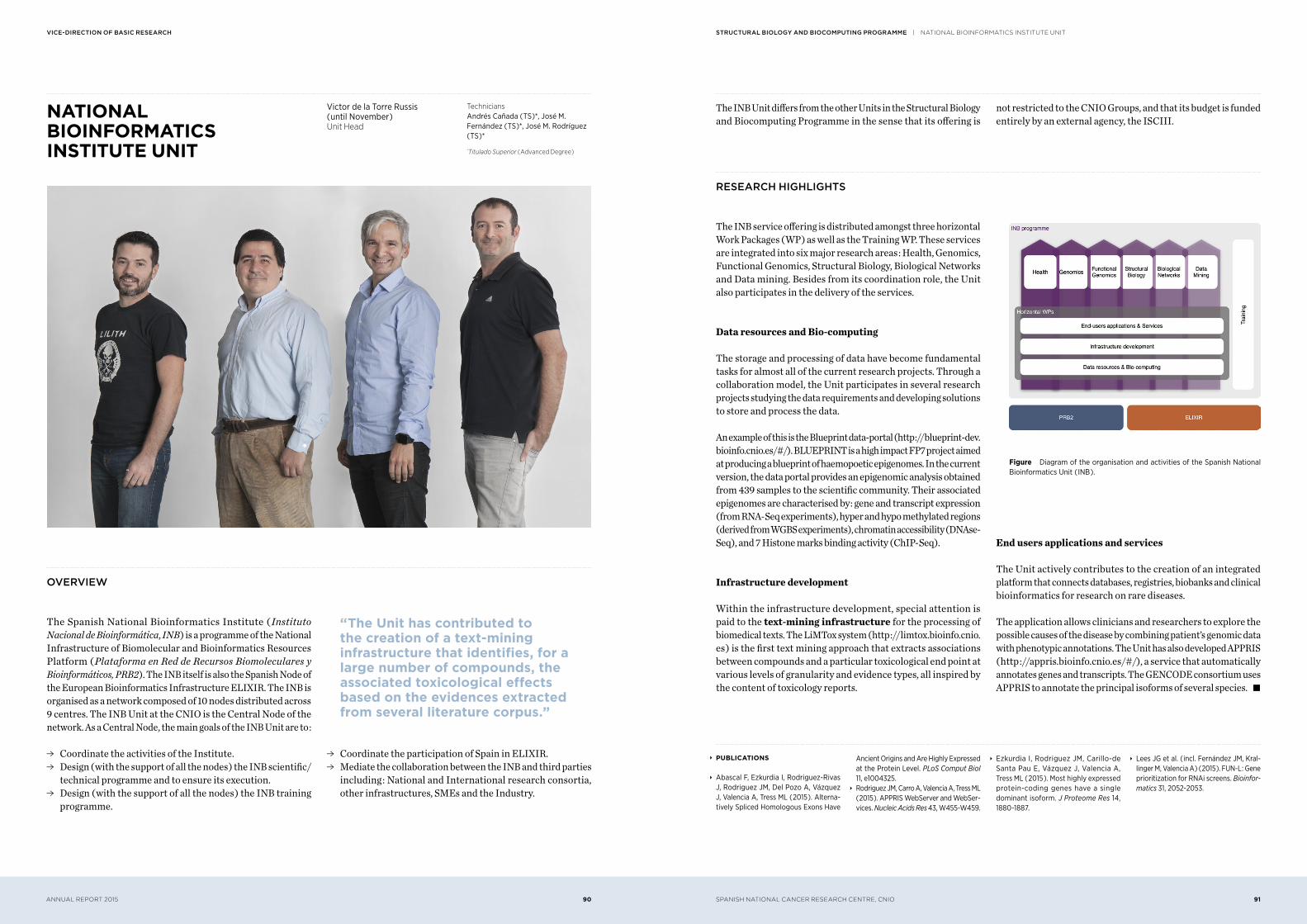
The INB service offering is distributed amongst three horizontal Work Packages ( WP ) as well as the Training WP. These services are integrated into six major research areas : Health, Genomics, Functional Genomics, Structural Biology, Biological Networks and Data mining. Besides from its coordination role, the Unit also participates in the delivery of the services.
Data resources and Bio-computing
The storage and processing of data have become fundamental tasks for almost all of the current research projects. Through a collaboration model, the Unit participates in several research projects studying the data requirements and developing solutions to store and process the data.
An example of this is the Blueprint data-portal ( http ://blueprint-dev.bioinfo.cnio.es/#/). BLUEPRINT is a high impact FP7 project aimed at producing a blueprint of haemopoetic epigenomes. In the current version, the data portal provides an epigenomic analysis obtained from 439 samples to the scientific community. Their associated epigenomes are characterised by : gene and transcript expression ( from RNA-Seq experiments ), hyper and hypo methylated regions ( derived from WGBS experiments ), chromatin accessibility ( DNAse-Seq ), and 7 Histone marks binding activity ( ChIP-Seq ).
Infrastructure development
Within the infrastructure development, special attention is paid to the text-mining infrastructure for the processing of biomedical texts. The LiMTox system ( http ://limtox.bioinfo.cnio.es ) is the first text mining approach that extracts associations between compounds and a particular toxicological end point at various levels of granularity and evidence types, all inspired by the content of toxicology reports.
End users applications and services
The Unit actively contributes to the creation of an integrated platform that connects databases, registries, biobanks and clinical bioinformatics for research on rare diseases.
The application allows clinicians and researchers to explore the possible causes of the disease by combining patient’s genomic data with phenotypic annotations. The Unit has also developed APPRIS ( http ://appris.bioinfo.cnio.es/#/), a service that automatically annotates genes and transcripts. The GENCODE consortium uses APPRIS to annotate the principal isoforms of several species. s
oVerVieW
The Spanish National Bioinformatics Institute ( Instituto Nacional de Bioinformática, INB ) is a programme of the National Infrastructure of Biomolecular and Bioinformatics Resources Platform ( Plataforma en Red de Recursos Biomoleculares y Bioinformáticos, PRB2 ). The INB itself is also the Spanish Node of the European Bioinformatics Infrastructure ELIXIR. The INB is organised as a network composed of 10 nodes distributed across 9 centres. The INB Unit at the CNIO is the Central Node of the network. As a Central Node, the main goals of the INB Unit are to :
ɗ Coordinate the activities of the Institute. ɗ Design ( with the support of all the nodes ) the INB scientific/
technical programme and to ensure its execution. ɗ Design ( with the support of all the nodes ) the INB training
programme.
“ The Unit has contributed to the creation of a text-mining infrastructure that identifies, for a large number of compounds, the associated toxicological effects based on the evidences extracted from several literature corpus.”
ɗ Coordinate the participation of Spain in ELIXIR. ɗ Mediate the collaboration between the INB and third parties
including : National and International research consortia, other infrastructures, SMEs and the Industry.
Victor de la Torre Russis ( until November )Unit Head
TechniciansAndrés Cañada ( TS )*, José M. Fernández ( TS )*, José M. Rodríguez ( TS )*
*Titulado Superior ( Advanced Degree )
Figure Diagram of the organisation and activities of the Spanish National Bioinformatics Unit ( INB ).
∞ PUBLICATIONS
∞ Abascal F, Ezkurdia I, Rodriguez-Rivas J, Rodriguez JM, Del Pozo A, Vázquez J, Valencia A, Tress ML ( 2015 ). Alterna-tively Spliced Homologous Exons Have
Ancient Origins and Are Highly Expressed at the Protein Level. PLoS Comput Biol 11, e1004325.
∞ Rodriguez JM, Carro A, Valencia A, Tress ML ( 2015 ). APPRIS WebServer and WebSer-vices. Nucleic Acids Res 43, W455-W459.
∞ Ezkurdia I, Rodriguez JM, Carillo-de Santa Pau E, Vázquez J, Valencia A, Tress ML ( 2015 ). Most highly expressed protein-coding genes have a single dominant isoform. J Proteome Res 14, 1880-1887.
∞ Lees JG et al. ( incl. Fernández JM, Kral-linger M, Valencia A ) ( 2015 ). FUN-L : Gene prioritization for RNAi screens. Bioinfor-matics 31, 2052-2053.
SPANISH NATIONAL CANCER RESEARCH CENTRE, CNIO 93ANNUAL REPORT 2015 92
structural Biology anD Biocomputing programme | ELECTRON mICROSCOPy UNITVice-Direction of Basic research
ELECTRON MICROSCOPY UNIT
oVerVieW
The Electron Microscopy ( EM ) Unit is a research laboratory and a central core facility that provides CNIO researchers, and the wider research community, with access to Transmission Electron Microscopy, as well as supplying expertise in EM image analysis. As a core facility, we offer standard specimen preparation techniques for proteins and protein complexes, data collection and data processing tailored to the specific needs of the users.
We also provide training for regular users on the use of equipment, as well as guidance regarding specimen preparation. For cell biology samples, we have established collaboration with the Centro de Investigaciones Biológicas ( CIB-CSIC, Madrid ), where the samples are prepared for further observation and imaging with our microscope.
research highlights
The Electron Microscopy Unit is a research facility that provides support for biological science projects at scales ranging from the cellular level to the macromolecular complex level. The Electron Microscopy Unit implements sample preparation protocols and data collection methods, as well as performing 2D and 3D data processing.
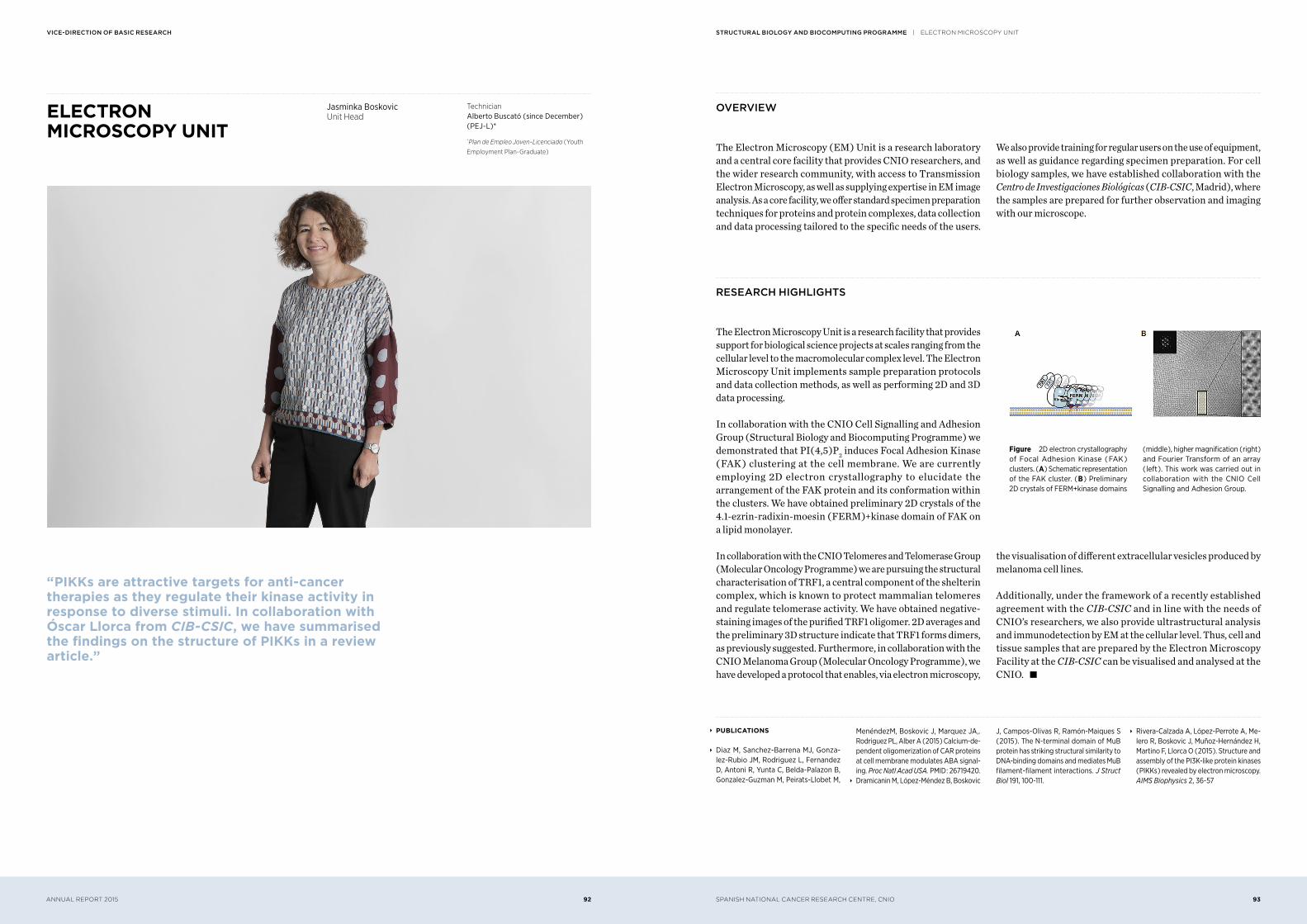
In collaboration with the CNIO Cell Signalling and Adhesion Group ( Structural Biology and Biocomputing Programme ) we demonstrated that PI( 4,5 )P2 induces Focal Adhesion Kinase ( FAK ) clustering at the cell membrane. We are currently employing 2D electron crystallography to elucidate the arrangement of the FAK protein and its conformation within the clusters. We have obtained preliminary 2D crystals of the 4.1-ezrin-radixin-moesin ( FERM )+kinase domain of FAK on a lipid monolayer.
In collaboration with the CNIO Telomeres and Telomerase Group ( Molecular Oncology Programme ) we are pursuing the structural characterisation of TRF1, a central component of the shelterin complex, which is known to protect mammalian telomeres and regulate telomerase activity. We have obtained negative-staining images of the purified TRF1 oligomer. 2D averages and the preliminary 3D structure indicate that TRF1 forms dimers, as previously suggested. Furthermore, in collaboration with the CNIO Melanoma Group ( Molecular Oncology Programme ), we have developed a protocol that enables, via electron microscopy,
the visualisation of different extracellular vesicles produced by melanoma cell lines.
Additionally, under the framework of a recently established agreement with the CIB-CSIC and in line with the needs of CNIO’s researchers, we also provide ultrastructural analysis and immunodetection by EM at the cellular level. Thus, cell and tissue samples that are prepared by the Electron Microscopy Facility at the CIB-CSIC can be visualised and analysed at the CNIO. s
“ PIKKs are attractive targets for anti-cancer therapies as they regulate their kinase activity in response to diverse stimuli. In collaboration with Óscar Llorca from CIB-CSIC, we have summarised the findings on the structure of PIKKs in a review article.”
Jasminka BoskovicUnit Head
TechnicianAlberto Buscató ( since December ) ( PEJ-L )*
*Plan de Empleo Joven-Licenciado ( Youth
Employment Plan-Graduate )
Figure 2D electron crystallography of Focal Adhesion Kinase ( FAK ) clusters. ( A ) Schematic representation of the FAK cluster. ( B ) Preliminary 2D crystals of FERM+kinase domains
( middle ), higher magnification ( right ) and Fourier Transform of an array ( left ). This work was carried out in collaboration with the CNIO Cell Signalling and Adhesion Group.
∞ PUBLICATIONS
∞ Diaz M, Sanchez-Barrena MJ, Gonza-lez-Rubio JM, Rodriguez L, Fernandez D, Antoni R, Yunta C, Belda-Palazon B, Gonzalez-Guzman M, Peirats-Llobet M,
MenéndezM, Boskovic J, Marquez JA,. Rodriguez PL, Alber A ( 2015 ) Calcium-de-pendent oligomerization of CAR proteins at cell membrane modulates ABA signal-ing. Proc Natl Acad USA. PMID : 26719420.
∞ Dramicanin M, López-Méndez B, Boskovic
J, Campos-Olivas R, Ramón-Maiques S ( 2015 ). The N-terminal domain of MuB protein has striking structural similarity to DNA-binding domains and mediates MuB filament-filament interactions. J Struct Biol 191, 100-111.
∞ Rivera-Calzada A, López-Perrote A, Me-lero R, Boskovic J, Muñoz-Hernández H, Martino F, Llorca O ( 2015 ). Structure and assembly of the PI3K-like protein kinases ( PIKKs ) revealed by electron microscopy. AIMS Biophysics 2, 36-57
SPANISH NATIONAL CANCER RESEARCH CENTRE, CNIO 95ANNUAL REPORT 2015 94
structural Biology anD Biocomputing programme | CRySTALLOgRAPHy UNITVice-Direction of Basic research
CRYSTALLOGRAPHY UNIT oVerVieW
The aim of the Crystallography Unit is to provide the CNIO Research Groups with a three-dimensional characterisation of the structure of biological macromolecules at high-resolution ( X-ray crystallography ) and low-resolution ( SAXS ). The knowledge of the 3D structure of proteins and protein complexes is essential for understanding their function in cellular processes. The crystal structures give us a picture – at atomic resolution − of the protein. With this knowledge, we know where to introduce mutations that can alter ( or improve ) the specificity of the protein and its affinity to other molecules. In turn, this can lead to the design of drugs to block or control the activity of proteins involved in
disease. Small-angle X-ray scattering ( SAXS ) is a complementary technique to X-ray crystallography. It permits delineation of the dynamic changes in shape and size undergone by the proteins in solution, giving a structural picture of the thermodynamic behaviour of these biological molecules, including changes induced upon ligand binding.
This Unit is shared between the Structural Biology and Biocomputing Programme and the Experimental Therapeutics Programme.
research highlights
The Crystallography Unit began its journey in February 2015, with the aim of providing state-of-the-art, high-throughput protein crystallization, X-ray crystallography and SAXS services to meet the demands of the Research Groups at the CNIO and special collaborative efforts outside our institute.
The full-service Unit provides access to sophisticated equipment and technologies, including the European synchrotron light sources. We also offer consultancies, guidance, and technical assistance at every stage of the 3D structure determination process. Non-crystallography groups benefit from expert aid in the use of existing 3D structures for the design and interpretation of experiments, including the possibility to crystallize their target proteins in the presence of inhibitors, for structure-based drug design.
Since its formation, the Unit works in close collaboration on the drug discovery projects led by the Experimental Therapeutics Programme. We also run a number of collaborations with different Groups at the CNIO involved in the following Programmes : Molecular Oncology ( Telomeres and Telomerase and Brain Metastasis Groups ), Clinical Research ( Gastrointestinal Cancer Clinical Research Unit ), and Cancer Cell Biology ( Epithelial Carcinogenesis Group ). Additionally, the Unit has initiated external collaborations with the Physical Chemistry Department ( University of Granada ), the Environmental Biology Department ( CIB-CSIC ), and the Pharmacology and Therapeutics Department ( Roswell Park Cancer Institute, USA ). s
“ Our goal is to provide CNIO investigators with 3D structural information of their target macromolecules to understand the mechanism( s ) that regulate their biological functions, including their modulation by novel therapeutic ligands.”
Inés MuñozUnit Head
TechnicianAlicia Virseda ( since December ) ( PEJ-L )*
*Plan de Empleo Joven-Licenciado ( Youth
Employment Plan-Graduate )
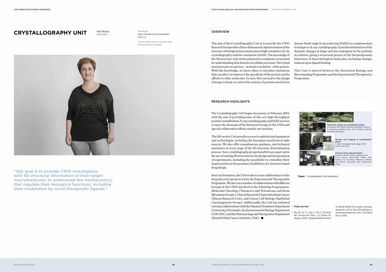
Figure Crystallography Unit Equipment.
∞ PUBLICATION
∞ Wu W, Xu C, Ling X, Fan C, Buckley BP, Chernov MV, Ellis L, Li F, Muñoz IG, Wang x ( 2015 ). Targeting RING domains
of Mdm2-MdmX E3 complex activates apoptotic arm of the p53 pathway in leukemia/lymphoma cells. Cell Death Dis 6, e2035.