Structural and Ultrastructural Aspects of Folliculogenesis ... · albiventris were described in...
8
113 Int. J. Morphol., 26(1):113-120, 2008. Structural and Ultrastructural Aspects of Folliculogenesis in Didelphis albiventris, the South-American Opossum Aspectos Estructurales y Ultraestructurales de la Foliculogénesis de Didelphis albiventris, la Zarigüeña Sudamericana * Maria Dalva Cesario & ** Selma M. Michelin Matheus CESARIO, M. D. & MATHEUS, S. M. M. Structural and ultrastructural aspects of folliculogenesis in Didelphis albiventris, the South- American opossum. Int. J. Morphol., 26(1):113-120, 2008. SUMMARY: The ovarian histology, the structural and the ultrastructural characteristics of the folliculogenesis in Didelphis albiventris were described in detail. Recent studies suggest that methatherian mammals have unusual reproductive cycle but there are few informations regarding the marsupials reproductive life. Despite of the opossum folliculogenesis pattern resembles methatherian and eutherian pattern in many aspects, the analysis shows some peculiar features of the oocyte structure and ultrastructure that make available new data on the reproductive biology of marsupials. KEY WORDS: Folliculogenesis, Oocytes; Opossum; Marsupial. INTRODUCTION Marsupials differ from eutherian mammals in their anatomical and physiological reproductive aspects although Mackay et al. (2004) have studied the postnatal reproductive tract development and have found that the mesonephric and paramesonephric ducts differentiation takes place after gonadal differentiation, according to the normal eutherian pattern. The opossum is a polyoestrous seasonal breeder (Fleming, 1973) and is sexually mature when in its 6 or 8 month-old and the females have two years of reproductive life. According to Collins (1973), Didelphis in Brazil has its first litter in August and September. Earlier studies on marsupial oogenesis had suggested that it follows the biphasic growth pattern, typical from eutherian mammals (Lintern-Moore et al., 1976; Lyne & Hollis, 1983; Kress et al. 2001). Because of the unusual reproductive cycle among the marsupials, data regarding oocyte organelles and nucleus, polyovular follicles, oocyte development, folliculogenesis control, embryonic membranes and implantation have been published (Harrison & Weir, 1977; Hughes, 1977). Lyne & Hollis described the Graafian follicles in Isoodon macrourus and Perameles nasuta. The grown follicles are usually very large but the oocyte size does not increase concomitantly. Among the marsupials, researches were undertaken in bandicoot ovary (Ullmann, 1979, 1981, 1989) and in Monodelphis opossum oocytes (Baggott et al., 1987, Mate et al., 1992). Oocytes maturation and in vitro fertilization studies in marsupials, especially Dasyuridae, were described by Selwood, (1982), Breed & Leigh, (1990), Breed, (1996) and Hickford et al. (2001). Kress et al. described the oogenesis pattern in Sminthopsis macroura and found that the timetable of oogenesis is accelerated as in other marsupials showing relatively early female maturation. The available literature suggests that researches have been developed related to eutherian mammals and few of them are related to methatherian ones (Wolgemuth et al., 1984). Especially in Didelphis albiventris, the South- American opossum, no description of the folliculogenesis pattern was found. The goal of this paper is the description of some structural and ultrastructural characteristics of the south-american opossum ovary, with special attention to the ovarian follicles. These findings may contribute to the better understanding of the folliculogenesis and the reproductive behavior of this animal. * Departamento de Morfologia, Instituto de Biociências, UNESP, São Paulo, Campus de Botucatu, Brasil. ** Departamento de Anatomia, Instituto de Biociências, UNESP, São Paulo, Campus de Botucatu, Brasil.
Transcript of Structural and Ultrastructural Aspects of Folliculogenesis ... · albiventris were described in...

113
Int. J. Morphol.,26(1):113-120, 2008.
Structural and Ultrastructural Aspects of Folliculogenesis inDidelphis albiventris, the South-American Opossum
Aspectos Estructurales y Ultraestructurales de la Foliculogénesisde Didelphis albiventris, la Zarigüeña Sudamericana
*Maria Dalva Cesario & **Selma M. Michelin Matheus
CESARIO, M. D. & MATHEUS, S. M. M. Structural and ultrastructural aspects of folliculogenesis in Didelphis albiventris, the South-American opossum. Int. J. Morphol., 26(1):113-120, 2008.
SUMMARY: The ovarian histology, the structural and the ultrastructural characteristics of the folliculogenesis in Didelphisalbiventris were described in detail. Recent studies suggest that methatherian mammals have unusual reproductive cycle but there are fewinformations regarding the marsupials reproductive life. Despite of the opossum folliculogenesis pattern resembles methatherian andeutherian pattern in many aspects, the analysis shows some peculiar features of the oocyte structure and ultrastructure that make availablenew data on the reproductive biology of marsupials.
KEY WORDS: Folliculogenesis, Oocytes; Opossum; Marsupial.
INTRODUCTION
Marsupials differ from eutherian mammals in theiranatomical and physiological reproductive aspects althoughMackay et al. (2004) have studied the postnatal reproductivetract development and have found that the mesonephric andparamesonephric ducts differentiation takes place after gonadaldifferentiation, according to the normal eutherian pattern. Theopossum is a polyoestrous seasonal breeder (Fleming, 1973) andis sexually mature when in its 6 or 8 month-old and the femaleshave two years of reproductive life. According to Collins (1973),Didelphis in Brazil has its first litter in August and September.
Earlier studies on marsupial oogenesis had suggestedthat it follows the biphasic growth pattern, typical fromeutherian mammals (Lintern-Moore et al., 1976; Lyne &Hollis, 1983; Kress et al. 2001). Because of the unusualreproductive cycle among the marsupials, data regardingoocyte organelles and nucleus, polyovular follicles, oocytedevelopment, folliculogenesis control, embryonicmembranes and implantation have been published (Harrison& Weir, 1977; Hughes, 1977). Lyne & Hollis described theGraafian follicles in Isoodon macrourus and Peramelesnasuta. The grown follicles are usually very large but theoocyte size does not increase concomitantly.
Among the marsupials, researches were undertakenin bandicoot ovary (Ullmann, 1979, 1981, 1989) and inMonodelphis opossum oocytes (Baggott et al., 1987, Mateet al., 1992). Oocytes maturation and in vitro fertilizationstudies in marsupials, especially Dasyuridae, weredescribed by Selwood, (1982), Breed & Leigh, (1990),Breed, (1996) and Hickford et al. (2001). Kress et al.described the oogenesis pattern in Sminthopsis macrouraand found that the timetable of oogenesis is accelerated asin other marsupials showing relatively early femalematuration.
The available literature suggests that researches havebeen developed related to eutherian mammals and few ofthem are related to methatherian ones (Wolgemuth et al.,1984). Especially in Didelphis albiventris, the South-American opossum, no description of the folliculogenesispattern was found. The goal of this paper is the descriptionof some structural and ultrastructural characteristics of thesouth-american opossum ovary, with special attention to theovarian follicles. These findings may contribute to the betterunderstanding of the folliculogenesis and the reproductivebehavior of this animal.
* Departamento de Morfologia, Instituto de Biociências, UNESP, São Paulo, Campus de Botucatu, Brasil.** Departamento de Anatomia, Instituto de Biociências, UNESP, São Paulo, Campus de Botucatu, Brasil.

114
MATERIAL AND METHOD
Adult females specimens of 400–500g weightwere trapped in Botucatu City, São Paulo State, Brazil(IBAMA license Nº 002252/97-24). The experimentswere conducted in accordance with the ethicalguidelines for animal use, determined by the BrazilianCollege of Animal Experimentation (COBEA). Theanimals were anaesthetized by ether inhalation andperfused by Karnovsky´s solution (2% glutaraldehyde- 4% paraformaldehyde in Sorensen´s phosphate bu-ffer 0.1 M, pH 7.3); through the left ventricle. Theovaries were removed and fixed overnight inKarnovsky´s solution.
For light microscopy, tissues were prepared toembed in Historesin (Historesin Jung 70-2218-500),according to standard methods and semithin sectionswere cut on Leica mod. RM 2065 microtome. Thesesections were stained with haematoxilyn and eosin andviewed under Zeiss Axiophot microscope.
For TEM, tissues were fixed for 24 hours inKarnovsky´s solution, washed in 0.1 M pH 7,3 PBS,post-fixed in 1% osmium tetroxide in PBS, for 2 hours,at the darkness, washed in distilled water (three changesof 5 minutes), stained in the block with 0.5% aqueoussolution of uranyl acetate for 2 hours, dehydratedthrough a graded series of acetone, embedded in 1:1Araldite - 100% acetone solution (12 hours) andembedded in Araldite. Ultrathin sections were doublestained with uranyl acetate and lead citrate. The sectionswere examined and photographed under a PHILIPS EM301.
RESULTS
The opossum has two ovaries located in the pel-vis cavity and histologically they are covered by acuboidal epithelium, which is squamous in someextension. Immediately underneath this epitheliumthere is the tunica albuginea of irregular denseconnective tissue. Each ovary is divided in two regions:a well-developed cortical region of dense stromal tissueand an internal medullary region of connective tissuewith many blood vessels. The ovarian follicles withthe oocytes are lying in the cortical region. Thehistological limit between both regions can readily beseen as the cortex is mainly acidophilic and the medullais eosinophilic (Fig. 1).
The primordial ovarian follicles are found near tothe tunica albuginea (Fig 1). A single layer of flattenedsquamous follicular cells around the oocyte maydistinguish them. Nuclei of these cells are flat in shapeand mitochondria and ribosomes are spread throughoutthe cytoplasm. The quiescent oocyte is spherical or ovalin shape and it has a large, round and eccentrically locatednucleus with a conspicuous and single nucleolus (Figs. 2and 3). Its envelope has many pores and it is commonlynamed germinal vesicle. The chromatin material, that isrich in euchromatin, proceeds to the terminal stage of thefirst meiotic prophase (diplothene stage). The oocytecytoplasm contains numerous rounded mitochondriashowing electron-dense matrix between the usualorganelles, like Golgi complex and endoplasmicreticulum. Electron-dense and multivesicular bodies arealso dispersed throughout the ooplasm (Fig. 4).
When the oocyte begins its growth phase, the sin-gle layer of follicular cells around it becomes cuboidalepithelial cells that form the primary follicle (Figs. 1 and5). As the oocyte develops the mitochondria take theelongated profile and are grouped under the plasmalemma.Electron-lucent vesicular bodies are common componentsin the ooplasm. In addition, the oocyte begins to besurrounded by a translucent noncellular material, the zonapellucida. A space develops between the oocyte and thesurrounding follicular epithelium into which irregularlyshaped microvilli project from the oolema and from theneighboring follicular cells, over the whole surface of theoocyte (Figs. 8 and 9).
The follicular growth is characterized by a steadyincrease in the number of follicular cells and the granulosabecomes a two-layer follicular cells (Fig. 6) and later, amultilayered envelope of follicular cells. The granulosa,than, acquires a theca tissue with two thecal layers: thecainterna, which is of dense connective tissue with severalblood vessels and theca externa of connective tissue (Figs.6, 7 and 8). Numerous lipid-like droplets are present inthe cytoplasm (Figs. 6 and 7).
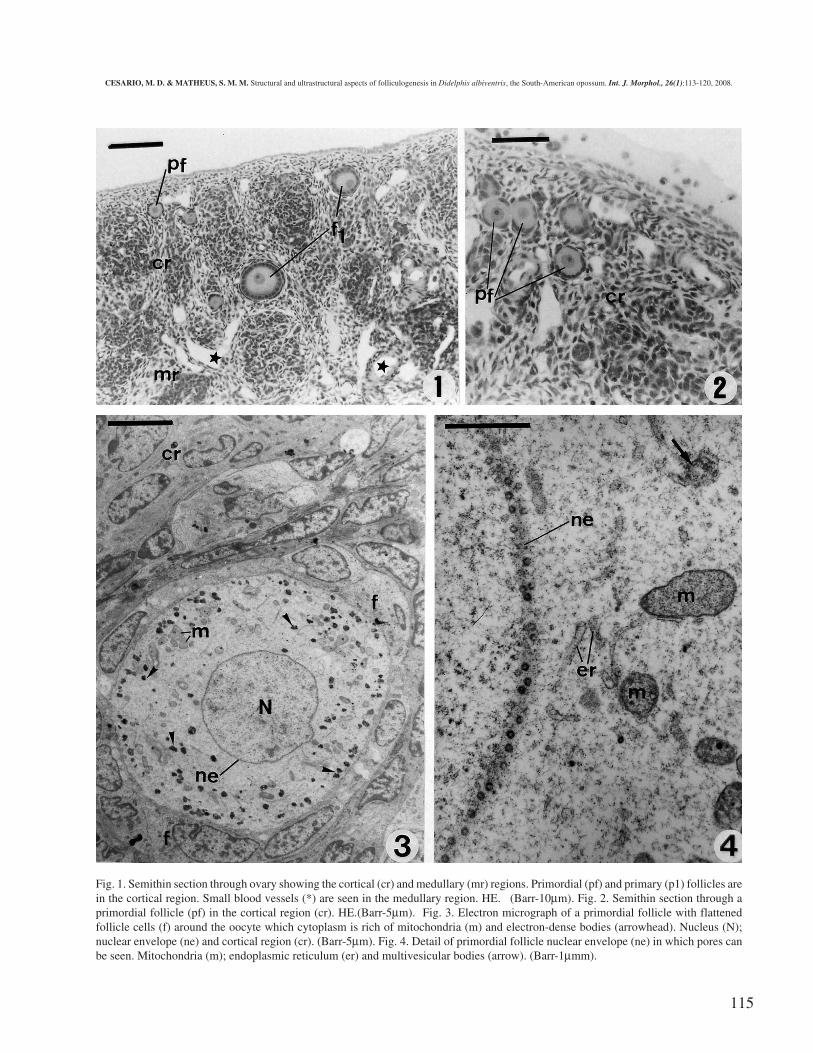
The Graafian follicle exhibits an incipient antrumat the late stage and as the antrum expands, the oocytetakes up an eccentric position surrounded by two or moregranulosa cell layers, the corona radiata and the cumuluscells with dilated intercellular spaces (Figs 10, 12 and13). The oocyte ooplasm becomes full of electron-lucentvesicular bodies and elongated mitochondria are groupedat the cortical region (Fig. 11).
CESARIO, M. D. & MATHEUS, S. M. M. Structural and ultrastructural aspects of folliculogenesis in Didelphis albiventris, the South-American opossum. Int. J. Morphol., 26(1):113-120, 2008.
115
Fig. 1. Semithin section through ovary showing the cortical (cr) and medullary (mr) regions. Primordial (pf) and primary (p1) follicles arein the cortical region. Small blood vessels (*) are seen in the medullary region. HE. (Barr-10µm). Fig. 2. Semithin section through aprimordial follicle (pf) in the cortical region (cr). HE.(Barr-5µm). Fig. 3. Electron micrograph of a primordial follicle with flattenedfollicle cells (f) around the oocyte which cytoplasm is rich of mitochondria (m) and electron-dense bodies (arrowhead). Nucleus (N);nuclear envelope (ne) and cortical region (cr). (Barr-5µm). Fig. 4. Detail of primordial follicle nuclear envelope (ne) in which pores canbe seen. Mitochondria (m); endoplasmic reticulum (er) and multivesicular bodies (arrow). (Barr-1µmm).
CESARIO, M. D. & MATHEUS, S. M. M. Structural and ultrastructural aspects of folliculogenesis in Didelphis albiventris, the South-American opossum. Int. J. Morphol., 26(1):113-120, 2008.

116
Fig. 5. Semithin section through cortical region of ovary showing a primary follicle (f1). HE (Barr-5µm). Fig. 6. Semithin section through cortical region ofovary showing a primary follicle (f1) and an early growing follicle (gf) surrounding by the theca interna (it) and externa (et). Its cytoplasm (c) shows manyvesicles, nucleus with nucleolus (N) and the zona pellucida (arrow). HE (Barr-5µm). Fig. 7. Semithin section showing a late growing follicle (gf) surroundingby the theca interna (it) and externa (et). (Barr-10µm). Fig. 8. Primary follicle. Zona pellucida (zp) with microvilli is taking place between the oocyte (Oo) andthe cubic follicle cells (f). Elongated mitochondria (m), lipid-like droples (arrowhead) and electron-dense bodies (arrow) are observed in the cytoplasm ofoocyte. Theca interna (it). (Barr-5µm). Fig. 9. Cortical cytoplasm area with elongated mitochondria (arrows) surrounding by the zona pellucida (zp) withmicrovilli (arrowheads) and follicular cells (f) of a growing follicle. (Barr-1µm).
117
Fig. 10. Semithin section of a Graafian follicle showing the oocyte cytoplasm (*) with the corona radiata cells (arrows) around and a largeantrum (A). Externally it is closed by follicular cells (f and theca layers (t). HE (Barr-10µm).Fig. 11. Graafian follicle. Mitochondria (m) are grouped in the cortical cytoplasm. Many electron-lucent vesicular bodies (vb) are spreadin the cytoplasm. The follicular cells (f) are around the zona pellucida (zp) and the oocyte. (Barr-10µm).Fig. 12. Graafian follicle. Follicular cells of the corona radiata (f), zona pellucida (zp) and the antrum (A). (Barr-10µm).Fig. 13. Follicular cells (f) from Graafian follicle showing many spaces (H) among them. (Barr-1µm).

118
DISCUSSION
Light and electron-microscopic studies providedstrong evidence that the ovarian histology and thefolliculogenesis in opossum resemble that of the eutherianpattern in some respects. There is a well-developed cortexof dense stromal tissue covered peripherally by a cuboidalgerminal epithelium, which is squamous in some extensionsand a medullary region with connective tissue and manyblood vessels. Among the methatheria Lyne & Hollis haveobserved the same structure in Isoodon macrourus andPerameles nasuta.
The ovarian follicles with the oocytes are in thecortical region and primordial follicles that have a monolayerof flattened follicular cells around the oocyte can be easilyidentified. Our data about oocytes ooplasm and nuclei are inagreement with Ullmann (1979) for bandicoot; Frankenberget al. (1996) for brushtail possum; Lucci et al. (1999) forgoat and Browder (1985) for mammals in general.
Numerous pore complexes perforate the nuclearenvelope of the opossum oocyte. According to Browder thesepore complexes are ubiquitous structures found in alleukaryotic cells and have a highly regular architecture. Theyare important gateways in the macromolecules translocation.
Hyttel et al. (1997), Lucci et al. and Kress et al. foundthat the communication between the oocyte and the granulosacells is apparently mediated through an endocytotic pathwayas signaled by the abundant coated pits and vesicles presentin the oocyte. We have found some vesicles in the analyzedoocytes but they are not in the same pattern described bythese authors. The electron-lucent vesicular bodies observedin the ooplasm as the oocytes increase, are probably yolkspheres, protein vesicles, lipid and carbohydrate, the eggnutritional substances constituents. The same structuralpattern was found in dogs and pigs oocytes and in pre-implantation embryos (Landin e Alvareng & Bicudo, 1997).The authors discuss that they are essential during the pre-implantation period as a nutritional source for these embryos.The similar pattern of oocyte ooplasm opossum leads us to
suppose that in opossum embryos the same nutritional sourceis required but a further study about the nature of this dropletsmust be conducted.
Electron-dense bodies are usually dispersedthroughout the ooplasm and they could be RNA and proteins,(secreted granulations). During oocytes growth the roundedmitochondria became elongated and gradually underwent aperipheral dislocation. Cui et al. (2005) studied oogenesis inthe common brushtail possum and found their oocytes with acytoplasm rich in mitochondria. Hyttel et al. observed thesame change in cattle and Lucci et al., in goat. Frankenberg& Selwood (2001) found that the brushtail possum ova areslightly larger on average than those of eutherian animals andthe same electron-lucent vesicles and electron-dense granularbodies were found in growing opossum oocytes. Harder &Jackson (2003) found in Monodelphis domestica, that theantral follicles diameters were 393 ± 4 mm.
Previous studies indicate that the zona pellucidaconsists of glycoproteins synthesized by the oocyte and thegranulosa cells during the onset of growth phase and the ZPglycoproteins appear only after multiple layers of these cellsare present (Selwood, 2000). Jewgenow & Rudolph (2001)concluded that the zona pellucida in cats is exclusivelyproduced by granulosa cells and its synthesis takes place atevery stage of follicular development. Our resultsdemonstrated that the opossum zona pellucida constructionoccurs in the same way as in those animals. Sharman, (1961)found that marsupial’s follicular envelope is typically thinnerthan that of other mammals. According to Hughes the zonapellucida, a protein- and carbohydrate-rich viteline coat, is ofovarian cell origin and is fully formed at the ovulation time.
Our results provided the first description of somestructural and ultrastructural features of folliculogenesis inDidelphis albiventris, the South-American opossum. Thisdescription makes available data that are fundamental to thestudies of its reproductive biology.
ACKNOWLEDGEMENTS. We would like to thank the Cen-tro de Microscopia Eletrônica, IB, UNESP, Botucatu, SP, Bra-sil, for the facilities in use.
CESARIO, M. D. & MATHEUS, S. M. M. Aspectos estructurales y ultraestructurales de la foliculogénesis de Didelphis albiventris, lazarigüeña sudamericana. Int. J. Morphol., 26(1):113-120, 2008.
RESUMEN: Fueron descritas con detalles la histología ovárica, las características estructurales y ultra-estructurales de lafoliculogénesis del Didelphis albiventris. Estudios recientes sugieren que mamíferos metaterios tienen un ciclo reproductivo inusual,pero existen pocas informaciones en relación a la vida reproductiva de los marsupiales. A pesar de que el modelo de foliculogénesis de lazarigüeya se parece al modelo metaterio y euterio en muchos aspectos, el análisis muestra algunos rasgos peculiares de las estructura yultra-estructura del oocito, que colocan a disposición nuevos datos en la biología reproductiva de los marsupiales.
PALABRAS CLAVE: Foliculogénesis; Oocitos; Zarigüeya; Marsupial.
CESARIO, M. D. & MATHEUS, S. M. M. Structural and ultrastructural aspects of folliculogenesis in Didelphis albiventris, the South-American opossum. Int. J. Morphol., 26(1):113-120, 2008.
119
REFERENCES
Baggot, L.; Davis-Butler, M.S. & Moore, H.D. M.Characterization of oestrus and timed collection ofoocytes in grey short-tailed opossum, Monodelphis do-mestica. J. Reprod. Fert., 79: 105-14, 1987.
Breed, W. G. Egg maturation and fertilization in marsupials.Reprod, Fertil. & Develop. 8(4):617-43,1996.
Breed, W. G. & Leigh, C. M. Morphological changes in theoocyte and its surrounding vestments during in vivofertilization in the Dasyurid Marsupial Sminthopsiscrassicaudata. J. Morphol. 204:177-96, 1990.
Browder, L.W. Developmental Biology. A ComprehensiveSynthesis. Oogenesis. 1ª ed. New York, Ed. Plenum Press,1985. V. 1.
Collins, L. R. Monotremes and marsupials. A reference forzoological institutions. Washington, D.C., Smithson Inst.Press. 1973.
Cui, S.; Nikolovski, S.; Nanayakkara, K. & Selwood, L.VAP1, with cystatin C motif, an oocyte protein encodedby a novel ovarian-specific gene during oogenesis in thecommon brushtail possum (Trichosurus vulpecula). Mol.Reprod., Dev. 71(1):19-28, 2005.
Fleming, T. H. The reproductive cycles of three species ofopossums and other mammals in the Panama Canal Zone.J. Mamm., 54:439-55, 1973.
Frankenberg, S.; Newell, G. & Selwood, L. A lightmicroscopic study of oogenesis in the brushtail possumTrichosurus vulpecula. Reprod. Fertil. & Develop., 8(4):541-546, 1996.
Frankenberg, S. & Selwood, L. Ultrastructure of oogenesisin the brushtail possum. Molecular Reproduction &Development, 58(3):297-306, 2001.
Harder, J. D. & Jackson, L. M. Male pheromone stimulatesovarian follicular development and body growth in ju-venil female opossums (Monodelphis domestica).Reprod. Biol. Endocrinol., 1(1):21, 2003.
Harrison, R. J. & Weir, B. J. Structure of mammalian ovary.In: Zuckerman, L. S., B. J. Weir, Eds. The ovary. 2nd edNew York, Academic Press, 1977. V. 1. pp. 113-217.
Hickford, D. E.; Merry, N. E.; Johnson, M. H. & Selwood,
L. Induced ovulation, mating sucess and embryonicdevelopment in the stripe-faced dunnart, Sminthopsismacroura. Reproduction, 122(5):777-83, 2001.
Hughes, R. L. Egg membranes and ovarian function duringpregnancy in the monotremes and marsupials. In: Calaby,J. H., C. H. Tyndale-Biscoe, (ed). Reproduction andEvolution Autralian Academy of Sciences, Camberra,1977. pp. 281-91
Hyttel, P.; Fair, T. H.; Callesen, T. & Greve, T. Oocyte growth,capacitation and final maturation in cattle.Theriogenology, 47:23-32, 1997.
Jewgenow K. & Rudolph, M. Timing and location of zonapellucida synthesis during oogenesis in domestic cats –an ultrastructural immunohistological investigation. J.Reprod. & Fert. 57 (Suppl):23-9, 2001.
Kress, A.; Merry, N. E. & Selwood, L. Oogenesis in themarsupial stripe – faced dunnart, Sminthopsis macroura.Cells Tissues Organs., 168(3):188-202, 2001.
Landin e Alvarenga, F. C. & Bicudo, S. D. Ultrastructuralstudy of pre-implantation dog embryos. Braz. J.Morphol. Sci. 14(2):213-7, 1997.
Lintern-Moore, S.; Moore, G. P. M.; Tyndale-Biscoe, C. H.& Poole, W.E. The growth of the oocyte and follicle inthe ovaries of monotremes and marsupials. Anat. Rec.,185: 325-32, 1976.
Lucci, C. M.; Carvalho, F. C. A.; Silva, J. R. V.; FigueiredoJ.R. & Báo, S.N. Study of goat ovarian preantral follicleswith light and transmission electron microscopy (TEM).Acta Microscópica. 8 (suppl. B oct.):295-6, 1999.
Lyne, A. G. & Hollis, D. E. Observation in Graafian folliclesand their oocytes during lactation and after removal ofpouch young in the marsupials Isoodon macrourus andPerameles nasuta. Am. J. Anat., 166:41-61, 1983.
Mackay, S.; Xie, Q.; Ullmann, S. L.; Gilmore, D. P. & Payne,A. P. Postnatal development of the reproductive systemin the grey short-tailed opossum. Monodelphis domesti-ca. Anat. Embryol. (Berl)., 208(2):121-33, 2004.
Mate, K. E.; Giles, I. & Rodger, J. C. Evidence that corticalgranule formation is a periovulatory event in marsupials.J. Reprod. Fert., 95:719-28,1992.
CESARIO, M. D. & MATHEUS, S. M. M. Structural and ultrastructural aspects of folliculogenesis in Didelphis albiventris, the South-American opossum. Int. J. Morphol., 26(1):113-120, 2008.

120
Novak, R.M. Walker’s Mammals of the World. (5th ed).London: The Johns Hopkins University Press, 1991. V.1. Cap. Marsupialia, pp. 10-113.
Selwood, L. A review of maturation and fertilization inmarsupials with special reference to the dasyurid:Antechinus stuartii. In Carnivorous Marsupials. Sydney:Ed M. Archer. Royal Zoological Society NSW, 1982. V.1. pp 65-76,
Selwood, L. Marsupial egg and embryo coats. Cells TissuesOrgans, 166(2): 208-19, 2000.
Sharman, G.B. The embryonic membranes and placentationin five genera of diprotodont marsupials. Proc. Zool. Soc.Lond., 137:197-220, 1961.
Ullmann, S. L. Observation on the primordial germ cells ofbandicoots (Peramelidae, Marsupialia). J. Anat.132(4):581-5, 1981.
Ullmann, S. L. Observation on the primordial oocyte of thebandicoot Isoodon macrourus (Peramelidae,Marsupialia). J. Anat., 128(3): 619-31, 1979.
Ullmann, S.L. Ovary development in bandicoots: sexualdifferentiation to follicle formation. J. Anat., 165:45-60,1989.
Wolgemuth, D. J.; Celenza, J.; Bundman, D. S. & Dunbar,B.S. Formation of the rabbit zona pellucida and itsrelationship to ovarian follicular development. Dev.Biol.106(1):1-14, 1984.
CESARIO, M. D. & MATHEUS, S. M. M. Structural and ultrastructural aspects of folliculogenesis in Didelphis albiventris, the South-American opossum. Int. J. Morphol., 26(1):113-120, 2008.
Correspondence to:Dr. Maria Dalva CesarioDepartamento de MorfologiaInstituto de BiociênciasUniversidade Estadual Paulista - UNESPCEP: 18618-000Botucatu - SPBRASIL
Email: [email protected]
Received: 14-08-2007Accepted: 08-01-2008