
Structural and Functional Properties of Platelet-Derived ...
21
Structural and Functional Properties of Platelet-Derived Growth Factor and Stem Cell Factor Receptors Carl-Henrik Heldin and Johan Lennartsson Ludwig Institute for Cancer Research, Uppsala University, SE-751 24 Uppsala, Sweden Correspondence: [email protected] The receptors for platelet-derived growth factor (PDGF) and stem cell factor (SCF) are members of the type III class of PTK receptors, which are characterized by five Ig-like domains extracellularly and a split kinase domain intracellularly. The receptors are activated by ligand-induced dimerization, leading to autophosphorylation on specific tyrosine resi- dues. Thereby the kinase activities of the receptors are activated and docking sites for down- stream SH2 domain signal transduction molecules are created; activation of these pathways promotes cell growth, survival, and migration. These receptors mediate important signals during the embryonal development, and control tissue homeostasis in the adult. Their over- activity is seen in malignancies and other diseases involving excessive cell proliferation, such as atherosclerosis and fibrotic diseases. In cancer, mutations of PDGFand SCF receptors— including gene fusions, point mutations, and amplifications—drive subpopulations of cer- tain malignancies, such as gastrointestinal stromal tumors, chronic myelomonocytic leuke- mia, hypereosinophilic syndrome, glioblastoma, acute myeloid leukemia, mastocytosis, and melanoma. T he type III tyrosine kinase receptor family consists of platelet-derived growth factor (PDGF) receptor a and b, stem cell factor (SCF) receptor (Kit), colony-stimulating fac- tor-1 (CSF-1) receptor, and Flt-3 (Blume-Jen- sen and Hunter 2001). Members of this receptor family are characterized by five Ig-like domains in their extracellular part, a single transmem- brane domain, and an intracellular part consist- ing of a rather well-conserved juxtamembrane domain, a tyrosine kinase domain with a char- acteristic inserted sequence without homology with kinases, and a less well-conserved carboxy- terminal tail. The ligands for these receptors are all dimeric molecules, and on binding they in- duce receptor dimerization. Although the over- all mechanisms for the activation of the type III tyrosine kinase receptors and the signaling pathways they induce are similar, the receptors are expressed on different cell types and thus have different functions in vivo. Here we will describe the structural and functional properties of the PDGF receptors and Kit. Editors: Joseph Schlessinger and Mark A. Lemmon Additional Perspectives on Signaling by Receptor Tyrosine Kinases available at www.cshperspectives.org Copyright # 2013 Cold Spring Harbor Laboratory Press; all rights reserved; doi: 10.1101/cshperspect.a009100 Cite this article as Cold Spring Harb Perspect Biol 2013;5:a009100 1 on December 9, 2021 - Published by Cold Spring Harbor Laboratory Press http://cshperspectives.cshlp.org/ Downloaded from
Transcript of Structural and Functional Properties of Platelet-Derived ...

Structural and Functional Properties ofPlatelet-Derived Growth Factor andStem Cell Factor Receptors
Carl-Henrik Heldin and Johan Lennartsson
Ludwig Institute for Cancer Research, Uppsala University, SE-751 24 Uppsala, Sweden
Correspondence: [email protected]
The receptors for platelet-derived growth factor (PDGF) and stem cell factor (SCF) aremembers of the type III class of PTK receptors, which are characterized by five Ig-likedomains extracellularly and a split kinase domain intracellularly. The receptors are activatedby ligand-induced dimerization, leading to autophosphorylation on specific tyrosine resi-dues. Thereby the kinase activities of the receptors are activated and docking sites for down-stream SH2 domain signal transduction molecules are created; activation of these pathwayspromotes cell growth, survival, and migration. These receptors mediate important signalsduring the embryonal development, and control tissue homeostasis in the adult. Their over-activity is seen in malignancies and other diseases involving excessive cell proliferation, suchas atherosclerosis and fibrotic diseases. In cancer, mutations of PDGF and SCF receptors—including gene fusions, point mutations, and amplifications—drive subpopulations of cer-tain malignancies, such as gastrointestinal stromal tumors, chronic myelomonocytic leuke-mia, hypereosinophilic syndrome, glioblastoma, acute myeloid leukemia, mastocytosis, andmelanoma.
The type III tyrosine kinase receptor familyconsists of platelet-derived growth factor
(PDGF) receptor a and b, stem cell factor(SCF) receptor (Kit), colony-stimulating fac-tor-1 (CSF-1) receptor, and Flt-3 (Blume-Jen-sen and Hunter 2001). Members of this receptorfamily are characterized by five Ig-like domainsin their extracellular part, a single transmem-brane domain, and an intracellular part consist-ing of a rather well-conserved juxtamembranedomain, a tyrosine kinase domain with a char-acteristic inserted sequence without homology
with kinases, and a less well-conserved carboxy-terminal tail. The ligands for these receptors areall dimeric molecules, and on binding they in-duce receptor dimerization. Although the over-all mechanisms for the activation of the typeIII tyrosine kinase receptors and the signalingpathways they induce are similar, the receptorsare expressed on different cell types and thushave different functions in vivo.
Here we will describe the structural andfunctional properties of the PDGF receptorsand Kit.
Editors: Joseph Schlessinger and Mark A. Lemmon
Additional Perspectives on Signaling by Receptor Tyrosine Kinases available at www.cshperspectives.org
Copyright # 2013 Cold Spring Harbor Laboratory Press; all rights reserved; doi: 10.1101/cshperspect.a009100
Cite this article as Cold Spring Harb Perspect Biol 2013;5:a009100
1
on December 9, 2021 - Published by Cold Spring Harbor Laboratory Press http://cshperspectives.cshlp.org/Downloaded from
PDGF RECEPTORS
Ligand-Binding Specificities of PDGFReceptors
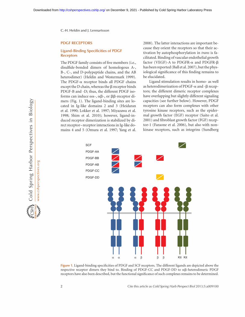
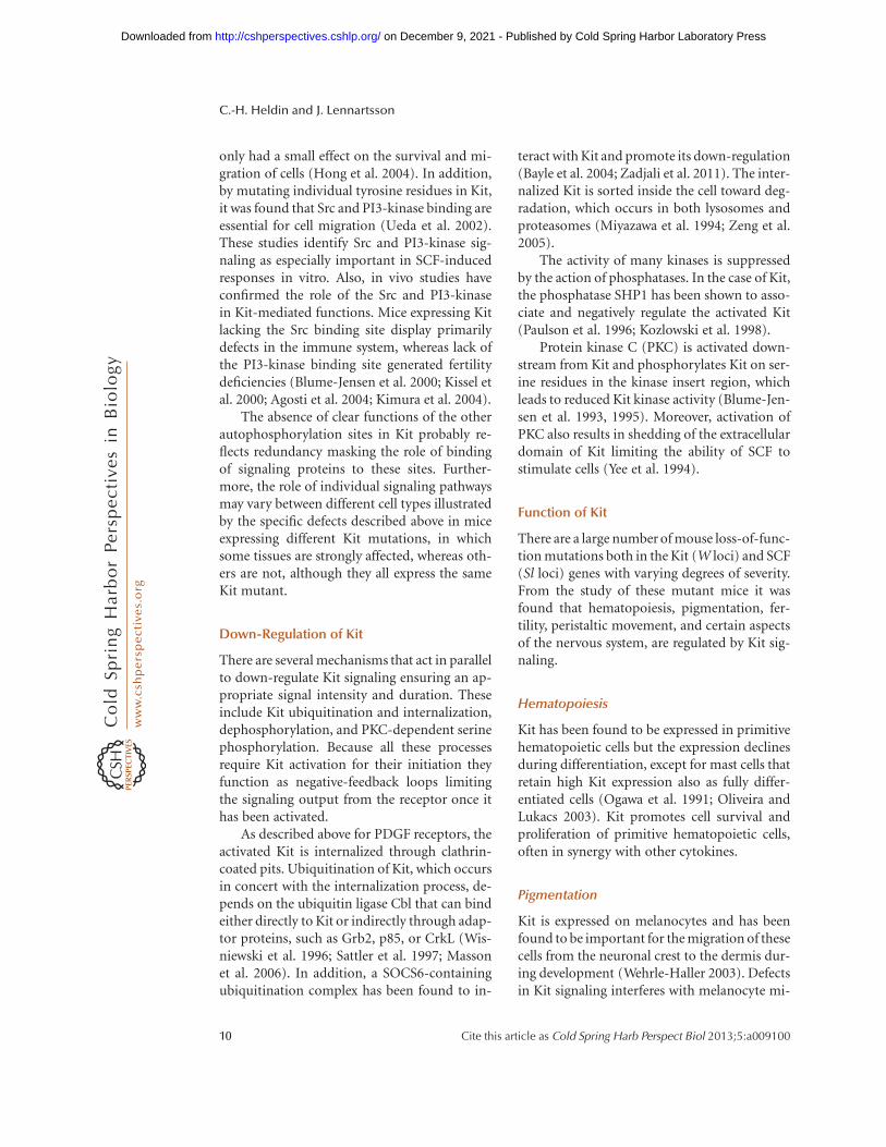
The PDGF family consists of five members (i.e.,disulfide-bonded dimers of homologous A-,B-, C-, and D-polypeptide chains, and the ABheterodimer) (Heldin and Westermark 1999).The PDGF-a receptor binds all PDGF chainsexcept the D chain, whereas theb receptor bindsPDGF-B and -D; thus, the different PDGF iso-forms can induce aa-, ab-, or bb-receptor di-mers (Fig. 1). The ligand-binding sites are lo-cated in Ig-like domains 2 and 3 (Heidaranet al. 1990; Lokker et al. 1997; Miyazawa et al.1998; Shim et al. 2010); however, ligand-in-duced receptor dimerization is stabilized by di-rect receptor–receptor interactions in Ig-like do-mains 4 and 5 (Omura et al. 1997; Yang et al.
2008). The latter interactions are important be-cause they orient the receptors so that their ac-tivation by autophosphorylation in trans is fa-cilitated. Binding of vascular endothelial growthfactor (VEGF)-A to PDGFR-a and PDGFR-bhas been reported (Ball et al. 2007), but the phys-iological significance of this finding remains tobe elucidated.
Ligand stimulation results in homo- as wellas heterodimerization of PDGF-a and -b recep-tors; the different dimeric receptor complexeshave overlapping but slightly different signalingcapacities (see further below). However, PDGFreceptors can also form complexes with othertyrosine kinase receptors, such as the epider-mal growth factor (EGF) receptor (Saito et al.2001) and fibroblast growth factor (FGF) recep-tor-1 (Faraone et al. 2006), but also with non-kinase receptors, such as integrins (Sundberg
SCF
PDGF-AA
PDGF-BB
PDGF-AB
PDGF-CC
PDGF-DD
α α α β β β Kit Kit
Figure 1. Ligand-binding specificities of PDGF and SCF receptors. The different ligands are depicted above therespective receptor dimers they bind to. Binding of PDGF-CC and PDGF-DD to ab-heterodimeric PDGFreceptors have also been described, but the functional significance of such complexes remains to be determined.
C.-H. Heldin and J. Lennartsson
2 Cite this article as Cold Spring Harb Perspect Biol 2013;5:a009100
on December 9, 2021 - Published by Cold Spring Harbor Laboratory Press http://cshperspectives.cshlp.org/Downloaded from
and Rubin 1996; Schneller et al. 1997), CD44 (Liet al. 2006), the low-density lipoprotein recep-tor-related protein (LRP) (Boucher et al. 2002;Loukinova et al. 2002), and the poliovirus re-ceptor Necl-5 (Minami et al. 2010). Such inter-actions modulate signaling via PDGF receptors.
Activation of PDGF Receptor Kinases
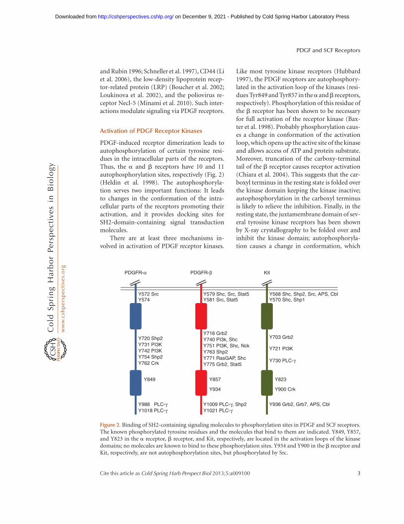
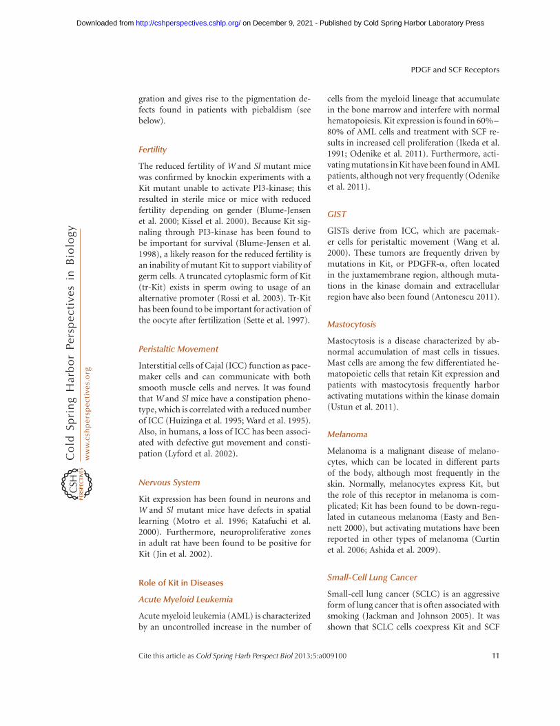
PDGF-induced receptor dimerization leads toautophosphorylation of certain tyrosine resi-dues in the intracellular parts of the receptors.Thus, the a and b receptors have 10 and 11autophosphorylation sites, respectively (Fig. 2)(Heldin et al. 1998). The autophosphoryla-tion serves two important functions: It leadsto changes in the conformation of the intra-cellular parts of the receptors promoting theiractivation, and it provides docking sites forSH2-domain-containing signal transductionmolecules.
There are at least three mechanisms in-volved in activation of PDGF receptor kinases.
Like most tyrosine kinase receptors (Hubbard1997), the PDGF receptors are autophosphory-lated in the activation loop of the kinases (resi-dues Tyr849 and Tyr857 in thea andb receptors,respectively). Phosphorylation of this residue ofthe b receptor has been shown to be necessaryfor full activation of the receptor kinase (Bax-ter et al. 1998). Probably phosphorylation caus-es a change in conformation of the activationloop, which opens up the active site of the kinaseand allows access of ATP and protein substrate.Moreover, truncation of the carboxy-terminaltail of the b receptor causes receptor activation(Chiara et al. 2004). This suggests that the car-boxyl terminus in the resting state is folded overthe kinase domain keeping the kinase inactive;autophosphorylation in the carboxyl terminusis likely to relieve the inhibition. Finally, in theresting state, the juxtamembrane domain of sev-eral tyrosine kinase receptors has been shownby X-ray crystallography to be folded over andinhibit the kinase domain; autophosphoryla-tion causes a change in conformation, which
Y572 SrcY574
Y720 Shp2Y731 PI3KY742 PI3KY754 Shp2Y762 Crk
Y988 PLC-γY1018 PLC-γ
Y849
Y579 Shc, Src, Stat5Y581 Src, Stat5
PDGFR-α PDGFR-β Kit
Y716 Grb2Y740 PI3k, ShcY751 PI3K, Shc, NckY763 Shp2Y771 RasGAP, ShcY775 Grb2, Stat5
Y1009 PLC-γ, Shp2Y1021 PLC-γ
Y857
Y934
Y568 Shc, Shp2, Src, APS, CbIY570 Shc, Shp1
Y703 Grb2
Y721 PI3K
Y730 PLC-γ
Y936 Grb2, Grb7, APS, Cbl
Y823
Y900 Crk
Figure 2. Binding of SH2-containing signaling molecules to phosphorylation sites in PDGF and SCF receptors.The known phosphorylated tyrosine residues and the molecules that bind to them are indicated. Y849, Y857,and Y823 in the a receptor, b receptor, and Kit, respectively, are located in the activation loops of the kinasedomains; no molecules are known to bind to these phosphorylation sites. Y934 and Y900 in the b receptor andKit, respectively, are not autophosphorylation sites, but phosphorylated by Src.
PDGF and SCF Receptors
Cite this article as Cold Spring Harb Perspect Biol 2013;5:a009100 3
on December 9, 2021 - Published by Cold Spring Harbor Laboratory Press http://cshperspectives.cshlp.org/Downloaded from
relieves the inhibition (Wybenga-Groot et al.2001; Hubbard 2004). A similar inhibitory func-tion has been shown in the PDGF-b receptor(Irusta et al. 2002). Moreover, in the context ofoncogenic fusion proteins of the PDGF recep-tors, the juxtamembrane domains have beenshown to have an inhibitory function (see fur-ther below), supporting the notion that the jux-tamembrane domain has an inhibitory functionalso in full-length PDGF receptors. Together,these mechanisms cooperate to keep the kinaseinactive; autophosphorylation of several tyro-sine residues is necessary for full activation ofthe receptor kinase.
Activation of Intracellular SignalTransduction Pathways by PDGFReceptors
The second important function of autophos-phorylation of the PDGF receptors is to allowbinding of signaling molecules containing SH2domains, which recognize phosphorylated tyro-sine residues (Heldin et al. 1998). Because dif-ferent SH2 domains have different preferencesregarding the three to six amino acid residuesdownstream from the phosphorylated tyrosine,there is a certain specificity in binding. ThePDGF receptors have been reported to bindabout 10 different families of SH2-domain-con-taining molecules, which initiates the activationof different signaling pathways (Fig. 2). Becausethe autophosphorylation pattern of PDGF-aand -b receptors differ depending on whetherthe receptors occur in homo- or heterodimericcomplexes, each of the three dimeric PDGF re-ceptor complexes have distinct signaling prop-erties (Ekman et al. 1999).
Certain of the SH2-domain signaling mol-ecules that bind to PDGF receptors have in-trinsic enzymatic activities (i.e., members ofthe Src family of tyrosine kinases, the GTPase-activating protein [GAP] for Ras, the tyrosinephosphatase SHP-2, and phospholipase C-g[PLC-g]) (reviewed in Heldin and Westermark1999). The respective enzymatic activities areactivated by binding to the receptors or by phos-phorylation by the receptor kinases. Alternative-ly, the enzymes are constitutively active and just
brought to the inner leaflet of the plasma mem-brane by the activated receptors.
There are also examples of SH2-domain-containing adaptor molecules, including Nck,Shc, and Crk, which bind to activated PDGFreceptors. They act by mediating interactionswith different downstream signaling molecules.Some of them form stable complexes with en-zymes, such as Grb2, which form a complex withthe nucleotide exchange molecule SOS1 activat-ing Ras. Moreover, the a- or b-regulatory p85subunits of the phosphatidylinositol 30-kinasebind to the receptors and to the a- or b-p110catalytic subunits. In addition, certain membersof the STAT family of transcription factors bindto and are activated by PDGF receptors (re-viewed by Heldin and Westermark 1999).
The intracellular parts of the PDGF receptorsalso interact with certain signaling moleculesindependent of autophosphorylation (e.g., thePDZ-domain protein NHERF), which bindsto the end of the carboxy-terminal tail of thePDGF receptors and enhances receptor signal-ing (Maudsley et al. 2000; Demoulin et al. 2003;Takahashi et al. 2006; Theisen et al. 2007), andthe adaptor molecule Alix, which binds to theregion around Tyr1021 of the carboxy-terminaltail and facilitates binding of the ubiquitin li-gase Cbl (Lennartsson et al. 2006).
In order for initiation of signaling via PDGFreceptors to be efficient, tyrosine phosphatasesneed to be inactivated (Sundaresan et al. 1995).This is achieved by a PI3-kinase-dependent ox-idation of a cysteine residue in the active site ofphosphatases (Bae et al. 2000).
Much effort has been put into the elucida-tion of which signaling pathways mediate thevarious effects of PDGF on cells (i.e., cell prolif-eration, survival, chemotaxis, and actin reorga-nization). Although cell-type differences havebeen reported, in general, PI3 kinase has beenfound to be important for the antiapoptotic andmotility responses of PDGF. Src via activation ofthe transcription factor Myc, and Ras via activa-tion of the Erk MAP kinase pathway, are impor-tant for the growth-stimulating effects. It shouldbe noted, however, that there is an extensivecross talk between different signaling pathways.Thus, each of the many signaling pathways in-
C.-H. Heldin and J. Lennartsson
4 Cite this article as Cold Spring Harb Perspect Biol 2013;5:a009100
on December 9, 2021 - Published by Cold Spring Harbor Laboratory Press http://cshperspectives.cshlp.org/Downloaded from
duced by the activated receptor can, to differentextents and in a cell-type-specific manner, con-tribute to most of the cellular effects of PDGF.
Modulation of Signaling via PDGF Receptors
Both aa- and bb-receptor complexes inducepowerful mitogenic signals. However, whereasbb-receptor homodimers andab heterodimersinduce chemotaxis, the aa homodimer inhibitschemotaxis, at least in certain cell types (Eriks-son et al. 1992). The ab-heterodimeric receptorcomplex appears to induce the most potent mi-togenic signal. A mechanism for this differencecould be that Tyr771, to which RasGAP binds,is efficiently phosphorylated in the bb homo-dimer, but not in the ab heterodimer (Ekmanet al. 1999). Thus, RasGAP, which deactivatesRas, cannot bind to the ab-receptor heterodi-mer leading to a more efficient activation of Rasby the ab-receptor heterodimer.
The simultaneous binding to activatedPDGF receptors of the Grb2/SOS1 complex,which activates Ras, and of RasGAP, which in-activates Ras, provides an example of how sig-naling via PDGF receptors can be modulated.Another example is the binding of the tyrosinephosphatase SHP-2, which dephosphorylatesthe b receptor and its substrates (Lechleideret al. 1993). However, in addition to negativelymodulating PDGF receptor signaling via de-phosphorylation, SHP-2 also positively contrib-utes to signaling through dephosphorylation ofthe carboxy-terminal tyrosine residue in Src,whereby Src is activated, and by functioning asan adaptor, which can bind Grb2/SOS1, thuspromoting activation of Ras (Dance et al. 2008).
Internalization and Sorting of PDGFReceptors
Following ligand binding, PDGF receptors areaccumulated in coated pit areas at the cell mem-brane, and then internalized in a clathrin- anddynamin-dependent manner, in a process thatpartly depends on the kinase activity of the re-ceptors (Sorkin et al. 1991). Signaling continuesin endosomes (Wang et al. 2004), until the pHdecreases enough to cause dissociation of PDGF
from its receptors. Most of the internalizedPDGF receptors are degraded by fusion of en-dosomes with multivesicular bodies and lyso-somes, or by degradation in proteasomes, pro-cesses that are promoted by polyubiquitinationof the receptors (Heldin et al. 1982; Mori et al.1992). Ubiquitination of the PDGF-b receptorcan be performed by the ubiquitin ligase Cbl(Miyake et al. 1999); degradation of Cbl is pro-moted by the adaptor molecule Alix, whichbinds to the PDGF-b receptor, thereby prevent-ing ubiquitination of the receptor and thus sta-bilizing it (Lennartsson et al. 2006).
Although most of PDGF receptors are de-graded after ligand-induced internalization,there are mechanisms that affect sorting andpromote recycling of the receptor to the plasmamembrane. One such mechanism was shown toinvolve overactivation of PLC-g and its down-stream effectors PKCa in cells deficient of thetyrosine phosphatase TC-PTP (Karlsson et al.2006); TC-PTP dephosphorylates preferential-ly Tyr1021 in the PDGF-b receptor, which is thebinding site of PLC-g. The overactivated PLC-g was found to promote recycling in a proteinkinase C-dependent manner (Hellberg et al.2009). Another mechanism was found to oper-ate in Ras-transformed fibroblasts; in these cells,PI3-kinase is overactivated leading to internali-zation of receptors via macropinocytosis, whichis accompanied by increased recycling (Schmeeset al. 2012). In both these cases, the inductionof recycling was associated with an increasedPDGF signal strength and duration. Thus, mod-ulation of intracellular sorting mechanisms canaffect PDGF signaling.
Function of PDGF and PDGF Receptorsduring Embryonal Development
Primarily based on gene knockout studies inmice, PDGF and PDGF receptors have beenshown to have important roles to promoteproliferation, migration, and differentiation ofspecific cell types during the embryonal devel-opment (reviewed by Andrae et al. 2008). Acommon theme that has emerged from thesestudies is that PDGF isoforms, secreted by epi-thelial or endothelial cells, act in a paracrine
PDGF and SCF Receptors
Cite this article as Cold Spring Harb Perspect Biol 2013;5:a009100 5
on December 9, 2021 - Published by Cold Spring Harbor Laboratory Press http://cshperspectives.cshlp.org/Downloaded from
manner on nearby mesenchymal cells, such asfibroblasts, pericytes, and smooth muscle cells(Hoch and Soriano 2003; Andrae et al. 2008).
Knockout of PDGFR-a and PDGF-A wasfound to affect early mesenchymal derivativesin both embryo and extraembryo tissues, and aproportion of these mice die before or at em-bryonic day 10.5 (Hoch and Soriano 2003).Knockout of PDGFR-a also causes defects inneural crest mesenchyme derivatives, includingthe cardiac outflow tract, the thymus and skel-etal components in the facial and other regions,and in the development of the palate and teeth(Soriano 1997; Tallquist et al. 2000; Tallquistand Soriano 2003; Xu et al. 2005).
PDGF-A knockout mice that survive birthdevelop lung emphysema, because PDGFR-a-positive alveolar myofibroblast precursors donot migrate to the alveolar saccules (Bostromet al. 1996; Lindahl et al. 1997b). Knockout ofPDGF-A or PDGFR-a in mice also leads toabnormal development of gastrointestinal villi(Karlsson et al. 2000), skin blistering (Soriano1997), reduced hair development (Karlsson etal. 1999), and defect development of Leydig cellsof the testis (Gnessi et al. 2000; Brennan et al.2003). In each case, the interaction between thePDGFR-a expressed by mesenchymal cells ofdifferent kinds, and the ligand expressed byneighboring epithelial cells, is perturbed.
PDGFR-a also mediates signals needed forproliferation and differentiation of oligoden-drocyte progenitor cells (Calver et al. 1998) andretinal astrocytes (Fruttiger et al. 1996), and de-termines the number of oligodendrocyte pro-genitor cells in the adult brain (Woodruff et al.2004). PDGF-AA is constitutively secreted fromneuronal cell bodies but not from axons (Frut-tiger et al. 2000).
PDGFR-b is expressed on pericytes and vas-cular smooth muscle cells and mediates recruit-ment of these cells to newly formed vessels inresponse to PDGF-BB secreted by endothelialcells (Lindahl et al. 1997a; Hellstrom et al.1999; Bjarnegard et al. 2004), and in particularby the tip cells that lead the angiogenic sprout(Gerhardt et al. 2003). PDGFR-b knockout em-bryos progressively develop abnormal glomeru-li in the kidney owing to defect development
of mesangial cells (Leveen et al. 1994; Soriano1994; Lindahl et al. 1998), capillary microaneur-ysm (Lindahl et al. 1997a), cardiac defects (Vanden Akker et al. 2008), and placental defects(Ohlsson et al. 1999), and die at E16–E19 fromhemorrhage. A role for PDGFR-b in the earlydevelopment of hematopoietic/endothelial pre-cursors has also been shown; activation ofPDGFR-b on these cells drives differentiationtoward endothelial cells (Rolny et al. 2006).
It is thus clear that PDGFR-a and PDGFR-b have different functions during embryonaldevelopment. To determine whether the differ-ences are owing to different expression patternsor to different signaling capacities, the intra-cellular parts of the receptors were swapped(Klinghoffer et al. 2001). Whereas loss of thecytoplasmic part of PDGFR-a could be rescuedby the cytoplasmic part of PDGFR-b, loss of theintracellular part of PDGFR-b was only part-ly rescued by the intracellular part of PDGFR-a. A partial rescue was also obtained when thePDGFR-a kinase domain was replaced with amore distant kinase domain (Hamilton et al.2003). These findings suggest that both expres-sion patterns and signaling capacities accountfor the differences in function of the two PDGFreceptors (Klinghoffer et al. 2001).
Function of PDGF and PDGF Receptorsin the Adult
In the adult, activation of PDGFR-b controlsthe intestinal fluid pressure of tissues and pre-vents formation of edema (Rodt et al. 1996).A probable mechanism is that fibroblasts andmyofibroblasts, which express PDGFR-b, makecontact with extracellular components via theirintegrins; activation of PDGFR-b induces con-traction of the cells, which controls the intersti-tial fluid pressure (Liden et al. 2006).
PDGF has also been shown to stimulatewound healing (Robson et al. 1992). PDGF re-ceptors are expressed by several cell types in-volved in wound healing, such as fibroblasts,smooth muscle cells, neutrophils, and macro-phages; on PDGF stimulation these cells arerecruited to the wounded area (e.g., by PDGFreleased from platelets). PDGF also contributes
C.-H. Heldin and J. Lennartsson
6 Cite this article as Cold Spring Harb Perspect Biol 2013;5:a009100
on December 9, 2021 - Published by Cold Spring Harbor Laboratory Press http://cshperspectives.cshlp.org/Downloaded from
to wound healing by stimulating the productionof different matrix molecules (reviewed by Hel-din and Westermark 1999).
A specific function for PDGFR-a to pro-mote proliferation of insulin-promoting b cellsin juvenile pancreatic islets was recently eluci-dated (Chen et al. 2011).
Role of PDGF Receptor Activationin Diseases
PDGF receptor activation has been observed incancer and in other diseases involving excesscell proliferation, such as atherosclerosis and fi-brotic conditions.
Overactivity of PDGF Receptorsin Tumor Cells
Certain malignancies are characterized by mu-tations in PDGF receptor genes. Thus, 5% ofgastrointestinal stromal tumors (GIST) showpoint mutations in the PDGFR-a gene (Hein-rich et al. 2003); in this tumor type, mutations inthe Kit gene are even more common (see below).The mutations affect the control mechanismsinvolved in keeping the receptor kinase inactiveand lead to a constitutively active kinase. Similaractivating point mutations in PDGFR-a havealso been observed in hypereosinophilic syn-drome (Elling et al. 2011).
In chronic myelomonocytic leukemia(CMML) the intracellular part of the PDGFR-b gene has been found to be fused to the TELgene (Golub et al. 1994) or other genes thathave in common that they encode proteins thatcan dimerize or oligomerize (Magnusson et al.2001). Similarly, the PDGFR-a gene is fused tothe FIP1L1 gene in hypereosinophilic syndrome(Cools et al. 2003; Griffin et al. 2003) and insystemic mastocytosis (Pardanani et al. 2003).The resulting fusion proteins have constitutive-ly active kinases as a result of juxtaposition ofthe kinases of the receptors, as well as by the lossof regulatory sequences in the juxtamembrane(Stover et al. 2006) and transmembrane (Toffa-lini et al. 2010) domains.
The PDGFR-a gene has been found to beamplified in subsets of glioblastomas (Fleming
et al. 1992; Kumabe et al. 1992; Puputti et al.2006), anaplastic oligodendrogliomas (Smith etal. 2000), esophageal squamous cell carcinoma(Arai et al. 2003), and pulmonary artery intimalsarcoma (Zhao et al. 2002). The increased num-ber of receptors on such cells makes the cellsvery sensitive to PDGF stimulation; moreover,at a sufficiently high receptor density, ligand-independent activation may occur. In addition,an activated deletion mutant of PDGFR-a hasbeen found in a human glioblastoma (Clarkeand Dirks 2003).
PDGF receptors may be activated also as aconsequence of mutation of ligand genes. Thus,in the rare skin tumor dermatofibrosarcomaprotuberans (DFSP), the PDGF-B gene is fusedto the collagen 1A1 gene (Simon et al. 1997;O’Brien et al. 1998), resulting in the productionof large amounts of a fusion protein, which afterprocessing becomes similar to mature PDGF-BB that activates its receptors in an autocrinemanner (Shimizu et al. 1999).
Epithelial tumors can undergo epithelial-mesenchymal transition (EMT), whereby theylose their epithelial characteristics and startto express mesenchymal components, such asPDGF receptors (Thiery et al. 2009). Thus,whereas epithelial tumors generally do not re-spond to PDGF, they may do so after they haveundergone EMT (Jechlinger et al. 2003). EMTcorrelates with increased invasiveness and meta-stasis; interestingly, inhibition of PDGF signalingwas shown to inhibit metastasis in mouse mod-els for breast cancer (Jechlinger et al. 2006), he-patocellular carcinoma (Gotzmann et al. 2006),and prostate cancer (Dolloff et al. 2005; Russellet al. 2009). PDGFR-a appears to be more im-portant than PDGFR-b in the promotion of me-tastasis of epithelial tumors.
A malignancy-dependent increase in expres-sion of PDGF isoforms and receptors has beenobserved in glioblastoma tumors. Thus, in thistumor type, PDGF appears to be involved in au-tocrine and paracrine stimulation, both in thetumor cells via PDGFR-a and in cells of thestroma via PDGFR-b (Hermanson et al. 1992).Moreover, in basal cell carcinoma, activation ofthe sonic hedgehog signaling pathway inducesthe expression of PDGFR-a (Xie et al. 2001).
PDGF and SCF Receptors
Cite this article as Cold Spring Harb Perspect Biol 2013;5:a009100 7
on December 9, 2021 - Published by Cold Spring Harbor Laboratory Press http://cshperspectives.cshlp.org/Downloaded from
Activation of PDGF Receptors in the StromalCompartment of Tumors
In addition to direct effects on the tumor cells,PDGF made by tumor cells and other cell typesin solid tumors acts in a paracrine manneron various cell types in the vasculature and inthe interstitial stroma of tumors (Pietras et al.2008). Thus, PDGF promotes tumor angio-genesis by stimulating perivascular progenitorcells, pericytes, and vascular smooth musclecells (Song et al. 2005), and by recruiting protu-morigenic inflammatory cells. Moreover, over-activity of PDGF contributes to the increasedinterstitial fluid pressure that characterizes amajority of solid tumors, which leads to a de-creased transcapillary transport and is thereforean obstacle in the treatment of tumors withchemotherapeutical drugs (Heldin et al. 2004).
Activation of PDGF Receptorsin Atherosclerosis
PDGF isoforms and receptors occur at increasedlevels in atherosclerotic lesions. According tothe response to injury hypothesis, PDGF iso-forms released by platelets at sites of endothel-ial cell injury stimulate vascular smooth musclecells to migrate from the vessel media into theintima and to proliferate (Ross 1993). Chronicinflammation involving release of PDGF fromdifferent types of immune cells also contributesto the narrowing of the vessel lumen (Hansson2005). PDGF and other growth factors and cy-tokines released then maintain the local inflam-mation in a vicious circle.
Activation of PDGF Receptors in FibroticDiseases
Increased PDGF signaling has also been linkedto various fibrotic conditions, such as lung fi-brosis, myelofibrosis, glomerulonephritis, andliver cirrhosis. During chronic inflammation,activated macrophages secrete PDGF isoformsas well as other inflammatory cytokines, whichup-regulate PDGF receptors on mesenchymalcells, thus promoting their proliferation andproduction of matrix molecules (Bonner 2004).In support of the notion that overactive PDGF
signaling drives fibrosis, knockin of constitu-tively active mutants of PDGFR-a was foundto lead to increased connective tissue growthand a progressive fibrotic phenotype in multipleorgans (Olson and Soriano 2009). Knockin of asimilar PDGFR-b mutant caused an enhancedwound healing response in the skin and the liver(Krampert et al. 2008).
PDGF Receptors as Binders of Viruses
PDGFR-a has been shown to be a receptor foradeno-associated virus type 5 (Di Pasquale et al.2003) and cytomegalovirus (Soroceanu et al.2008). Thus, PDGFR-a facilitates infections bythese viruses.
DEVELOPMENT AND CLINICAL USEOF PDGF ANTAGONISTS
Because overactivity of PDGF signaling is con-nected to many serious diseases, efforts havebeen made to develop antagonists of PDGF sig-naling. Several types of inhibitors are now avail-able, including DNA aptamers binding PDGFisoforms, inhibitory antibodies against the re-ceptors, and low molecular weight inhibitorsof PDGF receptor kinases (e.g., imatinib, suni-tinib, and sorafenib) (Demetri 2011). Beneficialeffects have been observed by treatment of therare malignancies that are driven by mutationsin genes for PDGF and PDGF receptors (i.e.,CMML, DFSP, hypereosinophilic syndrome,and GIST). However, treatment of glioblastomathat also often is characterized by overactivityof PDGF receptors, with PDGF antagonists, hasbeen less successful; probably these tumors haveundergone too many other genetic or epigeneticalterations and inhibition of PDGF receptorsignaling is therefore not enough. There are in-dications that anti-PDGF treatment could be ofmore general application for the targeting ofthe stroma compartment of solid tumors, toinhibit metastasis (Pietras et al. 2003; Catenaet al. 2010), and to lower the interstitial fluidpressure and thereby increase the transcapillaryflow and drug delivery (Heldin et al. 2004). Theside effects of anti-PDGF therapy have been rel-atively mild, but include a tendency to develop
C.-H. Heldin and J. Lennartsson
8 Cite this article as Cold Spring Harb Perspect Biol 2013;5:a009100
on December 9, 2021 - Published by Cold Spring Harbor Laboratory Press http://cshperspectives.cshlp.org/Downloaded from
edema, reflecting the important role of PDGF inregulation of the interstitial fluid pressure (Rodtet al. 1996), and heart failure, reflecting an im-portant role of PDGF in stress-induced cardiacangiogenesis (Chintalgattu et al. 2010). In pa-tients with advanced ovarian cancer, treatmentwith the inhibitory Fab fragment CDP860 led tothe development of significant ascites (Jaysonet al. 2005).
STEM CELL FACTOR RECEPTOR (Kit)
Activation of Kit by SCF
The biologically active SCF is a homodimericprotein that is primarily produced by fibroblastsand endothelial cells, and exists both as a solubleand membrane-bound form owing to alterna-tive splicing of exon 6 (Huang et al. 1992). Bothforms of SCF are initially produced as trans-membrane proteins, but the protein containingexon 6 becomes cleaved into the soluble SCFand the form lacking this exon remains mem-brane associated. Several proteases have beensuggested to be involved in the processing ofSCF, including MMP9, members of the ADAMfamily, and Chymase-1 (Longley et al. 1997;Heissig et al. 2002; Kawaguchi et al. 2007).
The SCF receptor is also called Kit, becauseit was first identified as an oncoprotein derivedfrom the Hardy-Zuckerman 4 feline sarcomavirus (Besmeret al. 1986). It is expressed in prim-itive hematopoietic cells and mast cells, but alsoin melanocytes and basal cells in the skin, germscells, interstitial cells of Cajal, and in certain re-gions of the brain. There are four human iso-forms of Kit. Two splice forms are characterizedby the presence or absence of a four-amino-acidsequence in the extracellular juxtamembrane re-gion (Reith et al. 1991). These two splice formsdisplay different signaling abilities with theshorter form being more strongly tyrosine phos-phorylated and more powerful in activation ofdownstream signaling, whereas the longer Kitsplice form signals less intensely but more per-sistently (Caruana et al. 1999; Voytyuk et al.2003). Kit isoforms are also characterized bythe absence or presence of a serine residue inthe kinase insert region (Crosier et al. 1993).
The process by which SCF activates Kit hasbeen studied both at a biochemical and struc-tural level, and occurs in a manner similar tothat described above for the PDGF receptor; thedimeric SCF ligand brings two Kit monomersin close proximity of each other, which enablesinteractions between the extracellular domainsof the receptors generating a stable dimer (Fig.1) (Lemmon et al. 1997; Zhang et al. 2000; Yu-zawa et al. 2007).
The crystal structure of the Kit kinase do-main revealed that the juxtamembrane regionis inserted between the two lobes of the kinasedomain thereby suppressing kinase activity, andthat this inhibition is released on phosphory-lation of tyrosine residues within the juxta-membrane segment (Mol et al. 2003, 2004). Akinetic study of the autophosphorylation pro-cess showed that the juxtamembrane region isfirst to be phosphorylated, consistent with therole of this region in suppressing the kinase ac-tivity (DiNitto et al. 2010).
Activation of Intracellular Pathways by Kit
The intracellular region of Kit contains sevenautophosphorylation sites that are involved inthe activation of the Kit kinase and can act asdocking sites for intracellular signaling pro-teins (Fig. 2). The signaling pathways activateddownstream from Kit overlap to a large extentwith those described for the PDGF receptor(see above), and include PI3-kinase, Src kinases,MAP kinase pathways, and phospholipase Cand D. Furthermore, it is well established thatthere exists a comprehensive cross talk betweendifferent pathways. For example, Src kinasescontribute to Erk1/2 activation, and Src in com-bination with PI3-kinase promotes Jnk activa-tion, in response to SCF (Timokhina et al. 1998;Lennartsson et al. 1999).
Attempts to study the role of individual sig-naling proteins include mutation of all tyro-sine residues in the intracellular region of Kitand then adding them back one at a time. Usingthis approach, it was found that the Src dockingsite on Kit is important for the migratory, sur-vival, and, partially, the proliferative response toSCF, whereas restoring coupling to PI3-kinase
PDGF and SCF Receptors
Cite this article as Cold Spring Harb Perspect Biol 2013;5:a009100 9
on December 9, 2021 - Published by Cold Spring Harbor Laboratory Press http://cshperspectives.cshlp.org/Downloaded from
only had a small effect on the survival and mi-gration of cells (Hong et al. 2004). In addition,by mutating individual tyrosine residues in Kit,it was found that Src and PI3-kinase binding areessential for cell migration (Ueda et al. 2002).These studies identify Src and PI3-kinase sig-naling as especially important in SCF-inducedresponses in vitro. Also, in vivo studies haveconfirmed the role of the Src and PI3-kinasein Kit-mediated functions. Mice expressing Kitlacking the Src binding site display primarilydefects in the immune system, whereas lack ofthe PI3-kinase binding site generated fertilitydeficiencies (Blume-Jensen et al. 2000; Kissel etal. 2000; Agosti et al. 2004; Kimura et al. 2004).
The absence of clear functions of the otherautophosphorylation sites in Kit probably re-flects redundancy masking the role of bindingof signaling proteins to these sites. Further-more, the role of individual signaling pathwaysmay vary between different cell types illustratedby the specific defects described above in miceexpressing different Kit mutations, in whichsome tissues are strongly affected, whereas oth-ers are not, although they all express the sameKit mutant.
Down-Regulation of Kit
There are several mechanisms that act in parallelto down-regulate Kit signaling ensuring an ap-propriate signal intensity and duration. Theseinclude Kit ubiquitination and internalization,dephosphorylation, and PKC-dependent serinephosphorylation. Because all these processesrequire Kit activation for their initiation theyfunction as negative-feedback loops limitingthe signaling output from the receptor once ithas been activated.
As described above for PDGF receptors, theactivated Kit is internalized through clathrin-coated pits. Ubiquitination of Kit, which occursin concert with the internalization process, de-pends on the ubiquitin ligase Cbl that can bindeither directly to Kit or indirectly through adap-tor proteins, such as Grb2, p85, or CrkL (Wis-niewski et al. 1996; Sattler et al. 1997; Massonet al. 2006). In addition, a SOCS6-containingubiquitination complex has been found to in-
teract with Kit and promote its down-regulation(Bayle et al. 2004; Zadjali et al. 2011). The inter-nalized Kit is sorted inside the cell toward deg-radation, which occurs in both lysosomes andproteasomes (Miyazawa et al. 1994; Zeng et al.2005).
The activity of many kinases is suppressedby the action of phosphatases. In the case of Kit,the phosphatase SHP1 has been shown to asso-ciate and negatively regulate the activated Kit(Paulson et al. 1996; Kozlowski et al. 1998).
Protein kinase C (PKC) is activated down-stream from Kit and phosphorylates Kit on ser-ine residues in the kinase insert region, whichleads to reduced Kit kinase activity (Blume-Jen-sen et al. 1993, 1995). Moreover, activation ofPKC also results in shedding of the extracellulardomain of Kit limiting the ability of SCF tostimulate cells (Yee et al. 1994).
Function of Kit
There are a large number of mouse loss-of-func-tion mutations both in the Kit (W loci) and SCF(Sl loci) genes with varying degrees of severity.From the study of these mutant mice it wasfound that hematopoiesis, pigmentation, fer-tility, peristaltic movement, and certain aspectsof the nervous system, are regulated by Kit sig-naling.
Hematopoiesis
Kit has been found to be expressed in primitivehematopoietic cells but the expression declinesduring differentiation, except for mast cells thatretain high Kit expression also as fully differ-entiated cells (Ogawa et al. 1991; Oliveira andLukacs 2003). Kit promotes cell survival andproliferation of primitive hematopoietic cells,often in synergy with other cytokines.
Pigmentation
Kit is expressed on melanocytes and has beenfound to be important for the migration of thesecells from the neuronal crest to the dermis dur-ing development (Wehrle-Haller 2003). Defectsin Kit signaling interferes with melanocyte mi-
C.-H. Heldin and J. Lennartsson
10 Cite this article as Cold Spring Harb Perspect Biol 2013;5:a009100
on December 9, 2021 - Published by Cold Spring Harbor Laboratory Press http://cshperspectives.cshlp.org/Downloaded from
gration and gives rise to the pigmentation de-fects found in patients with piebaldism (seebelow).
Fertility
The reduced fertility of W and Sl mutant micewas confirmed by knockin experiments with aKit mutant unable to activate PI3-kinase; thisresulted in sterile mice or mice with reducedfertility depending on gender (Blume-Jensenet al. 2000; Kissel et al. 2000). Because Kit sig-naling through PI3-kinase has been found tobe important for survival (Blume-Jensen et al.1998), a likely reason for the reduced fertility isan inability of mutant Kit to support viability ofgerm cells. A truncated cytoplasmic form of Kit(tr-Kit) exists in sperm owing to usage of analternative promoter (Rossi et al. 2003). Tr-Kithas been found to be important for activation ofthe oocyte after fertilization (Sette et al. 1997).
Peristaltic Movement
Interstitial cells of Cajal (ICC) function as pace-maker cells and can communicate with bothsmooth muscle cells and nerves. It was foundthat W and Sl mice have a constipation pheno-type, which is correlated with a reduced numberof ICC (Huizinga et al. 1995; Ward et al. 1995).Also, in humans, a loss of ICC has been associ-ated with defective gut movement and consti-pation (Lyford et al. 2002).
Nervous System
Kit expression has been found in neurons andW and Sl mutant mice have defects in spatiallearning (Motro et al. 1996; Katafuchi et al.2000). Furthermore, neuroproliferative zonesin adult rat have been found to be positive forKit (Jin et al. 2002).
Role of Kit in Diseases
Acute Myeloid Leukemia
Acute myeloid leukemia (AML) is characterizedby an uncontrolled increase in the number of
cells from the myeloid lineage that accumulatein the bone marrow and interfere with normalhematopoiesis. Kit expression is found in 60%–80% of AML cells and treatment with SCF re-sults in increased cell proliferation (Ikeda et al.1991; Odenike et al. 2011). Furthermore, acti-vating mutations in Kit have been found in AMLpatients, although not very frequently (Odenikeet al. 2011).
GIST
GISTs derive from ICC, which are pacemak-er cells for peristaltic movement (Wang et al.2000). These tumors are frequently driven bymutations in Kit, or PDGFR-a, often locatedin the juxtamembrane region, although muta-tions in the kinase domain and extracellularregion have also been found (Antonescu 2011).
Mastocytosis
Mastocytosis is a disease characterized by ab-normal accumulation of mast cells in tissues.Mast cells are among the few differentiated he-matopoietic cells that retain Kit expression andpatients with mastocytosis frequently harboractivating mutations within the kinase domain(Ustun et al. 2011).
Melanoma
Melanoma is a malignant disease of melano-cytes, which can be located in different partsof the body, although most frequently in theskin. Normally, melanocytes express Kit, butthe role of this receptor in melanoma is com-plicated; Kit has been found to be down-regu-lated in cutaneous melanoma (Easty and Ben-nett 2000), but activating mutations have beenreported in other types of melanoma (Curtinet al. 2006; Ashida et al. 2009).
Small-Cell Lung Cancer
Small-cell lung cancer (SCLC) is an aggressiveform of lung cancer that is often associated withsmoking (Jackman and Johnson 2005). It wasshown that SCLC cells coexpress Kit and SCF
PDGF and SCF Receptors
Cite this article as Cold Spring Harb Perspect Biol 2013;5:a009100 11
on December 9, 2021 - Published by Cold Spring Harbor Laboratory Press http://cshperspectives.cshlp.org/Downloaded from
suggesting the presence of an autocrine loop(Hibi et al. 1991). However, immunohisto-chemical staining of tumors for Kit expressionyielded varying results both regarding Kit ex-pression and correlation with clinical outcome(Fischer et al. 2007). Furthermore, no activat-ing Kit mutations could be found in SCLC tu-mors. Although imatinib inhibits growth ofSCLC cell lines, it failed to show efficiency inanimal models (Wolff et al. 2004). However,combining imatinib with chemotherapy (De-caudin et al. 2005), or using SU5416, whichinhibits several kinases including Kit, producedmore positive results in experimental models(Litz et al. 2004).
Piebaldism
In cancer, Kit is activated by gain-of-func-tion mutations or by autocrine stimulation. Incontrast, piebaldism involves germline loss-of-function mutations in Kit leading to patchesof skin and hair that are depigmented owingto lack of melanocytes (Spritz 1994).
Clinically Used Kit Antagonists
Overactivity of Kit has been found in severalmalignant diseases (see above) and inhibitingKit kinase activity is an approach to treat theseconditions. Imatinib, also discussed in thePDGF receptor context above, inhibits Kit andhas been used clinically. Because imatinib doesnot efficiently inhibit Kit with activating mu-tations within the kinase domain (Frost et al.2002; Ma et al. 2002), other drugs have beendeveloped that can inhibit Kit with such muta-tions, such as dasatinib and nilotinib (Demetri2011). Thus, depending on the mutational sta-tus of Kit, different drugs should be selected.Neither of the drugs mentioned above exclu-sively inhibit Kit, but inhibit also other kinasesand this may contribute to their clinical ef-fectiveness. Other multiselective drugs whosetargets include Kit are sunitinib and sorafenib.Masitinib is a potent inhibitor that targets Kitamong other kinases and is approved for treat-ing mast cell tumors in a veterinary setting.
FUTURE PERSPECTIVE
Studies over the last 30 years have revealed muchabout the structural properties, signaling capac-ities, normal functions, and role in diseases ofPDGF and SCF receptors. Moreover, treatmentregimens for malignant diseases in which thesereceptors are overactive are used routinely inthe clinic. Future studies will aim at elucidatingmechanisms for control of receptor signaling,including internalization, intracellular sorting,and termination of signaling. It is anticipatedthat such control mechanisms will include post-translational modifications of the receptors andtheir signaling mediators, other than the alreadycharacterized phosphorylation and ubiquitina-tion. Additional information is also needed re-garding cooperation of the receptors with co-receptors at the cell membrane, as well as theimportance of interactions with the receptorsand intracellular molecules, to enhance or con-trol their signaling capacities. It will also be im-portant to determine exactly where in the cellthe receptors induce their different signals: atthe plasma membrane, in endosomes, or in oth-er intracellular organelles. In addition, whereassome information is available regarding the in-volvement of PDGF and SCF receptors in dis-ease, a more detailed analysis of these receptorsin malignant as well as nonmalignant diseases ishighly warranted, as are more efficient and spe-cific methods to treat diseases in which the re-ceptors are overactive.
ACKNOWLEDGMENT
We thank Ingegard Schiller for valuable help inthe preparation of this manuscript.
REFERENCES
Agosti V, Corbacioglu S, Ehlers I, Waskow C, Sommer G,Berrozpe G, Kissel H, Tucker CM, Manova K, Moore MA,et al. 2004. Critical role for Kit-mediated Src kinase butnot PI 3-kinase signaling in pro T and pro B cell devel-opment. J Exp Med 199: 867–878.
Andrae J, Gallini R, Betsholtz C. 2008. Role of platelet-de-rived growth factors in physiology and medicine. GenesDev 22: 1276–1312.
Antonescu CR. 2011. The GIST paradigm: Lessons for otherkinase-driven cancers. J Pathol 223: 251–261.
C.-H. Heldin and J. Lennartsson
12 Cite this article as Cold Spring Harb Perspect Biol 2013;5:a009100
on December 9, 2021 - Published by Cold Spring Harbor Laboratory Press http://cshperspectives.cshlp.org/Downloaded from
Arai H, Ueno T, Tangoku A, Yoshino S, Abe T, Kawauchi S,Oga A, Furuya T, Oka M, Sasaki K. 2003. Detection ofamplified oncogenes by genome DNA microarrays inhuman primary esophageal squamous cell carcinoma:Comparison with conventional comparative genomic hy-bridization analysis. Cancer Genet Cytogenet 146: 16–21.
Ashida A, Takata M, Murata H, Kido K, Saida T. 2009. Path-ological activation of KIT in metastatic tumors of acraland mucosal melanomas. Int J Cancer 124: 862–868.
Bae YS, Sung J-Y, Kim O-S, Kim YJ, Hur KC, Kazlauskas A,Rhee SG. 2000. Platelet-derived growth factor-inducedH2O2 production requires the activation of phosphatidy-linositol 3-kinase. J Biol Chem 275: 10527–10531.
Ball SG, Shuttleworth CA, Kielty CM. 2007. Vascular endo-thelial growth factor can signal through platelet-derivedgrowth factor receptors. J Cell Biol 177: 489–500.
Baxter RM, Secrist JP, Vaillancourt RR, Kazlauskas A. 1998.Full activation of the platelet-derived growth factor b-receptor kinase involves multiple events. J Biol Chem273: 17050–17055.
Bayle J, Letard S, Frank R, Dubreuil P, De Sepulveda P. 2004.Suppressor of cytokine signaling 6 associates with KITand regulates KIT receptor signaling. J Biol Chem 279:12249–12259.
Besmer P, Murphy JE, George PC, Qiu FH, Bergold PJ,Lederman L, Snyder HW Jr, Brodeur D, Zuckerman EE,Hardy WD. 1986. A new acute transforming feline retro-virus and relationship of its oncogene v-kit with the pro-tein kinase gene family. Nature 320: 415–421.
Bjarnegard M, Enge M, Norlin J, Gustafsdottir S, Fredriks-son S, Abramsson A, Takemoto M, Gustafsson E, FasslerR, Betsholtz C. 2004. Endothelium-specific ablation ofPDGFB leads to pericyte loss and glomerular, cardiac andplacental abnormalities. Development 131: 1847–1857.
Blume-Jensen P, Hunter T. 2001. Oncogenic kinase signal-ling. Nature 411: 355–365.
Blume-Jensen P, Siegbahn A, Stabel S, Heldin C-H, Ronn-strand L. 1993. Increased Kit/SCF receptor induced mi-togenicity but abolished cell motility after inhibition ofprotein kinase C. EMBO J 12: 4199–4209.
Blume-Jensen P, Wernstedt C, Heldin C-H, Ronnstrand L.1995. Identification of the major phosphorylation sitesfor protein kinase C in Kit/stem cell factor receptor invitro and in intact cells. J Biol Chem 270: 14192–14200.
Blume-Jensen P, Janknecht R, Hunter T. 1998. The Kit re-ceptor promotes cell survival via activation of PI 3-kinaseand subsequent Akt-mediated phosphorylation of Badon Ser136. Curr Biol 8: 779–782.
Blume-Jensen P, Jiang G, Hyman R, Lee KF, O’Gorman S,Hunter T. 2000. Kit/stem cell factor receptor-inducedactivation of phosphatidylinositol 30-kinase is essentialfor male fertility. Nat Genet 24: 157–162.
Bonner JC. 2004. Regulation of PDGF and its receptors infibrotic diseases. Cytokine Growth Factor Rev 15: 255–273.
Bostrom H, Willetts K, Pekny M, Leveen P, Lindahl P, Hed-strand H, Pekna M, Hellstrom M, Gebre-Medhin S,Schalling M, et al. 1996. PDGF-A signaling is a criticalevent in lung alveolar myofibroblast development andalveogenesis. Cell 85: 863–873.
Boucher P, Liu P, Gotthardt M, Hiesberger T, Anderson RG,Herz J. 2002. Platelet-derived growth factor mediates ty-rosine phosphorylation of the cytoplasmic domain of thelow density lipoprotein receptor-related protein in cav-eolae. J Biol Chem 277: 15507–15513.
Brennan J, Tilmann C, Capel B. 2003. PDGFR-a mediatestestis cord organization and fetal Leydig cell developmentin the XY gonad. Genes Dev 17: 800–810.
Calver AR, Hall AC, Yu WP, Walsh FS, Heath JK, Betsholtz C,Richardson WD. 1998. Oligodendrocyte population dy-namics and the role of PDGF in vivo. Neuron 20: 869–882.
Caruana G, Cambareri AC, Ashman LK. 1999. Isoforms ofc-KIT differ in activation of signalling pathways andtransformation of NIH3T3 fibroblasts. Oncogene 18:5573–5581.
Catena R, Luis-Ravelo D, Anton I, Zandueta C, Salazar-Co-locho P, Larzabal L, Calvo A, Lecanda F. 2010. PDGFRsignaling blockade in marrow stroma impairs lung cancerbone metastasis. Cancer Res 71: 164–174.
Chen H, Gu X, Liu Y, Wang J, Wirt SE, Bottino R, Schorle H,Sage J, Kim SK. 2011. PDGF signalling controls age-de-pendent proliferation in pancreatic b-cells. Nature 478:349–355.
Chiara F, Bishayee S, Heldin C-H, Demoulin J-B. 2004.Autoinhibition of the platelet-derived growth factor b
receptor tyrosine kinase by its C-terminal tail. J BiolChem 279: 19732–19738.
Chintalgattu V, Ai D, Langley RR, Zhang J, Bankson JA, ShihTL, Reddy AK, Coombes KR, Daher IN, Pati S, et al. 2010.Cardiomyocyte PDGFR-b signaling is an essential com-ponent of the mouse cardiac response to load-inducedstress. J Clin Invest 120: 472–484.
Clarke ID, Dirks PB. 2003. A human brain tumor-derivedPDGFR-a deletion mutant is transforming. Oncogene22: 722–733.
Cools J, DeAngelo DJ, Gotlib J, Stover EH, Legare RD, Cor-tes J, Kutok J, Clark J, Galinsky I, Griffin JD, et al. 2003. Atyrosine kinase created by fusion of the PDGFRA andFIP1L1 genes as a therapeutic target of imatinib in idio-pathic hypereosinophilic syndrome. N Engl J Med 348:1201–1214.
Crosier PS, Ricciardi ST, Hall LR, Vitas MR, Clark SC, Cro-sier KE. 1993. Expression of isoforms of the human re-ceptor tyrosine kinase c-kit in leukemic cell lines andacute myeloid leukemia. Blood 82: 1151–1158.
Curtin JA, Busam K, Pinkel D, Bastian BC. 2006. Somaticactivation of KIT in distinct subtypes of melanoma. J ClinOncol 24: 4340–4346.
Dance M, Montagner A, Salles JP, Yart A, Raynal P. 2008.The molecular functions of Shp2 in the Ras/Mitogen-activated protein kinase (ERK1/2) pathway. Cell Signal20: 453–459.
Decaudin D, de Cremoux P, Sastre X, Judde JG, Nemati F,Tran-Perennou C, Freneaux P, Livartowski A, Pouillart P,Poupon MF. 2005. In vivo efficacy of STI571 in xeno-grafted human small cell lung cancer alone or combinedwith chemotherapy. Int J Cancer 113: 849–856.
Demetri GD. 2011. Differential properties of current tyro-sine kinase inhibitors in gastrointestinal stromal tumors.Semin Oncol 38: S10–S19.
PDGF and SCF Receptors
Cite this article as Cold Spring Harb Perspect Biol 2013;5:a009100 13
on December 9, 2021 - Published by Cold Spring Harbor Laboratory Press http://cshperspectives.cshlp.org/Downloaded from
Demoulin J-B, Seo JK, Ekman S, Grapengiesser E, HellmanU, Ronnstrand L, Heldin C-H. 2003. Ligand-inducedrecruitment of Naþ/Hþ exchanger regulatory factor tothe PDGF (platelet-derived growth factor) receptor reg-ulates actin cytoskeleton reorganization by PDGF. Bio-chem J 376: 505–510.
DiNitto JP, Deshmukh GD, Zhang Y, Jacques SL, Coli R,Worrall JW, Diehl W, English JM, Wu JC. 2010. Functionof activation loop tyrosine phosphorylation in the mech-anism of c-Kit auto-activation and its implication in su-nitinib resistance. J Biochem 147: 601–609.
Di Pasquale G, Davidson BL, Stein CS, Martins I, ScudieroD, Monks A, Chiorini JA. 2003. Identification of PDGFRas a receptor for AAV-5 transduction. Nat Med 9: 1306–1312.
Dolloff NG, Shulby SS, Nelson AV, Stearns ME, Johannes GJ,Thomas JD, Meucci O, Fatatis A. 2005. Bone-metastaticpotential of human prostate cancer cells correlates withAkt/PKB activation by a platelet-derived growth factorreceptor. Oncogene 24: 6848–6854.
Easty DJ, Bennett DC. 2000. Protein tyrosine kinases inmalignant melanoma. Melanoma Res 10: 401–411.
Ekman S, Rupp Thuresson E, Heldin C-H, Ronnstrand L.1999. Increased mitogenicity of an ab heterodimericPDGF receptor complex correlates with lack of RasGAPbinding. Oncogene 18: 2481–2488.
Elling C, Erben P, Walz C, Frickenhaus M, Schemionek M,Stehling M, Serve H, Cross NC, Hochhaus A, HofmannWK, et al. 2011. Novel imatinib-sensitive PDGFRA-acti-vating point mutations in hypereosinophilic syndromeinduce growth factor independence and leukemia-likedisease. Blood 117: 2935–2943.
Eriksson A, Siegbahn A, Westermark B, Heldin C-H,Claesson-Welsh L. 1992. PDGF a- and b-receptors acti-vate unique and common signal transduction pathways.EMBO J 11: 543–550.
Faraone D, Aguzzi MS, Ragone G, Russo K, Capogrossi MC,Facchiano A. 2006. Heterodimerization of FGF-receptor1 and PDGF-receptor-a: A novel mechanism underlyingthe inhibitory effect of PDGF-BB on FGF-2 in humancells. Blood 107: 1896–1902.
Fischer B, Marinov M, Arcaro A. 2007. Targeting receptortyrosine kinase signalling in small cell lung cancer(SCLC): What have we learned so far? Cancer Treat Rev33: 391–406.
Fleming TP, Saxena A, Clark WC, Robertson JT, OldfieldEH, Aaronson SA, Ali IU. 1992. Amplification and/oroverexpression of platelet-derived growth factor recep-tors and epidermal growth factor receptor in human glialtumors. Cancer Res 52: 4550–4553.
Frost MJ, Ferrao PT, Hughes TP, Ashman LK. 2002. Juxta-membrane mutant V560GKit is more sensitive to Imati-nib (STI571) compared with wild-type c-kit whereas thekinase domain mutant D816VKit is resistant. Mol CancerTher 1: 1115–1124.
Fruttiger M, Calver AR, Kruger WH, Mudhar HS, Micha-lovich D, Takakura N, Nishikawa SI, Richardson WD.1996. PDGF mediates a neuron-astrocyte interaction inthe developing retina. Neuron 17: 1117–1131.
Fruttiger M, Calver AR, Richardson WD. 2000. Platelet-de-rived growth factor is constitutively secreted from neuro-
nal cell bodies but not from axons. Curr Biol 10: 1283–1286.
Gerhardt H, Golding M, Fruttiger M, Ruhrberg C, Lund-kvist A, Abramsson A, Jeltsch M, Mitchell C, Alitalo K,Shima D, et al. 2003. VEGF guides angiogenic sproutingutilizing endothelial tip cell filopodia. J Cell Biol 161:1163–1177.
Gnessi L, Basciani S, Mariani S, Arizzi M, Spera G, Wang C,Bondjers C, Karlsson L, Betsholtz C. 2000. Leydig cell lossand spermatogenic arrest in platelet-derived growth fac-tor (PDGF)-A-deficient mice. J Cell Biol 149: 1019–1026.
Golub TR, Barker GF, Lovett M, Gilliland DG. 1994. Fusionof PDGF receptorb to a novel ets-like gene, tel, in chronicmyelomonocytic leukemia with t(5;12) chromosomaltranslocation. Cell 77: 307–316.
Gotzmann J, Fischer AN, Zojer M, Mikula M, Proell V,Huber H, Jechlinger M, Waerner T, Weith A, Beug H,et al. 2006. A crucial function of PDGF in TGF-b-medi-ated cancer progression of hepatocytes. Oncogene 25:3170–3185.
Griffin JH, Leung J, Bruner RJ, Caligiuri MA, Briesewitz R.2003. Discovery of a fusion kinase in EOL-1 cells andidiopathic hypereosinophilic syndrome. Proc Natl AcadSci 100: 7830–7835.
Hamilton TG, Klinghoffer RA, Corrin PD, Soriano P. 2003.Evolutionary divergence of platelet-derived growth factora receptor signaling mechanisms. Mol Cell Biol 23: 4013–4025.
Hansson GK. 2005. Inflammation, atherosclerosis, and cor-onary artery disease. N Engl J Med 352: 1685–1695.
Heidaran MA, Pierce JH, Jensen RA, Matsui T, Aaronson SA.1990. Chimeric a- and b-platelet-derived growth factor(PDGF) receptors define three immunoglobulin-like do-mains of the a-PDGF receptor that determine PDGF-AAbinding specificity. J Biol Chem 265: 18741–18744.
Heinrich MC, Corless CL, Duensing A, McGreevey L, ChenCJ, Joseph N, Singer S, Griffith DJ, Haley A, Town A,et al. 2003. PDGFRA activating mutations in gastrointes-tinal stromal tumors. Science 299: 708–710.
Heissig B, Hattori K, Dias S, Friedrich M, Ferris B, HackettNR, Crystal RG, Besmer P, Lyden D, Moore MA, et al.2002. Recruitment of stem and progenitor cells from thebone marrow niche requires MMP-9 mediated release ofkit-ligand. Cell 109: 625–637.
Heldin C-H, Westermark B. 1999. Mechanism of action andin vivo role of platelet-derived growth factor. Physiol Rev79: 1283–1316.
Heldin C-H, Wasteson A, Westermark B. 1982. Interactionof platelet-derived growth factor with its fibroblast recep-tor. Demonstration of ligand degradation and receptormodulation. J Biol Chem 257: 4216–4221.
Heldin C-H, Ostman A, Ronnstrand L. 1998. Signal trans-duction via platelet-derived growth factor receptors. Bio-chim Biophys Acta 1378: F79–F113.
Heldin C-H, Rubin K, Pietras K, Ostman A. 2004. Highinterstitial fluid pressure—An obstacle in cancer therapy.Nat Rev Cancer 4: 806–813.
Hellberg C, Schmees C, Karlsson S, Ahgren A, Heldin C-H.2009. Activation of protein kinase C a is necessary forsorting the PDGF b-receptor to Rab4a-dependent recy-cling. Mol Biol Cell 20: 2856–2863.
C.-H. Heldin and J. Lennartsson
14 Cite this article as Cold Spring Harb Perspect Biol 2013;5:a009100
on December 9, 2021 - Published by Cold Spring Harbor Laboratory Press http://cshperspectives.cshlp.org/Downloaded from
Hellstrom M, Kalen M, Lindahl P, Abramsson A, BetsholtzC. 1999. Role of PDGF-B and PDGFR-b in recruitmentof vascular smooth muscle cells and pericytes duringembryonic blood vessel formation in the mouse. Devel-opment 126: 3047–3055.
Hermanson M, Funa K, Hartman M, Claesson-Welsh L,Heldin C-H, Westermark B, Nister M. 1992. Platelet-de-rived growth factor and its receptors in human gliomatissue: Expression of messenger RNA and protein sug-gests the presence of autocrine and paracrine loops. Can-cer Res 52: 3213–3219.
Hibi K, Takahashi T, Sekido Y, Ueda R, Hida T, Ariyoshi Y,Takagi H. 1991. Coexpression of the stem cell factor andthe c-kit genes in small-cell lung cancer. Oncogene 6:2291–2296.
Hoch RV, Soriano P. 2003. Roles of PDGF in animal devel-opment. Development 130: 4769–4784.
Hong L, Munugalavadla V, Kapur R. 2004. c-Kit-mediatedoverlapping and unique functional and biochemical out-comes via diverse signaling pathways. Mol Cell Biol 24:1401–1410.
Huang EJ, Nocka KH, Buck J, Besmer P. 1992. Differentialexpression and processing of two cell associated forms ofthe kit-ligand: KL-1 and KL-2. Mol Biol Cell 3: 349–362.
Hubbard SR. 1997. Crystal structure of the activated insulinreceptor tyrosine kinase in complex with peptide sub-strate and ATP analog. EMBO J 16: 5572–5581.
Hubbard SR. 2004. Juxtamembrane autoinhibition in re-ceptor tyrosine kinases. Nat Rev Mol Cell Biol 5: 464–471.
Huizinga JD, Thuneberg L, Kluppel M, Malysz J, MikkelsenHB, Bernstein A. 1995. W/kit gene required for intersti-tial cells of Cajal and for intestinal pacemaker activity.Nature 373: 347–349.
Ikeda H, Kanakura Y, Tamaki T, Kuriu A, Kitayama H, Ishi-kawa J, Kanayama Y, Yonezawa T, Tarui S, Griffin JD.1991. Expression and functional role of the proto-onco-gene c-kit in acute myeloblastic leukemia cells. Blood 78:2962–2968.
Irusta PM, Luo Y, Bakht O, Lai CC, Smith SO, DiMaio D.2002. Definition of an inhibitory juxtamembrane WW-like domain in the platelet-derived growth factor b re-ceptor. J Biol Chem 277: 38627–38634.
Jackman DM, Johnson BE. 2005. Small-cell lung cancer.Lancet 366: 1385–1396.
Jayson GC, Parker GJ, Mullamitha S, Valle JW, Saunders M,Broughton L, Lawrance J, Carrington B, Roberts C, Issa B,et al. 2005. Blockade of platelet-derived growth factorreceptor-b by CDP860, a humanized, PEGylated di-Fab’, leads to fluid accumulation and is associated withincreased tumor vascularized volume. J Clin Oncol 23:973–981.
Jechlinger M, Grunert S, Tamir IH, Janda E, Ludemann S,Waerner T, Seither P, Weith A, Beug H, Kraut N. 2003.Expression profiling of epithelial plasticity in tumor pro-gression. Oncogene 22: 7155–7169.
Jechlinger M, Sommer A, Moriggl R, Seither P, Kraut N,Capodiecci P, Donovan M, Cordon-Cardo C, Beug H,Grunert S. 2006. Autocrine PDGFR signaling promotesmammary cancer metastasis. J Clin Invest 116: 1561–1570.
Jin K, Mao XO, Sun Y, Xie L, Greenberg DA. 2002. Stem cellfactor stimulates neurogenesis in vitro and in vivo. J ClinInvest 110: 311–319.
Karlsson L, Bondjers C, Betsholtz C. 1999. Roles for PDGF-A and sonic hedgehog in development of mesenchymalcomponents of the hair follicle. Development 126: 2611–2621.
Karlsson L, Lindahl P, Heath JK, Betsholtz C. 2000. Ab-normal gastrointestinal development in PDGF-A andPDGFR-a deficient mice implicates a novel mesenchy-mal structure with putative instructive properties in villusmorphogenesis. Development 127: 3457–3466.
Karlsson S, Kowanetz K, Sandin A, Persson C, Ostman A,Heldin C-H, Hellberg C. 2006. Loss of T-cell proteintyrosine phosphatase induces recycling of the platelet-derived growth factor (PDGF) b-receptor but not thePDGF a-receptor. Mol Biol Cell 17: 4846–4855.
Katafuchi T, Li AJ, Hirota S, Kitamura Y, Hori T. 2000. Im-pairment of spatial learning and hippocampal synapticpotentiation in c-kit mutant rats. Learn Mem 7: 383–392.
Kawaguchi N, Horiuchi K, Becherer JD, Toyama Y, Besmer P,Blobel CP. 2007. Different ADAMs have distinct influenc-es on Kit ligand processing: Phorbol-ester-stimulated ec-todomain shedding of Kitl1 by ADAM17 is reduced byADAM19. J Cell Sci 120: 943–952.
Kimura Y, Jones N, Kluppel M, Hirashima M, Tachibana K,Cohn JB, Wrana JL, Pawson T, Bernstein A. 2004. Target-ed mutations of the juxtamembrane tyrosines in the Kitreceptor tyrosine kinase selectively affect multiple celllineages. Proc Natl Acad Sci 101: 6015–6020.
Kissel H, Timokhina I, Hardy MP, Rothschild G, Tajima Y,Soares V, Angeles M, Whitlow SR, Manova K, Besmer P.2000. Point mutation in kit receptor tyrosine kinase re-veals essential roles for kit signaling in spermatogenesisand oogenesis without affecting other kit responses.EMBO J 19: 1312–1326.
Klinghoffer RA, Mueting-Nelsen PF, Faerman A, Shani M,Soriano P. 2001. The two PDGF receptors maintain con-served signaling in vivo despite divergent embryologicalfunctions. Mol Cell 7: 343–354.
Kozlowski M, Larose L, Lee F, Le DM, Rottapel R, Simino-vitch KA. 1998. SHP-1 binds and negatively modulatesthe c-Kit receptor by interaction with tyrosine 569 in thec-Kit juxtamembrane domain. Mol Cell Biol 18: 2089–2099.
Krampert M, Heldin C-H, Heuchel R. 2008. A gain-of-func-tion mutation in the PDGFR-b alters the kinetics of in-jury response in liver and skin. Lab Invest 88: 1204–1214.
Kumabe T, Sohma Y, Kayama T, Yoshimoto T, Yamamoto T.1992. Amplification of a-platelet-derived growth factorreceptor gene lacking an exon coding for a portion of theextracellular region in a primary brain tumor of glialorigin. Oncogene 7: 627–633.
Lechleider RJ, Sugimoto S, Bennett AM, Kashishian AS,Cooper JA, Shoelson SE, Walsh CT, Neel BG. 1993. Acti-vation of the SH2-containing phosphotyrosine phospha-tase SH-PTP2 by its binding site, phosphotyrosine 1009,on the human platelet-derived growth factor receptorb. JBiol Chem 268: 21478–21481.
Lemmon MA, Pinchasi D, Zhou M, Lax I, Schlessinger J.1997. Kit receptor dimerization is driven by bivalentbinding of stem cell factor. J Biol Chem 272: 6311–6317.
PDGF and SCF Receptors
Cite this article as Cold Spring Harb Perspect Biol 2013;5:a009100 15
on December 9, 2021 - Published by Cold Spring Harbor Laboratory Press http://cshperspectives.cshlp.org/Downloaded from

Lennartsson J, Blume-Jensen P, Hermanson M, Ponten E,Carlberg M, Ronnstrand L. 1999. Phosphorylation of Shcby Src family kinases is necessary for stem cell factorreceptor/c-kit mediated activation of the Ras/MAP ki-nase pathway and c-fos induction. Oncogene 18: 5546–5553.
Lennartsson J, Wardega P, Engstrom U, Hellman U, HeldinC-H. 2006. Alix facilitates the interaction between c-Cbland platelet-derived growth factor b-receptor and there-by modulates receptor downregulation. J Biol Chem 281:39152–39158.
Leveen P, Pekny M, Gebre-Medhin S, Swolin B, Larsson E,Betsholtz C. 1994. Mice deficient for PDGF B show renal,cardiovascular, and hematological abnormalities. GenesDev 8: 1875–1887.
Li L, Heldin C-H, Heldin P. 2006. Inhibition of platelet-derived growth factor–BB-induced receptor activationand fibroblast migration by hyaluronan activation ofCD44. J Biol Chem 281: 26512–26519.
Liden A, Berg A, Nedrebø T, Reed RK, Rubin K. 2006. Plate-let-derived growth factor BB-mediated normalization ofdermal interstitial fluid pressure after mast cell degranu-lation depends on b3 but not b1 integrins. CirculationRes 98: 635–641.
Lindahl P, Johansson BR, Leveen P, Betsholtz C. 1997a. Peri-cyte loss and microaneurysm formation in PDGF-B-de-ficient mice. Science 277: 242–245.
Lindahl P, Karlsson L, Hellstrom M, Gebre-Medhin S, Wil-letts K, Heath JK, Betsholtz C. 1997b. Alveogenesis failurein PDGF-A-deficient mice is coupled to lack of distalspreading of alveolar smooth muscle cell progenitorsduring lung development. Development 124: 3943–3953.
Lindahl P, Hellstrom M, Kalen M, Karlsson L, PeknyM, Pekna M, Soriano P, Betsholtz C. 1998. ParacrinePDGF-B/PDGF-Rb signaling controls mesangial cell de-velopment in kidney glomeruli. Development 125: 3313–3322.
Litz J, Sakuntala Warshamana-Greene G, Sulanke G, LipsonKE, Krystal GW. 2004. The multi-targeted kinase inhib-itor SU5416 inhibits small cell lung cancer growth andangiogenesis, in part by blocking Kit-mediated VEGFexpression. Lung Cancer 46: 283–291.
Lokker NA, O’Hare JP, Barsoumian A, Tomlinson JE, Ra-makrishnan V, Fretto LJ, Giese NA. 1997. Functionalimportance of platelet-derived growth factor (PDGF)receptor extracellular immunoglobulin-like domains.Identification of PDGF binding site and neutralizingmonoclonal antibodies. J Biol Chem 272: 33037–33044.
Longley BJ, Tyrrell L, Ma Y, Williams DA, Halaban R, Lang-ley K, Lu HS, Schechter NM. 1997. Chymase cleavage ofstem cell factor yields a bioactive, soluble product. ProcNatl Acad Sci 94: 9017–9021.
Loukinova E, Ranganathan S, Kuznetsov S, Gorlatova N,Migliorini MM, Loukinov D, Ulery PG, Mikhailenko I,Lawrence DA, Strickland DK. 2002. Platelet-derivedgrowth factor (PDGF)-induced tyrosine phosphoryla-tion of the low density lipoprotein receptor-related pro-tein (LRP). Evidence for integrated co-receptor functionbetwenn LRP and the PDGF. J Biol Chem 277: 15499–15506.
Lyford GL, He CL, Soffer E, Hull TL, Strong SA, SenagoreAJ, Burgart LJ, Young-Fadok T, Szurszewski JH, Farrugia
G. 2002. Pan-colonic decrease in interstitial cells of Cajalin patients with slow transit constipation. Gut 51: 496–501.
Ma Y, Zeng S, Metcalfe DD, Akin C, Dimitrijevic S, Butter-field JH, McMahon G, Longley BJ. 2002. The c-KIT mu-tation causing human mastocytosis is resistant to STI571and other KIT kinase inhibitors; kinases with enzymaticsite mutations show different inhibitor sensitivity pro-files than wild-type kinases and those with regulatory-type mutations. Blood 99: 1741–1744.
Magnusson MK, Meade KE, Brown KE, Arthur DC, KruegerLA, Barrett AJ, Dunbar CE. 2001. Rabaptin-5 is a novelfusion partner to platelet-derived growth factor b re-ceptor in chronic myelomonocytic leukemia. Blood 98:2518–2525.
Masson K, Heiss E, Band H, Ronnstrand L. 2006. Directbinding of Cbl to Tyr568 and Tyr936 of the stem cellfactor receptor/c-Kit is required for ligand-inducedubiquitination, internalization and degradation. BiochemJ 399: 5–67.
Maudsley S, Zamah AM, Rahman N, Blitzer JT, Luttrell LM,Lefkowitz RJ, Hall RA. 2000. Platelet-derived growth fac-tor receptor association with Naþ/Hþ exchanger regula-tory factor potentiates receptor activity. Mol Cell Biol 20:8352–8363.
Minami A, Mizutani K, Waseda M, Kajita M, Miyata M,Ikeda W, Takai Y. 2010. Necl-5/PVR enhances PDGF-induced attraction of growing microtubules to the plas-ma membrane of the leading edge of moving NIH3T3cells. Genes Cells 15: 1123–1135.
Miyake S, Mullane-Robinson KP, Lill NL, Douillard P, BandH. 1999. Cbl-mediated negative regulation of platelet-derived growth factor receptor-dependent cell prolifera-tion—A critical role for Cbl tyrosine kinase-binding do-main. J Biol Chem 274: 16619–16628.
Miyazawa K, Toyama K, Gotoh A, Hendrie PC, Mantel C,Broxmayer HE. 1994. Ligand-dependent polyubiquitina-tion of c-kit product: A possible mechanism of receptordown regulation in M107e cells. Blood 83: 137–145.
Miyazawa K, Backstrom G, Leppanen O, Persson C, Wern-stedt C, Hellman U, Heldin C-H, Ostman A. 1998. Roleof immunoglobulin-like domains 2-4 of the platelet-de-rived growth factor a-receptor in ligand-receptor com-plex assembly. J Biol Chem 273: 25495–25502.
Mol CD, Lim KB, Sridhar V, Zou H, Chien EY, Sang BC,Nowakowski J, Kassel DB, Cronin CN, McRee DE. 2003.Structure of a c-kit product complex reveals the basis forkinase transactivation. J Biol Chem 278: 31461–31464.
Mol CD, Dougan DR, Schneider TR, Skene RJ, Kraus ML,Scheibe DN, Snell GP, Zou H, Sang BC, Wilson KP. 2004.Structural basis for the autoinhibition and STI-571 inhi-bition of c-Kit tyrosine kinase. J Biol Chem 279: 31655–31663.
Mori S, Heldin C-H, Claesson-Welsh L. 1992. Ligand-in-duced polyubiquitination of the platelet-derived growthfactor b-receptor. J Biol Chem 267: 6429–6434.
Motro B, Wojtowicz JM, Bernstein A, van der Kooy D. 1996.Steel mutant mice are deficient in hippocampal learningbut not long-term potentiation. Proc Natl Acad Sci 93:1808–1813.
O’Brien KP, Seroussi E, Dal Cin P, Sciot R, Mandahl N,Fletcher JA, Turc-Carel C, Dumanski JP. 1998. Various
C.-H. Heldin and J. Lennartsson
16 Cite this article as Cold Spring Harb Perspect Biol 2013;5:a009100
on December 9, 2021 - Published by Cold Spring Harbor Laboratory Press http://cshperspectives.cshlp.org/Downloaded from
regions within the a-helical domain of the COL1A1 geneare fused to the second exon of the PDGFB gene in der-matofibrosarcomas and giant-cell fibroblastomas. GeneChrom Cancer 23: 187–193.
Odenike O, Thirman MJ, Artz AS, Godley LA, Larson RA,Stock W. 2011. Gene mutations, epigenetic dysregulation,and personalized therapy in myeloid neoplasia: Are wethere yet? Semin Oncol 38: 196–214.
Ogawa M, Matsuzaki Y, Nishikawa S, Hayashi S, Kunisada T,Sudo T, Kina T, Nakauchi H. 1991. Expression and func-tion of c-kit in hemopoietic progenitor cells. J Exp Med174: 63–71.
Ohlsson R, Falck P, Hellstrom M, Lindahl P, Bostrom H,Franklin G, Ahrlund-Richter L, Pollard J, Soriano P,Besholtz C. 1999. PDGFB regulates the development ofthe labyrinthine layer of the mouse fetal placenta. DevBiol 212: 124–136.
Oliveira SH, Lukacs NW. 2003. Stem cell factor: A hemo-poietic cytokine with important targets in asthma. CurrDrug Targets Inflamm Allergy 2: 313–318.
Olson LE, Soriano P. 2009. Increased PDGFRa activationdisrupts connective tissue development and drives sys-temic fibrosis. Dev Cell 16: 303–313.
Omura T, Heldin C-H, Ostman A. 1997. Immunoglobulin-like domain 4-mediated receptor-receptor interactionscontribute to platelet-derived growth factor-induced re-ceptor dimerization. J Biol Chem 272: 12676–12682.
Pardanani A, Ketterling RP, Brockman SR, Flynn HC, Pater-noster SF, Shearer BM, Reeder TL, Li CY, Cross NC, CoolsJ, et al. 2003. CHIC2 deletion, a surrogate for FIP1L1-PDGFRA fusion, occurs in systemic mastocytosis associ-ated with eosinophilia and predicts response to imatinibmesylate therapy. Blood 102: 3093–3096.
Paulson RF, Vesely S, Siminovitch KA, Bernstein A. 1996.Signalling by the W/Kit receptor tyrosine kinase is neg-atively regulated in vivo by the protein tyrosine phospha-tase Shp1. Nat Genet 13: 309–315.
Pietras K, Sjoblom T, Rubin K, Heldin C-H, Ostman A.2003. PDGF receptors as cancer drug targets. CancerCell 3: 439–443.
Pietras K, Pahler J, Bergers G, Hanahan D. 2008. Functionsof paracrine PDGF signaling in the proangiogenic tumorstroma revealed by pharmacological targeting. PLoS Med5: e19.
Puputti M, Tynninen O, Sihto H, Blom T, Maenpaa H, IsolaJ, Paetau A, Joensuu H, Nupponen NN. 2006. Amplifi-cation of KIT, PDGFRA, VEGFR2, and EGFR in gliomas.Mol Cancer Res 4: 927–934.
Reith AD, Ellis C, Lyman SD, Anderson DM, Williams DW,Bernstein A, Pawson T. 1991. Signal transduction by nor-mal isoforms and W mutant variants of the Kit receptortyrosine kinase. EMBO J 10: 2451–2459.
Robson MC, Phillips LG, Thomason A, Robson LE, PierceGF. 1992. Platelet-derived growth factor BB for the treat-ment of chronic pressure ulcers. Lancet 339: 23–25.
Rodt SA, Ahlen K, Berg A, Rubin K, Reed RK. 1996. A novelphysiological function for platelet-derived growth factor-BB in rat dermis. J Physiol 495: 193–200.
Rolny C, Nilsson I, Magnusson P, Armulik A, Jakobsson L,Wentzel P, Lindblom P, Norlin J, Betsholtz C, Heuchel R,et al. 2006. Platelet-derived growth factor receptor-b pro-
motes early endothelial cell differentiation. Blood 108:1877–1886.
Ross R. 1993. The pathogenesis of atherosclerosis: A per-spective for the 1990s. Nature 362: 801–809.
Rossi P, Dolci S, Sette C, Geremia R. 2003. Molecular mech-anisms utilized by alternative c-kit gene products in thecontrol of spermatogonial proliferation and sperm-me-diated egg activation. Andrologia 35: 71–78.
Russell MR, Jamieson WL, Dolloff NG, Fatatis A. 2009. Thea-receptor for platelet-derived growth factor as a targetfor antibody-mediated inhibition of skeletal metastasesfrom prostate cancer cells. Oncogene 28: 412–421.
Saito Y, Haendeler J, Hojo Y, Yamamoto K, Berk BC. 2001.Receptor heterodimerization: Essential mechanism forplatelet-derived growth factor-induced epidermal growthfactor receptor transactivation. Mol Cell Biol 21: 6387–6394.
Sattler M, Salgia R, Shrikhande G, Verma S, Pisick E, PrasadKV, Griffin JD. 1997. Steel factor induces tyrosine phos-phorylation of CRKL and binding of CRKL to a complexcontaining c-kit, phosphatidylinositol 3-kinase, andp120(CBL). J Biol Chem 272: 10248–10253.
Schmees C, Villasenor R, Zheng W, Ma H, Zerial M, HeldinC-H, Hellberg C. 2012. Macropinocytosis of the PDGFb-receptor promotes fibroblast transformation by H-RasG12V. Mol Biol Cell 23: 2571–2582.
Schneller M, Vuori K, Ruoslahti E. 1997. avb3 integrinassociates with activated insulin and PDGFb receptorsand potentiates the biological activity of PDGF. EMBOJ 16: 5600–5607.
Sette C, Bevilacqua A, Bianchini A, Mangia F, Geremia R,Rossi P. 1997. Parthenogenetic activation of mouse eggsby microinjection of a truncated c-kit tyrosine kinasepresent in spermatozoa. Development 124: 2267–2274.
Shim AH, Liu H, Focia PJ, Chen X, Lin PC, He X. 2010.Structures of a platelet-derived growth factor/propeptidecomplex and a platelet-derived growth factor/receptorcomplex. Proc Natl Acad Sci 107: 11307–11312.
Shimizu A, O’Brien KP, Sjoblom T, Pietras K, Buchdunger E,Collins VP, Heldin C-H, Dumanski JP, Ostman A. 1999.The dermatofibrosarcoma protuberans-associated colla-gen type Ia1/platelet-derived growth factor (PDGF) B-chain fusion gene generates a transforming protein that isprocessed to functional PDGF-BB. Cancer Res 59: 3719–3723.
Simon M-P, Pedeutour F, Sirvent N, Grosgeorge J, MinolettiF, Coindre J-M, Terrier-Lacombe M-J, Mandahl N, CraverRD, Blin N, et al. 1997. Deregulation of the platelet-de-rived growth factor B-chain gene via fusion with collagengene COL1A1 in dermatofibrosarcoma protuberans andgiant-cell fibroblastoma. Nat Genet 15: 95–98.
Smith JS, Wang XY, Qian J, Hosek SM, Scheithauer BW,Jenkins RB, James CD. 2000. Amplification of the plate-let-derived growth factor receptor-A (PDGFRA) geneoccurs in oligodendrogliomas with grade IV anaplasticfeatures. J Neuropathol Exp Neurol 59: 495–503.
Song S, Ewald AJ, Stallcup W, Werb Z, Bergers G. 2005.PDGFRbþ perivascular progenitor cells in tumours reg-ulate pericyte differentiation and vascular survival. NatCell Biol 7: 870–879.
PDGF and SCF Receptors
Cite this article as Cold Spring Harb Perspect Biol 2013;5:a009100 17
on December 9, 2021 - Published by Cold Spring Harbor Laboratory Press http://cshperspectives.cshlp.org/Downloaded from
Soriano P. 1994. Abnormal kidney development and hema-tological disorders in PDGF b-receptor mutant mice.Genes Dev 8: 1888–1896.
Soriano P. 1997. The PDGFa receptor is required for neuralcrest cell development and for normal patterning of thesomites. Development 124: 2691–2700.
Sorkin A, Westermark B, Heldin C-H, Claesson-Welsh L.1991. Effect of receptor kinase inactivation on the rateof internalization and degradation of PDGF and thePDGF b-receptor. J Cell Biol 112: 469–478.
Soroceanu L, Akhavan A, Cobbs CS. 2008. Platelet-derivedgrowth factor-a receptor activation is required for hu-man cytomegalovirus infection. Nature 455: 391–395.
Spritz RA. 1994. Molecular basis of human piebaldism. JInvest Dermatol 103: 137S–140S.
Stover EH, Chen J, Folens C, Lee BH, Mentens N, MarynenP, Williams IR, Gilliland DG, Cools J. 2006. Activation ofFIP1L1-PDGFRa requires disruption of the juxtamem-brane domain of PDGFRa and is FIP1L1-independent.Proc Natl Acad Sci 103: 8078–8083.
Sundaresan M, Yu ZX, Ferrans VJ, Irani K, Finkel T. 1995.Requirement for generation of H2O2 for platelet-derivedgrowth factor signal transduction. Science 270: 296–299.
Sundberg C, Rubin K. 1996. Stimulation of b1 integrins onfibroblasts induces PDGF independent tyrosine phos-phorylation of PDGF b-receptors. J Cell Biol 132: 741–752.
Takahashi Y, Morales FC, Kreimann EL, Georgescu MM.2006. PTEN tumor suppressor associates with NHERFproteins to attenuate PDGF receptor signaling. EMBO J25: 910–920.
Tallquist MD, Soriano P. 2003. Cell autonomous require-ment for PDGFRa in populations of cranial and cardiacneural crest cells. Development 130: 507–518.
Tallquist MD, Weismann KE, Hellstrom M, Soriano P. 2000.Early myotome specification regulates PDGFA expres-sion and axial skeleton development. Development 127:5059–5070.
Theisen CS, Wahl JK III, Johnson KR, Wheelock MJ. 2007.NHERF links the N-cadherin/catenin complex to theplatelet-derived growth factor receptor to modulate theactin cytoskeleton and regulate cell motility. Mol Biol Cell18: 1220–1232.
Thiery JP, Acloque H, Huang RY, Nieto MA. 2009. Epithe-lial-mesenchymal transitions in development and dis-ease. Cell 139: 871–890.
Timokhina I, Kissel H, Stella G, Besmer P. 1998. Kit signal-ing through PI3-kinase and Src kinase pathways: An es-sential role for Rac1 and JNK activation in mast cell pro-liferation. EMBO J 17: 6250–6262.
Toffalini F, Hellberg C, Demoulin J-B. 2010. Critical role ofthe platelet-derived growth factor receptor (PDGFR) b
transmembrane domain in the TEL-PDGFRb cytosoliconcoprotein. J Biol Chem 285: 12268–12278.
Ueda S, Mizuki M, Ikeda H, Tsujimura T, Matsumura I,Nakano K, Daino H, Honda Zi Z, Sonoyama J, Shi-bayama H, et al. 2002. Critical roles of c-Kit tyrosineresidues 567 and 719 in stem cell factor-induced chemo-taxis: Contribution of src family kinase and PI3-kinaseon calcium mobilization and cell migration. Blood 99:3342–3349.
Ustun C, DeRemer DL, Akin C. 2011. Tyrosine kinase in-hibitors in the treatment of systemic mastocytosis. LeukRes 35: 1143–1152.
Van den Akker NM, Winkel LC, Nisancioglu MH, MaasS, Wisse LJ, Armulik A, Poelmann RE, Lie-Venema H,Betsholtz C, Gittenberger-de Groot AC. 2008. PDGF-Bsignaling is important for murine cardiac development:Its role in developing atrioventricular valves, coronaries,and cardiac innervation. Dev Dyn 237: 494–503.
Wang L, Vargas H, French SW. 2000. Cellular origin of gas-trointestinal stromal tumors: A study of 27 cases. ArchPathol Lab Med 124: 1471–1475.
Wang Y, Pennock SD, Chen X, Kazlauskas A, Wang Z. 2004.Platelet-derived growth factor receptor-mediated signaltransduction from endosomes. J Biol Chem 279: 8038–8046.
Ward SM, Burns AJ, Torihashi S, Harney SC, Sanders KM.1995. Impaired development of interstitial cells and in-testinal electrical rhythmicity in steel mutants. Am J Phys-iol 269: C1577–C1585.
Wehrle-Haller B. 2003. The role of Kit-ligand in melanocytedevelopment and epidermal homeostasis. Pigment CellRes 16: 287–296.
Wisniewski D, Strife A, Clarkson B. 1996. c-kit ligand stim-ulates tyrosine phosphorylation of the c-Cbl protein inhuman hematopoietic cells. Leukemia 10: 1436–1442.
Wolff NC, Randle DE, Egorin MJ, Minna JD, Ilaria RL Jr.2004. Imatinib mesylate efficiently achieves therapeuticintratumor concentrations in vivo but has limited activityin a xenograft model of small cell lung cancer. Clin CancerRes 10: 3528–3534.
Woodruff RH, Fruttiger M, Richardson WD, Franklin RJ.2004. Platelet-derived growth factor regulates oligoden-drocyte progenitor numbers in adult CNS and their re-sponse following CNS demyelination. Mol Cell Neurosci25: 252–262.
Voytyuk O, Lennartsson J, Mogi A, Caruana G, CourtneidgeSA, Ashman LK, Ronnstrand L. 2003. Src family kinasesare involved in the differential signaling from two spliceforms of c-Kit. J Biol Chem 278: 9159–9166.
Wybenga-Groot LE, Baskin B, Ong SH, Tong J, Pawson T,Sicheri F. 2001. Structural basis for autoinhibition of theEphB2 receptor tyrosine kinase by the unphosphorylatedjuxtamembrane region. Cell 106: 745–757.
Xie J, Aszterbaum M, Zhang X, Bonifas JM, Zachary C,Epstein E, McCormick F. 2001. A role of PDGFRa inbasal cell carcinoma proliferation. Proc Natl Acad Sci98: 9255–9259.
Xu X, Bringas P Jr, Soriano P, Chai Y. 2005. PDGFR-a sig-naling is critical for tooth cusp and palate morphogene-sis. Dev Dyn 232: 75–84.
Yang Y, Yuzawa S, Schlessinger J. 2008. Contacts betweenmembrane proximal regions of the PDGF receptor ecto-domain are required for receptor activation but not forreceptordimerization.ProcNatlAcadSci 105:7681–7686.
Yee NS, Hsiau CW, Serve H, Vosseller K, Besmer P. 1994.Mechanism of down-regulation of c-kit receptor. Roles ofreceptor tyrosine kinase, phosphatidylinositol 30-kinase,and protein kinase C. J Biol Chem 269: 31991–31998.
Yuzawa S, Opatowsky Y, Zhang Z, Mandiyan V, Lax I, Schles-singer J. 2007. Structural basis for activation of the recep-
C.-H. Heldin and J. Lennartsson
18 Cite this article as Cold Spring Harb Perspect Biol 2013;5:a009100
on December 9, 2021 - Published by Cold Spring Harbor Laboratory Press http://cshperspectives.cshlp.org/Downloaded from
tor tyrosine kinase KIT by stem cell factor. Cell 130:323–334.
Zadjali F, Pike AC, Vesterlund M, Sun J, Wu C, Li SS, Ronn-strand L, Knapp S, Bullock AN, Flores-Morales A. 2011.Structural basis for c-KIT inhibition by the suppressorof cytokine signaling 6 (SOCS6) ubiquitin ligase. J BiolChem 286: 480–490.
Zeng S, Xu Z, Lipkowitz S, Longley JB. 2005. Regulation ofstem cell factor receptor signaling by Cbl family proteins(Cbl-b/c-Cbl). Blood 105: 226–232.
Zhang Z, Zhang R, Joachimiak A, Schlessinger J, KongXP. 2000. Crystal structure of human stem cell factor:Implication for stem cell factor receptor dimerizationand activation. Proc Natl Acad Sci 97: 7732–7737.
Zhao J, Roth J, Bode-Lesniewska B, Pfaltz M, Heitz PU,Komminoth P. 2002. Combined comparative genomichybridization and genomic microarray for detection ofgene amplifications in pulmonary artery intimal sarco-mas and adrenocortical tumors. Gene Chromosome Canc34: 48–57.
PDGF and SCF Receptors
Cite this article as Cold Spring Harb Perspect Biol 2013;5:a009100 19
on December 9, 2021 - Published by Cold Spring Harbor Laboratory Press http://cshperspectives.cshlp.org/Downloaded from
2013; doi: 10.1101/cshperspect.a009100Cold Spring Harb Perspect Biol Carl-Henrik Heldin and Johan Lennartsson and Stem Cell Factor ReceptorsStructural and Functional Properties of Platelet-Derived Growth Factor
Subject Collection Signaling by Receptor Tyrosine Kinases
CSF-1 Receptor Signaling in Myeloid CellsE. Richard Stanley and Violeta Chitu
The Genesis of Tyrosine PhosphorylationTony Hunter
Tyrosine KinasesThe EGFR Family: Not So Prototypical Receptor
Kathryn M. FergusonMark A. Lemmon, Joseph Schlessinger and
the Plasma Membrane of Living CellsStructure-Function Relationships of ErbB RTKs in
Thomas M. JovinDonna J. Arndt-Jovin, Michelle G. Botelho and
and SignalingTie2 and Eph Receptor Tyrosine Kinase Activation
Seegar, et al.William A. Barton, Annamarie C. Dalton, Tom C.M.
Two DecadesReceptor Tyrosine Kinases: Legacy of the First
Joseph Schlessinger
Receptors: Insights from MicroscopyThe Spatiotemporal Organization of ErbB
LidkeChristopher C. Valley, Keith A. Lidke and Diane S.
Kinases in Wnt Signal TransductionThe Role of Ryk and Ror Receptor Tyrosine
AmerongenJennifer Green, Roel Nusse and Renée van
Insulin-Resistant StatesInsulin Receptor Signaling in Normal and
Ronald KahnJérémie Boucher, André Kleinridders and C.
ProcessingRegulation of Receptor Tyrosine Kinase Ligand
Colin Adrain and Matthew Freeman
Central Role of RET in Thyroid CancerMassimo Santoro and Francesca Carlomagno
Tyrosine Kinase SignalingPTB-Domain-Containing Proteins in Receptor Molecular Mechanisms of SH2- and
Liu, et al.Melany J. Wagner, Melissa M. Stacey, Bernard A.
Receptor Tyrosine Kinase-Mediated Angiogenesis
Saharinen, et al.Michael Jeltsch, Veli-Matti Leppänen, Pipsa
Eph Receptor Signaling and Ephrins
PasqualeErika M. Lisabeth, Giulia Falivelli and Elena B.
http://cshperspectives.cshlp.org/cgi/collection/ For additional articles in this collection, see
Copyright © 2013 Cold Spring Harbor Laboratory Press; all rights reserved
on December 9, 2021 - Published by Cold Spring Harbor Laboratory Press http://cshperspectives.cshlp.org/Downloaded from
Biology of the TAM ReceptorsGreg Lemke Receptor Tyrosine Kinases
Effects of Membrane Trafficking on Signaling by
Marta Miaczynska
http://cshperspectives.cshlp.org/cgi/collection/ For additional articles in this collection, see
Copyright © 2013 Cold Spring Harbor Laboratory Press; all rights reserved
on December 9, 2021 - Published by Cold Spring Harbor Laboratory Press http://cshperspectives.cshlp.org/Downloaded from


![4,800 122,000 135Mthe clotting process [29], [30].Therefore, PRP, platelet lysates and other platelet-derived products can substitute FBS in cell culture. As the platelets are present](https://static.fdocuments.net/doc/165x107/61023ba464c6e21bcf52906d/4800-122000-135m-the-clotting-process-29-30therefore-prp-platelet-lysates.jpg)
