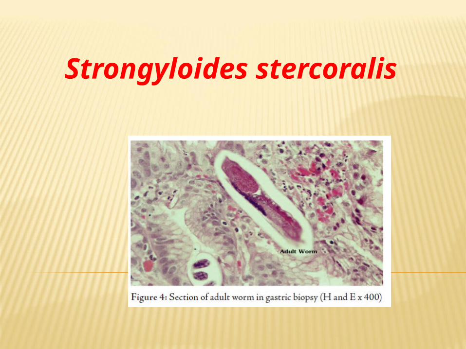
Strongyloides stercoralis. Habitat: females live in the superficial tissues of the small intestine...
30
Strongyloides stercoralis
-
Upload
cristina-hache -
Category
Documents
-
view
222 -
download
5
Transcript of Strongyloides stercoralis. Habitat: females live in the superficial tissues of the small intestine...
Strongyloides stercoralis
Strongyloides stercoralis
Habitat: females live in the superficial tissues of the small intestine (duodenum and jejunum)
Definitive host: Human, dogs and cats Route of infection: Filariform larvae penetrate the skin of human. Infective stage: Third stage larvae ( filariform). Diagnostic stage: First stage larvae(Rhabditiform) in feces.Geographical distribution: - cosmopolitan parasite, mainly in moist
and warm areas of low hygiene
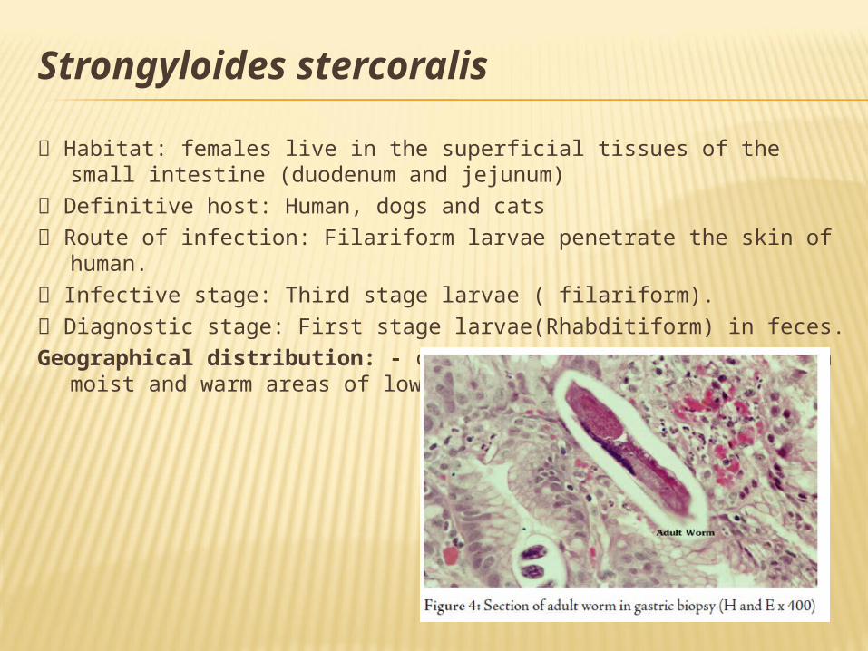
Morphology
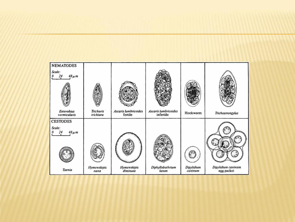
Egg:Size : 55 x 30 um. Shape: oval . Clear, thin shelled Similar to hookworm but are smaller .
Eggs are laid in the mucosa, hatch into rhabditiform larvae that penetrate the glandular epithelium and pass into the lumen of the intestine and out the feces
(Eggs are seldom seen in stools).
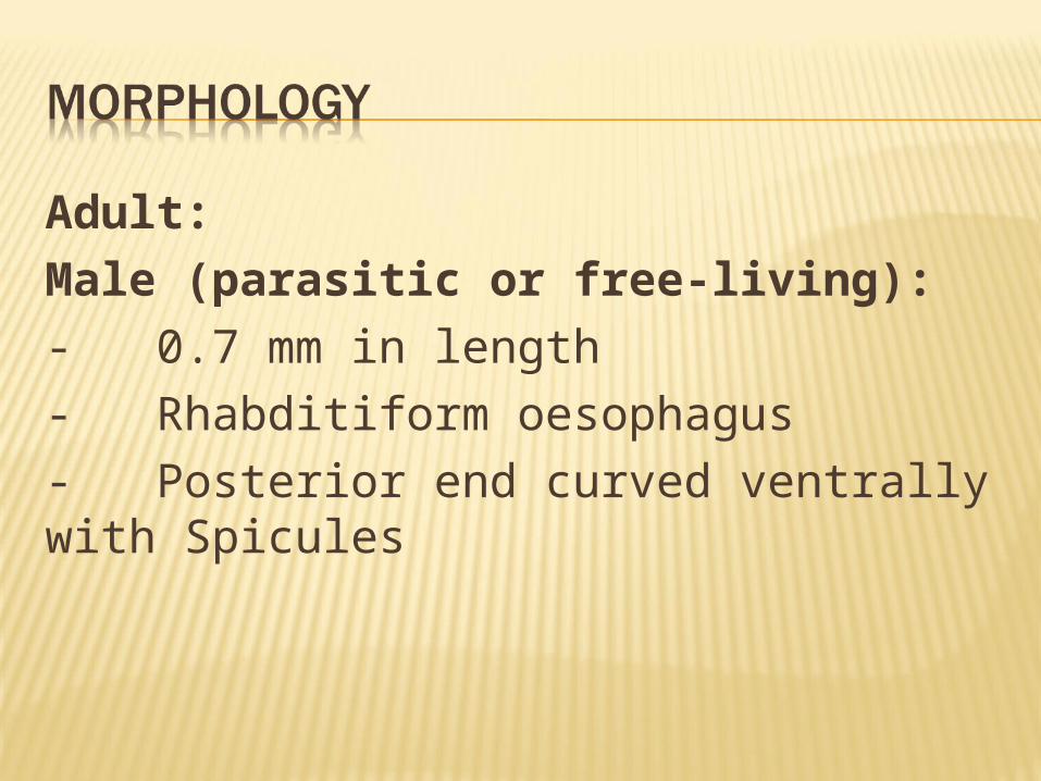
Adult:Male (parasitic or free-living):- 0.7 mm in length- Rhabditiform oesophagus- Posterior end curved ventrally with Spicules
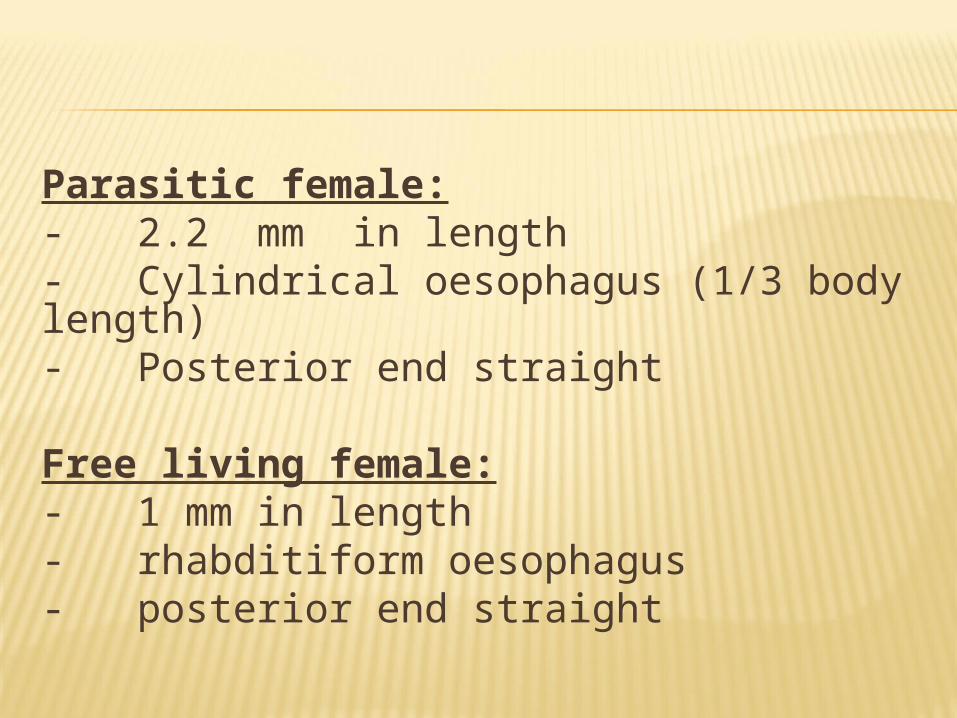
Parasitic female:- 2.2 mm in length- Cylindrical oesophagus (1/3 body length)- Posterior end straight
Free living female:- 1 mm in length- rhabditiform oesophagus- posterior end straight
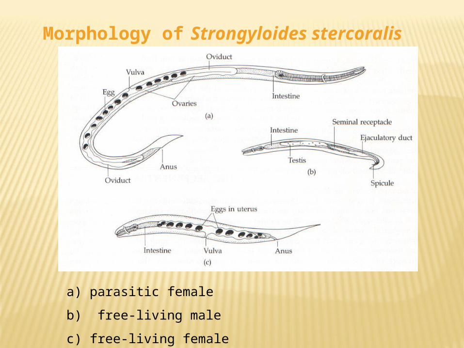
Morphology of Strongyloides stercoralis
a) parasitic female
b) free-living male
c) free-living female
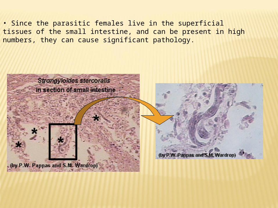
• Since the parasitic females live in the superficial tissues of the small intestine, and can be present in high numbers, they can cause significant pathology.
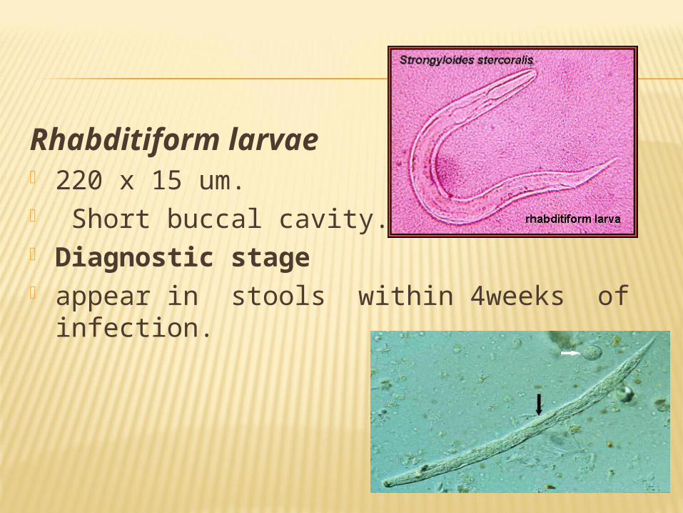
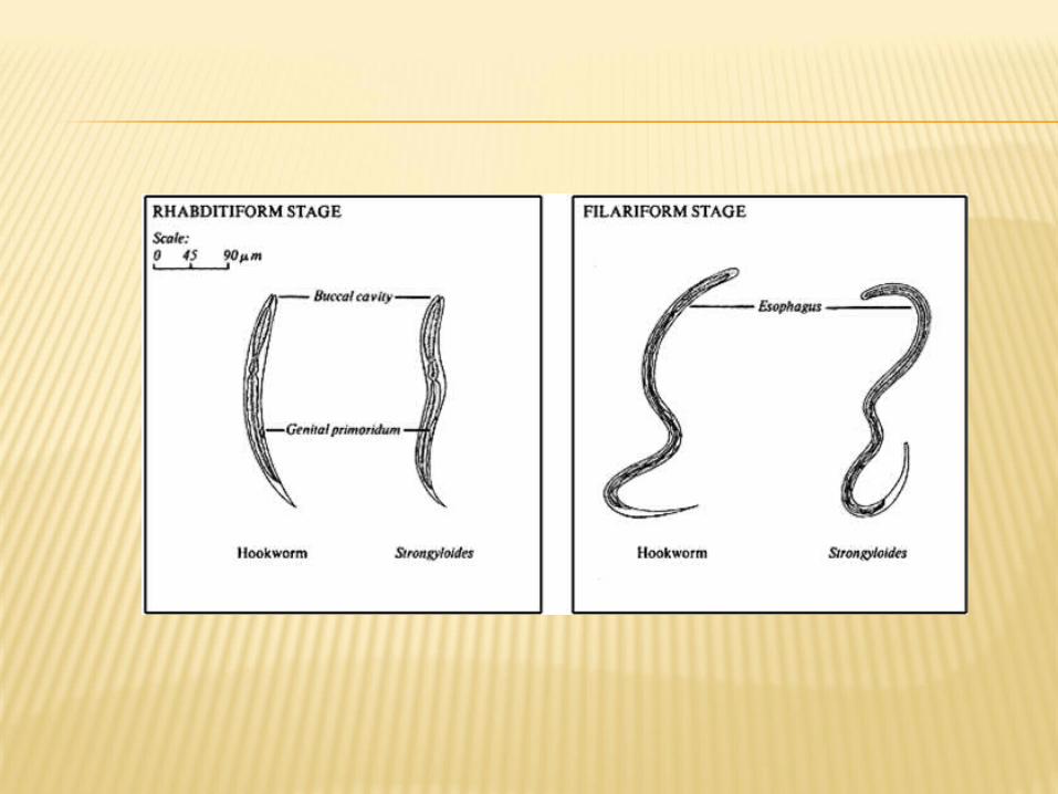
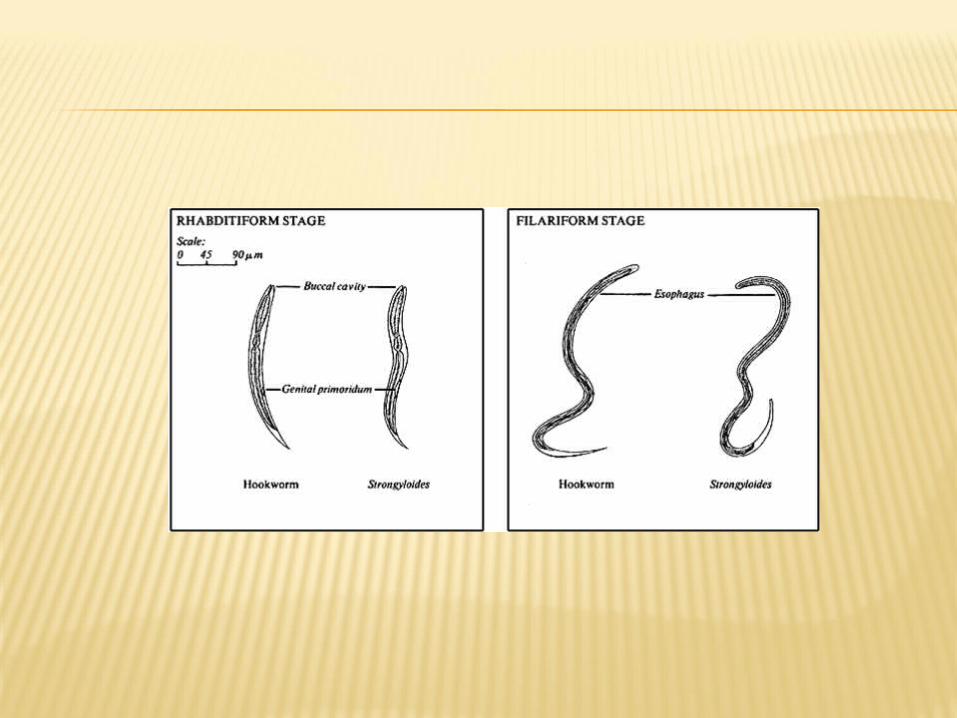
Rhabditiform larvae 220 x 15 um. Short buccal cavity. Diagnostic stage appear in stools within 4weeks of
infection.
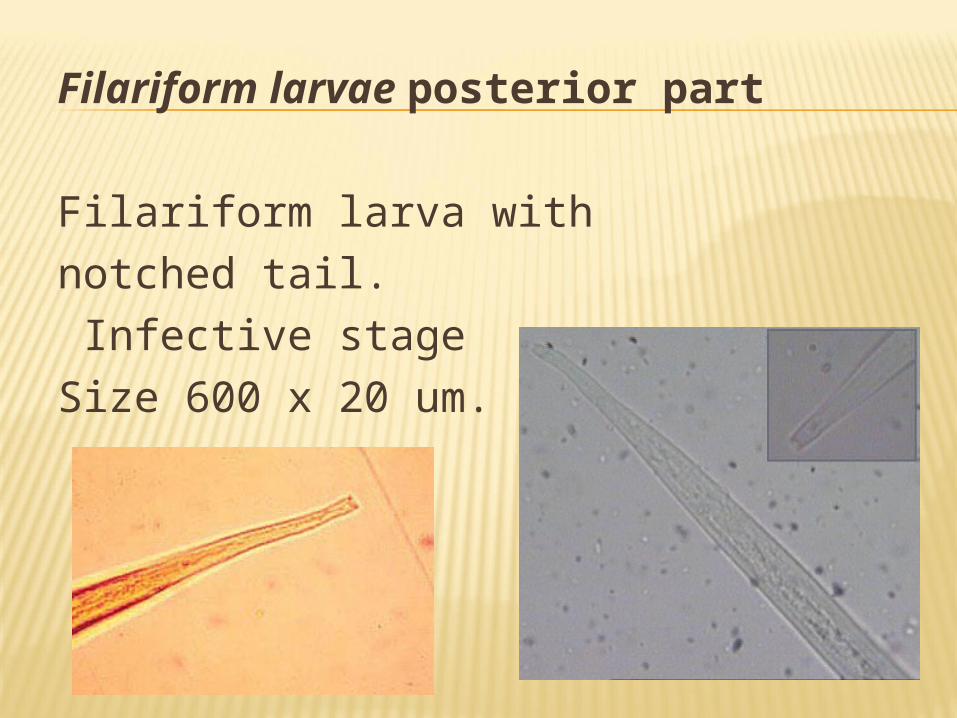
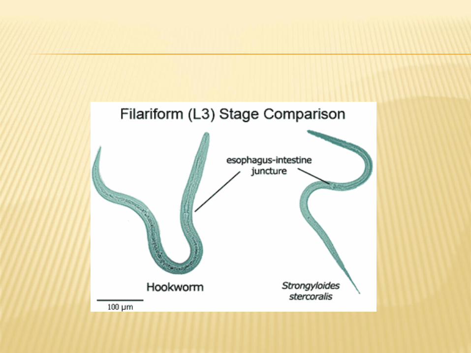
Filariform larvae posterior part
Filariform larva withnotched tail. Infective stageSize 600 x 20 um.
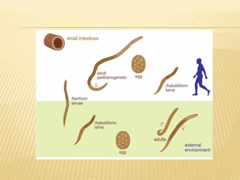
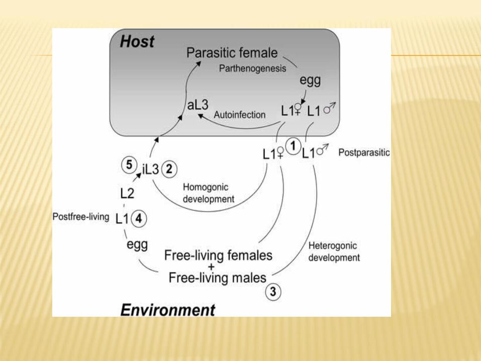
Life Cycle:Free-living cycle Parasitic cycle: In the parasitic stage,
no male form of this organism has been reliably identified, and the female reproduce in a parthinogenitic manner.
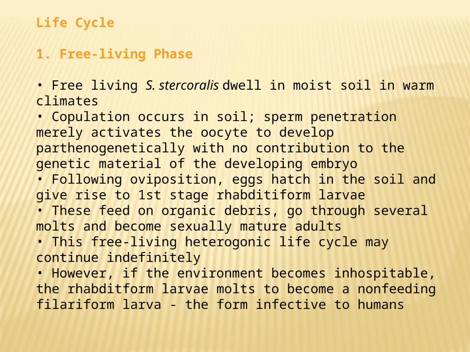
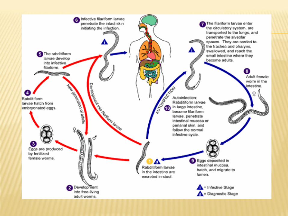
Life Cycle
1. Free-living Phase
• Free living S. stercoralis dwell in moist soil in warm climates• Copulation occurs in soil; sperm penetration merely activates the oocyte to develop parthenogenetically with no contribution to the genetic material of the developing embryo• Following oviposition, eggs hatch in the soil and give rise to 1st stage rhabditiform larvae• These feed on organic debris, go through several molts and become sexually mature adults• This free-living heterogonic life cycle may continue indefinitely• However, if the environment becomes inhospitable, the rhabditform larvae molts to become a nonfeeding filariform larva - the form infective to humans
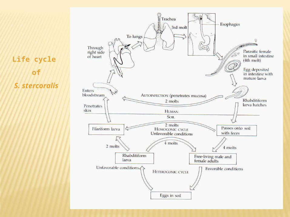
Life cycle
of
S. stercoralis
Life Cycle cont.
2. Parasitic Phase
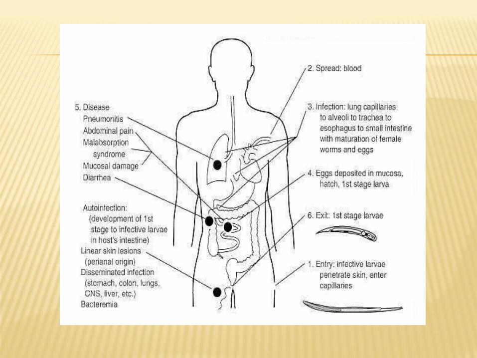
• When filariform larvae encounter a human or another suitable host (e.g. cats and dogs), they penetrate the skin and are carried by cutaneous veins to the vena cava
• They enter the right side of the heart and are carried to the lungs via the pulmonary artery
• In the lungs, following a 3rd molt, the larvae rupture from the pulmonary capillaries and enter the alveoli• From the alveoli, the larvae move up the respiratory tree to the epiglottis• Abetted by coughing and subsequent swallowing by the host, they migrate over the epiglottis to the esophagus and down into the small intestine, where they undergo a final molt and become sexually mature females
Parasitic Phase cont.
• Females produce embryonated eggs parhenogenetically• These eggs hatch in the mucosa into 1st stage rhabditiform larvae• These exit the intestine with the feces, feeding down the length of the intestine• Larvae become established in the soil, undergo several molts and become free-living adults• Under adverse conditions they can revert to being filariform larvae
3. Autoinfection
• During passage through the host digestive system, rhabditiform larvae may undergo 2 molts to filariform larvae and by penetrating the intestinal mucosa, enter the circulatory system and continue their parasitic lives without leaving the host•Autoinfection can also occur when larvae remain on and penetrate the perianal skin.• Autoinfection often leads to very high worm burdens in humans
Clinical Aspects:
•Cutaneous reaction due to skin penetration “ ground itch•Pulmonary symptoms (including Loeffler’s syndrome) can occur
during pulmonary migration of the filariform larvae.•Gastrointestinal symptoms include abdominal pain, vomiting,
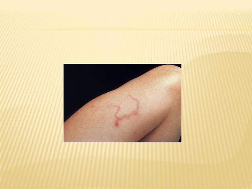
diarrhea, weight loss, malabsorption,.•Dermatologic manifestations include urticarial rashes in the
buttocks and waist areas (larva migrans).
•Autoinfection within the human host can lead to the (Hyperinfection Syndrome) or disseminated strongyloidiasis occurs in
immunosuppressed patients, neurologic(CNS), heart, lung , liver complications and septicemia. This syndrome is potentially fatal.
Laboratory Diagnosis
Direct stool smears (larvae) Cultivation of stool. (Damp charcoal or Harada-Mori
mediums).
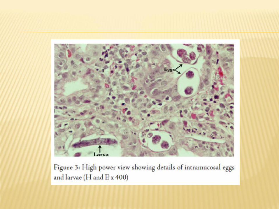
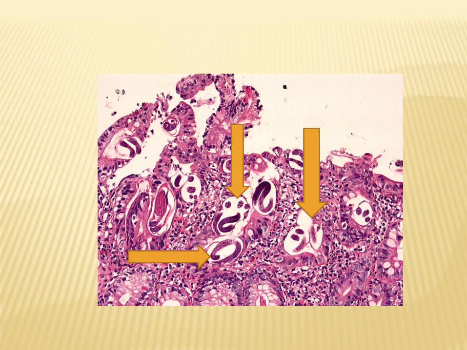
Histological examination of duodenal or jejunal biopsy specimens obtained by endoscopy can demonstrate adult worms embedded in the mucosa.
Eosinophilia, is present in uncomplicated strongyloidiasis, but is lost in hyper infection
For population screening in endemic areas, an ELISA for IgG anfi-Strongyloides antibodies is effective.

nausea, cramping, abdominal pain, distension, and watery diarrhea.