Stroke Mimics Bruce Campbell - ED Central
48
Stroke management Dr Bruce Campbell Neurologist Royal Melbourne Hospital
Transcript of Stroke Mimics Bruce Campbell - ED Central
Stroke management
Dr Bruce Campbell
Neurologist
Royal Melbourne Hospital

• Focused neurological exam –what are the key neuro exam findings that distinguish stroke from other neuro events
• Blood pressure management, glycaemic control management, temperature control and swallow assessment
• Preventing secondary complications and the importance of stroke unit care

Recognition of stroke and mimics
Stroke and important related diagnoses:
• Patients with focal neurology, altered conscious state, headache
• Differential diagnoses: – Stroke (infarct/intracerebral hemorrhage)
– Sub-arachnoid hemorrhage
– Seizure with Todd’s paresis
– Migraine
– Meningitis/encephalitis
– Metabolic eg sepsis, hypoglycaemia
– Bell’s palsy
– Vestibular neuronitis
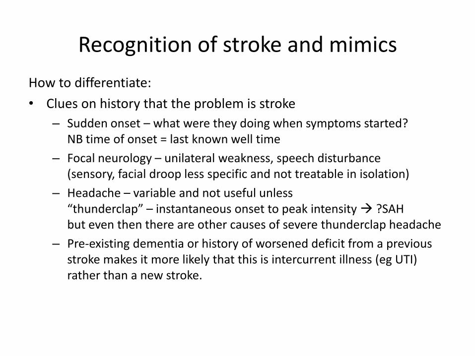
Recognition of stroke and mimics
How to differentiate:
• Clues on history that the problem is stroke
– Sudden onset – what were they doing when symptoms started? NB time of onset = last known well time
– Focal neurology – unilateral weakness, speech disturbance (sensory, facial droop less specific and not treatable in isolation)
– Headache – variable and not useful unless “thunderclap” – instantaneous onset to peak intensity ?SAH but even then there are other causes of severe thunderclap headache
– Pre-existing dementia or history of worsened deficit from a previous stroke makes it more likely that this is intercurrent illness (eg UTI) rather than a new stroke.
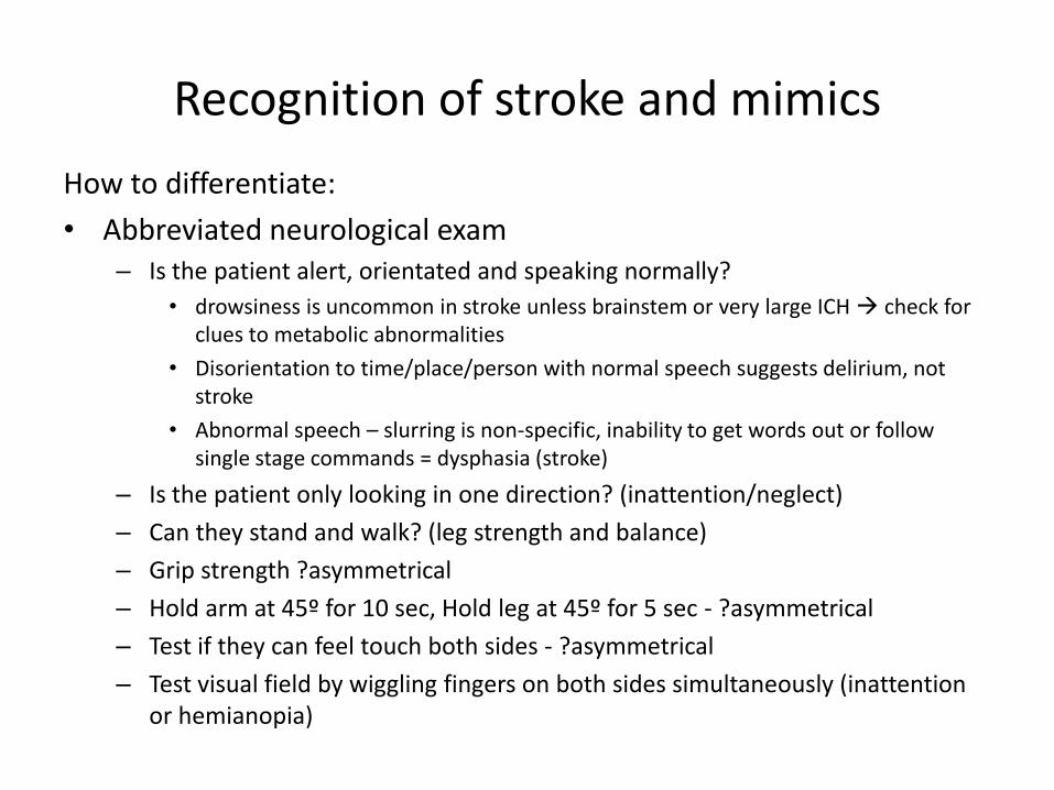
Recognition of stroke and mimics
How to differentiate:
• Abbreviated neurological exam – Is the patient alert, orientated and speaking normally?
• drowsiness is uncommon in stroke unless brainstem or very large ICH check for clues to metabolic abnormalities
• Disorientation to time/place/person with normal speech suggests delirium, not stroke
• Abnormal speech – slurring is non-specific, inability to get words out or follow single stage commands = dysphasia (stroke)
– Is the patient only looking in one direction? (inattention/neglect)
– Can they stand and walk? (leg strength and balance)
– Grip strength ?asymmetrical
– Hold arm at 45º for 10 sec, Hold leg at 45º for 5 sec - ?asymmetrical
– Test if they can feel touch both sides - ?asymmetrical
– Test visual field by wiggling fingers on both sides simultaneously (inattention or hemianopia)
Recognition of stroke and mimics
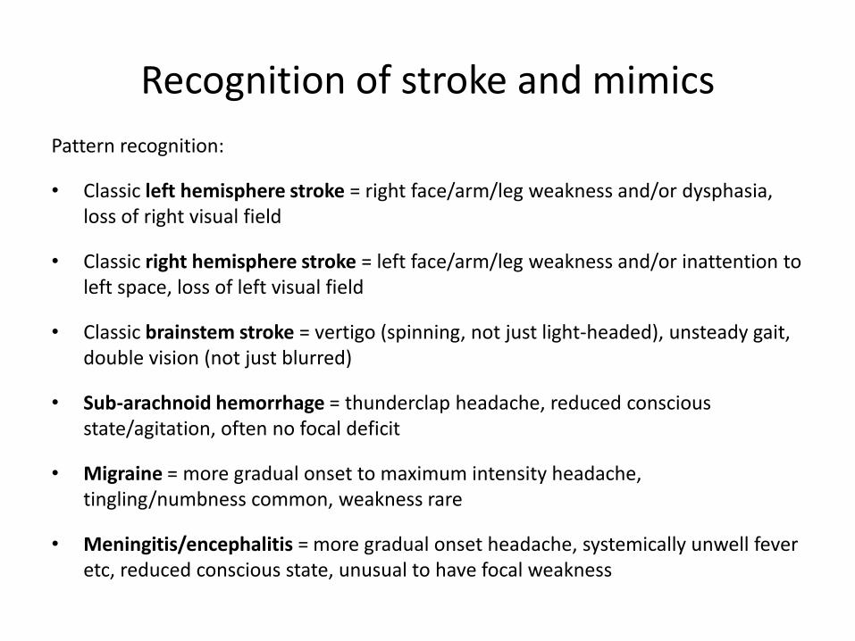
Pattern recognition:
• Classic left hemisphere stroke = right face/arm/leg weakness and/or dysphasia, loss of right visual field
• Classic right hemisphere stroke = left face/arm/leg weakness and/or inattention to left space, loss of left visual field
• Classic brainstem stroke = vertigo (spinning, not just light-headed), unsteady gait, double vision (not just blurred)
• Sub-arachnoid hemorrhage = thunderclap headache, reduced conscious state/agitation, often no focal deficit
• Migraine = more gradual onset to maximum intensity headache, tingling/numbness common, weakness rare
• Meningitis/encephalitis = more gradual onset headache, systemically unwell fever etc, reduced conscious state, unusual to have focal weakness
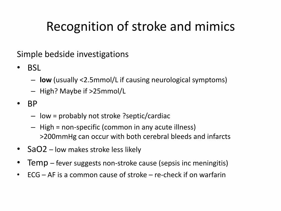
Recognition of stroke and mimics
Simple bedside investigations
• BSL – low (usually <2.5mmol/L if causing neurological symptoms)
– High? Maybe if >25mmol/L
• BP – low = probably not stroke ?septic/cardiac
– High = non-specific (common in any acute illness) >200mmHg can occur with both cerebral bleeds and infarcts
• SaO2 – low makes stroke less likely
• Temp – fever suggests non-stroke cause (sepsis inc meningitis)
• ECG – AF is a common cause of stroke – re-check if on warfarin
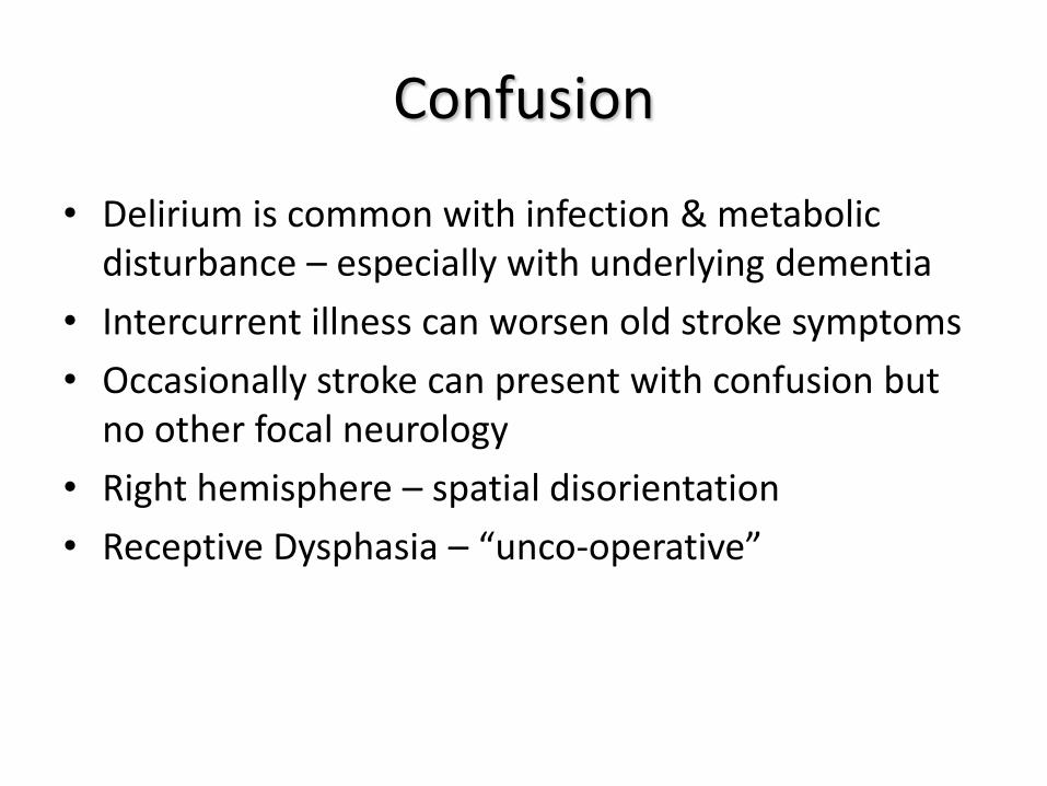
Confusion
• Delirium is common with infection & metabolic disturbance – especially with underlying dementia
• Intercurrent illness can worsen old stroke symptoms
• Occasionally stroke can present with confusion but no other focal neurology
• Right hemisphere – spatial disorientation
• Receptive Dysphasia – “unco-operative”
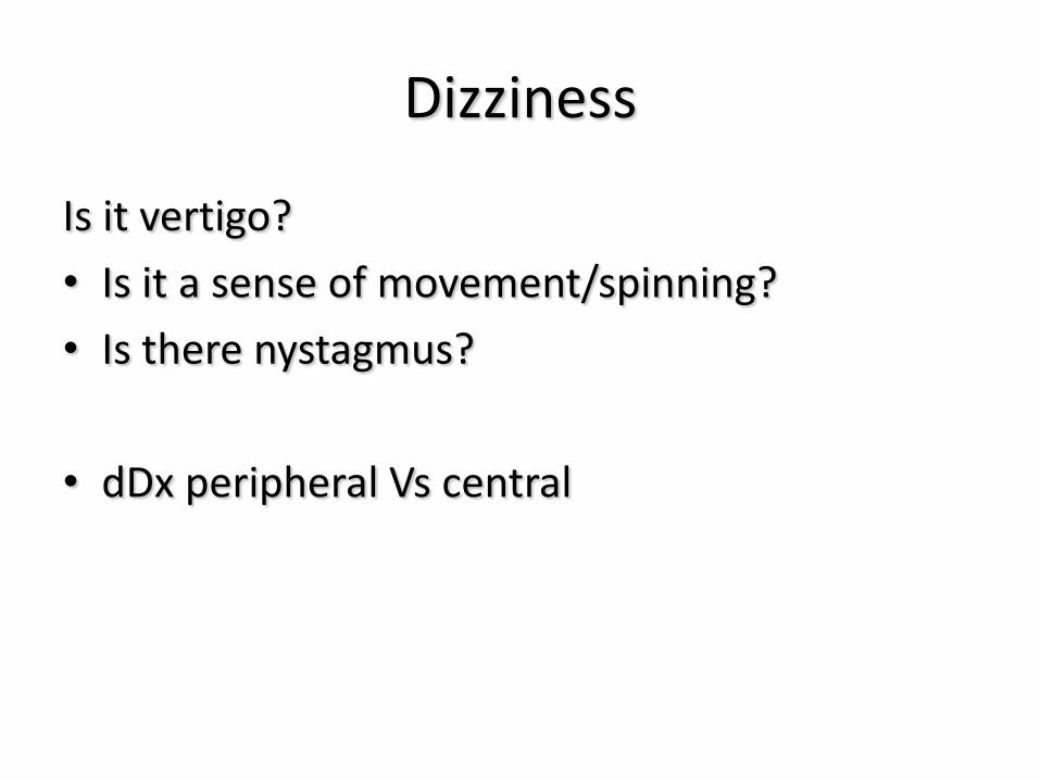
Dizziness
Is it vertigo?
• Is it a sense of movement/spinning?
• Is there nystagmus?
• dDx peripheral Vs central
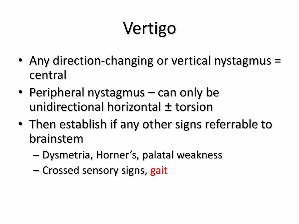
Vertigo
• Any direction-changing or vertical nystagmus = central
• Peripheral nystagmus – can only be unidirectional horizontal ± torsion
• Then establish if any other signs referrable to brainstem – Dysmetria, Horner’s, palatal weakness
– Crossed sensory signs, gait
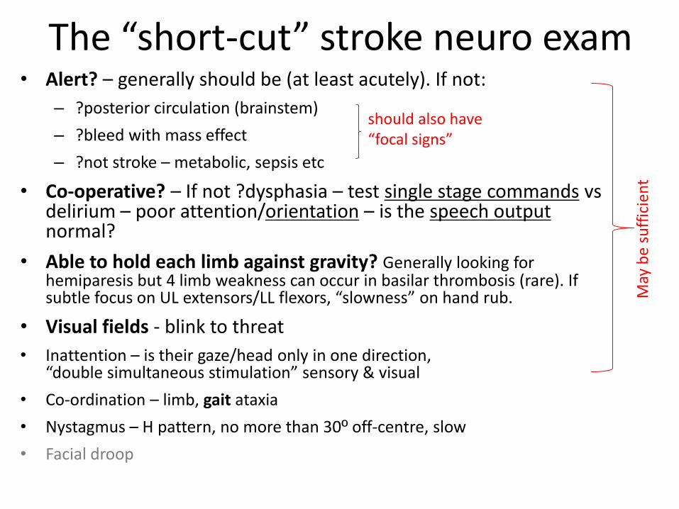
The “short-cut” stroke neuro exam • Alert? – generally should be (at least acutely). If not:
– ?posterior circulation (brainstem)
– ?bleed with mass effect
– ?not stroke – metabolic, sepsis etc
• Co-operative? – If not ?dysphasia – test single stage commands vs delirium – poor attention/orientation – is the speech output normal?
• Able to hold each limb against gravity? Generally looking for hemiparesis but 4 limb weakness can occur in basilar thrombosis (rare). If subtle focus on UL extensors/LL flexors, “slowness” on hand rub.
• Visual fields - blink to threat
• Inattention – is their gaze/head only in one direction, “double simultaneous stimulation” sensory & visual
• Co-ordination – limb, gait ataxia
• Nystagmus – H pattern, no more than 30º off-centre, slow
• Facial droop
May
be
suff
icie
nt
should also have “focal signs”
Interactive Cases
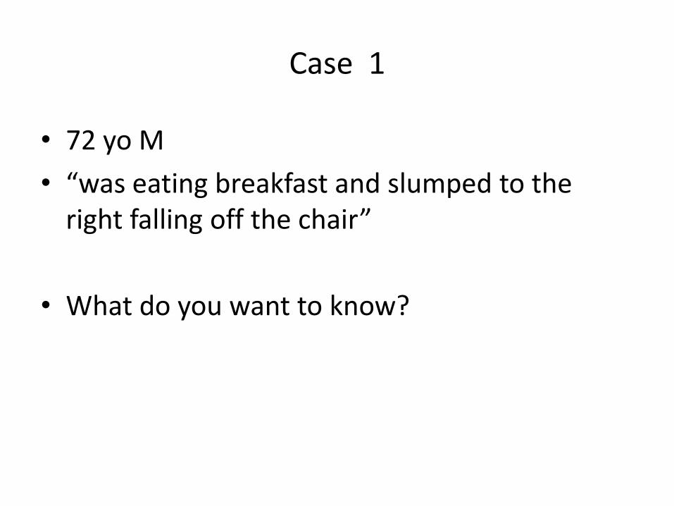
Case 1
• 72 yo M
• “was eating breakfast and slumped to the right falling off the chair”
• What do you want to know?
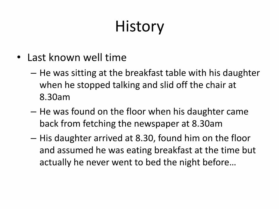
History
• Last known well time
– He was sitting at the breakfast table with his daughter when he stopped talking and slid off the chair at 8.30am
– He was found on the floor when his daughter came back from fetching the newspaper at 8.30am
– His daughter arrived at 8.30, found him on the floor and assumed he was eating breakfast at the time but actually he never went to bed the night before…
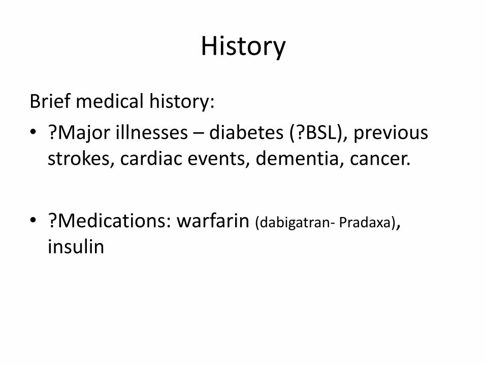
History
Brief medical history:
• ?Major illnesses – diabetes (?BSL), previous strokes, cardiac events, dementia, cancer.
• ?Medications: warfarin (dabigatran- Pradaxa), insulin
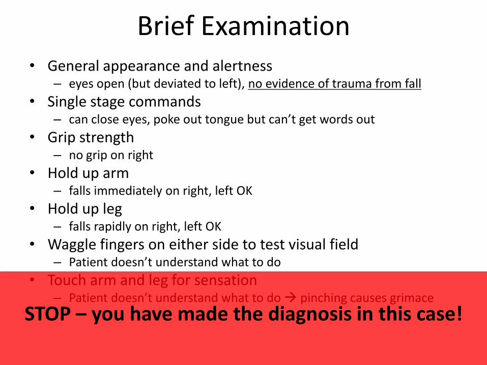
Brief Examination • General appearance and alertness
– eyes open (but deviated to left), no evidence of trauma from fall
• Single stage commands – can close eyes, poke out tongue but can’t get words out
• Grip strength – no grip on right
• Hold up arm – falls immediately on right, left OK
• Hold up leg – falls rapidly on right, left OK
• Waggle fingers on either side to test visual field – Patient doesn’t understand what to do
• Touch arm and leg for sensation – Patient doesn’t understand what to do pinching causes grimace
STOP – you have made the diagnosis in this case!
ACTION
• What are you going to do now?
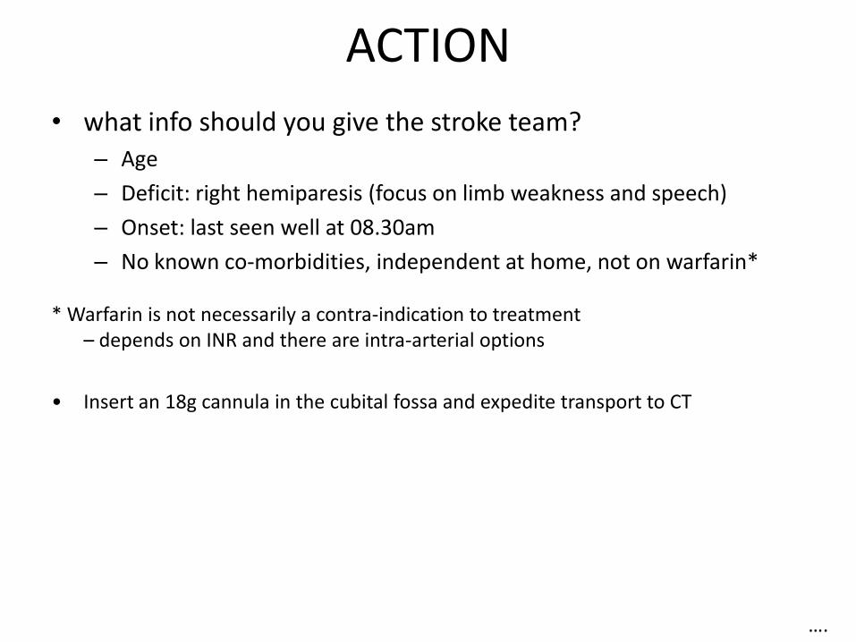
ACTION
• what info should you give the stroke team? – Age
– Deficit: right hemiparesis (focus on limb weakness and speech)
– Onset: last seen well at 08.30am
– No known co-morbidities, independent at home, not on warfarin*
* Warfarin is not necessarily a contra-indication to treatment – depends on INR and there are intra-arterial options
• Insert an 18g cannula in the cubital fossa and expedite transport to CT
….

Case 2:
• 84 yo F from a nursing home
• Staff report that she developed a facial droop this morning
• She has type 2 diabetes, ischemic heart disease, a previous stroke (details uncertain)
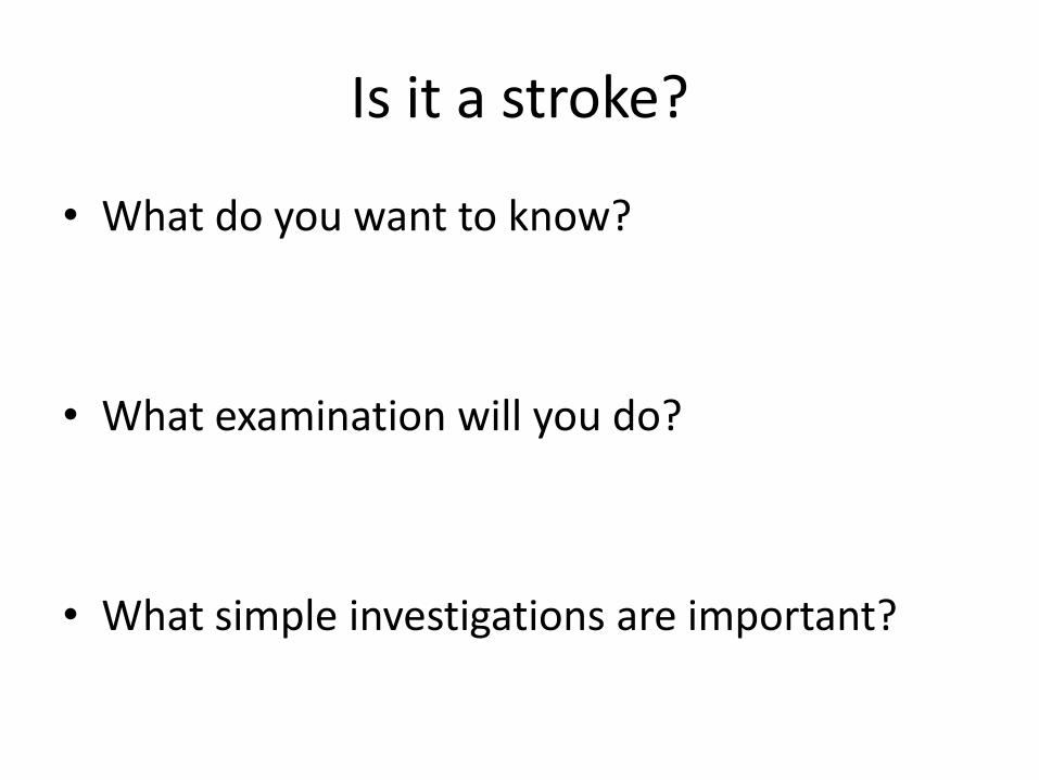
Is it a stroke?
• What do you want to know?
• What examination will you do?
• What simple investigations are important?
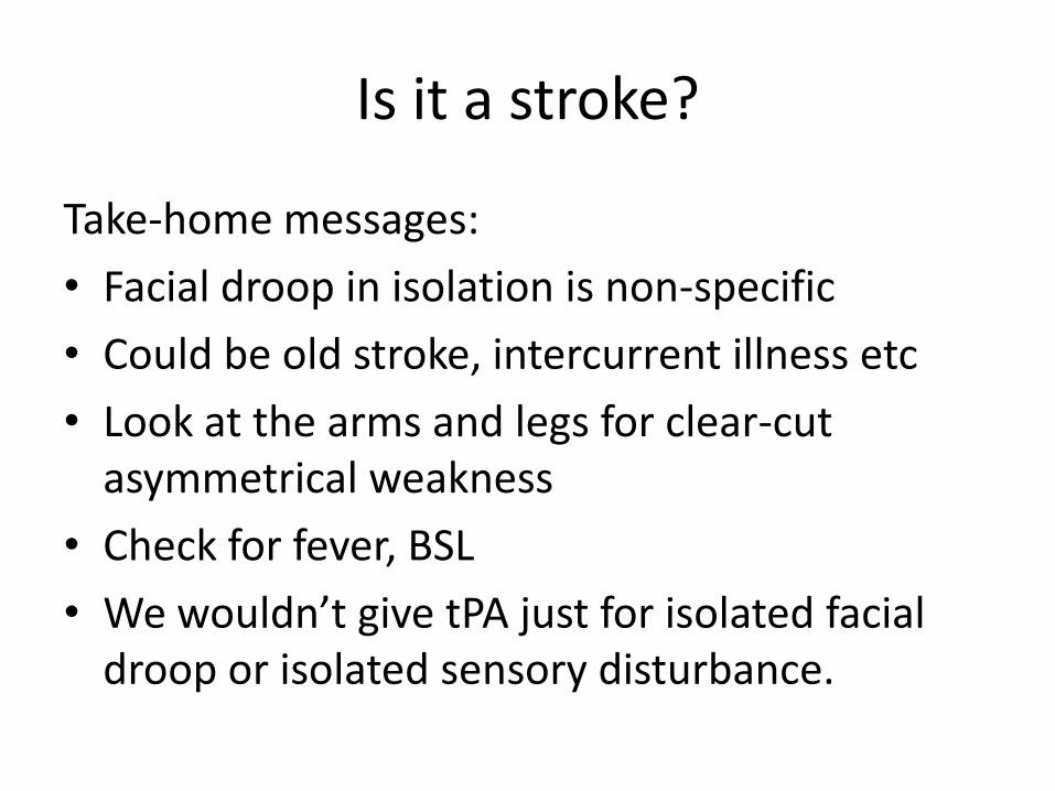
Is it a stroke?
Take-home messages:
• Facial droop in isolation is non-specific
• Could be old stroke, intercurrent illness etc
• Look at the arms and legs for clear-cut asymmetrical weakness
• Check for fever, BSL
• We wouldn’t give tPA just for isolated facial droop or isolated sensory disturbance.

Would we treat her?
• Significant deficit within 4.5 hours - maybe
• Quality of life judgment – mobility and communication
• >4.5 hours – only trials available
• Exclude premorbid disability (the trial outcome is usually “minimal disability” and sadly we rarely get people better than baseline
….
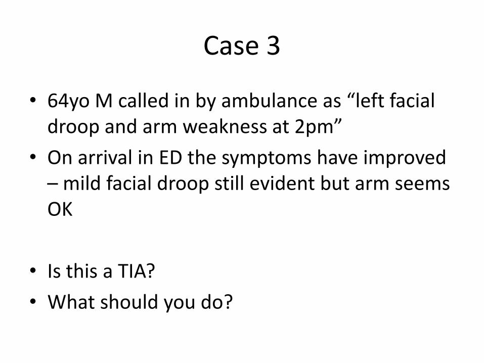
Case 3
• 64yo M called in by ambulance as “left facial droop and arm weakness at 2pm”
• On arrival in ED the symptoms have improved – mild facial droop still evident but arm seems OK
• Is this a TIA?
• What should you do?

Case 3
Take-home messages:
• TIA = completely resolved
(average duration ~10minutes)
• If they still have symptoms it’s probably stroke urgent transport
• If they have completely resolved they still need same-day assessment, investigation and treatment at a stroke centre (high risk of stroke in next couple of days).

Case 4
• A 56 year old man collapses at work. On arrival in ED he is GCS 9. Workmates say he had complained of double vision about 10 minutes before losing consciousness. He also had to leave work early after an episode of vertigo 2 days ago.
• What is going on here?
Case 4
• Basilar thrombosis:
• Often have preceding “brainstem symptoms”
– Vertigo/vomiting, ataxia, double vision, slurred speech
• Do you need to intubate?
….
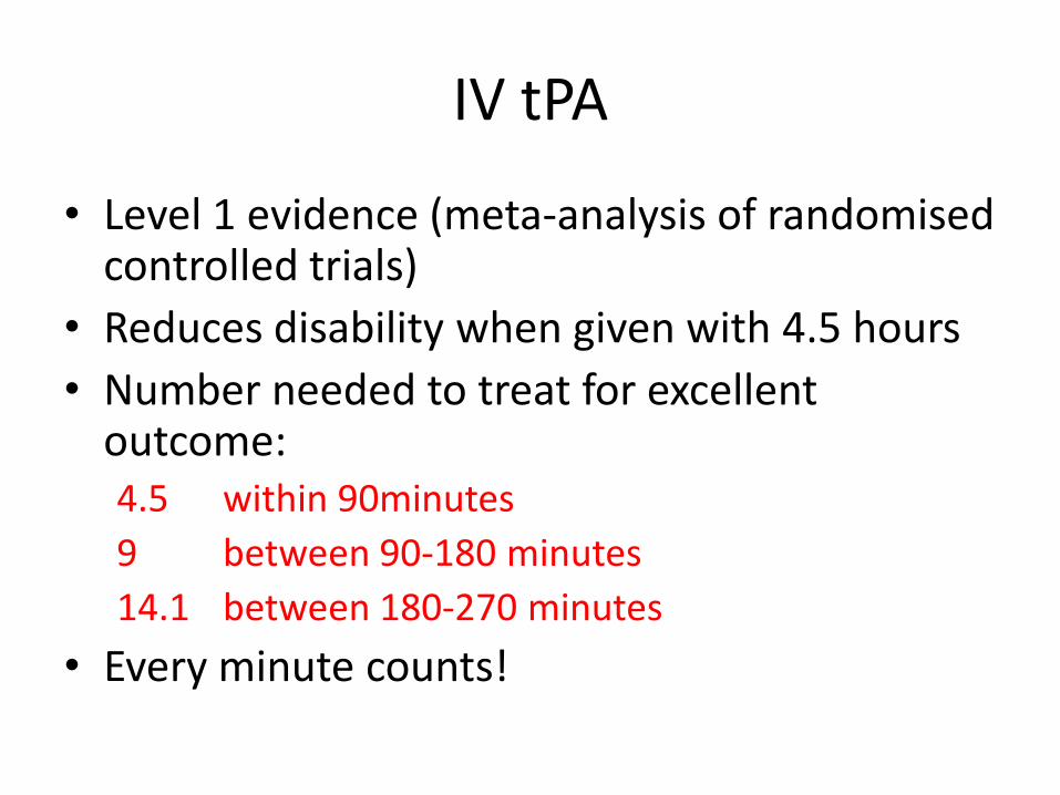
IV tPA
• Level 1 evidence (meta-analysis of randomised controlled trials)
• Reduces disability when given with 4.5 hours
• Number needed to treat for excellent outcome: 4.5 within 90minutes
9 between 90-180 minutes
14.1 between 180-270 minutes
• Every minute counts!
Stroke CT crash course
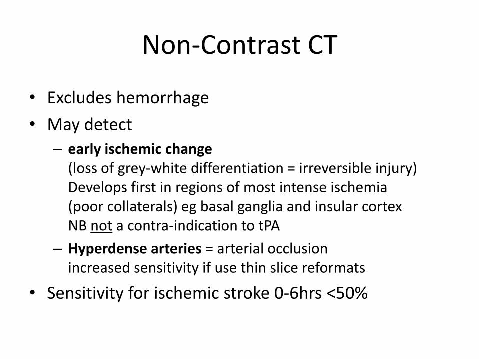
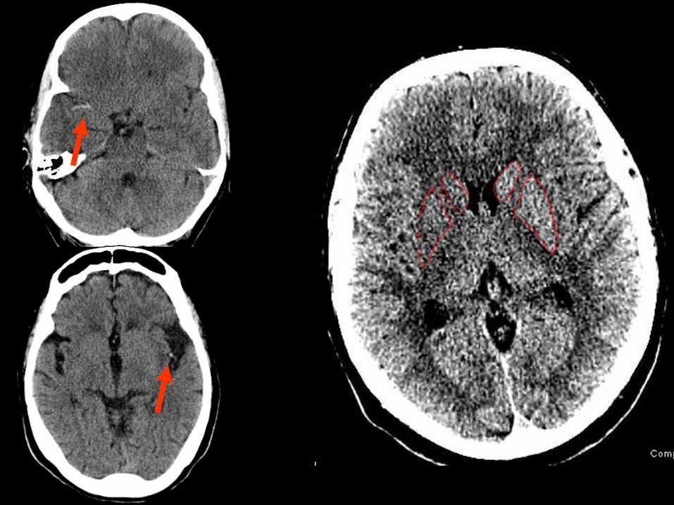
Non-Contrast CT
• Excludes hemorrhage
• May detect
– early ischemic change (loss of grey-white differentiation = irreversible injury) Develops first in regions of most intense ischemia (poor collaterals) eg basal ganglia and insular cortex NB not a contra-indication to tPA
– Hyperdense arteries = arterial occlusion increased sensitivity if use thin slice reformats
• Sensitivity for ischemic stroke 0-6hrs <50%
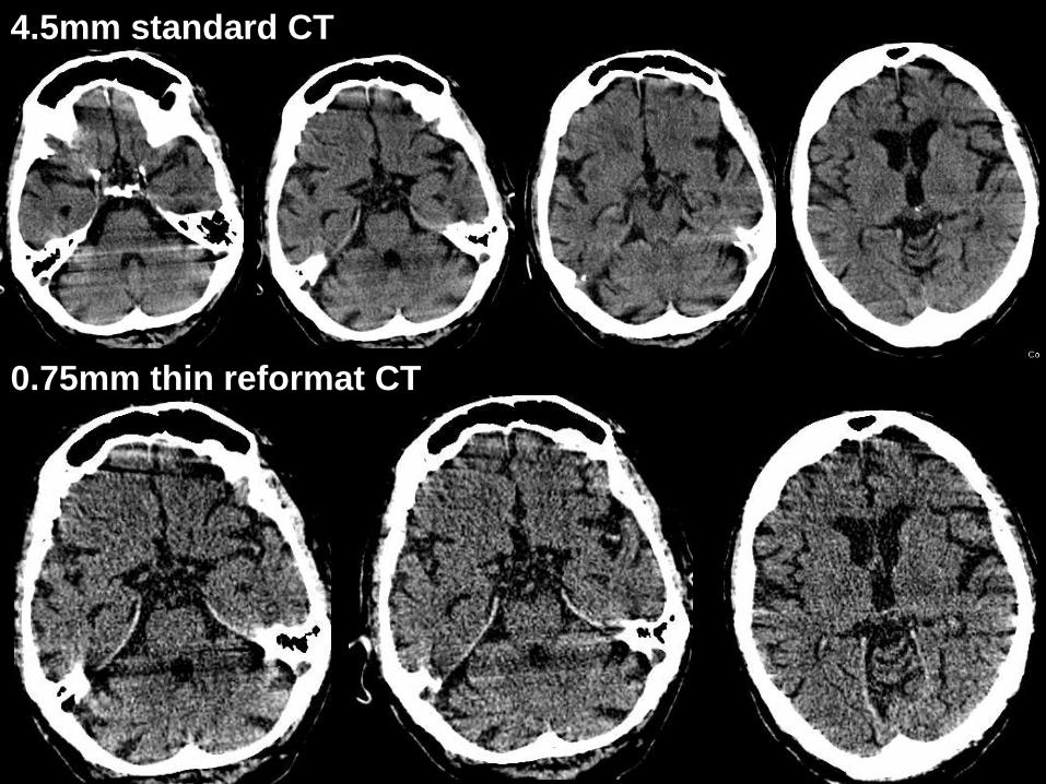
4.5mm standard CT
0.75mm thin reformat CT

CT Brain
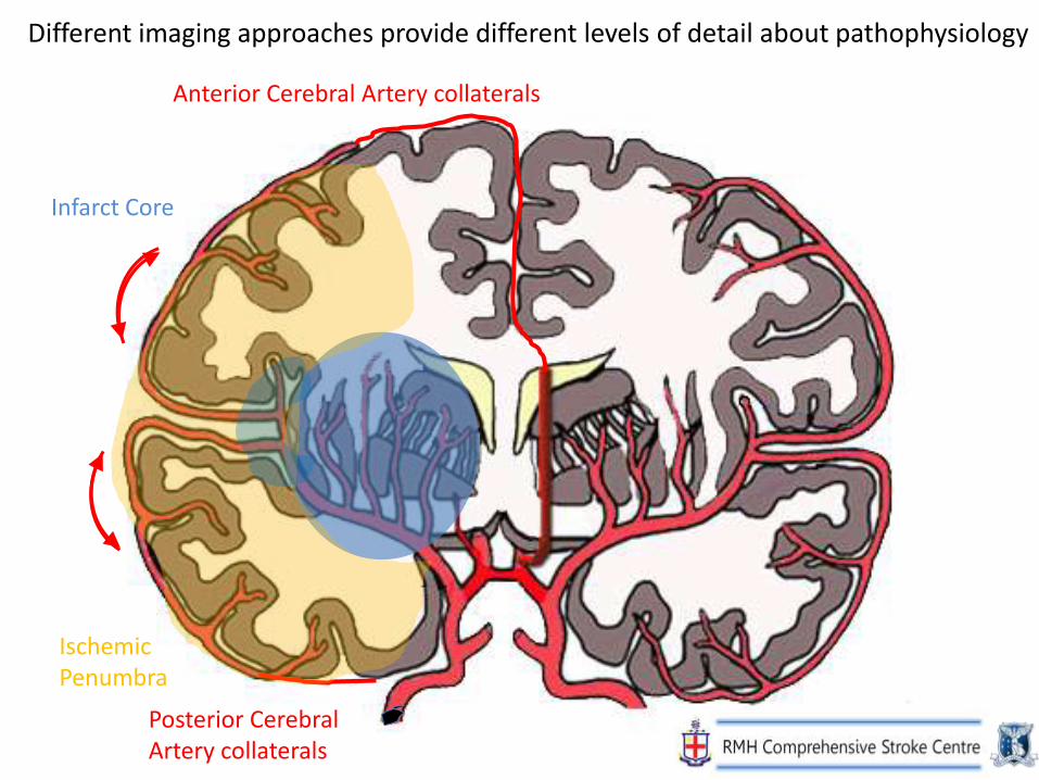
Anterior Cerebral Artery collaterals
Posterior Cerebral Artery collaterals
Infarct Core
Ischemic Penumbra
Different imaging approaches provide different levels of detail about pathophysiology
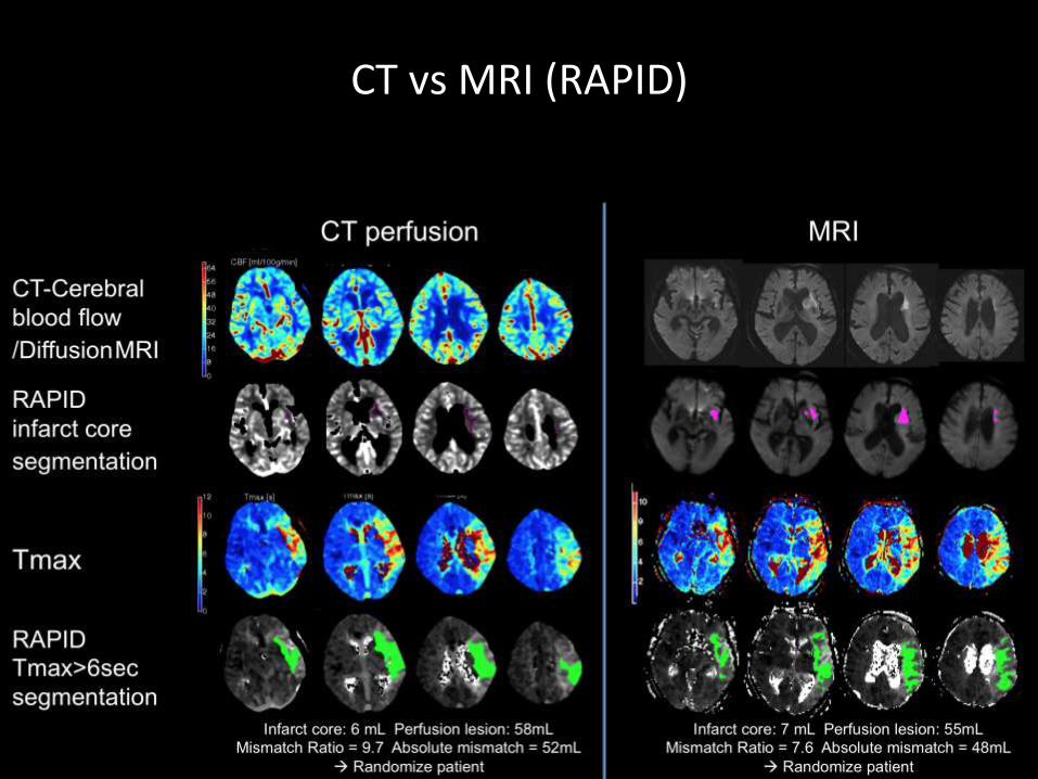
CT vs MRI (RAPID)
Thrombolysis
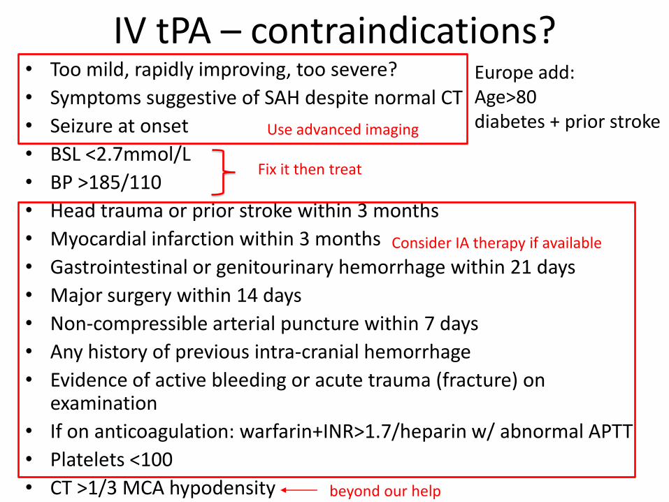
IV tPA – contraindications? • Too mild, rapidly improving, too severe?
• Symptoms suggestive of SAH despite normal CT
• Seizure at onset
• BSL <2.7mmol/L
• BP >185/110
• Head trauma or prior stroke within 3 months
• Myocardial infarction within 3 months
• Gastrointestinal or genitourinary hemorrhage within 21 days
• Major surgery within 14 days
• Non-compressible arterial puncture within 7 days
• Any history of previous intra-cranial hemorrhage
• Evidence of active bleeding or acute trauma (fracture) on examination
• If on anticoagulation: warfarin+INR>1.7/heparin w/ abnormal APTT
• Platelets <100
• CT >1/3 MCA hypodensity
Europe add: Age>80 diabetes + prior stroke Use advanced imaging
Consider IA therapy if available
Fix it then treat
beyond our help
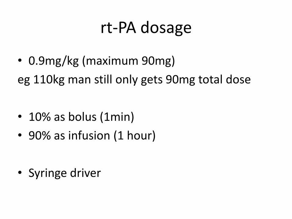
rt-PA dosage
• 0.9mg/kg (maximum 90mg)
eg 110kg man still only gets 90mg total dose
• 10% as bolus (1min)
• 90% as infusion (1 hour)
• Syringe driver
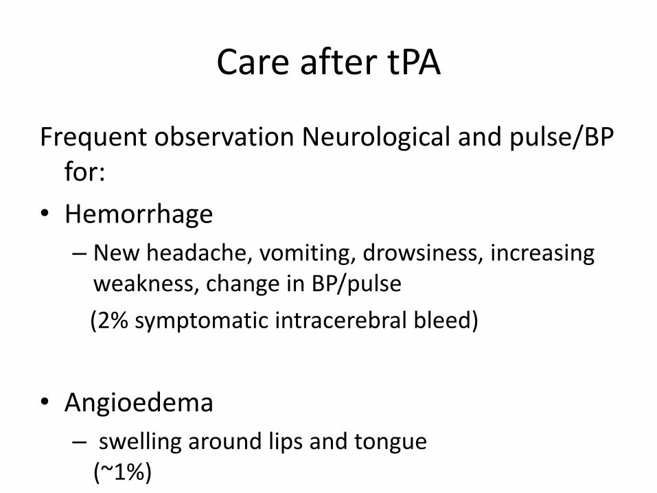
Care after tPA
Frequent observation Neurological and pulse/BP for:
• Hemorrhage
– New headache, vomiting, drowsiness, increasing weakness, change in BP/pulse
(2% symptomatic intracerebral bleed)
• Angioedema
– swelling around lips and tongue (~1%)

Care after tPA
Blood pressure
• Target <185/105mmHg (vs <220/120 if no tPA)
• High BP is associated with poor outcome and increased hemorrhage risk after tPA
• But we do not want to lower BP a lot!
• Dependent on “collateral” blood supply to keep tissue alive whilst we open the artery, excessive BP lowering may compromise this.
• Hydralazine 2.5-5mg IV, Labetolol 10mg IV ….
History of stroke care units
• Organised care for patients with ischemic chest pain – the CCU – was widely implemented in the late 60s & early 70s
• First Australian Stroke Unit was opened in 1978 at the Austin
• By the 1980s, there were many small trials of organised care with inconclusive results
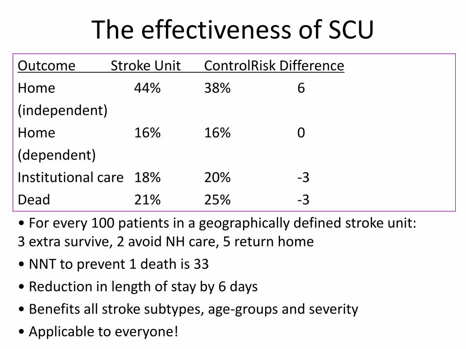
The effectiveness of SCU Outcome Stroke Unit ControlRisk Difference
Home 44% 38% 6
(independent)
Home 16% 16% 0
(dependent)
Institutional care 18% 20% -3
Dead 21% 25% -3
• For every 100 patients in a geographically defined stroke unit: 3 extra survive, 2 avoid NH care, 5 return home
• NNT to prevent 1 death is 33
• Reduction in length of stay by 6 days
• Benefits all stroke subtypes, age-groups and severity
• Applicable to everyone!
Assessment and Minimisation of Complications
• Assessment of aetiology and secondary prevention
• Glycaemic control (treat hyperglycaemia eg >11 but not aggressive euglycaemia)
• Aspiration pneumonia risk reduction (swallow assessment, clean mouth)
• Thromboembolism (DVT/PE) prophylaxis = LMW heparin (no TEDS)
• Neurological & vital sign monitoring
• Fever management (treat cause, paracetamol)
• Dehydration prevention
• Hypoxia prevention
• Pressure risk management
• Shoulder injury prevention
• Continence assessment
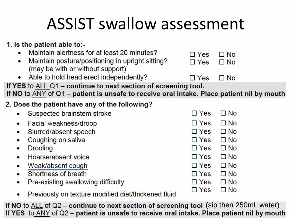
ASSIST swallow assessment
(sip then 250mL water)
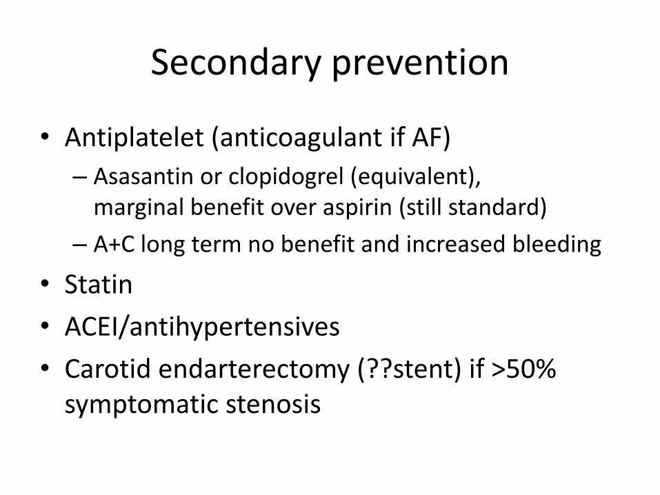
Secondary prevention
• Antiplatelet (anticoagulant if AF)
– Asasantin or clopidogrel (equivalent), marginal benefit over aspirin (still standard)
– A+C long term no benefit and increased bleeding
• Statin
• ACEI/antihypertensives
• Carotid endarterectomy (??stent) if >50% symptomatic stenosis
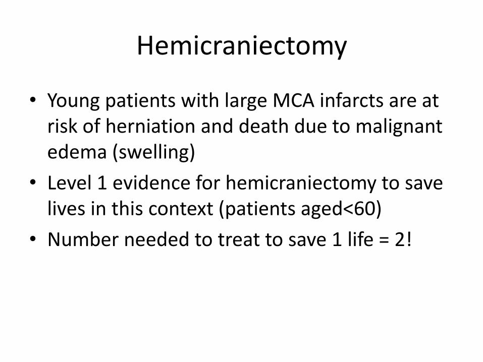
Hemicraniectomy
• Young patients with large MCA infarcts are at risk of herniation and death due to malignant edema (swelling)
• Level 1 evidence for hemicraniectomy to save lives in this context (patients aged<60)
• Number needed to treat to save 1 life = 2!

Hemicraniectomy
• A large bone flap is removed to allow the brain to expand (replaced after ~3 months)
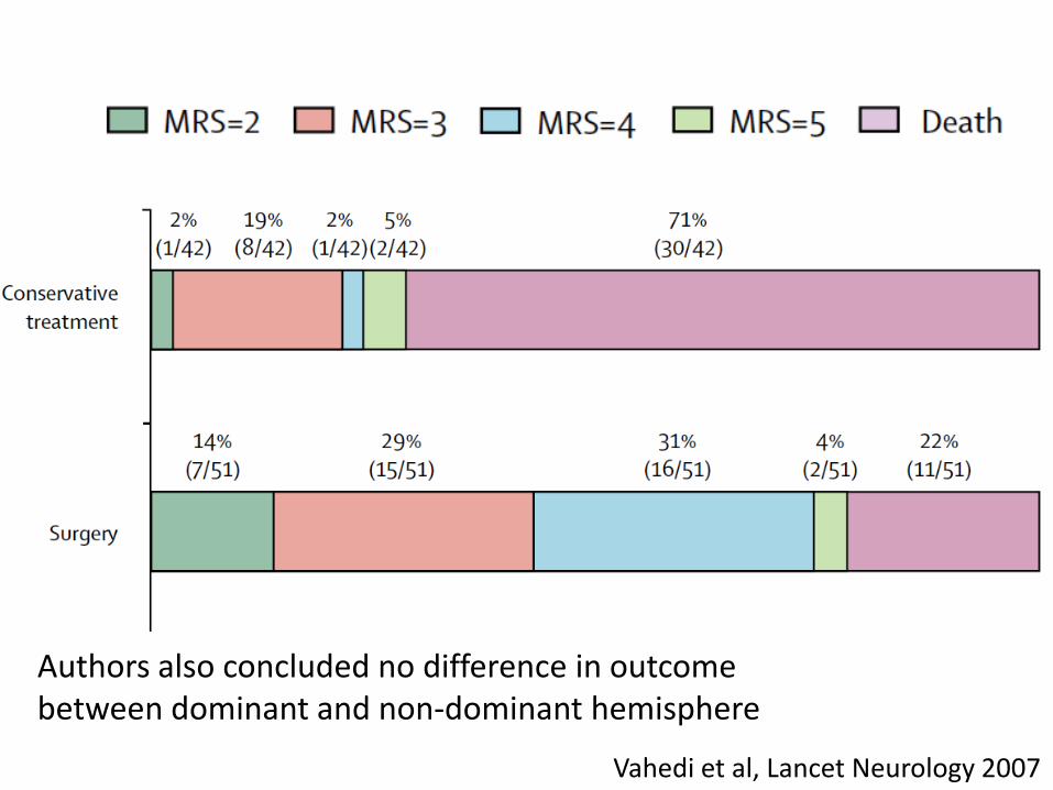
Vahedi et al, Lancet Neurology 2007
Authors also concluded no difference in outcome between dominant and non-dominant hemisphere