STROKE AND CEREBROVASCULAR DISEASE - CECity€¦ · Patient presents within 3 hours of neurologic...
41
1 STROKE AND CEREBROVASCULAR DISEASE
Transcript of STROKE AND CEREBROVASCULAR DISEASE - CECity€¦ · Patient presents within 3 hours of neurologic...

1
STROKE AND CEREBROVASCULAR
DISEASE

2
OBJECTIVES
Know and understand:
• The impact of cerebrovascular disease on older adults
• Treatment of acute ischemic stroke
• How to recognize a transient ischemic attack
• Prevention of ischemic stroke
• Prevention and treatment of hemorrhagic stroke

3
TOPICS COVERED
• Impact of Cerebrovascular Disease • Ischemic Stroke
Ø Small Vessel Disease Ø Large Vessel Disease Ø Cardioembolic and Cryptogenic Stroke Ø Treatment of Acute Ischemic Stroke Ø Transient Ischemic Attack Ø Primary and Secondary Ischemic Stroke Prevention
• Hemorrhagic Stroke Ø Intracerebral Hemorrhage Ø Subarachnoid Hemorrhage Ø Subdural Hematoma

4
IMPACT OF CEREBROVASCULAR DISEASE
• Incidence of stroke increases with advancing age, approximately doubles each decade
• Leading cause of disability and death among older adults
Ø 6 months after a stroke in those ≥65, 26% are dependent in ADLs and 46% have measurable cognitive deficits
Ø Fatality rate within 1 month of an acute stroke is 20%-30% across all age groups
Ø Common medical causes of death associated with stroke are myocardial infarction, arrhythmia, heart failure, aspiration pneumonia, and pulmonary embolism

5
ISCHEMIC STROKE
• Caused by occlusion of cerebral blood vessel
• Accounts for approximately 80%-85% of strokes
• Knowing the cause helps guide treatment and prognosis: Ø About 27% are due to cardiac emboli Ø About 19% are due to large vessel disease Ø About 17% are due to small vessel disease Ø About 35% cannot be classified

6
SMALL VESSEL DISEASE
• Causes occlusion of small penetrating vessels due to: Ø Lipohyalinosis (lipid deposition and hyalinization)
or Ø Local arteriosclerosis
• Causes ischemic strokes that are typically <1 cm, termed lacunar infarcts
• Risk factors: HTN, DM, smoking
• Lacunar strokes can occur concurrently with other mechanisms of ischemic stroke
7
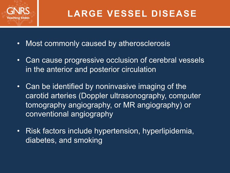
LARGE VESSEL DISEASE
• Most commonly caused by atherosclerosis
• Can cause progressive occlusion of cerebral vessels in the anterior and posterior circulation
• Can be identified by noninvasive imaging of the carotid arteries (Doppler ultrasonography, computer tomography angiography, or MR angiography) or conventional angiography
• Risk factors include hypertension, hyperlipidemia, diabetes, and smoking

8
CARDIOEMBOLIC STROKE
• Cardioembolism is an important, potentially preventable cause of ischemic stroke
• Atrial fibrillation (AF) is the most common cause
Ø Associated with a 4- to 5-fold increased risk because of thrombus formation in the left atrial appendage and cardioembolism to cerebral vessels
Ø Cardioembolic stroke due to AF accounts for 25%–30% of all ischemic strokes
• Cardioembolic stroke often occurs without preceding transient ischemic attack (TIA)

9
CRYPTOGENIC STROKE
• Ischemic stroke that does not have a clearly identifiable cause
• Use brain and vascular imaging to assess the size of the vessel(s) involved
• Consider atypical causes of stroke, such as vasculitis, coagulopathy, mitochondrial disorder
• Prolonged cardiac monitoring is sometimes needed to capture intermittent AF as a potential cause of cryptogenic stroke

10 IMMEDIATE TREATMENT OF ACUTE ISCHEMIC STROKE
• Quickly use head CT imaging to determine whether the patient is suffering from an ischemic or hemorrhagic event
• Assess the severity and pattern of neurologic deficits, such as by using the NIH Stroke Scale
• Identify non-stroke causes of acute neurologic dysfunction, such as migraine, seizure, and drug intoxication
11
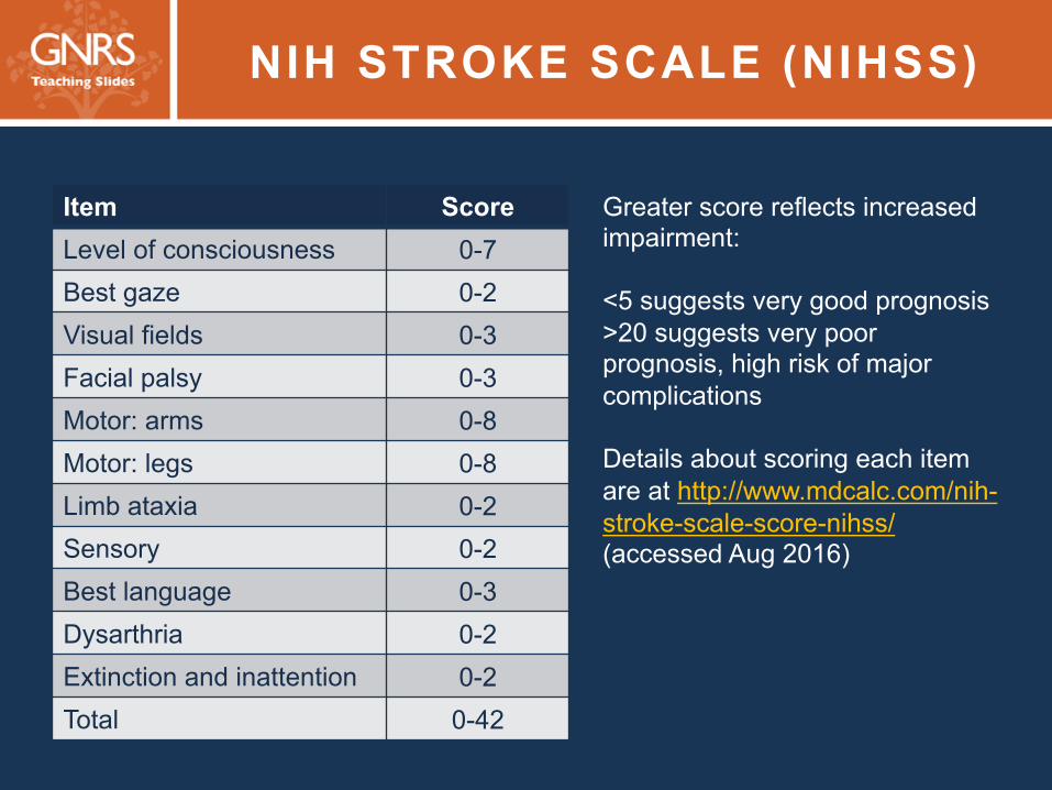
NIH STROKE SCALE (NIHSS)
Item Score Level of consciousness 0-7 Best gaze 0-2 Visual fields 0-3 Facial palsy 0-3 Motor: arms 0-8 Motor: legs 0-8 Limb ataxia 0-2 Sensory 0-2 Best language 0-3 Dysarthria 0-2 Extinction and inattention 0-2 Total 0-42
Greater score reflects increased impairment: <5 suggests very good prognosis >20 suggests very poor prognosis, high risk of major complications Details about scoring each item are at http://www.mdcalc.com/nih-stroke-scale-score-nihss/ (accessed Aug 2016)

12 ACUTE CARE OF THE OLDER STROKE PATIENT
• Optimize hydration status (gradually, to prevent cerebral edema)
• Control BP while avoiding hypotension
• Prevent deep-vein thrombosis
• Detect/treat coronary ischemia, heart failure, and cardiac arrhythmias
• Start long-term antiplatelet therapy or oral anticoagulation
• Normalize body temperature and blood glucose

13 RECOMBINANT TISSUE–PLASMINOGEN ACTIVATOR (rt-PA) (ALTEPLASE)
• Consider when: Ø Patient presents within 3 hours of neurologic deficit Ø CT confirms absence of intracranial hemorrhage
• AHA recommendation: Increase the window to 3-4.5 hours for patients ≤80 years old who have NIHSS score ≤25 and no prior history of stroke or diabetes
• Use requires careful assessment by a clinician experienced in treatment of stroke
• Carries a 6% risk of intracerebral hemorrhage, usually among patients with severe strokes
14
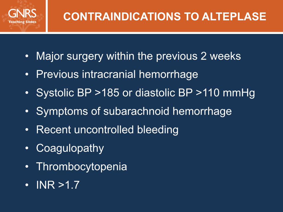
CONTRAINDICATIONS TO ALTEPLASE
• Major surgery within the previous 2 weeks • Previous intracranial hemorrhage
• Systolic BP >185 or diastolic BP >110 mmHg
• Symptoms of subarachnoid hemorrhage
• Recent uncontrolled bleeding
• Coagulopathy
• Thrombocytopenia
• INR >1.7

15 rt -PA PLUS ENDOVASCULAR THROMBECTOMY
In 4 recent randomized clinical trials:
• Endovascular thrombectomy in patients with proximal, large vessel occlusions of the anterior cerebral circulation provided significant benefit when treatment began within 6 hours of stroke onset
• Most patients received rt-PA (given IV) before endovascular thrombectomy
• Benefits were seen: Ø At 24 h, with 50% reduction in NIHSS score Ø At 90 days with improvements in functional outcomes

16
TRANSIENT ISCHEMIC ATTACK
• Defined as a brief episode of neurologic dysfunction caused by focal ischemia to the brain or spinal cord that does not result in acute infarction
• Traditionally thought to last up to 24 hours
• Newer imaging makes it clear: TIA typically lasts <1 to 2 hours Ø Longer episodes are commonly associated with acute
infarction
• Major risk factor for subsequent stroke

17
MANAGEMENT FOLLOWING TIA
• TIA symptoms should be evaluated emergently
• Brain imaging, ideally with MRI, is important to determine whether there has been an acute stroke and, if one is found, its location and type
• Noninvasive imaging of the carotid arteries, EKG, and echocardiography are important to determine the most likely cause of ischemia
• Initiate treatment for secondary stroke prevention based on the results of the evaluation

18
PREVENTING ISCHEMIC STROKE (1 of 4)
• BP therapy to maintain systolic BP <140 mmHg and diastolic BP <90 mmHg for secondary stroke prevention (SOE=A) and for primary prevention in those with other risk factors, such as diabetes
• Statin therapy for primary prevention in patients estimated to have a high 10-year risk of cardiovascular events based on AHA guidelines (SOE=A) Ø High-intensity statin therapy for patients who have had a TIA
or stroke believed to be of atherosclerotic origin, regardless of the LDL-C level (SOE=B)

19
PREVENTING ISCHEMIC STROKE (2 of 4)
• Several studies have confirmed a 2- to 4-fold increased risk of stroke in individuals with diabetes mellitus (SOE=A) Ø Aggressive treatment of hypertension and hyperlipidemia in
the diabetic population is crucial
Ø Some research suggests that tight control of blood glucose levels in healthier adults with diabetes might reduce the risk of stroke, although the evidence for reduction of other vascular complications (eg, retinopathy, nephropathy) is more compelling
• Support cessation of smoking, which increases the risk of stroke as much as 3-fold (SOE=A)
20
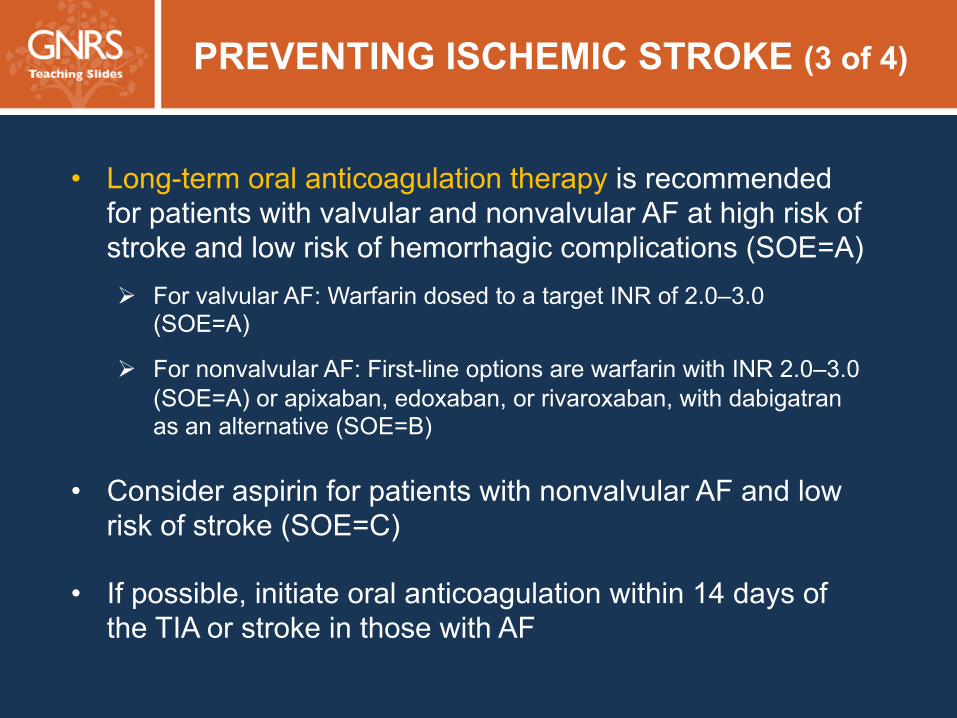
PREVENTING ISCHEMIC STROKE (3 of 4)
• Long-term oral anticoagulation therapy is recommended for patients with valvular and nonvalvular AF at high risk of stroke and low risk of hemorrhagic complications (SOE=A) Ø For valvular AF: Warfarin dosed to a target INR of 2.0–3.0
(SOE=A)
Ø For nonvalvular AF: First-line options are warfarin with INR 2.0–3.0 (SOE=A) or apixaban, edoxaban, or rivaroxaban, with dabigatran as an alternative (SOE=B)
• Consider aspirin for patients with nonvalvular AF and low risk of stroke (SOE=C)
• If possible, initiate oral anticoagulation within 14 days of the TIA or stroke in those with AF
21
PREVENTING ISCHEMIC STROKE (4 of 4)
• Following noncardioembolic ischemic stroke, aspirin is the mainstay of antiplatelet therapy for secondary prevention (SOE=A)
• The minimal necessary dosage has not been determined
• Other antiplatelet medications have not shown consistent superiority to aspirin Ø Sustained-release dipyridamole combined with aspirin (SOE=A) Ø Clopidogrel (SOE=B) (but it is an option for patients who cannot
tolerate aspirin) Ø Combining clopidogrel and aspirin (SOE=B)
• In two large trials, no benefit was found for the use of warfarin over aspirin for secondary prevention (SOE=A) in the setting of large vessel disease, including intracranial atherosclerotic disease

22
MANAGING ISCHEMIC STROKE
• All patients with cerebral atherosclerotic disease: Target modifiable risk factors; prescribe daily antiplatelet agent and statin (SOE=B)
• Patients with ≥70% symptomatic carotid stenosis: Carotid endarterectomy (CEA)
• Patients with recent TIA or ischemic stroke and ipsilateral moderate (50%–69%) carotid stenosis: Consider CEA
• Asymptomatic patients who have >70% stenosis of the internal carotid artery: Consider CEA, or endovascular treatment with carotid artery angioplasty and stenting, but only if the perioperative risk of stroke, myocardial infarction, and death is very low (<3%) (SOE=B)
23
TREATING CRYPTOGENIC STROKE
• Evaluate and treat modifiable stroke risk factors and initiate antiplatelet therapy
• If a cardioembolic cause is suspected, then prolonged (eg, 30 days) cardiac rhythm monitoring for AF is reasonable (SOE=B), and oral anticoagulant therapy should be initiated if intermittent AF is found
• In the setting of patent foramen ovale (PFO) and venous source of embolism, oral anticoagulation is recommended (SOE=A)
Ø If anticoagulation is contraindicated in such a patient, then an inferior vena cava filter (SOE=C) or transcatheter PFO closure (SOE=C) can be considered
24
HEMORRHAGIC STROKE (INTRACRANIAL HEMORRHAGE)
• Accounts for 15%–20% of all strokes
• 4 main subtypes: intracerebral hemorrhage, subarachnoid hemorrhage, subdural hematoma, and epidural hematoma
• Presents with abrupt onset of focal neurologic symptoms
• Often associated with severe headache, vomiting, very high blood pressure, and coma or decreased level of consciousness
• Carries a higher risk of mortality than ischemic stroke and is best treated in comprehensive stroke centers
• Emergent brain imaging (eg, head CT) is needed to determine whether a stroke is hemorrhagic or ischemic
25
ACUTE CARE OF
INTRACEREBRAL HEMORRHAGE (ICH)
• Patients with systolic BP of 150-220 mmHg: Lower systolic BP to 140 mmHg (SOE=A)
• Patients on warfarin: Intravenous vitamin K and therapy to replace vitamin K–dependent clotting factors to normalize the INR (SOE=B)
• Patients with severe coagulation factor deficiency or severe thrombocytopenia: Factor replacement therapy or platelets as needed (SOE=B)
• All anticoagulant and antiplatelet therapies should be stopped in the acute period
26
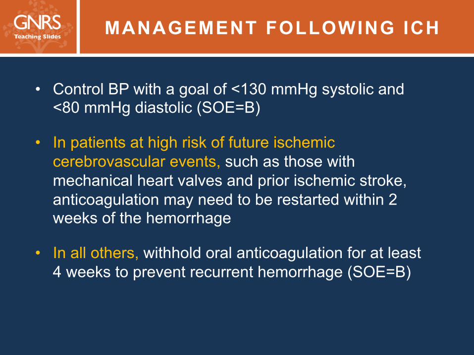
MANAGEMENT FOLLOWING ICH
• Control BP with a goal of <130 mmHg systolic and <80 mmHg diastolic (SOE=B)
• In patients at high risk of future ischemic cerebrovascular events, such as those with mechanical heart valves and prior ischemic stroke, anticoagulation may need to be restarted within 2 weeks of the hemorrhage
• In all others, withhold oral anticoagulation for at least 4 weeks to prevent recurrent hemorrhage (SOE=B)

27
SUBARACHNOID HEMORRHAGE (SAH)
• Caused by a rupture of an intracranial aneurysm (80%–85% of cases) or another abnormality of a cerebral vessel (eg, arteriovenous malformation)
• Risk factors for rupture: size of the aneurysm, patient age, female gender, posterior circulation location, change in size of aneurysm over time, prior SAH
• Patients with an aneurysm at higher risk of rupture should be referred to a neurosurgeon
• Acute management of SAH is similar to that of ICH but includes surgical intervention, and focuses on detection and treatment of arterial vasospasm, a common complication

28
SUBDURAL HEMATOMA
• Defined as collection of blood between the dura and the arachnoid
• Usually due to head trauma, although the trauma may be mild, particularly in older adults
• Bilateral in approximately 15% of cases
• Some older adults suffer from chronic subdural hematoma that may be symptomatic

29 INCIDENCE OF CHRONIC SUBDURAL HEMATOMA INCREASES WITH AGE
• No history of head injury: 50% of cases
• Other risk factors:
Ø Clotting disorders
Ø Shunting procedures
Ø Seizures
0.13
7.4
0 1 2 3 4 5 6 7 8
20-29 70-79
Inci
denc
e pe
r 100
,000
Age
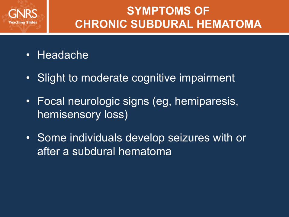
30 SYMPTOMS OF CHRONIC SUBDURAL HEMATOMA
• Headache
• Slight to moderate cognitive impairment
• Focal neurologic signs (eg, hemiparesis, hemisensory loss)
• Some individuals develop seizures with or after a subdural hematoma
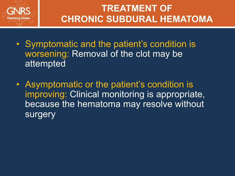
31 TREATMENT OF CHRONIC SUBDURAL HEMATOMA
• Symptomatic and the patient’s condition is worsening: Removal of the clot may be attempted
• Asymptomatic or the patient’s condition is improving: Clinical monitoring is appropriate, because the hematoma may resolve without surgery

32
CHOOSING WISELY®
Recommendation for Stroke and Cerebrovascular Disease,
based on the American Board of Internal Medicine Foundation’s Choosing Wisely® Campaign:
Do not recommend carotid endarterectomy for asymptomatic carotid stenosis unless the complication rate is low (<3%).

33
SUMMARY
• Cerebrovascular disease is a leading cause of
disability and death among older people
• Acute stroke treatments can effectively improve outcomes in patients, especially when care is provided soon after stroke symptoms begin and in a multidisciplinary stroke center
• Primary and secondary stroke prevention can significantly decrease the burden of cerebrovascular disease in older populations

34
CASE 1 (1 of 3)
• A 75-year-old woman comes to the office for follow-up. Ø Two days ago she was taken to the ER after a 30-minute episode of mild
left-sided hemiparesis and right transient monocular blindness. Ø Radiography in ER
v Diffusion-weighted MRI: no signs of stroke v MRA: 85% stenosis of the right carotid artery
Ø The patient was discharged from the ER with instructions to begin aspirin 81 mg/d and follow-up with her primary care doctor.
• History: hypertension, hyperlipidemia • Baseline medications: lisinopril, hydrochlorothiazide, atorvastatin • Physical examination at follow-up:
Ø Blood pressure 136/78 mmHg Ø Remainder of examination, including neurologic findings, is unremarkable.
35
CASE 1 (2 of 3)
Which one of the following is the most appropriate next step for long-term prevention of stroke?
A. Refer the patient for carotid artery stenting.
B. Refer the patient for carotid endarterectomy.
C. Add clopidogrel.
D. Increase dosage of aspirin.
36
CASE 1 (3 of 3)
Which one of the following is the most appropriate next step for long-term prevention of stroke?
A. Refer the patient for carotid artery stenting.
B. Refer the patient for carotid endarterectomy.
C. Add clopidogrel.
D. Increase dosage of aspirin.
37
CASE 2 (1 of 4)
• An 84-year-old man has newly diagnosed nonvalvular atrial fibrillation.
• History Ø Hypertension, diabetes, hyperlipidemia, CKD (est.
creatinine clearance 20 mL/min) v No history of stroke
Ø He broke his hip in a fall 3 years ago. v After repair and rehabilitation, he continues to live
independently. v He walks with a cane.
• Medications: metformin, lisinopril, atorvastatin, aspirin
38
CASE 2 (2 of 4)
• Physical examination
Ø Heart rate: 86 bpm, irregularly irregular
Ø Blood pressure: 130/75 mmHg
Ø Decreased proximal muscle strength when he rises from a chair
Ø Gait is stable with a cane.
• Radiography
Ø Recent transthoracic echo: no atrial clot, normal systolic and diastolic function
39
CASE 2 (3 of 4)
Which one of the following is the most appropriate management for reducing his risk of stroke?
A. Continue aspirin.
B. Stop aspirin, and begin warfarin.
C. Stop aspirin, and begin apixaban.
D. Continue aspirin, and add warfarin.

40
CASE 2 (4 of 4)
Which one of the following is the most appropriate management for reducing his risk of stroke?
A. Continue aspirin.
B. Stop aspirin, and begin warfarin.
C. Stop aspirin, and begin apixaban.
D. Continue aspirin, and add warfarin.

41
GNRS5 Teaching Slides Editor: Barbara Resnick, PhD, CRNP, FAAN, FAANP, AGSF
GNRS5 Teaching Slides modified from GRS9 Teaching Slides
based on chapter by Daniel L. Murman, MD, MS, FAAN
and questions by Nina J. Solenski, MD
Managing Editor: Andrea N. Sherman, MS
Copyright © 2016 American Geriatrics Society