Huntingtin-Associated Protein HAP1 trafficking and orexin neuronal function

Striatal neurons expressing full-length mutanthuntingtin exhibit decreased N-cadherin andaltered neuritogenesis
Surya A. Reis1,{, Morgan N. Thompson1, Jong-Min Lee1, Elisa Fossale1, Hyung-Hwan Kim4,
James K. Liao4, Michael A. Moskowitz5, Stanley Y. Shaw2, Linda Dong1, Stephen J. Haggarty3,
Marcy E. MacDonald1 and Ihn Sik Seong1,∗
1Molecular Neurogenetics Unit, Center for Human Genetic Research, 2Center for Systems Biology and 3Chemical
Biology Program, Center for Human Genetic Research, Massachusetts General Hospital, 185 Cambridge Street,
Boston, MA 02114, USA, 4Vascular Medicine Research Unit, Brigham and Women’s Hospital and Harvard Medical
School, Cambridge, MA 02139, USA and 5Stroke and Neurovascular Regulation, Massachusetts General Hospital,
Charlestown, MA 02129, USA
Received February 8, 2011; Revised March 16, 2011; Accepted March 23, 2011
The expanded CAG repeat that causes striatal cell vulnerability in Huntington’s disease (HD) encodes a poly-glutamine tract in full-length huntingtin that is correlated with cellular [ATP] and [ATP/ADP]. Since striatal neur-ons are vulnerable to energy deficit, we have investigated, in Hdh CAG knock-in mice and striatal cells, thehypothesis that decreased energetics may affect neuronal (N)-cadherin, a candidate energy-sensitive adhesionprotein that may contribute to HD striatal cell sensitivity. In vivo, N-cadherin was sensitive to ischemia and to theeffects of full-length mutant huntingtin, progressively decreasing in HdhQ111 striatum with age. In culturedstriatal cells, N-cadherin was decreased by ATP depletion and STHdhQ111 striatal cells exhibited dramaticallydecreased N-cadherin, due to decreased Cdh2 mRNA and enhanced N-cadherin turnover, which waspartially normalized by adenine supplementation to increase [ATP] and [ATP/ADP]. Consistent with decreasedN-cadherin function, STHdhQ111 striatal cells displayed profound deficits in calcium-dependent N-cadherin-mediated cell clustering and cell–substratum adhesion, and primary HdhQ111 striatal neuronal cells exhibiteddecreased N-cadherin and an abundance of immature neurites, featuring diffuse, rather than clustered, stainingfor N-cadherin and synaptic vesicle markers, which was partially rescued by adenine treatment. Thus, mutantfull-length huntingtin, via energetic deficit, contributes to decreased N-cadherin levels in striatal neurons, withdetrimental effects on neurite maturation, strongly suggesting that N-cadherin-mediated signaling meritsinvestigation early in the HD pathogenic disease process.
INTRODUCTION
The CAG expansion mutation that causes Huntington’sdisease (HD) elongates a polymorphic polyglutaminesegment in the huntingtin protein. Full-length huntingtinwith a polyglutamine region of more than �37 residuesinitiates a disease process that culminates in the loss ofneurons, especially in the striatum, and the onset ofthe motor, psychiatric and cognitive symptoms (1,2).
Understanding the rate-limiting events that contribute to theearly vulnerability of striatal neurons would guide efforts totrack the natural history of the disease and may provide newavenues for therapeutic development.
Studies investigating the earliest consequences of full-length mutant huntingtin, in HD patient cells and tissues andin genetically accurate Hdh CAG knock-in mouse cells andtissues, have revealed perturbations in membrane vesicle traf-ficking, gene transcription, intracellular signaling pathways
†Present address: Chemical Biology Programs, Center for Human Genetic Research, Massachusetts General Hospital, 185 Cambridge Street, Boston,MA 02114, USA.
∗To whom correspondence should be addressed. Tel: +1 6176439851; Fax: +1 6176433202; Email: [email protected]
# The Author 2011. Published by Oxford University Press. All rights reserved.For Permissions, please email: [email protected]
Human Molecular Genetics, 2011, Vol. 20, No. 12 2344–2355doi:10.1093/hmg/ddr127Advance Access published on March 29, 2011
Dow
nloaded from https://academ
ic.oup.com/hm
g/article/20/12/2344/2527052 by guest on 27 Novem
ber 2021

(3–6), as well as altered energetics, characterized bydecreased [ATP] and [ATP/ADP], which is correlated withthe size of the polyglutamine repeat (7–9). We have beenstudying the effects of altered energetics because the corre-lation of energetic measures with the polyglutamine repeatin full-length huntingtin implies a dominant effect thatconforms to the genetic features of the HD trigger mechanism,and energetic defects, thought to be important to striatal cells,may be evident throughout the lifetime of the cell (7–9). Cer-tainly, early weight loss in HD and a systemic metabolicdefect in branched chain amino acids are consistent with a sys-temic attempt to compensate for an early energy deficit (10).
Neuronal (N)-cadherin, which is intimately involved inneuronal cell adhesion, signaling, differentiation and synapsefunction (11,12), is a prime candidate for being affected byenergy deficit. Members of the cadherin family exhibit selec-tive degradation in response to renal ischemia and ATPdepletion (13–15), and in normal rat kidney cells via cleavageby membrane-type 1 matrix metalloprotease (MT-MMP) (16).However, N-cadherin has not been studied either in acuteneuronal ischemia and ATP depletion or in response to theHD mutation, which elicits a chronic energy deficit in aprocess that culminates in neurodegeneration.
N-cadherin is a transmembrane cell adhesion glycoproteincomposed of an extracellular domain, a single-pass transmem-brane region and a cytoplasmic tail (17). N-cadherin mol-ecules make calcium-dependent homophilic bonds betweentheir extracellular domains (18). The cytoplasmic domain con-tains two main binding regions, the C-terminal domain (CTD)and the juxtamembrane domain (JMD). The CTD binds b- andg-catenin, which in turn associate with the actin cytoskeletonto modulate cell adhesion and mobility via a-catenin (19,20).The JMD interacts with p120-catenin and with presenilin 1,which has emerged as a potential regulator of cell adhesionand neuronal physiology (21,22).
Here, we have assessed the candidacy of N-cadherin as anenergy-sensitive contributor to the striatal cell vulnerabilitythat ensues from the HD mutation. Specifically, we haveinvestigated N-cadherin in HdhQ111 CAG knock-in mousestriatum and cultured HdhQ111 striatal neuronal cells, whichexpress endogenous full-length 111-glutamine mutant hun-tingtin. We first tested whether N-cadherin was sensitive toacute ATP depletion/ischemia and to the chronic effects offull-length mutant huntingtin protein and then we evaluatedimmortalized STHdhQ111 and primary HdhQ111 striatalneurons to explore N-cadherin ATP sensitivity and the pheno-typic consequences of decreased N-cadherin function. Ourfindings reveal that N-cadherin is an ATP-sensitive proteinthat is associated with altered HD CAG striatal cell adhesionand neuritogenesis.
RESULTS
Striatal N-cadherin was sensitive to acute ischemia and tothe HD CAG mutation
To assess whether N-cadherin might be affected in a mildischemic brain injury paradigm where the striatum displaysearly vulnerability, we performed transient middle cerebralartery (MCA) occlusion with 12-month-old wild-type HdhQ7/Q7
and mutant HdhQ111/Q111 knock-in mice. For both genotypes,immunoblot of protein extracts at 24 h after reperfusion(Fig. 1A and B) revealed robustly decreased N-cadherin inthe ischemic, compared with the contralateral, striatal hemi-spheres. Both the reduction in N-cadherin and the infarctsize (at 30 and 60 min) were similar in wild-type and mutantbrain, consistent with a previous report demonstrating thatCAG expansion did not sensitize to acute ischemic injury(23). In contrast, full-length wild-type 7-glutamine huntingtinand 111-glutamine mutant huntingtin cleaved by calpains inresponse to severe ischemia (24), a-catenin and b-cateninwere not decreased, though p120 was slightly reduced byischemia, attesting to the mildness of the lesion and demon-strating the exquisite sensitivity of N-cadherin in the striatumto ischemia.
The immunoblot results also revealed reduced N-cadherinin contralateral HdhQ111/Q111 striata compared with wild-typestriata, suggesting an effect of the CAG mutation. This wasconfirmed by immunoblot analysis of wild-type andHdhQ111/Q111 striatal tissues at different ages, which revealeda progressive decrease in N-cadherin (normalized toa-tubulin) from 3 to 5 months of age that reached statisticalsignificance by 12 months of age (Supplementary Material,Fig. S1).
Notably, N-cadherin (Cdh2) mRNA levels were not reducedin total striatal tissue, following ischemic reperfusion or inresponse to the CAG mutation (data not shown), implyingthat cell-specific and/or multiple mechanisms may contributeto the energy-dependent N-cadherin decrease in vivo.
Striatal cell N-cadherin was sensitive to ATP depletion andthe HD CAG mutation
We then assessed whether N-cadherin might be sensitiveto ATP depletion and to the CAG mutation incultured STHdhQ7/Q7 and STHdhQ111/Q111 striatal neuronalcells expressing wild-type (7-glutamine) and mutant(111-glutamine) full-length huntingtin, respectively. Cellswere treated with 2-deoxyglucose and antimycin A, toinhibit both glycolysis and mitochondrial respiration, andnucleotides in cellular extracts were measured by HPLCanalysis. As reported previously (7,8), the baseline [ATP/ADP] was significantly lower in mutant, compared with wild-type, striatal cells, and, for both genotypes, [ATP/ADP] wasdramatically decreased within 2 h of energy depletion (Sup-plementary Material, Fig. S2A), although cell viability wasnot significantly changed (Supplementary Material, Fig.S2B). Immunoblot analysis revealed that N-cadherin wasdecreased at baseline in mutant, compared with wild-type,striatal cell extracts, with a progressive reduction over thetime course of ATP depletion for both genotypes (Fig. 1C).In contrast, a-catenin, b-catenin and p120 levels weresimilar for cells of either genotype and were not changed byATP depletion (Fig. 1C). Thus, the N-cadherin level wasreduced concomitant with decreased [ATP/ADP], both dueto purposeful energy depletion and in response to ATPdeficit due to full-length mutant huntingtin.
Further analyses demonstrated that decreased N-cadherin inmutant, compared with wild-type, striatal cells reflecteddecreased Cdh2 mRNA, as demonstrated by the results of
Human Molecular Genetics, 2011, Vol. 20, No. 12 2345
Dow
nloaded from https://academ
ic.oup.com/hm
g/article/20/12/2344/2527052 by guest on 27 Novem
ber 2021

RT–PCR analysis (Supplementary Material, Fig. S3A), aswell as the increased turnover rate of the protein, which hada half-life of �2 h in STHdhQ111/Q111 cells compared with ahalf-life of .4 h in wild-type cells (Supplementary Material,Fig. S3B). To explore the energy sensitivity of these measures,mutant striatal cell culture medium was supplemented withadenine, a precursor of high-energy nucleotides. By 2 days,[ATP/ADP] was mildly but significantly elevated (Supplemen-tary Material, Fig. S2C), and by 4 days of treatment, N-cadherin was modestly but consistently increased (Fig. 1D),though Cdh2 mRNA levels were not altered over the entire6 day time course (data not shown). These results suggestedthat enhanced N-cadherin protein turnover in the mutant stria-tal cells may involve an energy-sensitive process, whilealtered Cdh2 mRNA may reflect a different underlyingprocess. However, assays to explore the involvement of
metalloproteases, activated in ischemia, failed to discloseelevated MT-MMP, and MT-MMP inhibition with GM-6001did not alter the half-life of N-cadherin in mutant striatalcells (data not shown).
STHdhQ111/Q111 cells exhibited deficits inN-cadherin-mediated cell–cell adhesion
The potential functional consequences of decreasedN-cadherin in mutant striatal cells were then evaluated, begin-ning with confocal microscopy to investigate N-cadherin sub-cellular localization. Wild-type STHdhQ7/Q7 cells exhibitedprominent N-cadherin immunostain at cell–cell contacts,whereas mutant striatal cells displayed a decreased N-cadherinsignal that was not prominent at cell–cell contacts (Fig. 2A),strongly implying a defect in calcium-dependent
Figure 1. ATP-sensitive N-cadherin stability. (A) A representative coronal section of wild-type HdhQ7/Q7 mouse brain, following MCA occlusion/reperfusion,stained with 2,3,5-triphenyltetrazolium chloride (TTC), to demonstrate the infarct region (white area) in the left hemisphere (L), as well as the contralateral righthemisphere (R). The infarct sizes (mm3 and mean+ standard errors) were measured from mice brains of both genotypes at 30 and 60 min MCA occlusion as33.9+3.64 and 35.2+3.55 for wild-type brain at 30 and 60 min and 41.5+5.97 and 43.1+6.81 for mutant brain and showed no significant differences bygenotypes and times (n ¼ 3), consistent with the previous report (23). (B) Immunoblot showing bands of N-cadherin, b-catenin, a-catenin and p120 detected inprotein extracts of striata from the MCA-occluded hemisphere (L) and contralateral hemisphere (R) for wild-type HdhQ7/Q7 (Hdh7/7) and mutant HdhQ111/Q111
(Hdh111/111) mice, with 30 min MCA occlusion (30′) or 60 min MCA occlusion (60′), demonstrating decreased N-cadherin on the lesion side, relative to thea-tubulin loading control band. However, the reduction in N-cadherin was not statistically different between two genotypes or two occlusion times (n ¼ 3). (C)Immunoblot showing bands of N-cadherin, b-catenin, a-catenin and p120 detected in protein extracts of wild-type STHdhQ7/Q7 (ST7/7) and mutant STHdhQ111/
Q111 (ST111/111) cells, at time 0 (cont), and 0.5, 1 and 2 h in ATP-depletion medium. The adjacent graph plots the relative band intensity of N-cadherin normal-ized to a-tubulin in the same lane (y-axis), with the time in ATP-depleting medium (x-axis), illustrating decreased N-cadherin in mutant cells at baseline andATP-sensitive degradation of N-cadherin in cells of both genotypes. Bars denote the standard deviation from three independent experiments. (D) Immunoblotshowing N-cadherin in extracts of STHdhQ111/Q111 cells, after incubation in medium supplemented with 10 mM adenine for 0, 2, 4 and 6 days (D), relative to thea-tubulin band. The adjacent bar graph plots the relative N-cadherin intensities normalized to the a-tubulin band intensities (y-axis) for the time points analyzed(x-axis). Relative to the untreated control, N-cadherin levels were significantly elevated by day 4 (∗P , 0.05, n ¼ 3).
2346 Human Molecular Genetics, 2011, Vol. 20, No. 12
Dow
nloaded from https://academ
ic.oup.com/hm
g/article/20/12/2344/2527052 by guest on 27 Novem
ber 2021
N-cadherin-mediated cell–cell adhesion (25,26). Toassess this possibility, wild-type STHdhQ7/Q7 and mutantSTHdhQ111/Q111 cells were evaluated in a cell-clusteringassay, which measured the time taken for detached singlecells to form clusters (Fig. 2B). Within 30 min of dispersalto single cells, 70% of the wild-type cells, but only 30%of the mutant striatal cells, formed clusters in Ca2+-containingmedia, though cells of both genotypes exhibited similar Ca2+-independent (in EGTA) cell clustering. This phenotype wasN-cadherin dependent, as exogenous expression ofN-cadherin, after transfection, raised N-cadherin levelsto �50% of wild-type levels (Supplementary Material,Fig. S3C) and partially rescued the mutant cell deficit in Ca2+-dependent cluster formation. At 30 min after being dispersed,�20% more N-cadherin-transfected mutant striatal cellsformed clusters in Ca2+ media, compared with mutant cells
transfected with the control vector (Fig. 2C). In the absenceof Ca2+ (in EGTA), the transfection of N-cadherin had noeffect on cell–cell adhesion.
STHdhQ111/Q111 cells exhibited deficits in cell–substratumadhesion
N-cadherin is known to regulate cell–substratum adhesion(27) and actin cytoskeleton dynamics (28,29). Consequently,co-stained striatal cells were monitored by epifluorescencemicroscopy to detect DAPI-stained nuclei and filamentousactin (F-actin) cytoskeletal-binding proteins (Fig. 2D). Com-pared with wild-type STHdhQ7Q/7 cells, the mutantSTHdhQ111/Q111 striatal cells appeared smaller with elongatedshapes, and decreased stress fiber-like rhodamine–phalloidinsignal, though in the inset the pattern of the phalloidin
Figure 2. STHdhQ111/Q111 striatal cells exhibited decreased N-cadherin function. (A) Confocal images of the N-cadherin immunostain signal (red) in wild-typeSTHdhQ7/Q7 (ST7/7) and STHdhQ111/Q111 (ST111/111) striatal cells, indicating decreased N-cadherin signal at STHdhQ111/Q111cell–cell contacts. (B) Plot sum-marizing the results of three independent cell-clustering assays, showing the proportion of single cells compared with time zero (Nt/N0) (y-axis) relative to time ofclustering (x-axis) for STHdhQ7/Q7 (square) and STHdhQ111/Q111 (circle) striatal cells, in the absence (open symbols) and presence (closed symbols) of Ca2+,illustrating that mutant striatal cells exhibit impaired calcium-dependent clustering. Error bars denote the standard deviation. (C) Plot summarizing theresults of three independent STHdhQ111/Q111 cell-clustering assays for cells transfected with the phN-cad human N-cadherin expression vector (squares) orwith the control vector (circles), performed in the absence (open symbols) and presence (closed symbols) of Ca2+. Error bars denote the standard deviation.(D) Fluorescence images of STHdhQ7/Q7 (ST7/7) and STHdhQ111/Q111 (ST111/111) striatal cells, showing the pattern of DAPI (blue) nuclei, TRITC–phalloidin(red) F-actin signal and anti-cofilin (green) immunostain, illustrating decreased nuclear size, paucity of actin stress fibers and bright perinuclear cofilin stain ofthe mutant cells, consistent with an elongated rounded-up (less flat) morphology. The adjacent bar graph shows the average phalloidin signal and the total ornuclear area cofilin intensities. The average intensity of the phalloidin signal was significantly reduced in mutant striatal cells (∗P , 0.001) and the inset wastaken at different exposure conditions to examine the pattern of the phalloidin signal in mutant cells. The average intensity of the total cofilin signal was notaltered but its localization was shifted to the nuclear and perinuclear region (∗∗P , 0.001). Error bars represent standard errors.
Human Molecular Genetics, 2011, Vol. 20, No. 12 2347
Dow
nloaded from https://academ
ic.oup.com/hm
g/article/20/12/2344/2527052 by guest on 27 Novem
ber 2021
signal resembled that of wild-type cells (Fig. 2D). Consistentwith the elongated (less flat) morphology, mutant striatalcells exhibited smaller DAPI-stained nuclei and the significantintense cofilin stain of the mutant cells was localized to thenuclear and perinuclear region, although quantificationdemonstrated that the total cofilin signal in mutant and wild-type cells was similar (Fig. 2D). The mutant striatal cellsalso exhibited a similar nuclear/perinuclear immunostainingpattern for profilin, another F-actin-binding protein, which,like cofilin, is involved in actin filament structure anddynamics (30) (data not shown). Notably, immunoblotrevealed similar levels of F-actin, and actin monomer(G-actin), in extracts of mutant and wild-type striatal cells(data not shown). Thus, rather than lacking an actin cytoskele-ton, the altered subcellular patterns of actin-associated pro-teins, along with decreased N-cadherin, were consistent withthe elongated, less flat morphology of the striatal cells expres-sing full-length mutant huntingtin and strongly implied deficitsin cell–substrate adhesion, as well as cell–cell adhesion.
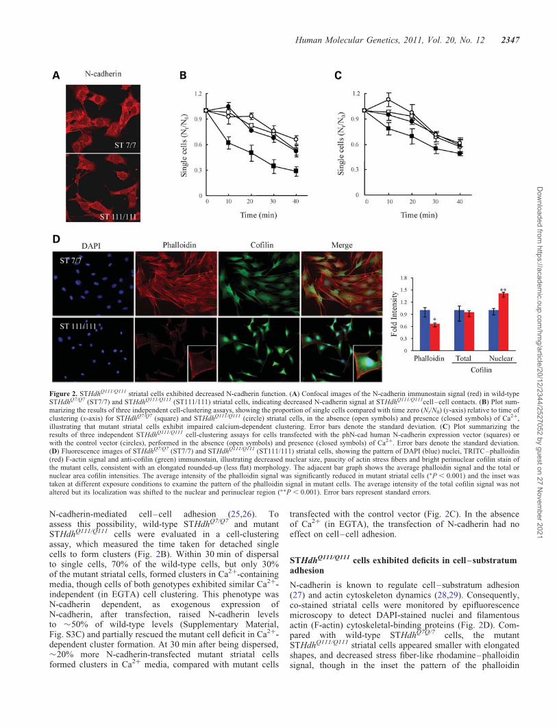
HdhQ111/Q111 primary striatal neuronal cells exhibiteddecreased N-cadherin and immature neuritis
N-cadherin-mediated cell–substrate adhesion promotesneurite outgrowth (27) and N-cadherin is required for propervesicle clustering essential for neurite formation and matu-ration (31–34). To evaluate the potential impact of full-lengthmutant huntingtin on N-cadherin function in developing stria-tal neurons, we examined primary neuronal cell cultures fromthe striatum of E14 control and HdhQ111/Q111 knock-in mouseembryos. At day 10 of differentiation (days in vitro, div10),F-actin rhodamine–phalloidin binding was assessed by confo-cal microscopy, as a surrogate for proper actin cytoskeleton/cell–substratum adhesion, and cultures were immunostainedto detect MAP2, a neuronal cell microtubule-associatedprotein, to evaluate the developing neuronal cell projections.Compared with wild-type primary striatal neuronal cells, theprimary HdhQ111/Q111 cells exhibited decreased rhodamine–phalloidin stain and an abnormally robust network of fineMAP2-stained projections, which confirmed neuronal celldifferentiation, while implying altered adhesion and develop-ment (Fig. 3).
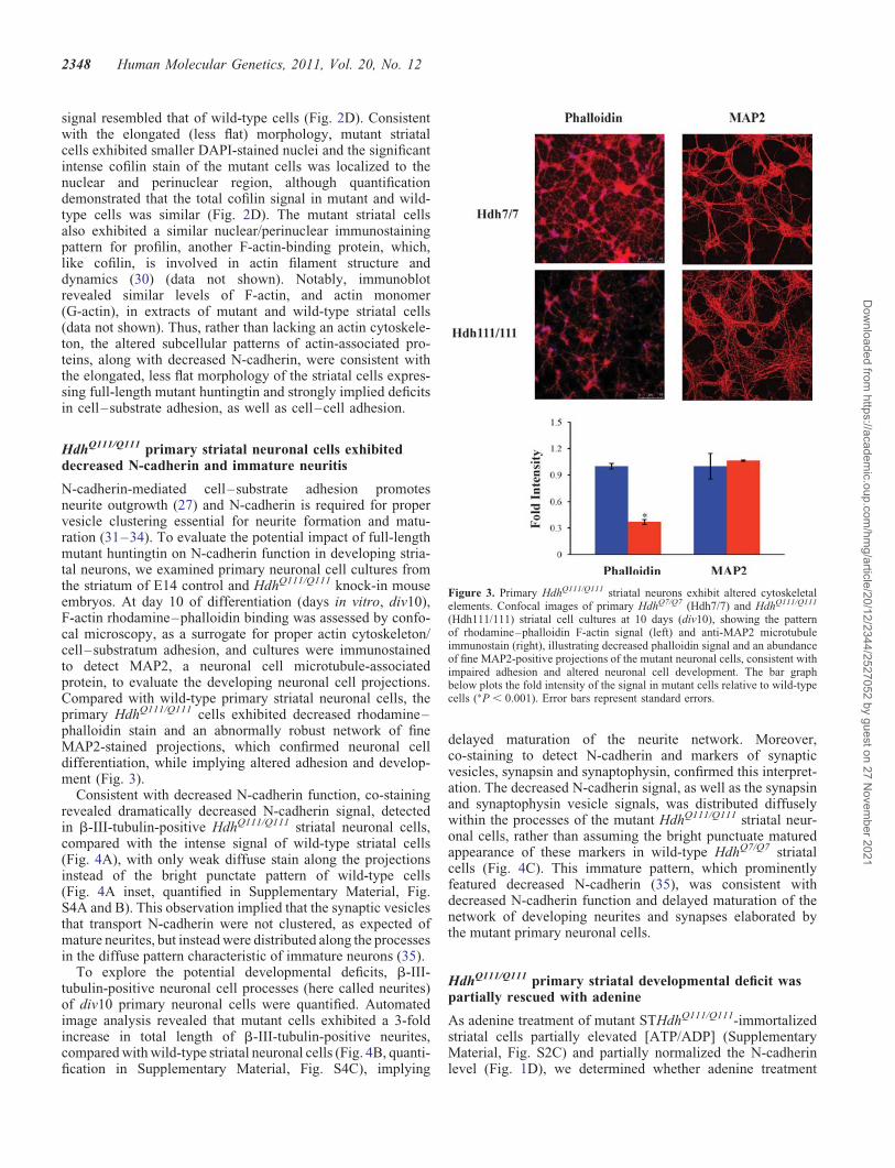
Consistent with decreased N-cadherin function, co-stainingrevealed dramatically decreased N-cadherin signal, detectedin b-III-tubulin-positive HdhQ111/Q111 striatal neuronal cells,compared with the intense signal of wild-type striatal cells(Fig. 4A), with only weak diffuse stain along the projectionsinstead of the bright punctate pattern of wild-type cells(Fig. 4A inset, quantified in Supplementary Material, Fig.S4A and B). This observation implied that the synaptic vesiclesthat transport N-cadherin were not clustered, as expected ofmature neurites, but instead were distributed along the processesin the diffuse pattern characteristic of immature neurons (35).
To explore the potential developmental deficits, b-III-tubulin-positive neuronal cell processes (here called neurites)of div10 primary neuronal cells were quantified. Automatedimage analysis revealed that mutant cells exhibited a 3-foldincrease in total length of b-III-tubulin-positive neurites,compared with wild-type striatal neuronal cells (Fig. 4B, quanti-fication in Supplementary Material, Fig. S4C), implying
delayed maturation of the neurite network. Moreover,co-staining to detect N-cadherin and markers of synapticvesicles, synapsin and synaptophysin, confirmed this interpret-ation. The decreased N-cadherin signal, as well as the synapsinand synaptophysin vesicle signals, was distributed diffuselywithin the processes of the mutant HdhQ111/Q111 striatal neur-onal cells, rather than assuming the bright punctuate maturedappearance of these markers in wild-type HdhQ7/Q7 striatalcells (Fig. 4C). This immature pattern, which prominentlyfeatured decreased N-cadherin (35), was consistent withdecreased N-cadherin function and delayed maturation of thenetwork of developing neurites and synapses elaborated bythe mutant primary neuronal cells.
HdhQ111/Q111 primary striatal developmental deficit waspartially rescued with adenine
As adenine treatment of mutant STHdhQ111/Q111-immortalizedstriatal cells partially elevated [ATP/ADP] (SupplementaryMaterial, Fig. S2C) and partially normalized the N-cadherinlevel (Fig. 1D), we determined whether adenine treatment
Figure 3. Primary HdhQ111/Q111 striatal neurons exhibit altered cytoskeletalelements. Confocal images of primary HdhQ7/Q7 (Hdh7/7) and HdhQ111/Q111
(Hdh111/111) striatal cell cultures at 10 days (div10), showing the patternof rhodamine–phalloidin F-actin signal (left) and anti-MAP2 microtubuleimmunostain (right), illustrating decreased phalloidin signal and an abundanceof fine MAP2-positive projections of the mutant neuronal cells, consistent withimpaired adhesion and altered neuronal cell development. The bar graphbelow plots the fold intensity of the signal in mutant cells relative to wild-typecells (∗P , 0.001). Error bars represent standard errors.
2348 Human Molecular Genetics, 2011, Vol. 20, No. 12
Dow
nloaded from https://academ
ic.oup.com/hm
g/article/20/12/2344/2527052 by guest on 27 Novem
ber 2021
might modulate the propensity of primary div10 E14 HdhQ111/Q111
striatal neurons to form abnormally extensive neurite net-works. As shown in Figure 5, after treatment with adeninefor 10 days, the div10 HdhQ111/Q111 striatal cultures exhibitedMAP2-positive neuronal cells with fewer neurites per cellbody compared with DMSO vehicle-treated cultures, thoughthe number of cell bodies was not appreciably altered, asshown by DAPI nuclear staining. Consistent with the ameli-orative effect of adenine on energy measures and N-cadherinlevels demonstrated in STHdhQ111/Q111 cells, these findingssuggest that the delayed development of mutant primary
neuronal cells may, at least in part, reflect decreased functionof the ATP-sensitive N-cadherin adhesion molecule.
DISCUSSION
HD is a progressive neurodegenerative disorder, with markedloss of the major population of neurons in the striatum(medium-sized spiny neurons), which are vulnerable to acuteenergetic challenge. The neuronal specificity of HD stemsfrom the effects of the HD CAG repeat, encoding a
Figure 4. Primary HdhQ111/Q111 striatal neurons displayed decreased N-cadherin signal and abnormally extensive neurite networks. (A) Confocal images ofprimary HdhQ7/Q7 (Hdh7/7) and HdhQ111/Q111 (Hdh111/111) striatal cell cultures, at 10 days (div10), showing the pattern of N-cadherin immunostain(green), b-III-tubulin microtuble immunostain (red) and DAPI-signal marking nuclei (blue), with an overlay of all layers (merge), showing in mutant cellsdecreased N-cadherin signal intensity and paucity of bright puncta (inset), concomitant with an extensive network of b-III-tubulin-positive extensions (neurites).(B) Bar graph plotting the total b-III-tubulin-positive neurite length per cell, which was significantly increased in the div10 primary HdhQ111/Q111 (Hdh111/111)striatal neuronal cells compared with the wild-type HdhQ7/Q7 (Hdh7/7) cells (∗P , 0.005). Error bars represent standard errors. (C) As a consequence of HDmutation, several markers of synaptic development showed decreased signal intensity, such as N-cadherin (green) and presynaptic markers, synapsin 1 (redin the left column) and synaptophysin (red in the right column). Along with a decrease in the markers for synaptic development in the HdhQ111/Q111
neurons, there was also a diffuse staining pattern in contrast to a punctuated pattern in the wild-type processes.
Human Molecular Genetics, 2011, Vol. 20, No. 12 2349
Dow
nloaded from https://academ
ic.oup.com/hm
g/article/20/12/2344/2527052 by guest on 27 Novem
ber 2021

polyglutamine repeat in the full-length huntingtin protein ofmore than �37 residues. We and others have discoveredthat the polyglutamine repeat modulates the, as yetunknown, role of full-length huntingtin in negatively regulat-ing measures of energy metabolism in human cells and ingenetically accurate CAG knock-in mice and striatal cells(7,8,36). In the current study, in order to guide in vivo inves-tigations of striatal cell vulnerability, we have explored thehypothesis that chronically decreased energetics, due toendogenous full-length mutant huntingtin, may affect criticalaspects of the biology of cultured striatal neuronal cells.
In a candidate approach, we have evaluated N-cadherin, anintegrator of adhesion and cytoskeletal signaling required forproper neuronal cell development and synaptic function. N-cadherin was sensitive to acute ATP depletion, in striataltissue and cultured striatal neuronal cells, and was strikinglysensitive to the effects of full-length mutant huntingtin. N-cadherin was progressively reduced with age in HdhQ111
knock-in mouse striatum and, consistent with a defect frombirth, was dramatically decreased both in cultured
STHdhQ111 immortalized neuronal cells, generated fromembryonic HdhQ111 striatal primordia (37), and in primary cul-tures of embryonic HdhQ111 striatal neurons.
The molecular basis of decreased N-cadherin in response tofull-length mutant huntingtin is not yet clear, but inSTHdhQ111 striatal cells appears to involve regulation of Cdh2mRNA, as well as N-cadherin protein stability. Chronicenergy deficit appears to contribute to the latter, but not theformer, as adenine nucleotide was able to elevate both [ATP/ADP] and N-cadherin levels, and the rate of N-cadherin turn-over was increased under ATP-depletion conditions, whilethese manipulations did not affect Cdh2 mRNA levels, whichinstead may reflect a role for full-length huntingtin in mediatingsome aspect of Cdh2 gene expression or mRNA stability.Enhanced N-cadherin turnover in STHdhQ111 striatal cellsdid not appear to involve MMP-1, which mediatesischemia-induced turnover of cadherin (15,16). MMP-1 levelswere not increased in mutant STHdhQ111 striatal cells andN-cadherin turnover was unaffected by GM-6001, a specificMMP inhibitor (data not shown). Rather, elevated
Figure 5. Adenine ameliorated altered HdhQ111/Q111 primary striatal neuron neuritogenesis. Confocal images of primary HdhQ111/Q111 striatal cell cultures at 10days (div10), after culture in media supplemented with vehicle (DMSO) or with adenine (Adenine), showing MAP2-immunostained microtubules (red) andDAPI-stained nuclei (blue) and overlapped images (merge), illustrating that adenine-treated neuronal cells displayed a less branched neurite network thanvehicle-treated cells. The bar graph below shows that adenine supplementation significantly decreased the total length of neurites/cell by 25% (∗P ¼ 0.001),while a similar number of DAPI-stained nuclei confirmed that adenine did not result in toxicity or loss of cells. Error bars represent standard errors.
2350 Human Molecular Genetics, 2011, Vol. 20, No. 12
Dow
nloaded from https://academ
ic.oup.com/hm
g/article/20/12/2344/2527052 by guest on 27 Novem
ber 2021

N-cadherin instability may entail altered GTPase-regulated N-cadherin phosphorylation (38,39), consistent with decreasedSTHdhQ111 striatal cell GTP and [GTP/GDP], as well as ATPand [ATP/ADP] (8). However, other possibilities include:altered calcium-dependent ligand or catenin binding and intern-alization, consistent with full-length huntingtin/syntaxin 1Aregulation of N-type calcium channels (40), impaired N-cadherin membrane association due to altered cholesterolmetabolism (41,42) or abnormal N-cadherin precursor proces-sing, trafficking and/or glycosylation, as implied by the findingsthat STHdhQ111 and primary HdhQ111 neuronal cells demon-strated a paucity of clustered N-cadherin and synaptic marker-positive vesicles. Certainly, STHdhQ111 striatal cells displaydiverse membrane trafficking defects, involving endocytic ves-icles (43,44), autophagic vacuole cargo engulfment (45) and theER/Golgi network (37), attesting to the impact of the polygluta-mine repeat on full-length huntingtin function in regulatingmembrane trafficking (37,43,46).
N-cadherin-mediated calcium-dependent adhesion/cyto-skeletal organization and signaling are especially needed fornormal neuronal cell development and functionality, forexample to achieve proper neurite outgrowth, synaptic vesicleclustering in maturing neurons, and synapse formation, matu-ration and dynamics (27,29,47–49). Mutant striatal cells exhib-ited impaired cell–cell, as well as cell–substrate adhesion,deficits in N-cadherin, synaptophysin and synapsin vesicle clus-tering and immature neurite networks, strongly suggestingdelayed developmental maturation, though this interpretationremains to be tested. In part, the chronic energetic deficit con-tributed to impaired N-cadherin function, perhaps via decreasedN-cadherin half-life. Adenine, which partially normalizedSTHdhQ111 striatal cell ATP and N-cadherin levels, signifi-cantly rescued primary HdhQ111 striatal cell neurite develop-ment, consistent with the hypothesis that the energetic statemay be a prominent factor in determining N-cadherin levelsand function in cells expressing full-length mutant huntingtin.
HD is typically assumed to be due to a disease process thatbegins later in life. However, our observation of decreased N-cadherin and delayed development of embryonic HdhQ111
striatal neurons in vitro offers a possible explanation forimpaired development of the striatum in E13.5–15.5HdhQ111 embryos (50), and for decreased measures of brainneurodevelopment in humans with expanded CAG repeats(51), which demonstrate effects of the HD CAG repeat thatbecome manifest even before birth. Later in life, N-cadherinprogressively decreased with age in HdhQ111 striatum, imply-ing chronic synaptic dysfunction, as N-cadherin orchestratesactivity-modulated CNS synapses (35). Indeed, decreased N-cadherin may contribute to impaired actin polymerizationand long-term potentiation detected in HdhQ92 mice (52,53).
Accumulated evidence now supports CAG-dependent ener-getic deficits in human cells and tissues, before onset of overtclinical symptoms, as well as in the brains of symptomaticindividuals (7,10,54–56). Therefore, it will be important todetermine whether decreased levels of N-cadherin may con-tribute to the HD pathogenic process, in a manner that isspecific to striatal neurons, thereby contributing to the earlyvulnerability of the striatum, compared with other brainregions. Furthermore, it will be of interest to determinewhether subtly altered development may sensitize striatal
cells, and perhaps other neuronal cells, to the diseaseprocess or may represent a rate-limiting step in the diseaseprocess that is initiated by the impact of the polyglutaminerepeat on full-length huntingtin.
MATERIALS AND METHODS
Mice and striatal neuronal cell cultures
HdhQ111/Q111 knock-in mice have been described previously(57). Striata were dissected from genotyped homozygousmutant HdhQ111/Q111 and wild-type HdhQ7/Q7 littermates fromheterozygous HdhQ111/Q7 matings, at various ages. Con-ditionally immortalized wild-type STHdhQ7/Q7 striatal cellsand homozygous mutant STHdhQ111/Q111 striatal cells, gener-ated from HdhQ111/Q111 and HdhQ7/Q7 littermate embryos,were described previously (37). The striatal cells were grownat 338C in Dulbecco’s modified Eagle’s medium (DMEM)supplemented with 10% fetal bovine serum, 1% non-essentialamino acids, 2 mM L-glutamine and 400 mg/ml G418 (Geneti-cin; Invitrogen). Primary neuronal cell cultures were set upfrom striata dissected from E14 wild-type HdhQ7/Q7 andmutant HdhQ111/Q111 embryos, from heterozygous HdhQ111/Q7
matings. The striata were dissociated with trypsin (0.5%),trypsin inhibitor (0.1%) and DNase (228 U/ml) treatments for1 min each in a 378C warm water bath. Dissociated cells wereplated onto poly-L-lysine (0.1 mg/ml) and laminin (50 mg/ml)(Sigma) double-coated coverslips in neurobasal medium(GIBCO) supplemented with 2% B27 and 1% penicillin/strepto-mycin/gentamycin. Cells were cultured for 10 days (div10) at378C and 5% CO2.
Focal ischemia
All focal ischemia experiments were conducted in accordancewith the National Institutes of Health and MassachusettsGeneral Hospital institutional guidelines on animal exper-imentation. Three wild-type HdhQ7/Q7 and three HdhQ111/
Q111 mice (12 months of age) were anesthetized with 2%halothane in 70% N2O and 30% O2, and then maintained on1% halothane in a similar gaseous mixture. Transient focalcerebral ischemia was performed using an 8-0 nylon monofila-ment coated with silicone, which was introduced into theinternal carotid artery via the external carotid artery andthen advanced 10 mm distal to the carotid bifurcation toocclude the MCA as described (58). Laser Doppler flowmetry(PF2B; Perimed Stockholm) of relative cerebral blood fluid(CBF) was used to verify successful occlusion (,20% base-line value). The MCA was occluded for 0.5 and 1 h followedby withdrawal of filament and reperfusion for 24 h. RelativeCBF returned to .95% of baseline values, indicating almostcomplete reperfusion without residual occlusion. After reper-fusion, right (control hemisphere) and left (infarct hemisphere)striata from each mouse brain were dissected for tissue extrac-tion and immunoblot analysis.
Immunoblot analysis
Whole-cell protein extracts were prepared from harvestedstriata and striatal cells by lysis on ice for 30 min in a buffer
Human Molecular Genetics, 2011, Vol. 20, No. 12 2351
Dow
nloaded from https://academ
ic.oup.com/hm
g/article/20/12/2344/2527052 by guest on 27 Novem
ber 2021

containing 20 mM HEPES (pH 7.6), 1 mM EDTA, 0.5% TritonX-100, protease inhibitor mixture (Roche, Indianapolis, IN,USA) and 1 mM phenylmethyl sulfonyl fluoride (PMSF),mixed by tapping every 10 min. The total lysates were clearedby centrifugation at 14 000g for 30 min and the supernatantswere collected. Protein concentration was determined by theBio-Rad (detergent compatible) protein assay. An amount of25 mg of protein extract was mixed with 4× SDS samplebuffer, boiled for 2 min and subjected to 6 or 10% SDS–PAGE. After electrophoresis, the proteins were transferred tonitrocellulose membranes (Schleicher & Schuell) and incubatedfor 30 min in blocking solution containing 5% non-fat pow-dered milk in TBS-T (50 mM Tris–HCl, 150 mM NaCl, pH7.4, 0.1% Tween 20). The blots were probed overnight at 48Cwith primary antibodies: MAB2166 for huntingtin (Chemicon),N-cadherin (BD-bioscience), a-catenin (Chemicon), b-catenin(Sigma), p120 (Sigma) and a-tubulin (Sigma). After fourwashes of 10 min each in TBS-T, the blots were incubated for1 h at room temperature with horseradish peroxidase-conjugated anti-mouse or anti-rabbit antisera. After a 30 minwash, the membranes were processed using an ECL chemilumi-nescence substrate kit (New England Biolabs, Beverly, MA,USA) and exposed to autoradiographic film (Hyperfilm ECL;Amersham Bioscience). Quantification of the immunoreactivebands was performed by scanning and analysis using theGS-800 Calibrated Densitometer and the Quantity One software(BioRad, Hercules, CA, USA).
Immunocytochemistry
Wild-type STHdhQ7/Q7 and homozygous mutant STHdhQ111/Q111
cells were grown on 4-chamber glass slides at a density of 2 ×10⁵ cells/well. The cells were fixed in 4% paraformaldehyde for20 min, permeabilized for 5 min in 0.1% Triton X-100 inphosphate-buffered saline (PBS), treated for 30 min in blockingbuffer [2% bovine serum albumin (BSA) in PBS] and incubatedfor 2 h in blocking solution containing anti-N-cadherin. Afterseveral washes in PBS (3 × 5 min), cells were incubated for1 h in blocking solution containing the anti-mouse fluorescentsecondary antibody. Cells were imaged with a laser confocalmicroscope (Leica) using a 60× oil objective. Primary cellswere fixed with 4% formaldehyde for 10 min, followed by20 min of 100% MeOH for permeabilization. Single or doubleincubation of neurons with various primary antibodies was per-formed overnight at 48C. The primary antibodies in this studywere N-cadherin antibody (Abcam; 1:200), b-III-tubulin (Che-micon; 1:500), Cofilin (Sigma: 1:1000), Synapsin-1 (SynapticSystems: 1:1000), Synaptophysin-1 (Synaptic Systems 1:200)and MAP2 (EnCor Biotechnology Inc: 1:10 000). For visualiza-tion, 1 h treatment with Alexa-488 and Alexa-568 secondaryantibodies (both Molecular Probes, 1:500) was performed.The Phalloidin–TRITC (Sigma: 1:1000) signal was visualizedwithout the secondary antibody. Coverslips were fixed ontoobject glasses using ProLong Gold antifade reagent containingDAPI (Invitrogen) for subsequent laser confocal microscopy(Leica) or epifluorescence microscopy (Zeiss).
Cell-clustering assay
Sub confluent striatal cell cultures were incubated for 10 minin cold PBS and the cells were then collected with a scraper.
After washing twice with PBS containing 10 mM HEPES–NaOH (pH 7.4), cells were resuspended in PBS containing10 mM HEPES–NaOH (pH 7.4), 1 mg/ml BSA, 1 mM EGTAand maintained at 48C. Before the start of clustering assays,cells were carefully resuspended to ensure single-cell suspen-sions, and viability of cells (.90%) was confirmed by trypanblue exclusion. Clustering assays, which were performed at338C with rotation in non-tissue culture 24-well Falcondishes to prevent cell–dish attachment, were started byaddition of 50 ml of cell suspension (5 × 10⁶ cells/ml) to500 ml of pre-warmed (338C) PBS containing 1 mg/ml BSA,10 mM HEPES–NaOH (pH 7.4) and either 2 mM CaCl2 or2 mM EGTA. Incubations were terminated by addition of500 ml of 5% glutaraldehyde in PBS and particle numberswere determined on a Coulter Counter Model Z2 (BeckmanCoulter, Fullerton, CA, USA). Clustering is expressed as thefractional loss of particle number, Nt/N0, where N0 is theparticle number at time 0 and Nt is the particle number afterany given time point.
RNA extraction and quantitative RT–PCR
Harvested striatal cells or dissected striata were extracted withTRIzol reagent (Invitrogen) to isolate total RNA according tothe manufacturer’s instructions. cDNA was synthesized usingOligo(dT)15 primer and Reverse Transcription System(Promega) according to the manufacturer’s instructions. Ampli-fication by PCR was performed with 10 ml aliquots of cDNA ina total volume of 50 ml using iQTM SYBR Green Supermix(Bio Rad) with a Bio-Rad iCycler (Hercules, CA, USA).Expression of Cdh2 was specifically detected by using twoprimers: Cdh2 forward, 5′-AGAGGCCTATCCATGCTGAG-3′ and Cdh2 reverse, 5′-AGCAGCTTTAAGGCCCTCAT-3′. The thermocycling program used began withincubation at 958C for 1 min, followed by 30 cycles of 958Cfor 15 s, 568C for 20 s and 728C for 15 s. A primer set ofb-actin, forward, 5′-GACGGCCAGGTCATCACTAT-3′,reverse, 5′-ATGCCACAGGATTCCATACC-3′, was used as apositive control to ensure the integrity and quantity of RNA.PCR amplification was performed for b-actin under the follow-ing conditions: 958C for 1 min; 30 cycles of 958C for 15 s, 548Cfor 20 s and 728C for 20 s. The DDCT method was used to cal-culate gene expression levels from quantitative RT–PCR (59).
N-cadherin half-life
STHdhQ7/Q7 and STHdhQ111/Q111 cells plated on 6-well disheswere incubated in growth medium containing cycloheximideat a final concentration of 30 mg/ml for 0, 1, 2 and 4 h. Ateach time point, the cells were washed once with ice-coldPBS and lysed by incubation for 30 min in a buffer containing20 mM HEPES (pH 7.6), 1 mM EDTA, 0.5% Triton X-100,protease inhibitor mixture (Roche, Indianapolis, IN, USA)and 1 mM PMSF, followed by tapping every 10 min. Thetotal lysates were then cleared by centrifugation at 14 000gfor 30 min and the supernatants were collected. The proteinconcentration was determined by the Bio-Rad (detergent com-patible) protein assay and equal amounts of protein from eachlysate were resolved by 10% SDS–PAGE. The proteins weretransferred to nitrocellulose membranes, blocked in 5%
2352 Human Molecular Genetics, 2011, Vol. 20, No. 12
Dow
nloaded from https://academ
ic.oup.com/hm
g/article/20/12/2344/2527052 by guest on 27 Novem
ber 2021

non-fat milk TBS-T and incubated overnight at 48C with amonoclonal anti N-cadherin antibody. The immunoblot wasthen probed with horseradish peroxidase-conjugated second-ary antibody and visualized by ECL reagents.
ATP depletion
STHdhQ7/Q7 and STHdhQ111/Q111 cells plated on 6-well disheswere washed twice with PBS and then were incubated at 338Cin either normal growth medium (control cells) or glucose-freeDMEM containing 10 mM 2-deoxyglucose and 10 mM antimy-cin A to yield ATP-depleted cells. After incubation for 0.5, 1and 2 h, cells were collected with a scraper and extracts weregenerated for immunoblot analysis.
After ATP depletion, viability was assessed by the MTS assaykit (Promega, Madison, WI, USA). Prior to the assay, cells werewashed three times with PBS. MTS [3-(4,5-dimethylthiazol-2-yl)-5-(3-carboxymethoxyphenyl)-2-(4-sulfophenyl)-2H-tetr-azolium, inner salt] solution was added to each well, followedby incubation at 338C for 2 h. Absorbance was measured at490 nm on a multi-well spectrophotometer (MolecularDevices). For measuring altered ATP nucleotide in cells afterATP depletion, HPLC analysis was performed as described pre-viously (7).
Image analysis of primary cultures
Quantification of immunostain was performed using ImageJ,image analysis software available through the NIH website(http://rsb.info.nih.gov/ij/). To determine the area for cells(cell bodies and neurites separately), the number ofb-III-positive pixels were counted within a field of view. Forcell bodies, b-III-tubulin staining defined the edges of the cellbody, so that the total area of the cell body could be determined.Four different fields per genotype were analyzed. For quantifi-cation of N-cadherin, intensity histograms of N-cadherin inb-III-tubulin-defined neurites and cell bodies were measured.To obtain the amounts of the N-cadherin signal for a specificgenotype and the respective cell part, the products of eachintensity over a threshold value multiplied by the percentageof the area covered by that intensity were added up∑i=maximum intensity
i=minimum intensity intensityi × pixeli/total pixel number( )
.
To compare the four different amounts with one another (wild-type cell body, wild-type neurites, mutant cell body and mutantcell neurites), the signal amount in mutant neurites was definedas one, and the other three signal amounts were expressed as aratio. The number of N-cadherin signal particles was deter-mined by using the Analyze Particle plug-in of ImageJ. Thirtyimages of neurites per genotype were analyzed. Images werethresholded six times for different signal intensity intervals(intensity 30–90; 40–90, 50–90, etc., until 80–90). For eachthresholded image, the number of particles was calculated(i.e. I30 – 90, I40 – 90, I50 – 90,. . ., I80 – 90). To obtain the number ofparticles with a specific highest intensity (40, 50, . . ., 90), thenumber of particles within one intensity interval was subtractedfrom the one of the next lower interval (i.e. N30 – 40 ¼ I30 – 90 –I40 – 90). The number of particles was also normalized to thelength of neurites. In order to determine the total neuritelength, in four fields of view per genotype of b-III-tubulin
images, the cell bodies were removed. The resulting imageswere skeletonized and the length of the skeleton was measured.Subsequently, this number was divided by the number of DAPI-positive nuclei in the appropriate field.
Statistical analysis
All cell images were quantified in 10 randomly chosen groupscomprising at least 100 cells and other experiments, such asimmunoblot and [ATP/ADP] measurement, in three indepen-dent experiments. The mean, standard deviation (SD) andstandard error (SE) were calculated and statistical significanceanalyzed using an unpaired two-sample t-test.
SUPPLEMENTARY MATERIAL
Supplementary Material is available at HMG online.
ACKNOWLEDGEMENTS
The authors thank Dr Anastasios Georgakopoulos from MountSinai School of Medicine for a gift of phN-cadherin vector.
Conflict of Interest statement. None declared.
FUNDING
This work was supported by the National Institute of Neuro-logical Disorders and Stroke (NS32765 to M.E.M.,NS070001 to J.K.L.); the Massachusetts HD Center WithoutWalls [NS16367 (Project 3) to M.E.M.]; the Huntington’sDisease Society of America Coalition for the Cure NormalFunction Team to M.E.M.; the de Gunzburg Family Foun-dation at Massachusetts General Hospital to S.Y.S.; NationalScientist Develoment Grant of the American Heart Associ-ation (0930202N to H.-H.K.); and an anonymous donor.
REFERENCES
1. Vonsattel, J.P. and DiFiglia, M. (1998) Huntington disease.J. Neuropathol. Exp. Neurol., 57, 369–384.
2. The Huntington’s Disease Collaborative Research Group. (1993) A novelgene containing a trinucleotide repeat that is expanded and unstable onHuntington’s disease chromosomes. Cell, 72, 971–983.
3. Gauthier, L.R., Charrin, B.C., Borrell-Pages, M., Dompierre, J.P., Rangone,H., Cordelieres, F.P., De Mey, J., MacDonald, M.E., Lessmann, V.,Humbert, S. et al. (2004) Huntingtin controls neurotrophic support andsurvival of neurons by enhancing BDNF vesicular transport alongmicrotubules. Cell, 118, 127–138.
4. Reddy, P.H., Mao, P. and Manczak, M. (2009) Mitochondrial structuraland functional dynamics in Huntington’s disease. Brain Res. Rev., 61,33–48.
5. Truant, R., Atwal, R. and Burtnik, A. (2006) Hypothesis: huntingtin mayfunction in membrane association and vesicular trafficking. Biochem. CellBiol., 84, 912–917.
6. Carnevale, F.A., Macdonald, M.E., Bluebond-Langner, M. andMcKeever, P. (2008) Using participant observation in pediatric healthcare settings: ethical challenges and solutions. J. Child Health Care, 12,18–32.
7. Seong, I.S., Ivanova, E., Lee, J.M., Choo, Y.S., Fossale, E., Anderson, M.,Gusella, J.F., Laramie, J.M., Myers, R.H., Lesort, M. et al. (2005) HDCAG repeat implicates a dominant property of huntingtin in mitochondrialenergy metabolism. Hum. Mol. Genet., 14, 2871–2880.
Human Molecular Genetics, 2011, Vol. 20, No. 12 2353
Dow
nloaded from https://academ
ic.oup.com/hm
g/article/20/12/2344/2527052 by guest on 27 Novem
ber 2021
8. Gines, S., Seong, I.S., Fossale, E., Ivanova, E., Trettel, F., Gusella, J.F.,Wheeler, V.C., Persichetti, F. and MacDonald, M.E. (2003) Specificprogressive cAMP reduction implicates energy deficit in presymptomaticHuntington’s disease knock-in mice. Hum. Mol. Genet., 12, 497–508.
9. Lee, J.M., Ivanova, E.V., Seong, I.S., Cashorali, T., Kohane, I., Gusella,J.F. and MacDonald, M.E. (2007) Unbiased gene expression analysisimplicates the huntingtin polyglutamine tract in extra-mitochondrialenergy metabolism. PLoS Genet., 3., e135.
10. Mochel, F., Charles, P., Seguin, F., Barritault, J., Coussieu, C., Perin, L.,Le Bouc, Y., Gervais, C., Carcelain, G., Vassault, A. et al. (2007) Earlyenergy deficit in Huntington disease: identification of a plasma biomarkertraceable during disease progression. PLoS One, 2, e647.
11. Benson, D.L., Colman, D.R. and Huntley, G.W. (2001) Molecules, mapsand synapse specificity. Nat. Rev. Neurosci., 2, 899–909.
12. Takeichi, M. (2007) The cadherin superfamily in neuronal connectionsand interactions. Nat. Rev. Neurosci., 8, 11–20.
13. Mandel, L.J., Doctor, R.B. and Bacallao, R. (1994) ATP depletion: anovel method to study junctional properties in epithelial tissues. II.Internalization of Na+,K(+)-ATPase and E-cadherin. J. Cell Sci., 107,3315–3324.
14. Bush, K.T., Tsukamoto, T. and Nigam, S.K. (2000) Selective degradationof E-cadherin and dissolution of E-cadherin-catenin complexes inepithelial ischemia. Am. J. Physiol. Renal Physiol., 278, F847–852.
15. Covington, M.D., Bayless, K.J., Burghardt, R.C., Davis, G.E. and Parrish,A.R. (2005) Ischemia-induced cleavage of cadherins in NRK cells:evidence for a role of metalloproteinases. Am. J. Physiol. Renal Physiol.,289, F280–F288.
16. Covington, M.D., Burghardt, R.C. and Parrish, A.R. (2006)Ischemia-induced cleavage of cadherins in NRK cells requires MT1-MMP(MMP-14). Am. J. Physiol. Renal Physiol., 290, F43–F51.
17. Hatta, K., Nose, A., Nagafuchi, A. and Takeichi, M. (1988) Cloning andexpression of cDNA encoding a neural calcium-dependent cell adhesionmolecule: its identity in the cadherin gene family. J. Cell Biol., 106, 873–881.
18. Takeichi, M. (1990) Cadherins: a molecular family important in selectivecell–cell adhesion. Annu. Rev. Biochem., 59, 237–252.
19. Aberle, H., Butz, S., Stappert, J., Weissig, H., Kemler, R. andHoschuetzky, H. (1994) Assembly of the cadherin–catenin complexin vitro with recombinant proteins. J. Cell Sci., 107, 3655–3663.
20. Knudsen, K.A., Soler, A.P., Johnson, K.R. and Wheelock, M.J. (1995)Interaction of alpha-actinin with the cadherin/catenin cell–cell adhesioncomplex via alpha-catenin. J. Cell Biol., 130, 67–77.
21. Baki, L., Marambaud, P., Efthimiopoulos, S., Georgakopoulos, A., Wen,P., Cui, W., Shioi, J., Koo, E., Ozawa, M., Friedrich, V.L. Jr et al. (2001)Presenilin-1 binds cytoplasmic epithelial cadherin, inhibits cadherin/p120association, and regulates stability and function of the cadherin/cateninadhesion complex. Proc. Natl Acad. Sci. USA, 98, 2381–2386.
22. Gumbiner, B.M. (2000) Regulation of cadherin adhesive activity. J. Cell
Biol., 148, 399–404.23. Namura, S., Hirt, L., Wheeler, V.C., McGinnis, K.M., Hilditch-Maguire,
P., Moskowitz, M.A., MacDonald, M.E. and Persichetti, F. (2002) TheHD mutation does not alter neuronal death in the striatum of Hdh(Q92)knock-in mice after mild focal ischemia. Neurobiol. Dis., 11, 147–154.
24. Kim, M., Roh, J.K., Yoon, B.W., Kang, L., Kim, Y.J., Aronin, N. andDiFiglia, M. (2003) Huntingtin is degraded to small fragments by calpainafter ischemic injury. Exp. Neurol., 183, 109–115.
25. Peyrieras, N., Hyafil, F., Louvard, D., Ploegh, H.L. and Jacob, F. (1983)Uvomorulin: a nonintegral membrane protein of early mouse embryo.Proc. Natl Acad. Sci. USA, 80, 6274–6277.
26. Yoshida, C. and Takeichi, M. (1982) Teratocarcinoma cell adhesion:identification of a cell-surface protein involved in calcium-dependent cellaggregation. Cell, 28, 217–224.
27. Bixby, J.L. and Zhang, R. (1990) Purified N-cadherin is a potent substratefor the rapid induction of neurite outgrowth. J. Cell Biol., 110, 1253–1260.
28. Goodwin, M. and Yap, A.S. (2004) Classical cadherin adhesionmolecules: coordinating cell adhesion, signaling and the cytoskeleton.J. Mol. Histol., 35, 839–844.
29. Bamji, S.X. (2005) Cadherins: actin with the cytoskeleton to formsynapses. Neuron, 47, 175–178.
30. Muhlrad, A., Ringel, I., Pavlov, D., Peyser, Y.M. and Reisler, E. (2006)Antagonistic effects of cofilin, beryllium fluoride complex, and phalloidin
on subdomain 2 and nucleotide-binding cleft in F-actin. Biophys. J., 91,4490–4499.
31. Regalado, M.P., Terry-Lorenzo, R.T., Waites, C.L., Garner, C.C. andMalenka, R.C. (2006) Transsynaptic signaling by postsynapticsynapse-associated protein 97. J. Neurosci., 26, 2343–2357.
32. Togashi, H., Abe, K., Mizoguchi, A., Takaoka, K., Chisaka, O. andTakeichi, M. (2002) Cadherin regulates dendritic spine morphogenesis.Neuron, 35, 77–89.
33. Bamji, S.X., Shimazu, K., Kimes, N., Huelsken, J., Birchmeier, W., Lu, B.and Reichardt, L.F. (2003) Role of beta-catenin in synaptic vesiclelocalization and presynaptic assembly. Neuron, 40, 719–731.
34. Bozdagi, O., Valcin, M., Poskanzer, K., Tanaka, H. and Benson, D.L.(2004) Temporally distinct demands for classic cadherins in synapseformation and maturation. Mol. Cell Neurosci., 27, 509–521.
35. Garner, C.C., Waites, C.L. and Ziv, N.E. (2006) Synapse development:still looking for the forest, still lost in the trees. Cell Tissue Res., 326,249–262.
36. Clabough, E.B. and Zeitlin, S.O. (2006) Deletion of the triplet repeatencoding polyglutamine within the mouse Huntington’s disease generesults in subtle behavioral/motor phenotypes in vivo and elevated levelsof ATP with cellular senescence in vitro. Hum. Mol. Genet., 15, 607–623.
37. Trettel, F., Rigamonti, D., Hilditch-Maguire, P., Wheeler, V.C., Sharp,A.H., Persichetti, F., Cattaneo, E. and MacDonald, M.E. (2000) Dominantphenotypes produced by the HD mutation in STHdh(Q111) striatal cells.Hum. Mol. Genet., 9, 2799–2809.
38. Braga, V.M. (1999) Small GTPases and regulation of cadherin dependentcell–cell adhesion. Mol. Pathol., 52, 197–202.
39. Watanabe, T., Sato, K. and Kaibuchi, K. (2009) Cadherin-mediatedintercellular adhesion and signaling cascades involving small GTPases.Cold Spring Harb. Perspect. Biol., 1, a003020.
40. Swayne, L.A., Chen, L., Hameed, S., Barr, W., Charlesworth, E., Colicos,M.A., Zamponi, G.W. and Braun, J.E. (2005) Crosstalk betweenhuntingtin and syntaxin 1A regulates N-type calcium channels. Mol. Cell
Neurosci., 30, 339–351.41. Markianos, M., Panas, M., Kalfakis, N. and Vassilopoulos, D. (2008) Low
plasma total cholesterol in patients with Huntington’s disease andfirst-degree relatives. Mol. Genet. Metab., 93, 341–346.
42. Valenza, M., Leoni, V., Karasinska, J.M., Petricca, L., Fan, J., Carroll, J.,Pouladi, M.A., Fossale, E., Nguyen, H.P., Riess, O. et al. (2010)Cholesterol defect is marked across multiple rodent models ofHuntington’s disease and is manifest in astrocytes. J. Neurosci., 30,10844–10850.
43. Pardo, R., Molina-Calavita, M., Poizat, G., Keryer, G., Humbert, S. andSaudou, F. (2010) pARIS-htt: an optimised expression platform to studyhuntingtin reveals functional domains required for vesicular trafficking.Mol. Brain, 3, 17.
44. Pal, A., Severin, F., Lommer, B., Shevchenko, A. and Zerial, M. (2006)Huntingtin-HAP40 complex is a novel Rab5 effector that regulates earlyendosome motility and is up-regulated in Huntington’s disease. J. Cell
Biol., 172, 605–618.45. Martinez-Vicente, M., Talloczy, Z., Wong, E., Tang, G., Koga, H.,
Kaushik, S., de Vries, R., Arias, E., Harris, S., Sulzer, D. et al. (2010)Cargo recognition failure is responsible for inefficient autophagy inHuntington’s disease. Nat. Neurosci., 13, 567–576.
46. Strehlow, A.N., Li, J.Z. and Myers, R.M. (2007) Wild-type huntingtinparticipates in protein trafficking between the Golgi and the extracellularspace. Hum. Mol. Genet., 16, 391–409.
47. Bruses, J.L. (2006) N-cadherin signaling in synapse formation andneuronal physiology. Mol. Neurobiol., 33, 237–252.
48. Tan, Z.J., Peng, Y., Song, H.L., Zheng, J.J. and Yu, X. (2010)N-cadherin-dependent neuron-neuron interaction is required for themaintenance of activity-induced dendrite growth. Proc. Natl Acad. Sci.
USA, 107, 9873–9878.49. Stan, A., Pielarski, K.N., Brigadski, T., Wittenmayer, N., Fedorchenko,
O., Gohla, A., Lessmann, V., Dresbach, T. and Gottmann, K. (2010)Essential cooperation of N-cadherin and neuroligin-1 in the transsynapticcontrol of vesicle accumulation. Proc. Natl Acad. Sci. USA, 107, 11116–11121.
50. Molero, A.E., Gokhan, S., Gonzalez, S., Feig, J.L., Alexandre, L.C. andMehler, M.F. (2009) Impairment of developmental stem cell-mediatedstriatal neurogenesis and pluripotency genes in a knock-in model ofHuntington’s disease. Proc. Natl Acad. Sci. USA, 106, 21900–21905.
2354 Human Molecular Genetics, 2011, Vol. 20, No. 12
Dow
nloaded from https://academ
ic.oup.com/hm
g/article/20/12/2344/2527052 by guest on 27 Novem
ber 2021

51. Nopoulos, P.C., Aylward, E.H., Ross, C.A., Mills, J.A., Langbehn, D.R.,Johnson, H.J., Magnotta, V.A., Pierson, R.K., Beglinger, L.J., Nance,M.A. et al. (2011) Smaller intracranial volume in prodromalHuntington’s disease: evidence for abnormal neurodevelopment. Brain,
134, 137–142.
52. Lynch, G., Kramar, E.A., Rex, C.S., Jia, Y., Chappas, D., Gall, C.M. andSimmons, D.A. (2007) Brain-derived neurotrophic factor restores synapticplasticity in a knock-in mouse model of Huntington’s disease.
J. Neurosci., 27, 4424–4434.
53. Simmons, D.A., Rex, C.S., Palmer, L., Pandyarajan, V., Fedulov, V., Gall,C.M. and Lynch, G. (2009) Up-regulating BDNF with an ampakinerescues synaptic plasticity and memory in Huntington’s disease knockin
mice. Proc. Natl Acad. Sci. USA, 106, 4906–4911.
54. Jenkins, B.G., Rosas, H.D., Chen, Y.C., Makabe, T., Myers, R.,MacDonald, M., Rosen, B.R., Beal, M.F. and Koroshetz, W.J. (1998) 1H
NMR spectroscopy studies of Huntington’s disease: correlations withCAG repeat numbers. Neurology, 50, 1357–1365.
55. Saft, C., Zange, J., Andrich, J., Muller, K., Lindenberg, K.,Landwehrmeyer, B., Vorgerd, M., Kraus, P.H., Przuntek, H. and Schols,L. (2005) Mitochondrial impairment in patients and asymptomaticmutation carriers of Huntington’s disease. Mov. Disord., 20, 674–679.
56. Sturrock, A. and Leavitt, B.R. (2010) The clinical and genetic features ofHuntington disease. J. Geriatr. Psychiatry Neurol., 23, 243–259.
57. Wheeler, V.C., Auerbach, W., White, J.K., Srinidhi, J., Auerbach, A.,Ryan, A., Duyao, M.P., Vrbanac, V., Weaver, M., Gusella, J.F. et al.(1999) Length-dependent gametic CAG repeat instability in theHuntington’s disease knock-in mouse. Hum. Mol. Genet., 8, 115–122.
58. Endres, M., Laufs, U., Huang, Z., Nakamura, T., Huang, P., Moskowitz,M.A. and Liao, J.K. (1998) Stroke protection by3-hydroxy-3-methylglutaryl (HMG)-CoA reductase inhibitors mediatedby endothelial nitric oxide synthase. Proc. Natl Acad. Sci. USA, 95,8880–8885.
59. Livak, K.J. and Schmittgen, T.D. (2001) Analysis of relative geneexpression data using real-time quantitative PCR and the 2(-Delta DeltaC(T)) method. Methods, 25, 402–408.
Human Molecular Genetics, 2011, Vol. 20, No. 12 2355
Dow
nloaded from https://academ
ic.oup.com/hm
g/article/20/12/2344/2527052 by guest on 27 Novem
ber 2021