
Stretch magnitude and frequency-dependent actin ...injury/Publications/DiPaolo_etal_2010.pdfStretch...
10
doi:10.1152/ajpcell.00379.2009 299:345-353, 2010. First published Jun 2, 2010; Am J Physiol Cell Physiol Margulies Brian C. DiPaolo, Guillaume Lenormand, Jeffrey J. Fredberg and Susan S. cytoskeleton remodeling in alveolar epithelia Stretch magnitude and frequency-dependent actin You might find this additional information useful... 69 articles, 39 of which you can access free at: This article cites http://ajpcell.physiology.org/cgi/content/full/299/2/C345#BIBL including high-resolution figures, can be found at: Updated information and services http://ajpcell.physiology.org/cgi/content/full/299/2/C345 can be found at: AJP - Cell Physiology about Additional material and information http://www.the-aps.org/publications/ajpcell This information is current as of August 20, 2010 . http://www.the-aps.org/. American Physiological Society. ISSN: 0363-6143, ESSN: 1522-1563. Visit our website at a year (monthly) by the American Physiological Society, 9650 Rockville Pike, Bethesda MD 20814-3991. Copyright © 2010 by the is dedicated to innovative approaches to the study of cell and molecular physiology. It is published 12 times AJP - Cell Physiology on August 20, 2010 ajpcell.physiology.org Downloaded from
Transcript of Stretch magnitude and frequency-dependent actin ...injury/Publications/DiPaolo_etal_2010.pdfStretch...

doi:10.1152/ajpcell.00379.2009 299:345-353, 2010. First published Jun 2, 2010;Am J Physiol Cell Physiol
Margulies Brian C. DiPaolo, Guillaume Lenormand, Jeffrey J. Fredberg and Susan S.cytoskeleton remodeling in alveolar epithelia Stretch magnitude and frequency-dependent actin
You might find this additional information useful...
69 articles, 39 of which you can access free at: This article cites http://ajpcell.physiology.org/cgi/content/full/299/2/C345#BIBL
including high-resolution figures, can be found at: Updated information and services http://ajpcell.physiology.org/cgi/content/full/299/2/C345
can be found at: AJP - Cell Physiologyabout Additional material and information http://www.the-aps.org/publications/ajpcell
This information is current as of August 20, 2010 .
http://www.the-aps.org/.American Physiological Society. ISSN: 0363-6143, ESSN: 1522-1563. Visit our website at a year (monthly) by the American Physiological Society, 9650 Rockville Pike, Bethesda MD 20814-3991. Copyright © 2010 by the
is dedicated to innovative approaches to the study of cell and molecular physiology. It is published 12 timesAJP - Cell Physiology
on August 20, 2010
ajpcell.physiology.orgD
ownloaded from
Stretch magnitude and frequency-dependent actin cytoskeleton remodeling inalveolar epithelia
Brian C. DiPaolo,1 Guillaume Lenormand,2 Jeffrey J. Fredberg,2 and Susan S. Margulies1
1Department of Bioengineering, University of Pennsylvania, Philadelphia, Pennsylvania; and 2Molecular and IntegrativePhysiological Sciences, School of Public Health, Harvard University, Boston, Massachusetts
Submitted 21 August 2009; accepted in final form 28 May 2010
DiPaolo BC, Lenormand G, Fredberg JJ, Margulies SS. Stretchmagnitude and frequency-dependent actin cytoskeleton remodeling inalveolar epithelia. Am J Physiol Cell Physiol 299: C345–C353, 2010.First published June 2, 2010; doi:10.1152/ajpcell.00379.2009.—Al-veolar epithelial cells (AEC) maintain integrity of the blood-gasbarrier with gasket-like intercellular tight junctions (TJ) that areanchored internally to the actin cytoskeleton. We hypothesize thatstretch rapidly reorganizes actin (�10 min) into a perijunctional actinring (PJAR) in a manner that is dependent on magnitude and fre-quency of the stretch, accompanied by spontaneous movement ofactin-anchored receptors at the plasma membrane. Primary AECmonolayers were stretched biaxially to create a change in surface area(�SA) of 12%, 25%, or 37% in a cyclic manner at 0.25 Hz for up to60 min, or held tonic at 25% �SA for up to 60 min, or left unstretched.By 10 min of stretch PJARs were evident in 25% and 37% �SA at0.25 Hz, but not for 12% �SA at 0.25 Hz, or at tonic 25% �SA, orwith no stretch. Treatment with 1 �M jasplakinolide abolishedstretch-induced PJAR formation, however. As a rough index ofremodeling rate, we measured spontaneous motions of 5-�m mi-crobeads bound to actin focal adhesion complexes on the apicalmembrane surfaces; within 1 min of exposure to �SA of 25% and37%, these motions increased substantially, increased with increasingstretch frequency, and were consistent with our mechanistic hypoth-esis. With a tonic stretch, however, the spontaneous motion ofmicrobeads attenuated back to unstretched levels, whereas PJARremained unchanged. Stretch did not increase spontaneous microbeadmotion in human alveolar epithelial adenocarcinoma A549 monolay-ers, confirming that this actin remodeling response to stretch was acell-type specific response. In summary, stretch of primary rat AECmonolayers forms PJARs and rapidly reorganized actin binding sitesat the plasma membrane in a manner dependent on stretch magnitudeand frequency.
mean square displacement; perijunctional actomyosin ring; epithelial;lung injury; mechanical ventilation
MECHANICAL VENTILATION is vital for treating specific life-threat-ening conditions but has been implicated in the etiology ofpulmonary barrier dysfunction. Ventilator-induced lung injuryoccurs in 5 to 15% of patients requiring mechanical ventilation(41, 65) and has a mortality rate of 34–60% in those patientswith acute respiratory distress syndrome (21). During mechan-ical ventilation, pulmonary alveolar epithelial cells (AEC)undergo biaxial stretch as the surface of the basement mem-brane increases (55), but the delivery of large gas volumes tolocalized lung regions has been implicated in the increase ofblood-gas barrier permeability (19, 27). Previously, rat type1-like AEC monolayers in culture were used to mimic the
alveolar epithelium in vitro (4, 12, 15, 30, 39). Cavanaugh andMargulies (10) demonstrated that high biaxial stretch (37%change in surface area, �SA), analogous to pathological ven-tilator volumes, results in an increase in paracellular perme-ability where tight junctions (TJ) offer primary resistance toepithelial paracellular transport (37). Investigators have dem-onstrated an integral role of the actin cytoskeleton in cell-celladhesion (61) and anchoring TJ protein (32) in other cell types.Others have shown that disruption of filamentous actin (F-actin) perturbs TJ functionality as a mediator of paracellularpermeability as well as TJ structure (11, 33). Moreover, cyclicstretch has been shown to alter F-actin distribution in alveolarepithelial cells (36). Taken together, these results lead to ourhypothesis that during biaxial stretch the actin cytoskeleton hasan integral effect on TJ-mediated paracellular permeability.
When a cell is stretched, the cell transduces the mechanicalsignal into a cascade of biochemical signals (16, 66) resultingin actin cytoskeleton rearrangement. During uniaxial stretch,F-actin cross-links with myosin and numerous actin-bindingproteins to form thick polymerized bundles or actin stressfibers. Human pulmonary artery endothelial cells (HPAEC)cyclically elongated uniaxially rapidly form actin stress fibersaligned perpendicular to stretch direction and enhanced F-actinat the cell periphery (3, 26, 49, 52, 63). When endothelial andepithelial monolayers are stretched biaxially, actin reorganizesinto stress fibers that form “tent-like” structures in the directionof least strain (11, 36, 63), forming perijunctional actin rings(PJAR) or perijunctional actomyosin rings (57) composed ofactin and myosin (20, 67). Lung cells experience biaxialloading routinely, but to date there is a paucity of data regard-ing the effect of biaxial stretch rate and magnitude on the actincytoskeleton of AEC monolayers.
The goals of our study are to test whether PJAR formationand PJAR intensity are dependent on biaxial stretch magnitude,frequency, and duration and to determine whether PJAR ismechanistically related to actin dynamics in monolayers of rattype 1-like AECs. Our overall hypothesis is that actin redis-tributes rapidly (within 10 min) such that PJAR formation andfluorescent intensity are both dependent on stretch magnitudeand frequency. Our observations suggest that the actin cy-toskeleton movement at the membrane increases rapidly (�1min), concurrent with a PJAR formation that is dependent onstretch magnitude, frequency, and time. Even with continuedstretch, actin cytoskeleton rearrangement rates slow over time,although PJAR remains.
MATERIALS AND METHODS
Primary rat type 1-like alveolar epithelial cell isolation. Alveolartype II cells were isolated from male Sprague-Dawley rats based on amethod reported by Dobbs et al. (17) with slight modification (56).
Address for reprint requests and other correspondence: S. S. Margulies,Dept. of Bioengineering, Univ. of Pennsylvania, 240 Skirkanich Hall, 210South 33rd St., Philadelphia, PA 19104-6321 (e-mail: [email protected]).
Am J Physiol Cell Physiol 299: C345–C353, 2010.First published June 2, 2010; doi:10.1152/ajpcell.00379.2009.
0363-6143/10 Copyright © 2010 the American Physiological Societyhttp://www.ajpcell.org C345
on August 20, 2010
ajpcell.physiology.orgD
ownloaded from

The animal protocols used in this study were reviewed and approvedby the University of Pennsylvania IACUC. Cells were seeded at 1.0million cells/cm2 onto fibronectin-coated (10 �g/cm2, Invitrogen,Carlsbad, CA) flexible Silastic membranes (Specialty Manufacturing,Saginaw, MI) in custom-designed wells (55). The cells were culturedfor 5 days at 37°C, 5% CO2 in MEM with 10% FBS replaced daily.After 5 days, the cells had adopted alveolar type I (ATI) features (4,12, 14, 15, 39, 45), including the expression of RTI40, and had grownto a confluent monolayer. Monolayers were then serum deprived inDulbecco’s MEM (DMEM, Mediatech, Manassas, VA) supplementedwith 20 mM HEPES for 3 h (unless stated otherwise) and stretchedbiaxially across a range of physiological relevant magnitudes includ-ing at 12%, 25%, or 37% �SA roughly corresponding to 64%, 86%,and 100% total lung capacity, respectively (55). Because stretch ratealso significantly affects the alveolar cell monolayer viability andpermeability (14, 56), both sustained tonic and cyclic stretch modeswere investigated. Sustained tonic stretch (0 Hz, held at stretch) wasused to model alveoli held at partial (residual) inflation. Cyclic(sinusoidal) stretch (0.25 Hz) was used to model ventilation.
PJAR quantification. Primary rat AEC monolayers were stretchedbiaxially at 12%, 25%, or 37% �SA cyclically at 0.25 Hz for 0(unstretched), 1, 10, or 60 min. An additional group of monolayerswere stretched at 25% �SA and held (sustained tonic, 0 Hz) at stretchfor the same durations. At the end of the stretch period, monolayerswere fixed with 1.5% paraformaldehyde (Electron Microscopy Sci-ences, Hatfield, PA) in phosphate-buffered saline (PBS) for 15 min,permeabilized using 0.1% Triton X-100 in PBS for 5 min, andblocked with 5% goat serum in PBS for 1 h. Wells were doublestained (in 5% goat serum in PBS for 1 h at 23°C) for F-actin(phalloidin, Invitrogen, Carlsbad, CA) to evaluate PJAR and zonaoccludens-1 (ZO-1; anti-ZO1 antibody, Invitrogen) to identify thelocation of the cell plasma membrane.
Both red (F-actin) and green (ZO-1) channels of two randommicroscope fields from each labeled monolayer were captured (�40objective) on an epifluorescent scope (Nikon) using identical exposuretimes for all images of each type. Each field was divided into a 3 �3 matrix of regions, and every other region (5 regions) was system-atically selected for analysis. In each region, all cells with at least 50%of its area residing in the region were evaluated, typically 16 cells perfield. The perijunctional F-actin fluorescent intensity of each cell wasanalyzed (ImageJ, version 1.43j) by tracing the peripheral ZO-1 (seeFig. 2, top inset), superimposing this ZO-1 trace onto the same cellstained for F-actin (see Fig. 2, bottom inset) and enlarging the traceline thickness radially inward and outward to create a boundary zonefor further analysis �1.6-�m thick (average PJAR thickness from asmall sample study, Fig. 2, bottom inset, white contours). MeanF-actin fluorescent intensity in this peripheral annulus (Ai) was mea-sured. Whole cell F-actin mean fluorescent intensity (Wi) was deter-mined, including annulus and cell interior. PJAR intensity (Pi) wasfound by taking the ratio of peripheral annulus mean intensity towhole cell mean intensity (Pi � Ai/Wi). For each experimental group,Pi was evaluated based on an average 32 cells (2 fields) per animalfrom at least 4 different animals. With the use of Dunnett’s test with0 min stretch (unstretched, UNS) as reference, mean Pi for eachsample (monolayer) was evaluated for each stretch magnitude acrosstime and at each time point across stretch magnitude (12, 25, 37%�SA) and frequency (dynamic and tonic). Significance was defined asP � 0.05.
Actin-mediated binding site movement. Spontaneous nanoscalemotions of microbeads attached to cell surface integrin receptors weremonitored to assess a mechanism of the molecular scale cytoskeletalrearrangement (2, 7, 8, 47). The receptors are linked internally to theactin cytoskeleton (2, 8, 53). Rat type 1-like AEC monolayers that hadbeen maintained in culture for 4 days were then serum deprivedovernight in DMEM � HEPES. Ferrimagnetic microbeads (5 �mdiameter, provided by Harvard School of Public Health, Boston, MA)were coated at 150 �g peptide per 1-mg beads with either Arg-Gly-
Asp (RGD, Sigma-Aldrich, St. Louis, MO) for adhesion to actinanchored (35) transmembrane integrin receptors (60, 64, 68) oracetylated low-density lipoprotein (AcLDL, Invitrogen), a proteincomplex that binds scavenger receptors but not focal adhesion com-plexes, as nonspecific control (43, 44, 64), then introduced onto cellmonolayers. Adherent microbeads (see Fig. 3B, inset top and bottom)were serially imaged through a �20 objective (Nikon) at 1 Hz on aphase-contrast epifluorescent scope in 5-min epochs, first in mono-layers at rest. The monolayers were then immediately stretchedbiaxially to 25% or 37% �SA and held (sustained tonic stretch) andimaged during stretch (5-min image acquisition, see top timeline inFig. 3A). Microbeads attached to unstretched (UNS) cells imaged atthe same intervals for the same duration served as controls (seebottom timeline, Fig. 3A). An additional group of rat type 1-like AECmonolayers were stretched cyclically (0.25 Hz) for a total of 40 min,imaged at similar intervals for 5 min at rest, and analyzed similarly(see middle timeline, Fig. 3A).
A separate group of monolayers were pretreated with 1 �Mjasplakinolide (Invitrogen) or 0.1 �M latrunculin-A (Invitrogen), orDMSO as vehicle control for 10 min during bead incubation and thenwashed to free nonadherent beads and end treatment. Five to ninemonolayers per group isolated from at least three rats were studiedunder each condition, each with an average of 125 analyzed mi-crobeads. To compare primary AEC with the human alveolar epithe-lial adenocarcinoma A549 cell line (ATCC, Manassas, VA), in sep-arate studies A549 cells seeded onto Silastic membranes at 0.25million cells/cm2 in MEM � 10% FBS at 37°C, 5% CO2 overnightthen serum deprived with DMEM � HEPES overnight grew toconfluent monolayers and were held at sustained tonic stretch, and thespontaneous nanoscale motion of microbeads was analyzed.
Each 5-min (300 frames) epoch of images was analyzed by usinga novel MATLAB (version 6.5 R13, The MathWorks, Natick, MA)program that determines the center of mass of each microbead,tracking it for the duration of the stretch (Fig. 3D, inset) whileremoving erroneous whole field displacement caused by microscopestage or Silastic membrane movement during image capture bysubtracting the median x and y displacement components of allmicrobeads from the x and y displacement components of eachmicrobead, respectively. Using bead coordinates, we calculated themean square displacement (MSD) of each microbead:
MSD(�t) � �r(t � �t) � r(t)�2
where r(t) is the microbead position at time t, and �t is the timebetween measurements (time lag).
When microbeads were coated with AcLDL, MSD was hypothe-sized to be a measure of binding site fluidity within the plasmamembrane; when microbeads were coated with RGD, MSD washypothesized to be a measure of actin remodeling within the cytoskel-eton (2, 7, 8, 47). MSD100, the total mean squared displacement over100 s (�t � 100 s, Fig. 3B), of each microbead was evaluated byaveraging total MSD from every 100 s long window of time containedwithin the 5-min image capture epoch (200 total windows). MedianMSD100 of all microbeads in each monolayer (average of 125 ana-lyzed microbeads per monolayer) was then calculated and used as themeasure of actin binding site movement. Median was used becauseMSD has a lognormal distribution (7) and to remove rare, yetpotentially mean confounding, erroneous bead tracks (beads attachedto monolayer impurities or adjacent beads, improperly identifiedbeads, nonadherent beads) from the sample. The median MSD100
value during stretch of each monolayer was divided by its respectivemedian MSD100 value just before stretch (stretch time � 0 min) todetermine normalized MSD100 (nMSD100) of the monolayer. nMSD100
was used to compare across stretch regimens, durations, and cell types.Normalization was performed at each stretch duration to standardize asample to help account for sample-to-sample variations in initial MSD100
as result of variation in microscope stage temperature and monolayerhandling.
C346 ALVEOLAR EPITHELIAL ACTIN REMODELS WITH STRETCH
AJP-Cell Physiol • VOL 299 • AUGUST 2010 • www.ajpcell.org
on August 20, 2010
ajpcell.physiology.orgD
ownloaded from

To test the effect of stretch and treatment, nMSD100 values werecompared with time-matched unstretched and untreated controls,respectively, using an ANOVA with Dunnett’s test. To test the effectof stretch time, nMSD100 values were compared with their prestretchvalues using a Dunnetts one-way ANOVA for repeated measures.
RESULTS
PJAR formation is rapid (�10 min) and dependent onstretch magnitude and frequency. The phalloidin-stained F-actin cytoskeleton in primary rat type 1-like AEC monolayersstretched biaxially reveals qualitative evidence of PJAR by 10min persisting up to 60 min in 25% �SA and 37% �SA 0.25Hz (Fig. 1A, panels 3 and 4, respectively) stretched monolay-ers. In contrast, UNS monolayers and monolayers stretched at12% �SA 0.25 Hz for 60 min displayed homogenous actinmorphology (Fig. 1A, panels 1 and 2, respectively). Monolay-ers held sustained tonic stretch at 25% �SA reveal evidence ofPJAR at 60 min (Fig. 1A, panel 5). For comparison withmicrobead tracking data, qualitative F-actin in images obtainedat 40 min stretch (not shown) are comparable with imagesobtained at 60 min stretch for each group.
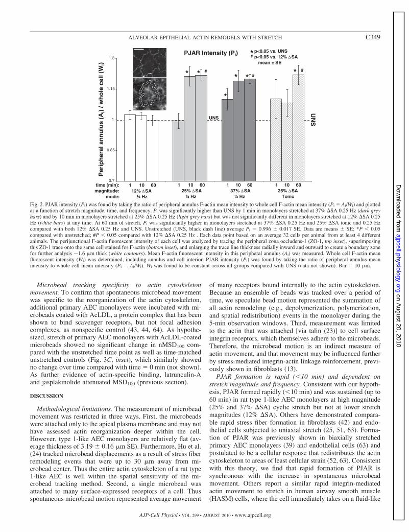
These qualitative observations of rapid PJAR formationcorrelated well with the quantitative metrics. PJAR intensity(Pi) was significantly higher than UNS by 1 min in monolayersstretched at 37% �SA 0.25 Hz and by 10 min in monolayersstretched at 25% �SA 0.25 Hz (Fig. 2, dark grey bars and lightgrey bars, respectively) but was not significantly different inmonolayers stretched at 12% �SA 0.25 Hz (white bars) at anytime. At 60 min of stretch, Pi was significantly higher inmonolayers stretched at 37% �SA 0.25 Hz and 25% �SA tonicand 0.25 Hz compared with both 12% �SA 0.25 Hz and UNS.Thus Pi showed a dependence on both stretch magnitude andstretch frequency.
We hypothesized that actin cytoskeleton remodeling duringformation of PJAR in stretched AEC monolayers would beaccompanied by an increase in the movement of microbeadscoated with the Arg-Gly-Asp (RGD) peptide sequence specif-ically binding to apical cell surface transmembrane integrinreceptors that anchor to the cytoskeleton. Qualitative PJARformation data corroborates well with microbead tracking data,showing significant actin remodeling in 25% and 37% �SAtonic (0 Hz) and dynamic (0.25 Hz) stretched monolayers after1 min (Fig. 3, C and D, respectively). Also, similar to quanti-tative Pi data, the microbeads adhered to the RGD receptorsshowed an effect of stretch magnitude and frequency at 1 minof stretch, such that nMSD100 (mean squared displacementduring 100 s normalized to unstretched values) was signifi-cantly higher in both 25% and 37% �SA held (sustained tonic)stretched (Fig. 3C) and cyclic (0.25 Hz) stretched monolayers(Fig. 3D) compared with their unstretched values. Further-more, at 1 min stretch nMSD100 was even greater in 37% �SAsustained tonic compared with 25% �SA sustained tonic (Fig.3C). Finally, at 1 min nMSD100 was greater in 25% �SA 0.25Hz when compared with 25% �SA sustained tonic stretch (Fig.3, D vs. C). Thus actin movement at the cell membranesignificantly increased at 1 min of stretch compared with theunstretched time point and, similar to Pi, was sensitive tostretch magnitude and frequency.
Although qualitative images and quantitative PJAR forma-tion showed PJAR persistence for stretch duration, the spon-taneous movement of anchored RGD-coated microbeads
dropped precipitously after 1 min, back to prestretch levels forall stretch conditions (Fig. 3, C and D). The one exemption wasat 40 min in the 25% �SA 0.25-Hz stretched monolayer groupwhere nMSD100 was also higher than unstretched at this timepoint. Also, at longer stretch durations (�10–40 min), spon-taneous microbead movement showed no dependence onstretch magnitude. Finally, nMSD100 in unstretched monolay-ers significantly decreased by 10 min, remaining constant(except at 30 min, which was not different from time � 0) forthe duration of time in the sustained tonic group, but not in thecyclic group.
Stretch-induced PJAR formation can be inhibited with jas-plakinolide and latrunculin-A. Treatment with jasplakinolidewas used to stabilize the actin cytoskeleton (5, 6). Monolayerstreated with 1 �M jasplakinolide for 10 min and then stretched25% �SA 0.25 Hz showed no qualitative evidence of PJARformation when fixed and labeled with F/G-actin antibody(Fig. 1B, bottom right). For comparison, consider monolayersstretched at the same magnitude and duration with vehiclecontrol (Fig. 1B, bottom left).
Qualitative images showing inhibited PJAR formation withjasplakinolide treatment corroborated well with quantitativemeasures of microbead tracking data. Treatment with 1 �Mjasplakinolide for 10 min also significantly attenuates themovement of integrin-adhered microbeads (MSD100 of 2,962 � 360nm2 SE) when compared with untreated monolayers at thesame (MSD100 of 5,891 � 743 nm2 SE). Thus we concludeactin stabilization with jasplakinolide pretreatment inhibitsactin binding site movement and formation of PJAR instretched monolayers.
Hypothesizing that actin reorganization requires depolymer-ization (inhibited by jasplakinolide) and repolymerization, weused latrunculin-A to inhibit actin repolymerization (70) instretched monolayers. Monolayers exposed to (sustained tonic)stretch of 25% �SA, 0.1 �M latrunculin-A pretreatment atten-uated the rapid (�1 min) increase in the spontaneous move-ment of microbeads attached to integrin receptors (Fig. 3C,inset) found in untreated monolayers. Thus pretreatment withlatrunculin-A abolished stretch-induced actin binding site re-modeling.
Actin remodeling response depends on cell type. Primary rattype 1-like AEC monolayer behavior was compared withmonolayers of a human alveolar epithelial adenocarcinomaA549 cell line (29). Qualitatively, A549 monolayers labeledwith phalloidin for F-actin exhibit PJAR in both unstretchedand held (sustained tonic) stretch of 37% �SA for up to 40 min(Fig. 4, inset). Thus A549 cells display no stretch-induced actinremodeling. Similarly, A549 monolayers held stretched at 25%and 37% �SA displayed no significant response in nMSD100
(Fig. 4). In unstretched A549 monolayers, tracking microbeadsattached to A549 integrin receptors display a significant de-crease in nMSD100 by 20 min and continuing for the durationof stretch compared with the 0 min time point (Fig. 4, also Fig.3B). Untreated, unstretched primary AEC monolayers from thesustained tonic group displayed a similar decrease in nMSD100
by 10 min lasting for the duration. However, unlike theprogressive decrease in nMSD100 in unstretched A549 cells,nMSD100 in untreated unstretched primary AEC monolayersdid not decline further.
C347ALVEOLAR EPITHELIAL ACTIN REMODELS WITH STRETCH
AJP-Cell Physiol • VOL 299 • AUGUST 2010 • www.ajpcell.org
on August 20, 2010
ajpcell.physiology.orgD
ownloaded from

Fig. 1. A: effect of biaxial stretch duration, magnitude, andfrequency on F-actin arrangement in type 1-like rat alveolarepithelial cell (AEC) monolayers before and after 1, 10, and60 min of stretch (time on x-axis). 1) Monolayers left un-stretched (UNS). 2) 12% change of surface area (�SA)0.25-Hz cyclic stretch at 60 min only. 3) 25% �SA 0.25-Hzcyclic stretch. 4) 37% �SA 0.25-Hz cyclic stretch. 5) 25%�SA sustained tonic (0 Hz ) stretch. Both 25% and 37% �SA0.25-Hz cyclic stretch produced actin stress fibers on the cellperiphery by 10 min, unlike monolayers stretched for 60 minat 12% �SA 0.25 Hz cyclic, which were similar to UNSmonolayers. Monolayers held sustained tonic 25% �SAstretch produced actin stress fibers on the cell periphery at 60min. Individual micrographs are 56 �m in width. Data at 60min stretch is comparable at 40 min (not shown). B: effect ofbiaxial stretch and jasplakinolide (JAS) on actin. Type 1-likerat AEC monolayers with antibody-labeled actin left un-stretched (top) or after 10 min of 25% �SA 0.25-Hz cyclicstretch (bottom). Vehicle control monolayers (left) stretched at25% �SA produce actin stress fibers on the cell periphery by10 min, whereas monolayers stretched at the same magnitudeand duration treated with 1 �M JAS for 10 min (right) tostabilize actin showed no perijunctional actin ring (PJAR)formation. Bar � 10 �m.
C348 ALVEOLAR EPITHELIAL ACTIN REMODELS WITH STRETCH
AJP-Cell Physiol • VOL 299 • AUGUST 2010 • www.ajpcell.org
on August 20, 2010
ajpcell.physiology.orgD
ownloaded from
Microbead tracking specificity to actin cytoskeletonmovement. To confirm that spontaneous microbead movementwas specific to the reorganization of the actin cytoskeleton,additional primary AEC monolayers were incubated with mi-crobeads coated with AcLDL, a protein complex that has beenshown to bind scavenger receptors, but not focal adhesioncomplexes, as nonspecific control (43, 44, 64). As hypothe-sized, stretch of primary AEC monolayers with AcLDL-coatedmicrobeads showed no significant change in nMSD100 com-pared with the unstretched time point as well as time-matchedunstretched controls (Fig. 3C, inset), which similarly showedno change over time compared with time � 0 min (not shown).As further evidence of actin-specific binding, latrunculin-Aand jasplakinolide attenuated MSD100 (previous section).
DISCUSSION
Methodological limitations. The measurement of microbeadmovement was restricted in three ways. First, the microbeadswere attached only to the apical plasma membrane and may nothave assessed actin reorganization deeper within the cell.However, type 1-like AEC monolayers are relatively flat (av-erage thickness of 3.19 � 0.16 �m SE). Furthermore, Hu et al.(24) tracked microbead displacements as a result of stress fiberremodeling events that were up to 30 �m away from mi-crobead center. Thus the entire actin cytoskeleton of a rat type1-like AEC is well within the spatial sensitivity of the mi-crobead tracking method. Second, a single microbead wasattached to many surface-expressed receptors of a cell. Thusspontaneous microbead motion represented average movement
of many receptors bound internally to the actin cytoskeleton.Because an ensemble of beads was tracked over a period oftime, we speculate bead motion represented the summation ofall actin remodeling (e.g., depolymerization, polymerization,and spatial redistribution) events in the monolayer during the5-min observation windows. Third, measurement was limitedto the actin that was attached [via talin (23)] to cell surfaceintegrin receptors, which themselves adhere to the microbeads.Therefore, the microbead motion is an indirect measure ofactin movement, and that movement may be influenced furtherby stress-mediated integrin-actin linkage reinforcement, previ-ously shown in fibroblasts (13).
PJAR formation is rapid (�10 min) and dependent onstretch magnitude and frequency. Consistent with our hypoth-esis, PJAR formed rapidly (�10 min) and was sustained (up to60 min) in rat type 1-like AEC monolayers at high magnitude(25% and 37% �SA) cyclic stretch but not at lower stretchmagnitudes (12% �SA). Others have demonstrated compara-ble rapid stress fiber formation in fibroblasts (42) and endo-thelial cells subjected to uniaxial stretch (25, 51, 63). Forma-tion of PJAR was previously shown in biaxially stretchedprimary AEC monolayers (39) and endothelial cells (63) andpostulated to be a cellular response that redistributes the actincytoskeleton to areas of least cellular strain (52, 63). Consistentwith this theory, we find that rapid formation of PJAR issynchronous with the increase in spontaneous microbeadmovement. Others report a similar rapid integrin-mediatedactin movement to stretch in human airway smooth muscle(HASM) cells, where the cell immediately takes on a fluid-like
Fig. 2. PJAR intensity (Pi) was found by taking the ratio of peripheral annulus F-actin mean intensity to whole cell F-actin mean intensity (Pi � Ai/Wi) and plottedas a function of stretch magnitude, time, and frequency. Pi was significantly higher than UNS by 1 min in monolayers stretched at 37% �SA 0.25 Hz (dark greybars) and by 10 min in monolayers stretched at 25% �SA 0.25 Hz (light grey bars) but was not significantly different in monolayers stretched at 12% �SA 0.25Hz (white bars) at any time. At 60 min of stretch, Pi was significantly higher in monolayers stretched at 37% �SA 0.25 Hz and 25% �SA tonic and 0.25 Hzcompared with both 12% �SA 0.25 Hz and UNS. Unstretched (UNS, black dash line) average Pi � 0.996 � 0.017 SE. Data are means � SE; *P � 0.05compared with unstretched; #P � 0.05 compared with 12% �SA 0.25 Hz . Each data point based on an average 32 cells per animal from at least 4 differentanimals. The perijunctional F-actin fluorescent intensity of each cell was analyzed by tracing the peripheral zona occludens-1 (ZO-1, top inset), superimposingthis ZO-1 trace onto the same cell stained for F-actin (bottom inset), and enlarging the trace line thickness radially inward and outward to create a boundary zonefor further analysis �1.6 �m thick (white contours). Mean F-actin fluorescent intensity in this peripheral annulus (Ai) was measured. Whole cell F-actin meanfluorescent intensity (Wi) was determined, including annulus and cell interior. PJAR intensity (Pi) was found by taking the ratio of peripheral annulus meanintensity to whole cell mean intensity (Pi � Ai/Wi). Wi was found to be constant across all groups compared with UNS (data not shown). Bar � 10 �m.
C349ALVEOLAR EPITHELIAL ACTIN REMODELS WITH STRETCH
AJP-Cell Physiol • VOL 299 • AUGUST 2010 • www.ajpcell.org
on August 20, 2010
ajpcell.physiology.orgD
ownloaded from

C350 ALVEOLAR EPITHELIAL ACTIN REMODELS WITH STRETCH
AJP-Cell Physiol • VOL 299 • AUGUST 2010 • www.ajpcell.org
on August 20, 2010
ajpcell.physiology.orgD
ownloaded from

behavior (8, 28, 53). Also, Trepat et al. (53) measured cellstiffness by using optical twisting cytometry and molecular-scale structural rearrangement using the spontaneous move-ment of beads and found a decrease in cell stiffness and anacceleration of remodeling kinetics with transient stretch. Inaddition, Krishnan et al. (28) measured cell traction stress byusing cell mapping rheometry and found a decrease in celltraction force following a biaxial stretch. Furthermore, we findthe increase in spontaneous microbead movement was depen-dent on stretch magnitude and frequency. Krishnan et al. (28)and Trepat et al. (53) also showed a stretch magnitude-depen-dent cell response.
With prolonged cyclic stretch, the initial spontaneous move-ment of microbeads was attenuated back to unstretched levelsfor the duration of stretch, and steady-state MSD levels wereunaffected by stretch magnitude or stretch frequency. Thisfinding suggests the mechanism of actin remodeling into PJARtook place rapidly (�1 min) during stretch and then ceased
with sustained stretch, despite persistence of PJAR. Thus, aftertransient fluidization at stretch onset, the cell returns to its moresolid-like state with sustained stretch, a finding similar to thatafter stretch release of HASM cells (8, 28, 53). Once formed,PJAR structure may require only a baseline actin remodelingrate for maintenance of the new organization, a rate similar tothe homogeneous actin structure found in unstretched mono-layers. Further investigation is needed to elucidate whetherrapid formation of PJAR is due to active biochemical signalingcascades, passive mechanical forces, or both.
The magnitude dependence of PJAR intensity correlateswith an increase in paracellular permeability at high magnitudestretch and no change in paracellular permeability at lowmagnitude stretch (9, 10). These findings strengthen the hy-pothesis that the actin cytoskeleton is integral to TJ barriermaintenance in primary AEC monolayers. Others have shownPJAR and TJ are intimately linked (34) and that modificationof the actin cytoskeleton results in changes in TJ-mediatedparacellular permeability (11, 31, 33). We speculate that thereorganization of actin might result in a physical separation ofactin and TJ protein, thus diminishing the cellular ability tomediate paracellular permeability.
Stretch-induced PJAR formation can be inhibited with jas-plakinolide and latrunculin-A. Previously, we reported thattreatment with 1 �M jasplakinolide reduced, but did notabolish, the stretch-induced increase in paracellular permeabil-ity in primary rat AEC monolayers stretched at 37% �SA (9).Here we show that we abolish formation of PJAR duringstretch to 25% �SA by pretreating monolayers with 1 �Mjasplakinolide, an actin-stabilizing agent that inhibits depoly-merization. In addition, treatment with latrunculin-A effec-tively inhibited the movement of actin-bound receptors inbiaxially stretched primary AEC monolayers. Similarly, Trepatet al. used 0.1 �M latrunculin-A in HASM cells and showedattenuation of stretch-induced decrease in cell stiffness (8, 53).Moreover Shen et al. (48) used latrunculin-A to depolymerizeactin in Madin-Darby canine kidney cells, finding a reductionin transepithelial resistance within 5 min, an internalization ofTJ protein occludin, and an elimination of PJAR within 20 min.Others have demonstrated the roles of protein kinase C (18),adenylate cyclase (50), Rho and Rac (69), Rho-kinase (1, 26,62), myosin light chain kinase (3, 22), and cofilin (38) on actininvolvement in TJ structure and function (22, 58, 59, 62) in
Fig. 3. A: applied stretch protocol in monolayers held in sustained tonic stretch (top), stretched cyclically (middle), or left unstretched (bottom). Monolayers wereleft on the scope to equilibrate for 30 min before start of stretch. Stretch starts at time � 0 min. Adherent microbeads were serially imaged in 5-min long epochslabeled MSD on the timeline. The median MSD100 (total mean squared displacement over 100 s) value during stretch of each monolayer was divided by itsrespective median MSD100 value just before stretch (stretch time � 0 min) to determine normalized MSD100 (nMSD100) of the monolayer. B: microbead MSDvs. time lag in A549 cells. MSD of unstretched cells during capture plotted against time lag (�t) at different times during 40 min of rest in A549 monolayers.MSD100 decreased in A549 cells during 40 min rest. Top inset: dark microbeads shown attached to A549 monolayer surface; bar � 50 �m. Bottom inset:Arg-Gly-Asp (RGD)-coated microbead (white arrow) bound to phalloidin-labeled F-actin cytoskeleton in rat type 1-like AEC monolayer; bar � 10 �m.C: nMSD100 in primary AEC monolayers. nMSD100 as a function of time for UNS samples and 25% and 37% �SA held in sustained tonic (0 Hz) stretchedsamples. nMSD100 compared with unstretched samples was significantly higher at 1 min 25% and 37% �SA sustained tonic stretch, attenuating back to baselineby 10 min. nMSD100 was greater in 37% �SA sustained tonic compared with 25% �SA sustained tonic. UNS monolayers show significant decrease in nMSD100
at 10 min, remaining constant (except at 30 min) for the duration of time. Inset: nMSD100 at 1 min of 25% �SA sustained tonic stretch (grey bars) or leftunstretched (white bars). Stretched monolayers either treated with 0.1 �M latrunculin-A or incubated with acetylated low-density lipoprotein (AcLDL)-coatedmicrobeads showed no significant change in nMSD100 compared with their corresponding unstretched time point as well as time-matched UNS controls.D: nMSD100 in primary AEC monolayers as a function of time for UNS samples and 25% and 37% �SA 0.25 Hz cyclic stretched samples. When comparedwith unstretched samples, nMSD100 was significantly higher at 1 min 25% and 37% �SA cyclic stretch, attenuating back to baseline by 10 min. nMSD100 in25% �SA 0.25 Hz stretched samples became significantly higher again at 40 min. Inset: illustrative bead motion traces over 5 min in monolayers left unstretched(left) or stretched 25% �SA 0.25 Hz for 1 min (right). Spontaneous bead displacement in stretched monolayers was higher. C and D: data are means � SE from9 monolayers per group with an average 125 beads analyzed per monolayer.
Fig. 4. Normalized MSD100 (nMSD100) as a function of stretch time in A549monolayers stretched 25% and 37% �SA held in sustained tonic (0 Hz) stretchor left unstretched. nMSD100 in stretched monolayers did not change signifi-cantly compared with unstretched monolayers. Unstretched monolayersshowed significantly lower nMSD100 at 20 min to 40 min. Data are means �SE from 5 monolayers per group with an average 125 beads analyzed permonolayer. Inset: A549 cells stained with phalloidin for F-actin, UNS, or heldat 37% �SA sustained tonic stretch for 40 min (compare with plot black datapoint). A549 cells exhibit PJAR with and without stretch. Bar � 10 �m.
C351ALVEOLAR EPITHELIAL ACTIN REMODELS WITH STRETCH
AJP-Cell Physiol • VOL 299 • AUGUST 2010 • www.ajpcell.org
on August 20, 2010
ajpcell.physiology.orgD
ownloaded from

other stretched cell types. Whereas our results show that PJARcan be modulated, further investigation will elucidate thespecific upstream pathways responsible for the formation andfunctional consequences of PJAR in biaxially stretched pri-mary AEC monolayers.
Actin remodeling response depends on cell type. Unlikeprimary cells, A549 cell monolayers exhibited PJAR in bothunstretched and stretched cell monolayers stained for F-actin.Also, the spontaneous movement of microbeads in unstretchedA549 monolayers was significantly lower by 20–40 min com-pared with time � 0 min, with nMSD100 at 40 min significantlyless than at 20 min. This progressive decrease in microbeadmovement in A549 cells, shown by others previously (54), isnot found in unstretched untreated rat type 1-like AEC mono-layers. The process of stiffening has been shown to exhibit aprogressive decrease in microbead movement and exhibit sim-ilarities to physical aging (8), a phenomenon found in someglassy materials (46). In aging systems, molecular networksconstantly advance to microconfigurations that are progres-sively more stable but do so at a speed that is slower than anyexponential process (40). We conclude that the actin arrange-ment and response to stretch of A549 cells is significantlydifferent from that in primary alveolar epithelial cells.
Summary. We have demonstrated that actin rearranges rap-idly in primary AEC monolayers to form perijunctional actinring during biaxial stretch and that formation depends onstretch magnitude and frequency. We have shown mechanisti-cally that PJAR formation was synchronous with an increase inactin binding site movement, which was attenuated to baselinelevels by 10 min. These data reveal that high-magnitude biaxialstretch within the physiological range increases the fluidity ofthe actin cytoskeleton, which reorganizes to form PJARs.Together with our previously published studies demonstratingthat similarly large stretch magnitudes and rates adverselyaffect monolayer permeability (9, 14), we further speculate thatrapid actin cytoskeleton reorganization has a deleterious effecton paracellular permeability. Future studies will investigate theeffect of actin remodeling pathway inhibitors on retainingparacellular barrier properties during stretch to explore oppor-tunities to prevent ventilator-induced lung injury.
ACKNOWLEDGMENTS
The authors thank Yoram Lanir for role in the development of the exper-imental design of the MSD studies and Anirudh Lingamaneni for assisting withPJAR intensity quantification.
GRANTS
This work was supported by the National Institutes of Health GrantR01-HL-057204 (to S. S. Margulies), from the University of PennsylvaniaAshton Fellowship program (to B. C. DiPaolo), and the Parker B. FrancisFellowship program (to G. Lenormand).
DISCLOSURES
No conflicts of interest, financial or otherwise, are declared by the author(s).
REFERENCES
1. Albinsson S, Nordstrom I, Hellstrand P. Stretch of the vascular wallinduces smooth muscle differentiation by promoting actin polymerization.J Biol Chem 279: 34849–34855, 2004.
2. An SS, Fabry B, Mellema M, Bursac P, Gerthoffer WT, Kayyali US,Gaestel M, Shore SA, Fredberg JJ. Role of heat shock protein 27 incytoskeletal remodeling of the airway smooth muscle cell. J Appl Physiol96: 1701–1713, 2004.
3. Birukov KG, Jacobson JR, Flores AA, Ye SQ, Birukova AA, VerinAD, Garcia JG. Magnitude-dependent regulation of pulmonary endothe-lial cell barrier function by cyclic stretch. Am J Physiol Lung Cell MolPhysiol 285: L785–L797, 2003.
4. Borok Z, Danto SI, Zabski SM, Crandall ED. Defined medium forprimary culture de novo of adult rat alveolar epithelial cells. In Vitro CellDev Biol Anim 30A: 99–104, 1994.
5. Bubb MR, Senderowicz AM, Sausville EA, Duncan KL, Korn ED.Jasplakinolide, a cytotoxic natural product, induces actin polymerizationand competitively inhibits the binding of phalloidin to F-actin. J BiolChem 269: 14869–14871, 1994.
6. Bubb MR, Spector I, Beyer BB, Fosen KM. Effects of jasplakinolide onthe kinetics of actin polymerization. An explanation for certain in vivoobservations. J Biol Chem 275: 5163–5170, 2000.
7. Bursac P, Fabry B, Trepat X, Lenormand G, Butler JP, Wang N,Fredberg JJ, An SS. Cytoskeleton dynamics: fluctuations within thenetwork. Biochem Biophys Res Commun 355: 324–330, 2007.
8. Bursac P, Lenormand G, Fabry B, Oliver M, Weitz DA, Viasnoff V,Butler JP, Fredberg JJ. Cytoskeletal remodelling and slow dynamics inthe living cell. Nat Mater 4: 557–561, 2005.
9. Cavanaugh KJ, Cohen TS, Margulies SS. Stretch increases alveolarepithelial permeability to uncharged micromolecules. Am J Physiol CellPhysiol 290: C1179–C1188, 2006.
10. Cavanaugh KJ Jr and Margulies SS. Measurement of stretch-inducedloss of alveolar epithelial barrier integrity with a novel in vitro method. AmJ Physiol Cell Physiol 283: C1801–C1808, 2002.
11. Cavanaugh KJ Jr Oswari J, and Margulies SS. Role of stretch on tightjunction structure in alveolar epithelial cells. Am J Respir Cell Mol Biol25: 584–591, 2001.
12. Cheek JM, Evans MJ, Crandall ED. Type I cell-like morphology intight alveolar epithelial monolayers. Exp Cell Res 184: 375–387, 1989.
13. Choquet D, Felsenfeld DP, Sheetz MP. Extracellular matrix rigiditycauses strengthening of integrin-cytoskeleton linkages. Cell 88: 39–48,1997.
14. Cohen TS, Cavanaugh KJ, Margulies SS. Frequency and peak stretchmagnitude affect alveolar epithelial permeability. Eur Respir J 32: 854–861, 2008.
15. Danto SI, Zabski SM, Crandall ED. Reactivity of alveolar epithelialcells in primary culture with type I cell monoclonal antibodies. Am JRespir Cell Mol Biol 6: 296–306, 1992.
16. Dewey CF Jr. Effects of fluid flow on living vascular cells. J BiomechEng 106: 31–35, 1984.
17. Dobbs LG, Gonzalez R, Williams MC. An improved method for isolat-ing type II cells in high yield and purity. Am Rev Respir Dis 134: 141–145,1986.
18. Dwyer-Nield LD, Miller AC, Neighbors BW, Dinsdale D, MalkinsonAM. Cytoskeletal architecture in mouse lung epithelial cells is regulatedby protein kinase C- and calpain II. Am J Physiol Lung Cell Mol Physiol270: L526–L534, 1996.
19. Egan EA. Lung inflation, lung solute permeability, and alveolar edema. JAppl Physiol 53: 121–125, 1982.
20. Fujiwara K, Pollard TD. Fluorescent antibody localization of myosin inthe cytoplasm, cleavage furrow, and mitotic spindle of human cells. J CellBiol 71: 848–875, 1976.
21. Haake R, Schlichtig R, Ulstad DR, Henschen Barotrauma RR. Patho-physiology, risk factors, and prevention. Chest 91: 608–613, 1987.
22. Hecht G, Pestic L, Nikcevic G, Koutsouris A, Tripuraneni J, LorimerDD, Nowak G, Guerriero V Jr, Elson EL, and Lanerolle PD. Expres-sion of the catalytic domain of myosin light chain kinase increasesparacellular permeability. Am J Physiol Cell Physiol 271: C1678–C1684,1996.
23. Horwitz A, Duggan K, Buck C, Beckerle MC, Burridge K. Interactionof plasma membrane fibronectin receptor with talin–a transmembranelinkage. Nature 320: 531–533, 1986.
24. Hu S, Chen J, Fabry B, Numaguchi Y, Gouldstone A, Ingber DE,Fredberg JJ, Butler JP, Wang N. Intracellular stress tomography revealsstress focusing and structural anisotropy in cytoskeleton of living cells. AmJ Physiol Cell Physiol 285: C1082–C1090, 2003.
25. Iba T, Sumpio BE. Morphological response of human endothelial cellssubjected to cyclic strain in vitro. Microvasc Res 42: 245–254, 1991.
26. Kaunas R, Nguyen P, Usami S, Chien S. Cooperative effects of Rho andmechanical stretch on stress fiber organization. Proc Natl Acad Sci USA102: 15895–15900, 2005.
C352 ALVEOLAR EPITHELIAL ACTIN REMODELS WITH STRETCH
AJP-Cell Physiol • VOL 299 • AUGUST 2010 • www.ajpcell.org
on August 20, 2010
ajpcell.physiology.orgD
ownloaded from

27. Kim KJ, Crandall ED. Effects of lung inflation on alveolar epithelialsolute and water transport properties. J Appl Physiol 52: 1498–1505,1982.
28. Krishnan R, Park CY, Lin YC, Mead J, Jaspers RT, Trepat X,Lenormand G, Tambe D, Smolensky AV, Knoll AH, Butler JP,Fredberg JJ. Reinforcement versus fluidization in cytoskeletal mechano-responsiveness. PLoS One 4: e5486, 2009.
29. Lieber M, Smith B, Szakal A, Nelson-Rees W, Todaro G. A continuoustumor-cell line from a human lung carcinoma with properties of type IIalveolar epithelial cells. Int J Cancer 17: 62–70, 1976.
30. Liebler JM, Borok Z, Li X, Zhou B, Sandoval AJ, Kim KJ, CrandallED. Alveolar epithelial type I cells express beta2-adrenergic receptors andG-protein receptor kinase 2. J Histochem Cytochem 52: 759–767, 2004.
31. Ma TY, Hollander D, Tran LT, Nguyen D, Hoa N, Bhalla D. Cytoskel-etal regulation of Caco-2 intestinal monolayer paracellular permeability. JCell Physiol 164: 533–545, 1995.
32. Madara JL. Intestinal absorptive cell tight junctions are linked to cy-toskeleton. Am J Physiol Cell Physiol 253: C171–C175, 1987.
33. Madara JL, Barenberg D, Carlson S. Effects of cytochalasin D onoccluding junctions of intestinal absorptive cells: further evidence that thecytoskeleton may influence paracellular permeability and junctionalcharge selectivity. J Cell Biol 102: 2125–2136, 1986.
34. Madara JL, Moore R, Carlson S. Alteration of intestinal tight junctionstructure and permeability by cytoskeletal contraction. Am J Physiol CellPhysiol 253: C854–C861, 1987.
35. Maksym GN, Fabry B, Butler JP, Navajas D, Tschumperlin DJ,Laporte JD, Fredberg JJ. Mechanical properties of cultured humanairway smooth muscle cells from 0.05 to 04 Hz . J Appl Physiol 89:1619–1632, 2000.
36. Margulies SS, Oswari J, Matthay MA, Tschumperlin DJ. Alveolarepithelial cytoskeleton and cell vulnerability to stretch. In: Proceedings ofthe American Society of Mechanical Engineers Bioengineering Division,1999, p. 517–518.
37. Mitic LL, Anderson JM. Molecular architecture of tight junctions. AnnuRev Physiol 60: 121–142, 1998.
38. Nagumo Y, Han J, Bellila A, Isoda H, Tanaka T. Cofilin mediatestight-junction opening by redistributing actin and tight-junction proteins.Biochem Biophys Res Commun 377: 921–925, 2008.
39. Oswari J, Matthay MA, Margulies SS. Keratinocyte growth factorreduces alveolar epithelial susceptibility to in vitro mechanical deforma-tion. Am J Physiol Lung Cell Mol Physiol 281: L1068–L1077, 2001.
40. Parisi G. Brownian motion. Nature 433: 221, 2005.41. Parker JC, Hernandez LA, Peevy KJ. Mechanisms of ventilator-in-
duced lung injury. Crit Care Med 21: 131–143, 1993.42. Pender N, McCulloch CA. Quantitation of actin polymerization in two
human fibroblast sub-types responding to mechanical stretching. J Cell Sci100: 187–193, 1991.
43. Plopper G, Ingber DE. Rapid induction and isolation of focal adhesioncomplexes. Biochem Biophys Res Commun 193: 571–578, 1993.
44. Puig-de-Morales M, Millet E, Fabry B, Navajas D, Wang N, Butler JP,Fredberg JJ. Cytoskeletal mechanics in adherent human airway smoothmuscle cells: probe specificity and scaling of protein-protein dynamics.Am J Physiol Cell Physiol 287: C643–C654, 2004.
45. Qiao R, Zhou B, Liebler JM, Li X, Crandall ED, Borok Z. Identifica-tion of three genes of known function expressed by alveolar epithelial typeI cells. Am J Respir Cell Mol Biol 29: 98–105, 2003.
46. Ramos L, Cipelletti L. Ultraslow dynamics and stress relaxation in theaging of a soft glassy system. Phys Rev Lett 87: 245503, 2001.
47. Raupach C, Zitterbart DP, Mierke CT, Metzner C, Muller FA, FabryB. Stress fluctuations and motion of cytoskeletal-bound markers. Phys RevE Stat Nonlin Soft Matter Phys 76: 011918, 2007.
48. Shen L, Turner JR. Actin depolymerization disrupts tight junctions viacaveolae-mediated endocytosis. Mol Biol Cell 16: 3919–3936, 2005.
49. Shikata Y, Rios A, Kawkitinarong K, DePaola N, Garcia JG, BirukovKG. Differential effects of shear stress and cyclic stretch on focal
adhesion remodeling, site-specific FAK phosphorylation, and small GT-Pases in human lung endothelial cells. Exp Cell Res 304: 40–49, 2005.
50. Shirinsky VP, Antonov AS, Birukov KG, Sobolevsky AV, RomanovYA, Kabaeva NV, Antonova GN, Smirnov VN. Mechano-chemicalcontrol of human endothelium orientation and size. J Cell Biol 109:331–339, 1989.
51. Takemasa T, Sugimoto K, Yamashita K. Amplitude-dependent stressfiber reorientation in early response to cyclic strain. Exp Cell Res 230:407–410, 1997.
52. Takemasa T, Yamaguchi T, Yamamoto Y, Sugimoto K, Yamashita K.Oblique alignment of stress fibers in cells reduces the mechanical stress incyclically deforming fields. Eur J Cell Biol 77: 91–99, 1998.
53. Trepat X, Deng L, An SS, Navajas D, Tschumperlin DJ, GerthofferWT, Butler JP, Fredberg JJ. Universal physical responses to stretch inthe living cell. Nature 447: 592–595, 2007.
54. Trepat X, Grabulosa M, Puig F, Maksym GN, Navajas D, Farre R.Viscoelasticity of human alveolar epithelial cells subjected to stretch. AmJ Physiol Lung Cell Mol Physiol 287: L1025–L1034, 2004.
55. Tschumperlin DJ, Margulies SS. Equibiaxial deformation-induced in-jury of alveolar epithelial cells in vitro. Am J Physiol Lung Cell MolPhysiol 275: L1173–L1183, 1998.
56. Tschumperlin DJ, Oswari J, Margulies AS. Deformation-induced injuryof alveolar epithelial cells. Effect of frequency, duration, and amplitude.Am J Respir Crit Care Med 162: 357–362, 2000.
57. Turner JR. “Putting the squeeze” on the tight junction: understandingcytoskeletal regulation. Semin Cell Dev Biol 11: 301–308, 2000.
58. Turner JR, Angle JM, Black ED, Joyal JL, Sacks DB, Madara JL.PKC-dependent regulation of transepithelial resistance: roles of MLC andMLC kinase. Am J Physiol Cell Physiol 277: C554–C562, 1999.
59. Turner JR, Rill BK, Carlson SL, Carnes D, Kerner R, Mrsny RJ,Madara JL. Physiological regulation of epithelial tight junctions isassociated with myosin light-chain phosphorylation. Am J Physiol CellPhysiol 273: C1378–C1385, 1997.
60. van der Flier A, Sonnenberg A. Function and interactions of integrins.Cell Tissue Res 305: 285–298, 2001.
61. Van Itallie CM, Anderson JM. Occludin confers adhesiveness whenexpressed in fibroblasts. J Cell Sci 110: 1113–1121, 1997.
62. Walsh SV, Hopkins AM, Chen J, Narumiya S, Parkos CA, Nusrat A.Rho kinase regulates tight junction function and is necessary for tightjunction assembly in polarized intestinal epithelia. Gastroenterology 121:566–579, 2001.
63. Wang JH, Goldschmidt-Clermont P, Wille J, Yin FC. Specificity ofendothelial cell reorientation in response to cyclic mechanical stretching.J Biomech 34: 1563–1572, 2001.
64. Wang N, Butler JP, Ingber DE. Mechanotransduction across the cellsurface and through the cytoskeleton. Science 260: 1124–1127, 1993.
65. Ware LB, Matthay MA. The acute respiratory distress syndrome. N EnglJ Med 342: 1334–1349, 2000.
66. Wechezak AR, Viggers RF, Sauvage LR. Fibronectin and F-actinredistribution in cultured endothelial cells exposed to shear stress. LabInvest 53: 639–647, 1985.
67. White GE, Gimbrone MA Jr, Fujiwara K. Factors influencing theexpression of stress fibers in vascular endothelial cells in situ. J Cell Biol97: 416–424, 1983.
68. Wiesner S, Legate KR, Fassler R. Integrin-actin interactions. Cell MolLife Sci 62: 1081–1099, 2005.
69. Wojciak-Stothard B, Potempa S, Eichholtz T, Ridley AJ. Rho and Racbut not Cdc42 regulate endothelial cell permeability. J Cell Sci 114:1343–1355, 2001.
70. Yarmola EG, Somasundaram T, Boring TA, Spector I, Bubb MR.Actin-latrunculin A structure and function. Differential modulation ofactin-binding protein function by latrunculin A. J Biol Chem 275: 28120–28127, 2000.
C353ALVEOLAR EPITHELIAL ACTIN REMODELS WITH STRETCH
AJP-Cell Physiol • VOL 299 • AUGUST 2010 • www.ajpcell.org
on August 20, 2010
ajpcell.physiology.orgD
ownloaded from


![Review Actin-targeting natural products: structures ... · actin-binding proteins actively break or ‘sever’ actin filaments [e.g. actin-depolymerizing factor (ADF) and cofilin].](https://static.fdocuments.net/doc/165x107/5f0f85bd7e708231d44494d0/review-actin-targeting-natural-products-structures-actin-binding-proteins-actively.jpg)


![miR-106b-5p contributes to the lung metastasis of breast ...€¦ · CNN1 can inhibit actin-activated myosin ATPase and Ca. 2+ dependent mobility of actin 20, 21, therefore ] [being](https://static.fdocuments.net/doc/165x107/6046980676569e3a7c6b1418/mir-106b-5p-contributes-to-the-lung-metastasis-of-breast-cnn1-can-inhibit-actin-activated.jpg)