Streptococcus sanguis - Infection and Immunity - American Society
7
INFECTION AND IMMUNITY, Apr. 1983, p. 303-309 0019-9567/83/040303-07$02.00/0 Copyright (© 1983, American Society for Microbiology Vol. 40, No. 1 Corncob Formation Between Fusobacterium nucleatum and Streptococcus sanguis P. LANCY, JR.,' J. M. DIRIENZO,I* B. APPELBAUM,' B. ROSAN,1 AND S. C. HOLT2 Department of Microbiology, School of Dental Medicine and Center for Oral Health Research, University of Pennsylvania, Philadelphia, Pennsylvania 19104,1 and Department of Microbiology, University of Massachusetts, Amherst, Massachusetts 010032 Received 12 November 1982/Accepted 4 January 1983 Corncob formation in dental plaque was believed to be limited to strains of Bacterionema matruchotii and Streptococcus sanguis. We observed recently that strains of Fusobacterium nucleatum also interacted with S. sanguis to form corncobs. Since the fusobacteria are among the first anaerobic filaments to colonize subgingival plaque, these interactions could serve as a connecting link between the transformation of supra- to subgingival plaque. To further character- ize these interactions, quantitative in vitro studies of the kinetics of corncob formation of the fusobacteria were undertaken. These studies indicated that fewer streptococci were needed to saturate F. nucleatum strain 364 compared to strain 10953. Corncob formation with both strains was enhanced with increasing pH up to pH 8, at which point autoaggregation of the streptococci occurred. Variation in ionic strength and divalent cations had little effect on the interaction, and EDTA suppressed aggregate formation only slightly. Detergents at concentrations above 0.05% also inhibited corncob formation. Electron micrographs suggested that attachment of the cocci to the fusiforms was mediated through localized tufts of fimbriae, as they are in the Bacterionema system. However, although both trypsin and heat treatment of the streptococci inhibited corncob formation with fusobac- teria, the effects were not as complete as those seen in Bacterionema species. Unlike the Bacterionema model, trypsin and heat treatment of the fusobacteria resulted in inhibition of corncob formation. These results suggest that several different receptors may be involved in corncob formation. Dental plaque is an accumulation of oral bac- teria in an adherent matrix of polysaccharide and other bacterial and host products on tooth surfaces. Although the quantitative microbial composition is highly variable, plaque formation is usually characterized by an ordered succes- sion of cocci followed by filamentous forms (1, 11, 21). A dominant organism in the initial colonization of the tooth surface is Streptococ- cus sanguis (3-5); this appears to be associated with the affinity of the organism for the salivary glycoproteins in the acquired pellicle on the tooth surface (27). The streptococci in these nidi multiply to form microcolonies (23) which sub- sequently are "invaded" by a mixture of gram- positive rods, anaerobic gram-negative cocci, and filaments characteristic of mature supragin- gival plaque (11). A number of environmental factors, including alterations in the redox poten- tial, appear to play a part in the appearance of filaments (21). An especially important factor may be the specific interaction of the surface of the filaments with the surfaces of the streptococ- ci. The most common species of oral streptococ- ci involved in these reactions also appears to be S. sanguis (23). S. sanguis forms aggregates with Actinomyces spp. (2, 6, 7, 12), Streptococcus mutans (24), Bacteroides spp. (25), and Bacterionema sp. (8- 10, 14-17, 26). The aggregates with Bacterion- ema sp. are of particular interest because they form highly specific morphological units re- ferred to as "corncobs" (8). These units consist of a filamentous organism surrounded by adherent cocci (8, 10, 14-17) and resemble an ear of corn. Although the corncob was originally thought to be associated only with specific strains of S. sanguis and Bacterionema matru- chotii, we have found that corncobs are also formed between S. sanguis and Fusobacterium nucleatum. The fusobacteria are major inhabi- tants of subgingival plaque (13), and corncob formation with fusobacteria may serve as a connecting link between supra- and subgingival plaque. Study of the factors affecting corncob formation with the fusobacteria could be impor- tant for understanding plaque maturation and for developing strategies of prevention. 303 Downloaded from https://journals.asm.org/journal/iai on 19 January 2022 by 103.168.199.218.
Transcript of Streptococcus sanguis - Infection and Immunity - American Society
INFECTION AND IMMUNITY, Apr. 1983, p. 303-3090019-9567/83/040303-07$02.00/0Copyright (© 1983, American Society for Microbiology
Vol. 40, No. 1
Corncob Formation Between Fusobacterium nucleatum andStreptococcus sanguis
P. LANCY, JR.,' J. M. DIRIENZO,I* B. APPELBAUM,' B. ROSAN,1 AND S. C. HOLT2Department of Microbiology, School of Dental Medicine and Center for Oral Health Research, University of
Pennsylvania, Philadelphia, Pennsylvania 19104,1 and Department of Microbiology, University ofMassachusetts, Amherst, Massachusetts 010032
Received 12 November 1982/Accepted 4 January 1983
Corncob formation in dental plaque was believed to be limited to strains ofBacterionema matruchotii and Streptococcus sanguis. We observed recently thatstrains of Fusobacterium nucleatum also interacted with S. sanguis to formcorncobs. Since the fusobacteria are among the first anaerobic filaments tocolonize subgingival plaque, these interactions could serve as a connecting linkbetween the transformation of supra- to subgingival plaque. To further character-ize these interactions, quantitative in vitro studies of the kinetics of corncobformation of the fusobacteria were undertaken. These studies indicated that fewerstreptococci were needed to saturate F. nucleatum strain 364 compared to strain10953. Corncob formation with both strains was enhanced with increasing pH upto pH 8, at which point autoaggregation of the streptococci occurred. Variation inionic strength and divalent cations had little effect on the interaction, and EDTAsuppressed aggregate formation only slightly. Detergents at concentrations above0.05% also inhibited corncob formation. Electron micrographs suggested thatattachment of the cocci to the fusiforms was mediated through localized tufts offimbriae, as they are in the Bacterionema system. However, although both trypsinand heat treatment of the streptococci inhibited corncob formation with fusobac-teria, the effects were not as complete as those seen in Bacterionema species.Unlike the Bacterionema model, trypsin and heat treatment of the fusobacteriaresulted in inhibition of corncob formation. These results suggest that severaldifferent receptors may be involved in corncob formation.
Dental plaque is an accumulation of oral bac-teria in an adherent matrix of polysaccharideand other bacterial and host products on toothsurfaces. Although the quantitative microbialcomposition is highly variable, plaque formationis usually characterized by an ordered succes-sion of cocci followed by filamentous forms (1,11, 21). A dominant organism in the initialcolonization of the tooth surface is Streptococ-cus sanguis (3-5); this appears to be associatedwith the affinity of the organism for the salivaryglycoproteins in the acquired pellicle on thetooth surface (27). The streptococci in these nidimultiply to form microcolonies (23) which sub-sequently are "invaded" by a mixture of gram-positive rods, anaerobic gram-negative cocci,and filaments characteristic of mature supragin-gival plaque (11). A number of environmentalfactors, including alterations in the redox poten-tial, appear to play a part in the appearance offilaments (21). An especially important factormay be the specific interaction of the surface ofthe filaments with the surfaces of the streptococ-ci. The most common species of oral streptococ-
ci involved in these reactions also appears to beS. sanguis (23).
S. sanguis forms aggregates with Actinomycesspp. (2, 6, 7, 12), Streptococcus mutans (24),Bacteroides spp. (25), and Bacterionema sp. (8-10, 14-17, 26). The aggregates with Bacterion-ema sp. are of particular interest because theyform highly specific morphological units re-ferred to as "corncobs" (8). These units consistof a filamentous organism surrounded byadherent cocci (8, 10, 14-17) and resemble anear of corn. Although the corncob was originallythought to be associated only with specificstrains of S. sanguis and Bacterionema matru-chotii, we have found that corncobs are alsoformed between S. sanguis and Fusobacteriumnucleatum. The fusobacteria are major inhabi-tants of subgingival plaque (13), and corncobformation with fusobacteria may serve as aconnecting link between supra- and subgingivalplaque. Study of the factors affecting corncobformation with the fusobacteria could be impor-tant for understanding plaque maturation and fordeveloping strategies of prevention.
303
Dow
nloa
ded
from
http
s://j
ourn
als.
asm
.org
/jour
nal/i
ai o
n 19
Jan
uary
202
2 by
103
.168
.199
.218
.

304 LANCY ET AL.
MATERIALS AND METHODS
Strains, media, storage, and growth conditions. Thestreptococcal strains used in this study, CC5A andG9B, have been described previously (1, 9). Strepto-cocci were grown in brain heart infusion broth at 37°C.F. nucleatum 10953 and B. matruchotii 14266 were
obtained from the American Type Culture Collection,Rockville, Md., and F. nucleatum 364 was obtainedfrom S. S. Socransky, Forsyth Dental Center, Boston,Mass. The Fusobacterium strains were grown in 100ml of brain heart infusion broth supplemented with0.2% yeast extract, 0.05% L-cysteine, and 0.5% sodi-um bicarbonate under anaerobic conditions (Gas-Pak;BBL Microbiology Systems, Cockeysville, Md.).
Culture harvesting. The streptococcal cultures wereharvested by centrifugation at 9,000 x g for 15 min at4°C. The cell pellets were suspended in 1/10 theoriginal volume in 0.15 M NaCl and pelleted. Thisprocedure was repeated once more, and the final pelletwas suspended in 0.15 M NaCl to a value of 1,000Klett units at 470 nm. The Fusobacterium cultureswere treated similarly, except that centrifugation was
at 12,000 x g. The cell suspensions were stored on iceuntil used (maximum, 7 days). No changes in theability of the cells to form corncobs were observedduring this time.
Radioactive labeling conditions. Streptococci werelabeled with [methyl-3H]thymidine at a concentrationof 1 to 2 ,uCi/ml of brain heart infusion broth as
described previously (1, 9). The specific activity of thelabeled streptococci was approximately 2.4 x 10⁴cells/cpm (CC5A) and 1.3 x 10⁵ cells/cpm (G9B).Corncob assay procedure. The reaction mixture con-
sisted of 50 of Fusobacterium suspension (contain-ing approximately S x 107 fusobacteria), 175 ,ul ofstreptococcal suspension (containing 1.8 x 10' strep-tococci), and 1,775 ,u1 of buffer in a polystyrene 12- by75-ml test tube (W. Sarstedt, Inc., Princeton, N.J.).The buffer was either 0.05 M Tris-hydrochloride (pH8.0) or 0.05 M sodium phosphate (pH 7.0); the reactionmixtures were incubated for 1 h at 37°C on a rockingplatform mixer (LabQuake; Labindustries, Inc.,Berkeley, Calif.). The presence of corncobs was as-
sayed both microscopically and quantitatively by re-tention on 5-p.m filters (Nucleopore Corp., Pleasan-ton, Calif.) as described previously (9). Kinetic datawere plotted as the mean values calculated fromtriplicate samples.
Effects of pH, salts, and detergents on corncob forma-tion. Sodium, potassium, magnesium, and calcium, as
either chloride or acetate salts, were tested for theireffects on corncob formation when added to the reac-
tion mixture in the range of 100 to 500 mM. EDTA was
also tested in the same range. Ionic effects were
evaluated in both sodium phosphate and Tris-hydro-chloride buffers with buffer concentrations rangingfrom 10 to 500 mM. The effects of pH and detergents(sodium dodecyl sulfate and Triton X-100) in theconcentration range of 0.01 to 1% (vol/vol) on corncobformation were determined.
Electron microscopy. For transmission electron mi-croscopy, cells and corncobs were fixed by a two-stepregimen. Entire corncob reaction mixtures were made0.25% in redistilled glutaraldehyde (Baker, ThomasScientific Co., Philadelphia, Pa.) and incubated at 4°Covernight in the dark. The mixtures were then made
2.5% in glutaraldehyde. The fixed corncobs wereenrobed in warm Nobel agar, and the resulting pelletswere washed four times in cold phosphate buffer andpostfixed in 2% (wt/vol) osmium tetroxide for 1 h at4°C. The pellets were then washed once in phosphatebuffer, dehydrated through a graded ethanol series,and embedded in Epon. Thin sections were cut on aPorter-Blum MT-2 ultramicrotome, stained with ura-nyl acetate and lead citrate (20), and examined in aJOEL 1OOS transmission electron microscope.
Samples for scanning electron microscopy wereprepared as described previously (19) and examined ina JOEL 25S scanning electron microscope.
Saliva-coated hydroxyapatite adherence assays. Sali-va-coated hydroxyapatite adherence assays were per-formed as previously described (1). Briefly, [3H]thy-midine-labeled S. sanguis G9B cells were used asreference cells in competition assays. Increasingamounts of unlabeled F. nucleatum 364 or 10953 cellswere added to a fixed concentration of G9B cells in thepresence of saliva-coated hydroxyapatite beads, andthe amount of radiolabeled cells adhering to the beadswas quantitated. The results were expressed as afunction of the percentage of bound labeled cells in thetotal cell mixture, relative to control assays with nofusobacteria added.
Heat and trypsin treatment. Samples of cells to beheat treated were centrifuged in a Microfuge (FisherScientific Co., Pittsburgh, Pa.) for 1 min at 13,000 x g.The pellets were resuspended in an equal volume ofbuffer and placed in a 70°C water bath. After appropri-ate time periods, tubes were removed and centrifugedonce more, the pellets were resuspended in freshbuffer, and the suspensions were assayed for ability toform corncobs. Samples of cells to be trypsin treatedwere similarly pelleted. The pellets were resuspendedin an equal volume of 0.05 M Tris-hydrochloride buffer(pH 8.0) containing 0.5 mg of trypsin per ml (SigmaChemical Co., St. Louis, Mo.) and incubated at 37°Cin a water bath. At the end of the incubation period,the tubes were centrifuged, and the pellets werewashed three times with buffer and resuspended inbuffer containing 0.5 mg of soybean trypsin inhibitor(Sigma) per ml. Controls indicated that the inhibitorhad no effect on corncob formation. The tubes wereincubated for 30 min at 37°C and centrifuged, and thepellets were washed as described above and resus-pended to the original volume before corncob assay.
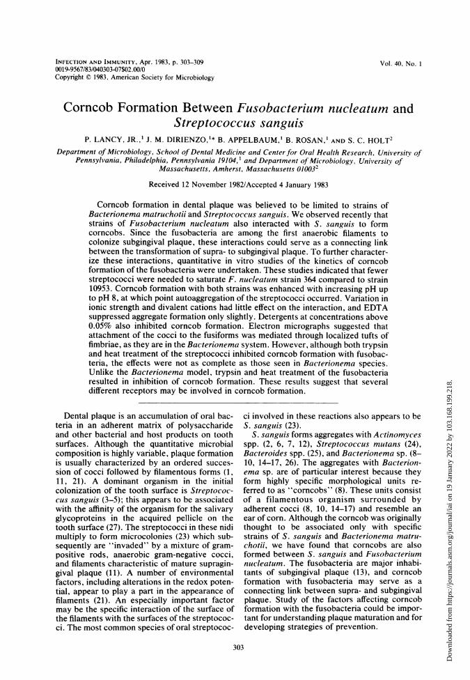
RESULTSMorphological observations. When strains of
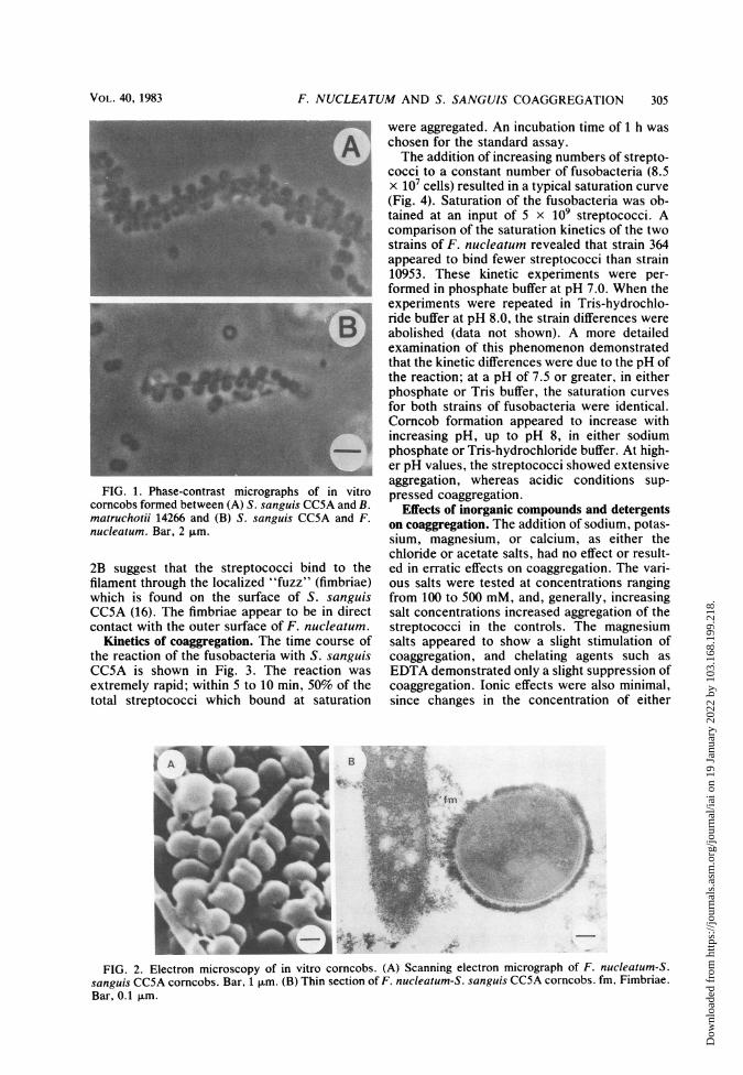
F. nucleatum were mixed with S. sanguis andexamined by phase-contrast microscopy, corn-cob formation similar to that observed withBacterionema sp. and S. sanguis was seen (Fig.1) (9). The streptococci were arranged along thesurface of the filament, and at saturation, thefilament was completely obscured. At subsatur-ating conditions, the arrangement of the strepto-cocci was random. Scanning electron micro-graphs revealed that the association of F.nucleatum and S. sanguis (Fig. 2A) had a mor-phology similar to that seen in the Bacterionemamodel. Thin sections of corncobs shown in Fig.
INFECT. IMMUN.
Dow
nloa
ded
from
http
s://j
ourn
als.
asm
.org
/jour
nal/i
ai o
n 19
Jan
uary
202
2 by
103
.168
.199
.218
.
F. NUCLEATUM AND S. SANGUIS COAGGREGATION 305
-.
FIG. 1. Phase-contrast micrographs of in vitrocorncobs formed between (A) S. sanguis CC5A and B.matruchotii 14266 and (B) S. sanguis CC5A and F.nucleatum. Bar, 2 ,um.
2B suggest that the streptococci bind to thefilament through the localized "fuzz" (fimbriae)which is found on the surface of S. sanguisCC5A (16). The fimbriae appear to be in directcontact with the outer surface of F. nucleatum.
Kinetics of coaggregation. The time course ofthe reaction of the fusobacteria with S. sanguisCC5A is shown in Fig. 3. The reaction wasextremely rapid; within 5 to 10 min, 50% of thetotal streptococci which bound at saturation
were aggregated. An incubation time of 1 h waschosen for the standard assay.The addition of increasing numbers of strepto-
cocci to a constant number of fusobacteria (8.5x 10⁷ cells) resulted in a typical saturation curve(Fig. 4). Saturation of the fusobacteria was ob-tained at an input of 5 x 10⁹ streptococci. Acomparison of the saturation kinetics of the twostrains of F. nucleatum revealed that strain 364appeared to bind fewer streptococci than strain10953. These kinetic experiments were per-formed in phosphate buffer at pH 7.0. When theexperiments were repeated in Tris-hydrochlo-ride buffer at pH 8.0, the strain differences wereabolished (data not shown). A more detailedexamination of this phenomenon demonstratedthat the kinetic differences were due to the pH ofthe reaction; at a pH of 7.5 or greater, in eitherphosphate or Tris buffer, the saturation curvesfor both strains of fusobacteria were identical.Corncob formation appeared to increase withincreasing pH, up to pH 8, in either sodiumphosphate or Tris-hydrochloride buffer. At high-er pH values, the streptococci showed extensiveaggregation, whereas acidic conditions sup-pressed coaggregation.
Effects of inorganic compounds and detergentson coaggregation. The addition of sodium, potas-sium, magnesium, or calcium, as either thechloride or acetate salts, had no effect or result-ed in erratic effects on coaggregation. The vari-ous salts were tested at concentrations rangingfrom 100 to 500 mM, and, generally, increasingsalt concentrations increased aggregation of thestreptococci in the controls. The magnesiumsalts appeared to show a slight stimulation ofcoaggregation, and chelating agents such asEDTA demonstrated only a slight suppression ofcoaggregation. Ionic effects were also minimal,since changes in the concentration of either
B
fm
FIG. 2. Electron microscopy of in vitro corncobs. (A) Scanning electron micrograph of F. nucleatum-S.sanguis CC5A corncobs. Bar, 1 pum. (B) Thin section of F. nucleatum-S. sanguis CC5A corncobs. fm, Fimbriae.Bar, 0.1 ,um.
VOL. 40, 1983
Dow
nloa
ded
from
http
s://j
ourn
als.
asm
.org
/jour
nal/i
ai o
n 19
Jan
uary
202
2 by
103
.168
.199
.218
.

306 LANCY ET AL.
4
'O
0
.5
0
N
0
3
2
0 5 10 15 20 25 30 35 40 45 50 55 60
Time (min)
FIG. 3. Number of S. sanguis CC5A cells bound to F. nucleatum as a function of time of incubation. Opencircles, F. nucleatum 364; closed circles, F. nucleatum 10953.
sodium phosphate or Tris-hydrochloride rangingfrom 10 to 500 mM neither stimulated nor de-pressed coaggregation. At concentrations above0.05%, detergents such as sodium dodecyl sul-fate and Triton X-100 abolished corncob forma-tion. On the basis of these results, no additionalsalts or reagents were added to the standardassay buffer.
Effect of heat and protease treatments on corn-
cob formation. The F. nucleatum strains were
heated at 70°C for various periods of time andthen assayed for corncob formation with un-
treated S. sanguis CCSA (Fig. 5). After beingheated for 30 min, strain 10953 showed 86%
inhibition of corncob formation, whereas strain364 reached a maximum value of inhibition(54%) after only 5 min of heating. Heat treat-ment of S. sanguis CCSA resulted in a maximuminhibition of corncob formation 5 min after expo-sure; a 32% inhibition was seen with strain 364and 35% with strain 10953. F. nucleatum 364 and10953 were also treated with trypsin, followedby trypsin inhibitor, and assayed for corncobactivity (Fig. 6A). Rapid inhibition of corncobformation was obtained after trypsin treatmentfor 30 min, and corncob formation was inhibitedbetween 50 and 60% for strains 364 and 10953.Trypsin also abolished the corncob activity of S.
150
'Dc
100
._
m.550
0
0
50
0 0.1 0.2 0.3 0.5 0.7 1.0
10 10 Cocci addedFIG. 4. Number of S. sanguis CC5A bound to a constant amount of F. nucleatwm as a function of the number
of cocci added. Symbols are explained in the legend to Fig. 3.
10
-0- - -- --
!0 q / _
10 2
ni I I I
INFECT. IMMUN.
51
1
Dow
nloa
ded
from
http
s://j
ourn
als.
asm
.org
/jour
nal/i
ai o
n 19
Jan
uary
202
2 by
103
.168
.199
.218
.
F. NUCLEATUM AND S. SANGUIS COAGGREGATION
25
c
0
.5
0
0
r-
20
15
10
51
0
012 5 10 20 30
Tkme at 70C (mhi)FIG. 5. Effects of heat treatment on corncob formation. Open squares, heat-treated F. nucleatum 364
assayed with untreated S. sanguis CCSA; closed squares, heat-treated F. nucleatum 10953 assayed withuntreated CC5A; open circles, treated S. sanguis CC5A assayed with untreated F. nucleatum 364; closed circles,treated S. sanguis CC5A assayed with untreated F. nucleatum 10953.
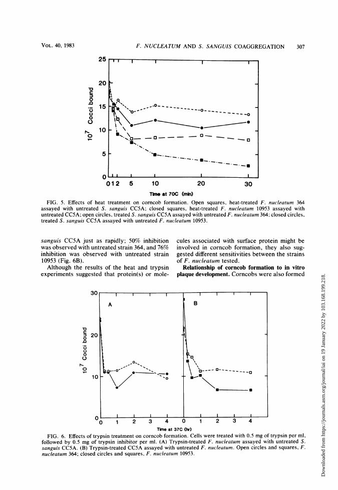
sanguis CC5A just as rapidly; 50% inhibitionwas observed with untreated strain 364, and 76%inhibition was observed with untreated strain10953 (Fig. 6B).Although the results of the heat and trypsin
experiments suggested that protein(s) or mole-
cules associated with surface protein might beinvolved in corncob formation, they also sug-gested different sensitivities between the strainsof F. nucleatum tested.
Relationship of corncob formation to in vitroplaque development. Corncobs were also formed
30
o200
0.2 3 V0 1 2 3 4
0
0
0..~~~~~~~~~~
10 'I
0~~ ~ ~ ~ ~ ~ ~
0 1 2 3 4 0 1 2 3 4
Time at 37C (hr)
FIG. 6. Effects of trypsin treatment on corncob formation. Cells were treated with 0.5 mg of trypsin per ml,followed by 0.5 mg of trypsin inhibitor per ml. (A) Trypsin-treated F. nucleatum assayed with untreated S.
sanguis CC5A. (B) Trypsin-treated CC5A assayed with untreated F. nucleatum. Open circles and squares, F.
nucleatum 364; closed circles and squares, F. nucleatum 10953.
U' I
- ..o-. - - - - - - - - -
jb
*.%. \- 0-..
.-0
II
VOL. 40, 1983 307
Dow
nloa
ded
from
http
s://j
ourn
als.
asm
.org
/jour
nal/i
ai o
n 19
Jan
uary
202
2 by
103
.168
.199
.218
.

308 LANCY ET AL.
with S. sanguis G9B, which has been usedextensively in studies of in vitro plaque forma-tion (1). To determine the effect of corncobformation on plaque formation, the F. nuclea-tum strains were mixed with 3H-labeled S. san-guis G9B in the presence of saliva-coated hy-droxyapatite, and adhesion was compared withthat of controls in the absence of the fusobac-teria. The results indicated a 60% increase in S.sanguis G9B adhesion in the presence of F.nucleatum 364 and a 178% increase in adhesionwhen strain G9B was mixed with strain 10953.
DISCUSSIONThere are some similarities and differences
between corncob formation involving Bacterio-nema sp. and Fusobacterium spp. The similar-ities are in their morphological resemblance toeach other, their relative tolerance to ionicstrength and various cations, and their sensitiv-ity to EDTA. The Bacterionema model exhibit-ed a distinct optimum at pH 6.5, whereas corn-cob formation in the Fusobacterium systemincreased as the pH was increased to 8, abovewhich streptococcal aggregation interfered withthe measurement of corncob formation. TheBacterionema corncobs seem to be relativelyinsensitive to detergents, whereas Fusobacter-ium corncobs are markedly inhibited by sodiumdodecyl sulfate and Triton X-100.Although morphologically both systems ap-
pear to be similar in that attachment of thestreptococci to the filaments appears to be medi-ated through the localized fimbriae on S. sanguisCC5A, strains such as S. sanguis G9B, which donot appear to have such localized tufts, can formcorncobs with fusobacteria but not with Bacteri-onema sp. However, the failure to detect local-ized tufts of fimbriae in some strains may be dueto technical difficulties. Attempts to overcomethese problems are being investigated, since ithas been reported that such localized tufts aremuch more common than formerly thought (P.Handley, personal communication). Indeed,Handley has suggested that at least four mor-phologically distinct types of localized tufts arefound in S. sanguis. However, differences in theconditions between corncob formation in Bac-terionema sp. and Fusobacterium spp. suggestthat different surface polymers or binding sitesare involved.
In contrast to the Bacterionema model, tryp-sin and heat treatment of fusobacteria result in a
reduction of corncob formation; similar treat-ments of Bacterionema sp. do not inhibit aggre-
gate formation. Although trypsin treatment ofthe streptococci does reduce corncob formationin both systems, inhibition in the Bacterionemamodel is greater. Mouton et al. (16) have shownthat lipoteichoic acid is present in the localized
tufts of S. sanguis CC5A, and their studies, aswell as those of others, have suggested that suchfimbriae may also contain protein and carbohy-drate (17). If the lipoteichoic acids are intimatelyassociated with fimbriae proteins, as suggestedby Ofek et al. (18), then trypsin treatment mightnot only remove surface proteins but could alsosolubilize the associated teichoic acids. Thus,these experiments still leave open the possibilitythat both protein and teichoic acids may play arole in corncob formation. Whatever the natureof the surface structures which are ultimatelyfound to be involved in the process, our studiessuggest that corncob formation is a more generalphenomenon and may indeed be one of themechanisms allowing for a change of the charac-teristic gram-positive supragingival plaque to amore gram-negative filamentous subgingivalplaque.
ACKNOWLEDGMENTS
This study was supported by Public Health Service grantsIF-32-DE-05215 (P.L.), DE-02623 (J.M.D.), DE-03180 (B.R.),and DE-05123 (S.C.H.) from the National Institute of DentalResearch.
LITERATURE CITED
1. Applebaum, B., E. Golub, S. C. Holt, and B. Rosan. 1979.In vitro studies of dental plaque formation: adsorption oforal streptococci to hydroxyapatite. Infect. Immun.25:717-728.
2. Bourgeau, G., and B. C. McBride. 1976. Dextran-mediat-ed interbacterial aggregation between dextran-synthesiz-ing streptococci and Actinomyces viscosus. Infect. Im-mun. 13:1228-1234.
3. Carlsson, J. 1967. Presence of various types of non-hemolytic streptococci in dental plaque and in other sitesin the oral cavity in man. Odontol. Revy 18:55-74.
4. Carlsson, J., H. Grahnen, and G. Jonsson. 1975. Lactoba-cilli and streptococci in the mouths of children. CariesRes. 9:333-339.
5. Carlsson, J., H. Grahnen, G. Jonsson, and S. Wikner.1970. Establishment of Streptococcus sanguis in themouths of infants. Arch. Oral Biol. 15:1143-1148.
6. Ellen, R. P., and I. P. Balcerzak-Raczkowski. 1977. Inter-bacterial aggregation of Actinomyces naeslundii and den-tal plaque streptococci. J. Periodontal Res. 12:11-20.
7. Gibbons, R. J., and N. Nygaard. 1970. Interbacterial ag-gregation of plaque bacteria. Arch. Oral Biol. 15:1397-1400.
8. Jones, S. J. 1972. A special relationship between sphericaland filamentous micro-organisms in mature human dentalplaque. Arch. Oral Biol. 17:613-616.
9. Lancy, P., Jr., B. Appelbaum, S. C. Holt, and B. Rosan.1980. Quantitative in vitro assay for "corncob" forma-tion. Infect. Immun. 29:663-670.
10. Listgarten, M. A., H. Mayo, and M. Amsterdam. 1973.Ultrastructure of the attachment device between coccaland filamentous microorganisms in "corncob" formationsof dental plaque. Arch. Oral Biol. 18:651-656.
11. Listgarten, M. A., H. E. Mayo, and R. Tremblay. 1975.Development of dental plaque on epoxy resin crowns inman. J. Periodontol. 46:10-25.
12. McIntire, F. C., A. E. Vatter, J. Baros, and J. Arnold.1978. Mechanism of coaggregation between Actinomycesviscosus T14V and Streptococcus sanguis 34. Infect.Immun. 21:978-988.
13. Moore, W. E. C., R. R. Ranney, and L. V. Holdeman.1982. Subgingival microflora in periodontal disease: cul-
INFECT. IMMUN.
Dow
nloa
ded
from
http
s://j
ourn
als.
asm
.org
/jour
nal/i
ai o
n 19
Jan
uary
202
2 by
103
.168
.199
.218
.
F. NUCLEATUM AND S. SANGUIS COAGGREGATION 309
tural studies, p. 13-26. In R. J. Genco and S. E. Mergen-hagen (ed.), Host-parasite interactions in periodontal dis-eases. American Society for Microbiology, Washington,D.C.
14. Mouton, C. 1974. Association bacterienne de types differ-ents au sein de la plaque dentaire: la formation en epi demais. J. Biol. Buccale 2:207-224.
15. Mouton, C., H. S. Reynolds, and R. J. Genco. 1977.Combined micromanipulation, culture and immunofluo-rescent techniques for isolation of the coccal organismscomprising the 'corn-cob" configuration of human dentalplaque. J. Biol. Buccale 5:321-332.
16. Mouton, C., H. S. Reynolds, and R. J. Genco. 1980.Characterization of tufted streptococci isolated from the"corncob' configuration of human dental plaque. Infect.Immun. 27:235-245.
17. Newman, H. N., and G. S. McKay. 1973. An unusualmicrobial configuration in human dental plaque. Micro-bios 8:117-128.
18. Ofek, I., W. A. Simpson, and E. H. Beachey. 1982. For-mation of molecular complexes between a structurallydefined M protein and acylated or deacylated lipoteichoicacid of Streptococcius pvogenes. J. Bacteriol. 149:426-433.
19. Poirier, T. P., S. J. Tonell, and S. C. Holt. 1979. Ultra-structure of gliding bacteria: scanning electron microsco-py of Capnocytophaga spltigena, Capnocytophaga gingi-valis, and Capnocytophaga ochrocea. Infect. Immun.26:1146-1158.
20. Reynolds, E. J. 1963. The use of lead citrate at high pH asan electron opaque stain for electron microscopy. J. Cell
Biol. 17:208-212.21. Ritz, H. L. 1967. Microbial population shifts in developing
human dental plaque. Arch. Oral Biol. 12:1561-1568.22. Rosan, B., B. Appelbaum, L. K. Campbell, K. W. Knox,
and A. J. Wicken. 1982. Chemostat studies of the effect ofenvironmental control on Streptococcus sanguis adher-ence to hydroxyapatite. Infect. Immun. 35:64-70.
23. Rosan, B., C. H. Lai, and M. A. Listgarten. 1976. Strepto-coccus sanguis: a model in the application in immunologi-cal analysis for the in-situ localization of bacteria in dentalplaque. J. Dent. Res. 55A:124-141.
24. Schachtele, C. F., S. K. Harlander, D. W. Fuller, P. K.Zollinger, and W. L. S. Leung. 1976. Bacterial interfer-ence with sucrose dependent adhesion of oral streptococ-ci, p. 401-412. In H. M. Stiles, W. J. Loesche, and T. C.O'Brien (ed.), Proceedings: Microbial Aspects of DentalCaries (a special supplement to Microbiology Abstracts),vol. II. Information Retrieval, Inc., Washington, D.C.
25. Slots, J., and R. J. Gibbons. 1978. Attachment of Bacte-roides melaniogenicuiis subsp. asaccharolvticus to oralsurfaces and its possible role in colonization of the mouthand of periodontal pockets. Infect. Immun. 19:254-264.
26. Takazoe, I., T. Matsukubo, and T. Kato. 1978. Experi-mental formation of "corn-cob" in vitro. J. Dent. Res.57:384-387.
27. van Houte, J. 1976. Oral bacterial colonization: mecha-nisms and implications, p. 3-32. In H. M. Stiles, W. J.Loesche. and T. C. O'Brien (ed.), Proceedings: MicrobialAspects of Dental Caries (a special supplement to Micro-biology Abstracts), vol. I. Information Retrieval, Inc.,Washington, D.C.
VOL. 40, 1983
Dow
nloa
ded
from
http
s://j
ourn
als.
asm
.org
/jour
nal/i
ai o
n 19
Jan
uary
202
2 by
103
.168
.199
.218
.