Streaming an image through the eye: The retina seen as a ... · PDF fileRESEARCH ISSN...
28
HAL Id: hal-00668076 https://hal.inria.fr/hal-00668076 Submitted on 9 Feb 2012 HAL is a multi-disciplinary open access archive for the deposit and dissemination of sci- entific research documents, whether they are pub- lished or not. The documents may come from teaching and research institutions in France or abroad, or from public or private research centers. L’archive ouverte pluridisciplinaire HAL, est destinée au dépôt et à la diffusion de documents scientifiques de niveau recherche, publiés ou non, émanant des établissements d’enseignement et de recherche français ou étrangers, des laboratoires publics ou privés. Streaming an image through the eye: The retina seen as a dithered scalable image coder Khaled Masmoudi, Marc Antonini, Pierre Kornprobst To cite this version: Khaled Masmoudi, Marc Antonini, Pierre Kornprobst. Streaming an image through the eye: The retina seen as a dithered scalable image coder. [Research Report] RR-7877, INRIA. 2012, pp.27. <hal-00668076>
Transcript of Streaming an image through the eye: The retina seen as a ... · PDF fileRESEARCH ISSN...

HAL Id: hal-00668076https://hal.inria.fr/hal-00668076
Submitted on 9 Feb 2012
HAL is a multi-disciplinary open accessarchive for the deposit and dissemination of sci-entific research documents, whether they are pub-lished or not. The documents may come fromteaching and research institutions in France orabroad, or from public or private research centers.
L’archive ouverte pluridisciplinaire HAL, estdestinée au dépôt et à la diffusion de documentsscientifiques de niveau recherche, publiés ou non,émanant des établissements d’enseignement et derecherche français ou étrangers, des laboratoirespublics ou privés.
Streaming an image through the eye: The retina seen asa dithered scalable image coder
Khaled Masmoudi, Marc Antonini, Pierre Kornprobst
To cite this version:Khaled Masmoudi, Marc Antonini, Pierre Kornprobst. Streaming an image through the eye: Theretina seen as a dithered scalable image coder. [Research Report] RR-7877, INRIA. 2012, pp.27.<hal-00668076>

ISS
N02
49-6
399
ISR
NIN
RIA
/RR
--78
77--
FR
+E
NG
RESEARCHREPORT
N° 7877February 2012
Project-Team NeuroMathCompProject Team
Streaming an imagethrough the eye:The retina seen as adithered scalable imagecoderKhaled Masmoudi, Marc Antonini, Pierre Kornprobst

RESEARCH CENTRESOPHIA ANTIPOLIS – MÉDITERRANÉE
2004 route des Lucioles - BP 93
06902 Sophia Antipolis Cedex
Streaming an image through the eye:
The retina seen as a dithered scalable image
coder
Khaled Masmoudi∗†, Marc Antonini†, Pierre Kornprobst‡
Project-Team NeuroMathComp Project Team
Research Report n° 7877 — February 2012 — 24 pages
Abstract: We propose the design of an original scalable image coder/decoder that is in-spired from the mammalians retina. Our coder accounts for the time-dependent and also non-deterministic behavior of the actual retina. The present work brings two main contributions: As afirst step, (i) we design a deterministic image coder mimicking most of the retinal processing stagesand then (ii) we introduce a retinal noise in the coding process, that we model here as a dithersignal, to gain interesting perceptual features. Regarding our first contribution, our main source ofinspiration will be the biologically plausible model of the retina called Virtual Retina. The coderthat we propose has two stages. The first stage is an image transform which is performed by theouter layers of the retina. Here we model it by filtering the image with a bank of difference ofGaussians with time-delays. The second stage is a time-dependent analog-to-digital conversionwhich is performed by the inner layers of the retina. The main novelty of this coder is to showthat the time-dependent behavior of the retina cells could ensure, in an implicit way, scalabilityand bit allocation. Regarding our second contribution, we reconsider the inner layers of the retina.We emit a possible interpretation for the non-determinism observed by neurophysiologists in theiroutput. For this sake, we model the retinal noise that occurs in these layers by a dither signal.The dithering process that we propose adds several interesting features to our image coder. Thedither noise whitens the reconstruction error and decorrelates it from the input stimuli. Further-more, integrating the dither noise in our coder allows a faster recognition of the fine details of theimage during the decoding process. Our present paper goal is twofold. First, we aim at mimickingas closely as possible the retina for the design of a novel image coder while keeping encouragingperformances. Second, we bring a new insight concerning the non-deterministic behavior of theretina.
Key-words: Static image compression, bio-inspired signal coding, retina, spiking neuron, A/Dconversion, dithering, scalability
∗ [email protected]† UNS–CNRS–I3S laboratory‡ INRIA–NeuroMathComp project team

Streaming an image through the eye
Résumé : Nous proposons un nouveau codeur/décodeur d’images dynamique qui est inspiréde la rétine des mammifères. Notre codeur tient compte du comportement à la fois dépen-dant du temps mais aussi non-déterministe de la rétine réelle. Le présent travail apporte deuxcontributions principales: (i) Dans un premier temps, nous concevons un codeur d’images àcomportement déterministe qui mime la plupart des étapes de traitement de la rétine et puis(ii), nous introduisons un bruit rétinien dans le processus de codage, que nous modélisons icicomme une signal de dithering, pour acquérir des fonctionnalités perceptuelles intéressantes. Ence qui concerne notre première contribution, notre principale source d’inspiration sera le modèlebiologiquement plausible de la rétine appelée Virtual Retina. Le codeur que nous proposonscomporte deux étapes. La première étape est une transformation de l’image qui est effectuéepar les couches externes de la rétine. Ici nous le modélisons par filtrage de l’image avec unebanc de différences de gaussiennes retardées dans le temps. La deuxième étape est une fonctiondu temps analogique-numérique de conversion qui est effectuée par les couches internes de larétine. La principale nouveauté de ce codeur est de montrer que le comportement dépendant dutemps de cellules de la rétine pourrait assurer, l’allocation de débit et la scalabilité d’une manièreimplicite. Concernant notre deuxième contribution, nous réexaminons les couches internes dela rétine. Nous émettons une interprétation possible pour le non-déterminisme de leur sortietel qu’observé observé par les neurophysiologistes. Pour cette cause, nous modélisons le bruitrétinienne qui survient dans ces couches par un signal de dithering. Le processus de ditheringque nous proposons ajoute plusieurs fonctionnalités intéressantes à notre codeur d’images. Lebruit de dithering rend blanc le spectre de l’erreur de reconstruction et la décorréle des stimulid’entrée. Par ailleurs, l’intégration du bruit de dithering dans notre codeur permet une recon-naissance plus rapide des détails fins de l’image pendant le processus de décodage. Notre objectifdans le présent travail est double. Tout d’abord, nous cherchons à imiter le plus fidèlement pos-sible la rétine pour la conception d’un codeur image original tout en gardant des performancesencourageantes. Deuxièmement, nous apportons une nouvelle vision concernant le comportementnon-déterministe de la rétine.
Mots-clés : Compression d’images, codage bio-inspiré du signal, rétine, neurone spikant,conversion A/D, dithering, scalabilité

Streaming an image through the eye 3
1 Introduction
Research in still image compression yielded several coding algorithms as the JPEG standards [2,5]. Though, these algorithms follow for their most a characteristic design schema. Namely, threemain stages are considered. First, a transform is applied to the image. Second, the transformeddata is quantized. Finally, an entropic coder compresses the bitstream to be transmitted. In-terestingly, we retrieve a similar behavior in the mammalians retina [7]. Indeed, accumulatedneurophysiologic evidence showed that the retina applies a transform to the image. Then, theretina binarizes the signal obtained to generate a set of uniformly shaped electrical impulses: thespikes [23]. Finally, several works tend to confirm that the retina generates a compact code to betransmitted to the visual cortex [22]. Thus, we are convinced that an interdisciplinary approachcombining the signal processing techniques and the knowledge acquired by neurophysiologistswould lead to novel coding algorithms beyond the standards.
In order to design a novel bio-inspired image coder, we need to capture the main propertiesof the retina processing. Though the action of perception seems effortless, neurophysiologicalexperiments proved that the mechanisms involved in the retina are highly complex and demand-ing. Recent studies such as [11] confirmed that the retina is doing non-trivial operations tothe input signal before transmission to the visual cortex. Besides, the retina appears to be anon-deterministic system. The retinal code is characterized by random fluctuations. Indeed,recordings made at the output of the retina show that a single stimulus leads to different codesacross trials. So that we have to deal with two issues. The first one is the complexity of theretinal processing and the need to decipher the code generated. The second issue is the non-determinism of the retinal response and its possible perceptual role. Therefore, our present paperwill be structured around two parts.
In a first step, our goal is to reproduce the main stages of the retina processing for the designof a retina-inspired coder with a deterministic behavior. Our main source of inspiration will bethe bio-plausible Virtual Retina model [36] whose goal was to find the best compromise betweenthe biological reality and the possibility to make large-scale simulations. Based on this model, wepropose a coding scheme following the architecture and functionalities of the retina. Unlike mostof the efforts mimicking the retina behavior, here we focus explicitly on the coding application.So that, we do some adaptations to the model in order to be able to conceive a decoding schemeand retrieve the original stimulus. We keep our design as close as possible to biological realitytaking into account the retina processing complexity, while keeping an interesting rate/distortiontrade-off. The coder/decoder that we design offers several interesting features such as scalability.
In a second step, we tackle the issue of reproducing the trial-to-trial variability in the retinaand its possible role. Here, we make the hypothesis that the non-determinism observed is adithering process. So that, we identify the deep retina layers behavior to a non-subtractivedithered A/D converter. We elaborate a multiscale model for the distribution of the dither noisethat we integrate in our coder. The hypothesis that we emit is seducing because of the perceptualimpact it induces. Still, our model obeys the biological plausibility constraint. Interestingly, thedither noise provides our coder with perceptual properties such as the enhancement of the imagecontours and singularities as well as the reconstruction error whitening.
This paper is organized into two parts. The first part consists of the Sections 2 to 5 andpresents the design of our bio-inspired scalable image coder/decoder with a deterministic behav-ior. This part is organized as follows. In Section 2 we revisit the retina model called VirtualRetina [36]. In Section 3, we show how this retina model can be used as the basis of a novel bio-inspired image coder. In Section 4, we present the decoding pathway. In Section 5, we show the
RR n° 7877

4 Masmoudi & Antonini & Kornprobst
main results that demonstrate the properties of our model. The second part consists of the Sec-tions 6 to 7. In Section 6, we show how we integrated the dithering process in our coder/decoder.Then, in Section 7, we detail the perceptual impact of it. We show, on two test images withdifferent properties, the ability of our dithered scalable coder to accelerate the recognition of theimage details and singularities during the decoding process. Finally, in Section 8, we summarizeour main conclusions.
2 Virtual Retina : A bio-plausible retina model
One first motivation for our work is to investigate the retina functional architecture and use it asa design basis to devise new codecs. So, it is essential to understand what are the main functionalprinciples of the retina processing. The literature in computational neuroscience dealing with theretina proposes different models (see, e.g., [35] for a review). These models are very numerous,ranking from detailed models of a specific physiological phenomenon, to large-scale models ofthe whole retina.
In this article, we focused on the category of large-scale models as we are interested in a modelthat gathers the main features of the mammalians retina. Within this category, we consideredthe retina model called Virtual Retina [36]. This model is one of the most complete ones, in theliterature, as it encompasses the major features of the actual mammalians retina. This model ismostly state-of-the-art and the authors confirmed its relevance by reproducing accurately actualretina cell recordings for several experiments.
The architecture of the Virtual Retina model follows the structure of the mammalians retinaas schematized in Figure 1(a). The model has several interconnected layers and three mainprocessing steps can be distinguished:
• The outer layers: The first processing step is described by non-separable spatio-temporalfilters, behaving as time-dependent edge detectors. This is a classical step implemented inseveral retina models.
• The inner layers: A non-linear contrast gain control is performed. This step models mainlybipolar cells by control circuits with time-varying conductances.
• The ganglionic layer: Leaky integrate and fire neurons are implemented to model theganglionic layer processing that finally converts the stimulus into spikes.
Given this model as a basis, our goal is to adapt it to conceive the new codec presented inthe next sections.
3 The coding pathway
The coding pathway is schematized in Figure 1(b). It follows the same architecture as VirtualRetina. However, since we have to define also a decoding pathway, we need to think aboutthe invertibility of each processing stage. For this reason some adaptations are required anddescribed in this section. The coder design, presented in this section, is an enhancement of ourprevious effort in [17].
3.1 The image transform: The outer layers of the retina
In Virtual Retina, the outer layers were modelled by a non-separable spatio-temporal filtering.This processing produces responses corresponding to spatial or temporal variations of the signal
Inria

Streaming an image through the eye 5
Figure 1: (a) Schematic view of the Virtual Retina model proposed by [36]. (b) and (c): Overviewof our bio-inspired codec. Given an image, the static DoG-based multi-scale transform generatesthe subbands {Fk}. DoG filters are sorted from the lowest frequency-band filter DoG0 to thehighest one DoGN−1. Each subband Fk is delayed using a time-delay circuit Dtk , with tk < tk+1.The time-delayed multi-scale output is then made available to the subsequent coder stages. Thefinal output of the coder is a set of spike series, and the coding feature adopted will be the spikecount nkij(tobs) recorded for each neuron indexed by (kij) at a given time tobs.
RR n° 7877

6 Masmoudi & Antonini & Kornprobst
because it models time-dependent interactions between two low-pass filters: this is termed center-surround differences. This stage has the property that it responds first to low spatial frequenciesand later to higher frequencies. This time-dependent frequency integration was shown for VirtualRetina [37] and it was confirmed experimentally (see, e.g., [26]). This property is interesting asa large amount of the total signal energy is contained in the low frequencies subbands, whereashigh frequencies bring further details. This idea already motivated bit allocation algorithms toconcentrate the resources for a good recovery on lower frequencies.
However, it appears that inverting this non-separable spatio-temporal filtering is a complexproblem [37, 38]. To overcome this difficulty, we propose to model differently this stage whilekeeping its essential features. To do so, we decomposed this process into two steps: The first oneconsiders only center-surround differences in the spatial domain (through differences of Gaus-sians) which is justified by the fact that our coder here gets static images as input. The secondstep reproduces the time-dependent frequency integration by the introduction of time-delays.
Center-surround differences in the spatial domain: The DoG model
Neurophysiologic experiments have shown that, as for classical image coders, the retina encodesthe stimulus representation in a transform domain. The retinal stimulus transform is performedin the cells of the outer layers, mainly in the outer plexiform layer (OPL). Quantitative studiessuch as [10, 24] have proven that the OPL cells processing can be approximated by a linearfiltering. In particular, the authors in [10] proposed the largely adopted DoG filter which is aweighted difference of spatial Gaussians that is defined as follows:
DoG(x, y) = wcGσc(x, y)− wsGσs
(x, y), (1)
where wc and ws are the respective weights of the center and surround components of thereceptive fields, and σc and σs are the standard deviations of the Gaussian kernels Gσc
and Gσs.
In terms of implementation, as in [28], the DoG cells can be arranged in a dyadic grid tosweep all the stimulus spectrum as schematized in Figure 2(a). Each layer k in the grid, is tiledwith DoGk cells having a scale k and generating a transform subband Fk, where σsk+1
= 12σsk
and σck+1= 1
2σck . So, in order to measure the degree of activation Ioplkij of a given DoGk cell
at the location (i, j) with a scale k, we compute the convolution of the original image f by theDoGk filter:
Ioplkij =
∞∑
x,y=−∞
DoGk(i− x, j − y) f(x, y). (2)
This transform generates a set of (43N2 − 1) coefficients for an N2-sized image, as it works
in the same fashion as a Laplacian pyramid [4]. An example of such a bio-inspired multi-scaledecomposition is shown in Figure 2(b). Note here that we added to this bank of filters a Gaussianlow-pass scaling function that represents the state of the OPL filters at the time origin. This yieldsa low-pass coefficient I
opl000 and enables the recovery of a low-pass residue at the reconstruction
level [8, 19].
Integrating time dynamics through time-delay circuits
Of course, the model described in (2) has no dynamical properties. In the actual retina, thesurround Gσs
in (1) appears progressively across time driving the filter passband from low fre-quencies to higher ones. Our goal is to reproduce this phenomenon that we called time-dependentfrequency integration. To do so, we added in the coding pathway of each subband Fk a time-delay circuit Dtk . The value of tk is specific to Fk and is an increasing function of k. The tk
Inria

Streaming an image through the eye 7
(a) (b) (c)
Figure 2: (a) Input image cameraman. (b) Example of a dyadic grid of DoG’s used for the imageanalysis (from [28]). (c) Example on image (a) of DoG coefficients generated by the retina model(the subbands are shown in the logarithmic scale)
Figure 3: Time delays Dtk introduced in the coding process. The time-dependent frequencyintegration is reproduced by delaying the coding process start of the subband Fk by tk. Theseries tk is represented as a function of the scale k. The progression law is exponential with atime constant τopl = 65ms.
delay causes the subband Fk to be transmitted to the subsequent stages of the coder startingfrom the time tk. The time-delayed activation coefficient I
oplkij (t) computed at the location (i, j)
for the scale k at time t is now defined as follows:
Ioplkij (t) = I
oplkij 1{t>tk}(t), (3)
where 1{t>tk} is the indicator function such that, 1{t>tk}(t) = 0 if t < tk and 1 otherwise.While in our previous work [17] tk is increasing linearly as a function of k, we changed the lawgoverning tk to an exponential one with a time constant denoted by τopl. This change is intendedto bring more biological plausibility to our new coder as the time behavior of the outer layerscells is exponential [10, 36]. Indeed, in the actual retina, the passband of the DoG cells runsthrough the low frequencies at a fast pace, then decelerates in an exponential fashion. So, thetime-dependent frequency integration is not a linear phenomenon. The evolution of time delaystk with respect to the scale k, in the present work, is detailed in Figure 3.
RR n° 7877

8 Masmoudi & Antonini & Kornprobst
3.2 The A/D converter: The inner and ganglionic layers of the retina
The retinal A/D converter is defined based on the processing occurring in the inner and ganglioniclayers, namely a contrast gain control, a non-linear rectification and a discretization based onleaky integrate and fire neurons (LIF) [15]. A different treatment will be performed for eachdelayed subband, and this produces a natural bit allocation mechanism. Indeed, as each subbandFk is presented at a different time tk, it will be subject to a transform according to the state ofour dynamic A/D converter at tk.
3.2.1 Contrast gain control
The retina adjusts its operational range to match the input stimuli magnitude range. This isdone by an operation called contrast gain control mainly performed in the bipolar cells. Indeed,real bipolar cells conductance is time varying, resulting in a phenomenon termed as shuntinginhibition. This shunting avoids the system saturation by reducing high magnitudes.In Virtual Retina, given the scalar magnitude I
oplkij of the input step current Ioplkij (t), the contrast
gain control is a non-linear operation on the potential of the bipolar cells. This potential variesaccording to both the time and the magnitude value I
oplkij ; and will be denoted by V b
kij(t, Ioplkij ).
This phenomenon is modelled, for a constant value of Ioplkij , by the following differential equation:
cbdV b
kij(t, Ioplkij )
dt+ gb(t)V b
kij(t, Ioplkij ) = I
oplkij (t), for t > 0,
gb(t) = Eτb
t∗ Q(V b
kij(t, Ioplkij )),
(4)
where Q(V bkij) = gb0 + λb
(
V bkij(t)
)2
and Eτb =1
τbexp
−t
τb , for t > 0. Figure 4(a) shows the time
behavior of V bkij(t, I
oplkij ) for different magnitude values I
oplkij of Ioplkij (t).
3.2.2 Non-linear rectification
In the next processing step, the potential V bkij(t, I
oplkij ) is subject to a non-linear rectification
yielding the so-called ganglionic current Igkij(t, I
oplkij ). Virtual Retina models it, for a constant
scalar value Ioplkij , by:
Igkij(t, I
oplkij ) = N
(
Twg,τg(t) ∗ V bkij(t, I
oplkij )
)
, for t > 0, (5)
where wg and τg are constant scalar parameters, Twg,τg is the linear transient filter defined byTwg,τg = δ0(t)− wgEτg(t), and N is defined by:
N(v) =
ig0
ig0 − λg(v − v
g0)
, if v < vg0
ig0 + λg(v − v
g0), if v > v
g0 ,
where ig0, v
g0 , and λg are constant scalar parameters. Figure 4(b) shows the time behavior of
Igkij(t, I
oplkij ) for different values of Ioplkij .
As the currents Ioplkij are delayed with times {tk}, our goal is to catch the instantaneous
behavior of the inner layers at these times {tk}. This amounts to infer the transforms Igtk(Ioplkij )
that maps a given scalar magnitude Ioplkij into a rectified current Irkij as the modelled inner layers
Inria

Streaming an image through the eye 9
(a) (b)
(c) (d)
Figure 4: 4(a): V bkij(t) as a function of time for different values of Iopl; 4(b): I
gkij as a function
of time for different values of Iopl; 4(c): The functions fgtk
that map Ioplkij into I
gkij for different
values of tk; 4(d): The functions fntobs
that map Irkij into nkij for different values of tobs
would generate it at tk. To do so, we start from the time-varying curves of Igkij(t, I
oplkij ) in
Figure 4(b) and we do a transversal cut at each time tk. We show in Figure 4(c) the resulting
maps fgtk
such that Igkij(tk, I
oplkij ) = f
gtk(Ioplkij ).
As for Ioplkij (t) (see Equation (3)), we introduce the time dimension using the indicator function1{t>tk}(t). The final output of this stage is the set of step functions Irkij(t) defined by:
Irkij(t) = Irkij 1{t>tk}(t), with Irkij = fgtk(Ioplkij ). (6)
This non-linear rectification is analogous to a widely-used telecommunication technique: thecompanding [6]. Companders are used to make the quantization steps unequal after a linear gaincontrol stage. Though, unlike A − law or µ − law companders that amplify low magnitudes,the inner layers emphasize high magnitudes in the signal. Indeed, the bio-inspired compander,defined here, emphasizes high energy signals rather than high probability ones. This tendencyis accentuated as the gain control gets higher across time. Besides, the inner layers stage havea time dependent behavior, whereas a usual gain controller/compander is static, and this makesour A/D converter go beyond the standards.
RR n° 7877

10 Masmoudi & Antonini & Kornprobst
3.2.3 Leaky integrate-and-fire quantization:
The ganglionic layer is the deepest one tiling the retina: it transforms a continuous signal Irkij(t)into discrete sets of spike trains. As in Virtual Retina, this stage is modelled by leaky integrateand fire neurons (LIF) which is a classical model. One LIF neuron is associated to every positionin each subband Fk. The time-behavior of a LIF neuron is governed by the fluctuation of itsvoltage Vkij(t). Whenever Vkij(t) reaches a predefined δ threshold, a spike is emitted and the
voltage goes back to a resting potential V 0R. Between two spike emission times, t
(l)kij and t
(l+1)kij ,
the potential evolves according to the following differential equation:
cldVkij(t)
dt+ glVkij(t) = Irkij(t), for t ∈ [t
(l)kij , t
(l+1)kij ], (7)
where gl is a constant conductance, and cl is a constant capacitance. In the literature, neuronsactivity is commonly characterized by the count of spikes emitted during an observation timebin [0, tobs], which we denote by nkij(tobs) [34]. Obviously, as nkij(tobs) encodes for the value ofIrkij(t), there is a loss of information as nkij(tobs) is an integer. The LIF is thus performing aquantization. If we observe the instantaneous behavior of the ganglionic layer at different timestobs, we get a quasi-uniform scalar quantizer that refines in time. We can do this by a similarprocess to the one described in the previous paragraph. We show in Figure 4(d) the resultingmaps fn
tobssuch that:
nkij(tobs) = fntobs
(Irkij). (8)
Based on the set of spike counts {nkij(tobs)}, measured at the output of our coder, we describein the next section the decoding pathway to recover the initial image f(x, y).
4 The decoding pathway
The decoding pathway is schematized in Figure 1(c). It consists in inverting, step by step, eachcoding stage described in Section 3. At a given time tobs, the coding data is the set of (43N
2− 1)
spike counts nkij(tobs), this section describes how we can recover an estimation ftobs of the N2-
sized input image f(x, y). Naturally, the recovered image ftobs(x, y) depends on the time tobswhich ensures time-scalability: the quality of the reconstruction improves as tobs increases. Theganglionic and inner layers are inverted using look-up tables constructed off-line and the imageis finally recovered by a direct reverse transform of the outer layers processing.
Recovering the input of the ganglionic layer:
First, given a spike count nkij(tobs), we recover Irkij(tobs), the estimation of Irkij(tobs). To do so,
we compute off-line the look-up table ntobs(Irkij) that maps the set of current magnitude values
Irkij into spike counts at a given observation time tobs (see Figure 4(d)). The reverse mapping is
done by a simple interpolation in the reverse-look up table denoted LUTLIFtobs
. Here we draw thereader’s attention to the fact that, as the input of the ganglionic layer is delayed, each coefficientof the subband Fk is decoded according to the reverse map LUTLIF
tobs−tk. Obviously, the recovered
coefficients do not match exactly the original ones due to the quantization performed in theLIF’s.
Recovering the input of the inner layers:
Second, given a rectified current value Irkij(tobs), we recover Ioplkij (tobs), the estimation of Ioplkij (tobs).In the same way as for the preceding stage, we infer the reverse “inner layers mapping” through
Inria

Streaming an image through the eye 11
the pre-computed look up table LUTCGtobs
. The current intensities Ioplkij (tobs), corresponding to
the retinal transform coefficients, are passed to the subsequent retinal transform decoder.
Recovering the input stimulus:
Finally, given the set of (43N2 − 1) coefficients {Ioplkij (tobs)}, we recover ftobs(x, y), the estimation
of the original image stimulus f(x, y). Though the dot product of every pair of DoG filters isapproximately equal to 0, the set of filters considered is not strictly orthonormal. We provedin [18] that there exists a dual set of vectors enabling an exact reconstruction. Hence, thereconstruction estimate f of the original input f can be obtained as follows:
ftobs(x, y) =∑
{kij}
Ioplkij (tobs) DoGk(i− x, j − y), (9)
where {kij} is the set of possible scales and locations in the considered dyadic grid and DoGk
are the duals of the DoGk filters obtained as detailed in [18]. Equation (9) defines a progres-sive reconstruction depending on tobs. This provides our code with an important feature: thescalability. Despite the fact that the input of our coder is a static image, we will be referring tothis feature as time-scalability. Indeed, in our case different levels of rate and quality levels areachievable thanks to the observation time tobs.
5 Results: Case of the bio-inspired and noiseless scalable
image coder
We show examples of image reconstruction using our bio-inspired coder at different times1. Then,we study these results in terms of quality and bit-cost.Quality is assessed by classical image quality criteria (PSNR and mean SSIM [31]). The costis measured by the Shannon entropy H(tobs) upon the population of {nkij(tobs)}. The entropycomputed in bits per pixel (bpp), for an N2-sized image, is defined by:
H(tobs) =1
N2
K−1∑
k=0
22kH({
nskij(tobs), (i, j) ∈ J0, 2k − 1K2})
, (10)
where K is the number of analyzing subbands. Figure 5 shows two examples of progressivereconstruction obtained with our new coder. Bit-rate/Quality are computed for each image interms of the triplet (bit-rate in bpp/ PSNR quality in dB/ mean SSIM quality). Progressivereconstruction of cameraman in the left column yields: From top to bottom (0.006 bpp/ 16.02dB/ 0.48), (0.077 bpp/ 18.34 dB/ 0.55), (0.23 bpp/ 21.20 dB/ 0.65), and (1.39 bpp/ 26.30 dB/0.84). Progressive reconstruction of baboon in the right column yields: From top to bottom(0.037 bpp/ 16.98 dB/ 0.18), (0.32 bpp/ 19.07 dB/ 0.35), (0.63 bpp/ 20.33 dB/ 0.49), and (2.24bpp/ 27.37 dB/ 0.92).
The new concept of time scalability is an interesting feature as it introduces time dynamics inthe design of the coder. Figure 6 illustrates this concept. This is a consequence of the mimickingof the actual retina. We also notice that, as expected, low frequencies are transmitted first toget a first approximation of the image, then details are added progressively to draw its contours.
1In all experiments, the model parameters are set to biologically realistic values: gb0
= 810−10S, τb =
12 10−3 s, λb
= 910−7, cb = 1.5 10−10 F , v
g0
= 410−3 V , i
g0
= 15 10−12A, wg
= 810−1, τg = 16 10
−3 s;
λg = 12 10−9 S, δ = 210−3 V, gL = 210−9 S, V 0
R= 0V , t0 = 10 10−3s, tK−1 = 38 10−3s, τopl = 65 10−3s.
RR n° 7877

12 Masmoudi & Antonini & KornprobstCameraman reconstruction Baboon reconstruction
20
ms
30
ms
40
ms
50
ms
Figure 5: Progressive image reconstruction of cameraman and baboon using our new bio-inspiredcoder. The coded/decoded image is shown at: 20 ms, 30 ms, 40 ms, and 50 ms.
Inria

Streaming an image through the eye 13
(a) Bit-rate as a function of time (b) Quality as a function of time
Figure 6: Illustration of the concept of time scalability. the test image is cameraman. 6(a) showsthe bit-rate variation of the encoded image as a function of the observation time tobs. The bit-rate is measured by means of the entropy in bits per pixel (bpp). 6(b) shows the reconstructionquality variation as a function of the observation time tobs. The quality is measured by means ofthe mean structural similarity index (mean SSIM). The only parameter that is tuned by the useris tobs. Both quality and cost increase in accordance with tobs. We talk about time-scalability.
The bit-cost of the coded image is slightly high. This can be explained by the fact that Shannonentropy is not the most relevant metric in our case as no context is taken into consideration,especially the temporal context. Indeed, one can easily predict the number of spikes at a giventime t knowing nkij(t−dt). Note also that no compression techniques, such that bit-plane coding,are yet employed. Our paper aims mainly at setting the basis of new bio-inspired coding designs.
For the reasons cited above, the performance of our coding scheme in terms of bit-cost havestill to be improved to be competitive with the well established JPEG and JPEG 2000 standards.Thus we show no comparison in this paper. Though primary results are encouraging, noting thatoptimizing the bit-allocation mechanism and exploiting coding techniques as bit-plane coding [27]would improve considerably the bit-cost. Besides, the image as reconstructed with our bio-inspired coder shows no ringing and no block effect as in JPEG. Finally our codec enablesscalability in an original fashion through the introduction of time dynamics within the codingmechanism.
Note also that differentiation in the processing of subbands, introduced through time-delaysin the retinal transform, ensures an implicit bit-allocation mechanism. In particular the non-linearity in the inner layers stage amplifies singularities and contours, and these provide crucialinformation for the analysis of the image.
6 Introducing the noise in the coder: The non-subtractive
dither hypothesis
In the preceding Sections 3 and 4, we presented the design of an image coder based on a bio-plausible model of the retina. We especially emphasized the deep retina layers analogy with A/Dconverters. Despite the fact that our coder takes into account several features of the actual retina
RR n° 7877

14 Masmoudi & Antonini & Kornprobst
as its time-dependent behavior, still it follows a deterministic law. Though, the actual neuralcode of the retina is clearly non-deterministic [9, 25]. Thus, in this section, we tackle the issue ofthe coding non-determinism in the retina. We make the proposal that the processing stages priorto the ganglionic layer yield a special type of noise: the dither noise. We then experience theperceptual impact of such a noise in our coder and give an original and plausible interpretationof its role in the stimuli coding process.
6.1 The non-deterministic nature of the neural code of the retina
One major issue encountered by neuroscientists is the non-determinism of the retinal neuralcode. Indeed, given a single visual stimulus, spikes timings in the retina output are not exactlyreproducible across trials. Yet, no clear evidence is established about the phenomena at theorigin of this trial-to-trial variability. Several hypotheses were discussed in the literature andyielded two different points of view. The first hypothesis is that the precise timings of individualspikes convey a large amount of information [21, 20]. This hypothesis suggests that the stimuluscoding process in the retina is deterministic and reports detailed information about the stimuluswith a high temporal fidelity. In this case, each single spike timing makes sense. The secondhypothesis is that only a few statistical quantities measured over the spike-based output conveythe relevant information about the stimulus to the visual cortex [3]. For instance, since [1] it waswidely assumed that the variable spike patterns corresponding to a single stimulus are randominstantiations of a desired firing rate. In this case, the precise timing of each single spike maynot be meaningful and thus spikes may carry some amount of noise. The spike based-outputshould then be averaged to reveal meaningful signals [9].
The role of spikes timings variability in the neural code of the retina is still an open issueand no clear evidence establishes whether this variability conveys precise information or randomnoise [25]. Here, we make the proposal that the non-determinism in the retinal processing priorto the ganglionic layer yields a dither noise [14, 16]. This noise, while corrupting the inputof the ganglionic layer by a completely random signal, brings interesting features to the spike-based output of the retina. We will notice that the dithering process helps us to recognize thefine details of the stimulus earlier than with the noiseless coder. For this to be possible, thedistribution of the noise that we introduce obeys specific constraints defined in [33]. Obviously,the dither noise hypothesis is one possible assumption among several others and we do not claimits biological exactness. Still, our present effort aims at bridging the differences between thedifferent points of view reported above by exploring the hypothesis of a “retinal useful noise”.
6.2 Multiscale non-subtractive dithering
We introduce in this section a multiscale dithering process that will be integrated in our bio-inspired image coder. Indeed, the coder that we designed has a multiscale architecture. So thatthe dither noise to be introduced must take into consideration the different scales of the retinamodel cells used for the image analysis.
We will assume that the processing stages of the retina that precede the ganglionic layerintroduce a noise. As this noise is prior to the quantization done in the ganglionic layer, it isreferred to as a dither noise. As, furthermore, this dither noise takes into consideration themultiscale architecture of the retina model, we will be talking about a multiscale dithering. Thepresent work extends our previous efforts in [14, 16] to the multiscale case.
A few techniques referred to as multiscale dithering have been described in the literature. For
Inria

Streaming an image through the eye 15
example, in [30] the authors considered a hierarchical wavelet transform. The sibling subbands,ie. lying in the same level, are decorrelated by applying a series of rotations. The transformapplied on the subbands is loosely referred to as dithering because it introduces a change onthe wavelet coefficients prior to quantization. The resulting image is meant to reduce entropywhile keeping the same perceptual quality. Another example is given in [12]. The authors usedan image hierarchical quadtree representation and employ an error diffusion algorithm to get abinary halftone image. The distribution of binary pixels over the image space gives the impres-sion of a multi-gray level image while using only two quantization levels. Although interesting,these state-of-the-art algorithms have one major drawback regarding the goals of our presentwork. Indeed, the techniques described rely on a totally deterministic algorithm. No randombehavior is introduced during the coding process. Whereas in our case, we need to consider acoding process that may lead to different codes across trials for a single image. Besides the twoalgorithms are iterative and time consuming and this is contradicts the speed of processing inthe retina.
In order to define the dither noise that corrupts the current Irkij (cf. Equation (6)) at theinput of the ganglionic layer, we reconsider the ganglion cell as a noisy leaky integrate and fireneuron (nLIF), that behaves according to the following equation:
cldVkij(t)
dt+ glVkij(t) = Irkij(t) + ηkij , for t ∈ [t
(l)kij , t
(l+1)kij ], (11)
The choice of the noise ηkij distribution model to apply must obey two constraints: the biologicalplausibility and the mathematical constraints that provide our coder with interesting perceptualproperties.
First, let us consider the biological plausibility constraint. Our aim is to mimic as closely aspossible the actual retina behavior while modelling the multiscale dithering ηkij . So that, onemust consider the nature of the dependency (if any) between the scale and the noise strengthaccording to neurophysiologists observations. In this context, the authors in [13] stated that:“The main difference between small and large cells is that the larger ones have lower peak sen-sitivity”. This means that the large retina cells have a low reactivity to stimulus variations andthus are poorly affected by noise. On the contrary, small cells are extremely sensitive to stimulusvariations and thus could be highly affected by noise. Our aim is to reproduce this phenomenonof noise strength variability as a function of retina cells scale. So that, we will corrupt the cur-rents (Irkij) at the input of the retina ganglionic layer with noise coefficients ηkij , such that thedynamic range of this noise distribution depends on the cells scale k. The larger the subband Fk
cells are, the lower the noise dynamic range is. Thus, we will have to generate K noise subbands,with an increasing dynamic range, to corrupt K subbands of rectified currents Irkij .
Second, let us consider the mathematical constraint. Indeed we must consider the statisticalproperties that have to be verified by the added noise to provide our coder with interestingperceptual features. To this end, we refer to the results established in [33], and recall the followingfundamental theorem of dither noise distribution for the case of a uniform scalar quantizer:
Theorem 1 The choice of zero-mean dither probability distribution function (pdf) which rendersthe first and second moments of the total error independent of the input, such that the firstmoment is zero and the second is minimized, is unique and is a triangular pdf of 2 LSB peak-to-peak amplitude.
Thus, we suppose that (i) ηkij has a triangular probability distribution function with no loss ofbiological plausibility, and (ii) that the dynamic range of ηkij is twice wider than the quantizationstep of the considered ganglion cell. Having these two conditions we verify the theorem. In this
RR n° 7877

16 Masmoudi & Antonini & Kornprobst
Figure 7: Estimation of the LIF neuron quantization step Qlif as a function of the observationtime tobs. The abscissa shows the observation times tobs between 0ms and 100ms. The ordinateaxis shows the mean quantization step Qlif estimated at a given tobs in Amperes.
way, we identify the retinal noise ηkij to a dither signal. As we do not subtract the dither signalin the decoding process, our coder is said to be a non-subtractive dithered system (NSD) [33, 32].
According to the discussion above we will consider that the noise ηkij dynamic range (i) is anincreasing function of the scale k of the considered DoG retina cell, and (ii) is twice the widthof the quantization step of the sussequent ganglion cell. Here we remind the reader that theganglion cells are modelled, in our coder, by LIF neurons that are dynamic quantizers. Indeedthe ganglionic layer evolves from a coarse to a fine quantizer. The quantization step of a LIFneuron will be denoted Qlif . Obviously, Qlif is a decreasing function of the observation time tobsas shown in Figure 7. Furthermore, according to our original retina transform (cf. Section 3.1),the coding process of each subband Fk is delayed in time by tk. So that, the ganglion cells willhave different levels of progression at a given time tobs depending on the subband scale k. Weset the dithering parameters for an optimal observation time t∗obs. So that, each subband will becorrupted by a noise subband having a triangular pdf which dynamic range ∆k depends on thescale k such that:
{
Qlif (t∗obs − tk) = Q∗k
∆k = 2Q∗k
(12)
An example of a multiscale dither noise thus defined is given in Figure 8. The test image iscameraman and the optimal observation time chosen is t∗obs = 52ms. The rectified currents Irkijin each subband of scale k are subject to a dither noise ηkij that has a triangular distribution witha dynamic range ∆k. We can notice that large cells in the low frequency subbands are poorlycorrupted with noise while tight cells in the high frequency subbands are highly corrupted withnoise. This is due to the fact that ∆k+1 > ∆k, ∀0 6 k < K − 2. Interestingly, we remark thatthe time delays introduced in our model of the retinal transform allow us to implicitly satisfythe constraint of noise dynamic range ∆k being an increasing function of the cells scale k.
Adding such a dither noise to the input of the ganglionic layer Irkij induces interesting fea-tures. As specified in the theorem above, one important feature is the decorrelation betweenthe reconstruction error at the output of the de-quantizer and the original signal at the input ofthe corresponding quantizer. The results of the theorem were demonstrated for uniform scalar
Inria

Streaming an image through the eye 17
(a) Original rectified coefficients (b) Dithered rectified coefficients
Figure 8: Example of dither noise introduced at the input of the ganglionic layer. The testimage is cameraman. 8(a) shows the noiseless rectified coefficients (Irkij). 8(b) shows the rectifiedcoefficients (Irkij + ηkij) with the dither noise ηkij added. The noise parameters are set for theoptimal observation time is t∗obs = 52ms. The dither noise has a triangular distribution with adynamic range ∆k that depends on the subband Fk considered. The larger the subband cellsare, the lower the noise dynamic range is. The high frequencies are more corrupted with noisethan the low frequencies.
quantizers. Whereas in our coder the ganglionic layer is not strictly a scalar quantizer but ratheran approximation of it and, furthermore, the bio-inspired A/D converter that we designed isnot uniform due to the preceding gain control and non-linear rectification stages. So that, wemust verify the relevance of our approach. As the dithering process occurs in the DoG transformdomain, we measure the error/input correlation in the transform domain. The error that we will
denote by εkij is defined, in this case, as the difference between the output of the OPL layer Ioplkij
and the estimation of it after decoding Ioplkij , such that:
εkij = Ioplkij − I
oplkij (13)
We can experimentally verify that, in fact, εkij and the input stimuli Ioplkij are decorrelated.
This feature is clearly demonstrated when computing the cross correlation between εkij and Ioplkij
as shown in Figures 9(a) and 9(b) for the test image cameraman and the highest frequencysubband FK−1. Comparable observations are made on the other subbands. Figure 9(a) shows
the cross-correlation between εkij and Ioplkij measured for the noiseless case. The correlation is
high especially when the spatial lag is small. 9(b) shows the same cross-correlation measures forthe dithered case. We observe a very high decrease in the correlation even for the small spatiallags cases. Then, we can conclude that the signals εkij and I
oplkij are clearly decorrelated.
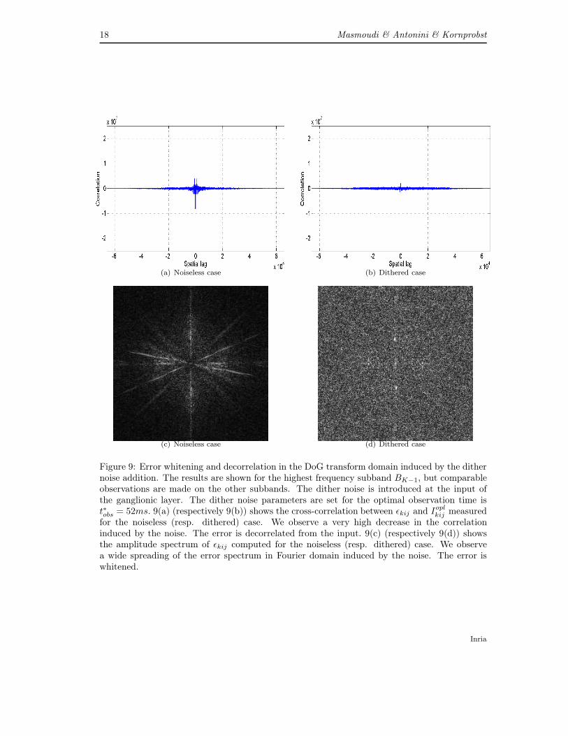
Another perceptually important feature that is induced by the dithering process is the errorwhitening. We verify also this feature in our case. As shown in Figures 9(c), the spectrum ofthe error obtained when using our coder with no addition of noise is non-uniform. This denotesstrong geometric correlations in the error image which yields annoying artefacts. On the contrary,we notice in Figure 9(d) that the error spectrum is equally dispatched in the Fourier domain ifwe add a dither noise. Thus our new dithered scalable image coder gained interesting featuresthrough the integration of a dithering process.
RR n° 7877
18 Masmoudi & Antonini & Kornprobst
(a) Noiseless case (b) Dithered case
(c) Noiseless case (d) Dithered case
Figure 9: Error whitening and decorrelation in the DoG transform domain induced by the dithernoise addition. The results are shown for the highest frequency subband BK−1, but comparableobservations are made on the other subbands. The dither noise is introduced at the input ofthe ganglionic layer. The dither noise parameters are set for the optimal observation time ist∗obs = 52ms. 9(a) (respectively 9(b)) shows the cross-correlation between εkij and I
oplkij measured
for the noiseless (resp. dithered) case. We observe a very high decrease in the correlationinduced by the noise. The error is decorrelated from the input. 9(c) (respectively 9(d)) showsthe amplitude spectrum of εkij computed for the noiseless (resp. dithered) case. We observea wide spreading of the error spectrum in Fourier domain induced by the noise. The error iswhitened.
Inria

Streaming an image through the eye 19
The whitening and de-correlation features yield a greater reconstruction error in terms ofmean squared error [33]. Though, the error whitening and decorrelation features acquired in thetransform domain are perceptually important. Indeed, a strong correlation between the codingerror and the original signal implies annoying artefacts. Besides the error whitening is importantbecause all frequencies are affected by the same noise. The perceptual impact of dithering onthe final image reconstruction ftobs is shown in the next section.
7 Results: Case of the bio-inspired and dithered scalable
image coder
We show in this section the perceptual impact of the dithering on the reconstructed imagesusing our decoder. Our experiments demonstrate the ability of the dither noise to accelerate therecognition of the image details and singularities during the decoding process.
A first example is given in Figure 10. The left column shows the evolution of the reconstruc-tion ftobs with increasing times tobs, in the case of noiseless coding. The right column showsthe evolution of the reconstruction ftobs with increasing times tobs, in the case of addition of adither noise to the input of the ganglionic layer. The central column shows a filtered version ofcameraman. Cameraman is sharpened to enhance the image details. The comparison betweenthe noiseless case reconstruction (on the left) and the dithered reconstruction (on the right)demonstrates perceptual importance of noise in the image coding process in the retina. Withthe addition of noise, details of cameraman are well rendered “before date”. For example, thehand of cameraman and the tower in the background appear since tobs = 44ms for the ditheredcase while still invisible in the noiseless case at the same observation time. We can also noticethat the horizontal stripes in the background, the grass details, the pant folds, and the handare well rendered since tobs = 48ms. On the contrary these details are still invisible or highlyblurry in the noiseless case at the same observation time. Finally, at the optimal observationtobs = t∗obs = 52ms all the fine details of the image, including the coat and the backgrounddetails, are clearly distinguished in the dithered case while still blurry or invisible in the noiselesscase.
A second example is given in Figure 11 for the baboon test image. This image is rich ofdetails and singularities and thus particularly challenging. Though, our dithered coder stillrenders the image details “before date in this case” (with another adequate parametrization forthe dither noise). As for the preceding example, the left image shows the reconstruction ftobsin the case of noiseless coding. The right image shows the reconstruction ftobs in the case ofaddition of a dither noise to the input of the ganglionic layer. The central image is a sharpenedversion of baboon. The observation time shown in this figure is also the optimal observationtobs = t∗obs = 44ms. The comparison between the noiseless case reconstruction (on the left) andthe dithered reconstruction (on the right) confirms the observations made in the first example.The dither noise helps the recognition of fine details “before date”. While in the noiseless caseface and bear details of baboon are still blurry, these details are well rendered in the ditheredreconstruction case.
On one hand, the integration of a dither noise in the coding process yield a greater recon-struction error in terms of mean squared error [33]. Besides, as the dither noise is a disorderedsignal, it also increases the entropy of the image code. On the other hand, the error whiteningand de-correlation features acquired by our system are perceptually important. This is a crucialpoint because our current results may prove that the retina conveys a code that is optimizedfor the tasks to be performed by the visual cortex as categorization. While the rate/distortiontrade-off remains an important goal for a coding scheme it may not be the central performance
RR n° 7877

20 Masmoudi & Antonini & Kornprobst
Ditherless reconstruction Sharpened cameraman Dithered reconstruction
40
ms
44m
s48m
s52m
s
Figure 10: Perceptual impact of the multiscale dithering on the reconstruction of cameraman.The observation times tobs are shown on the left. From top to bottom, tobs take successively thevalues of: 40ms, 44ms, 48ms, and 52ms. The observation time shown in this figure is also theoptimal observation tobs = t∗obs = 44ms. Inria

Streaming an image through the eye 21
Ditherless reconstruction Sharpened baboon Dithered reconstruction
44
ms
Figure 11: Perceptual impact of the multiscale dithering on the reconstruction of baboon. Theobservation time shown in this figure is also the optimal observation tobs = t∗obs = 44ms.
criterion for the retina.
8 Conclusion
The work that we presented brings two main contributions. As a first step, we proposed a bio-inspired codec for static images with a deterministic behavior. The image coder is based on twostages. The first stage is the image transform as performed by the outer layers of the retina.In order to integrate time dynamics, we added to this transform time delays that are subbandspecific so that, each subband is processed differently. The second stage is a succession of twodynamic processing steps mimicking the deep retina layers behavior. These latter perform anA/D conversion and generate a spike-based, invertible, retinal code for the input image in anoriginal fashion.
In a second step, we investigated the issue of non-determinism in the retina neural code. Weproposed to model the retinal noise by a multiscale dither signal with specific statistical proper-ties. The dithering process that we proposed whitens the reconstruction error and decorrelatesit from the input stimuli. Besides, from a perceptual point of view, our coder allows an earlierrecognition of the image details and singularities during the decoding process.
In conclusion, our coding scheme offers interesting features such as (i) time-scalability, asthe choice of the observation time of our codec enables different reconstruction qualities, and (ii)bit-allocation, as each subband of the image transform is separately mapped according to the cor-responding state of the inner layers. In addition, when integrating a dithering process our codergained interesting perceptual features. These features, if the dithering hypothesis is confirmed,help the visual cortex recognize the fine details of the image. This latter point is interestingbecause it may prove that the retina conveys a code that is optimized for the tasks to be per-formed by the visual cortex. Interestingly, our dithering hypothesis found an echo recently in thecomputational neurosciences community [29]. We are convinced that further neurophysiologicinvestigations may also confirm the relevance of dithering in the retinal processing.
In terms of rate/distortion, the results accomplished by our coding scheme are encouraging.Though the rate/distortion performance is not the primary goal of this work, our coder couldstill be improved to be competitive with the well established JPEG and JPEG 2000 standards.
RR n° 7877

22 Masmoudi & Antonini & Kornprobst
Optimizing techniques as bit-plane coding are to be investigated.
This work is at the crossroads of diverse hot topics in the fields of neurosciences, brain-machine interfaces, and signal processing and tries to bridge the gap between these differentdomains towards the conception of new biologically inspired coders.
References
[1] E.D. Adrian. The impulses produced by sensory nerve endings. Journal of Physiology,61(1):49–72, 1926.
[2] M. Antonini, M. Barlaud, P. Mathieu, and I. Daubechies. Image coding using wavelettransform. IEEE Transactions on Image Processing, 1992.
[3] E.N. Brown, R.E. Kass, and P.P. Mitra. Multiple neural spike train data analysis: state-of-the-art and future challenges. Nature Neuroscience, 7(5):456–461, 2004.
[4] P. Burt and E. Adelson. The Laplacian pyramid as a compact image code. IEEE Transac-tions on Communications, 31(4):532–540, 1983.
[5] C. Christopoulos, A. Skodras, and T. Ebrahimi. The JPEG 2000 still image coding system:An overview. IEEE Transactions on Consumer Electronics, 16(4):1103–1127, 2000.
[6] A.B. Clark and et al. Electrical picture-transmitting system. US Patent assigned to AT&T, 1928.
[7] C.E. Connor, S.L. Brincat, and A. Pasupathy. Transformation of shape information in theventral pathway. Current Opinion in Neurobiology, 17(2):140–147, 2007.
[8] J.L. Crowley and R.M. Stern. Fast computation of the difference of low-pass transform.IEEE Transactions on Pattern Analysis and Machine Intelligence, (2):212–222, 2009.
[9] R.R. de Ruyter van Steveninck, G.D. Lewen, S.P. Strong, R. Koberle, and W. Bialek.Reproducibility and variability in neural spike trains. Science, 275(5307):1805, 1997.
[10] D.J. Field. What is the goal of sensory coding? Neural Computation, 6(4):559–601, 1994.
[11] T. Gollisch and M. Meister. Eye smarter than scientists believed: Neural computations incircuits of the retina. Neuron, 65(2):150–164, 2010.
[12] I. Katsavounidis and C.C. Jay Kuo. A multiscale error diffusion technique for digital halfton-ing. IEEE Transactions on Image Processing, 6(3):483–490, 1997.
[13] C.K. Kier, G. Buchsbaum, and P. Sterling. How retinal microcircuits scale for ganglion cellsof different size. The Journal of Neuroscience, 15(11):7673–7683, 1995.
[14] K. Masmoudi, M. Antonini, and P. Kornprobst. Another look at the retina as an imagedithered scalar quantizer. In International Workshop on Image Analysis for MultimediaInteractive Services (WIAMIS 2010), pages 1–4. IEEE, 2010.
[15] K. Masmoudi, M. Antonini, and P. Kornprobst. Another look at the retina as an imagescalar quantizer. In Proceedings of the International Symposium on Circuits and Systems(ISCAS 2010), pages 3076–3079. IEEE, 2010.
Inria

Streaming an image through the eye 23
[16] K. Masmoudi, M. Antonini, and P. Kornprobst. Encoding and decoding stimuli using abiologically realistic model: The non-determinism in spike timings seen as a dither signal.In Proceedings of Research in Encoding And Decoding of Neural Ensembles (AREADNE2010), page 75, 2010.
[17] K. Masmoudi, M. Antonini, and P. Kornprobst. A bio-inspired image coder with temporalscalability. In Advanced Concepts for Intelligent Vision Systems, pages 447–458. SpringerBerlin/Heidelberg, 2011.
[18] K. Masmoudi, M. Antonini, and P. Kornprobst. Frames for exact inversion of the rank ordercoder. to appear in IEEE Transactions on Neural Networks, 23, 2012.
[19] K. Masmoudi, M. Antonini, P. Kornprobst, and L. Perrinet. A novel bio-inspired staticimage compression scheme for noisy data transmission over low-bandwidth channels. InProceedings of the International Conference on Acoustics, Speech, and Signal Processing(ICASSP 2010), pages 3506–3509. IEEE, 2010.
[20] S. Panzeri, R.S. Petersen, S.R. Schultz, M. Lebedev, and M.E. Diamond. The role of spiketiming in the coding of stimulus location in rat somatosensory cortex. Neuron, 29(3):769–777, 2001.
[21] D. Perkell and T. Bullock. Neural coding. Neurosciences Research Program Bulletin, 6:221–343, 1968.
[22] L. Perrinet, M. Samuelides, and S. Thorpe. Coding static natural images using spiking eventtimes: do neurons cooperate? IEEE Transactions on Neural Networks, 15(5):1164–1175,2004.
[23] F. Rieke, D. Warland, R. de Ruyter van Steveninck, and W. Bialek. Spikes: Exploring theNeural Code. The MIT Press, Cambridge, MA, USA, 1997.
[24] R.W. Rodieck. Quantitative analysis of the cat retinal ganglion cells response to visualstimuli. Vision Research, 5(11):583–601, 1965.
[25] M.N. Shadlen and W.T. Newsome. Noise, neural codes and cortical organization. Findingsand Current Opinion Cognitive Neuroscience, 4:569–79, 1998.
[26] P. Sterling, E. Cohen, R.G. Smith, and Y. Tsukamoto. Retinal circuits for daylight: whyballplayers don’t wear shades. Analysis and Modeling of Neural Systems, pages 143–162,1992.
[27] D. Taubman. High performance scalable image compression with ebcot. IEEE transactionson Image Processing, 9(7):1158–1170, 2000.
[28] R. Van Rullen and S. Thorpe. Rate coding versus temporal order coding: What the retinalganglion cells tell the visual cortex. Neural Computation, 13:1255–1283, 2001.
[29] M. Vidne, Y. Ahmadian, J. Shlens, J.W. Pillow, J. Kulkarni, A.M. Litke, EJ Chichilnisky,E. Simoncelli, and L. Paninski. Modeling the impact of common noise inputs on the networkactivity of retinal ganglion cells. to appear in Journal of Computational Neuroscience, 2012.
[30] Chung-Neng Wang, Chi-Min Liu, and Tihao Chiang. Perceptual dithering for octave sub-band image coding. Journal of Visual Communication and Image Representation, 15(2):163– 184, 2004.
RR n° 7877
24 Masmoudi & Antonini & Kornprobst
[31] Z. Wang, A.C. Bovik, H.R. Sheikh, and E.P. Simoncelli. Image quality assessment: Fromerror visibility to structural similarity. IEEE Transactions on Image Processing, 13(4):600–612, 2004.
[32] R.A. Wannamaker. Subtractive and nonsubtractive dithering: A mathematical comparison.J. Audio Eng. Soc, 52(12):1211–1227, 2004.
[33] R.A. Wannamaker, S.P. Lipshitz, J. Vanderkooy, and J.N. Wright. A theory of nonsubtrac-tive dither. IEEE Transactions on Signal Processing, 48(2):499–516, 2000.
[34] W.Gerstner and W.Kistler. Spiking Neuron Models : Single Neurons, Populations, Plastic-ity. Cambridge University Press, 2002.
[35] A. Wohrer. Model and large-scale simulator of a biological retina, with contrast gain control.PhD thesis, Graduate School of Information and Communication Sciences, University ofNice-Sophia Antipolis, 2008.
[36] A. Wohrer and P. Kornprobst. Virtual retina : A biological retina model and simulator,with contrast gain control. Journal of Computational Neuroscience, 26(2):219–249, 2009.
[37] Adrien Wohrer, Pierre Kornprobst, and Marc Antonini. Retinal filtering and image recon-struction. Research Report RR-6960, INRIA, 2009.
[38] Y. Zhang, A. Ghodrati, and D.H. Brooks. An analytical comparison of three spatio-temporalregularization methods for dynamic linear inverse problems in a common statistical frame-work. Inverse Problems, 21:357, 2005.
Contents
1 Introduction 3
2 Virtual Retina : A bio-plausible retina model 4
3 The coding pathway 4
3.1 The image transform: The outer layers of the retina . . . . . . . . . . . . . . . . 43.2 The A/D converter: The inner and ganglionic layers of the retina . . . . . . . . . 8
3.2.1 Contrast gain control . . . . . . . . . . . . . . . . . . . . . . . . . . . . . 83.2.2 Non-linear rectification . . . . . . . . . . . . . . . . . . . . . . . . . . . . . 83.2.3 Leaky integrate-and-fire quantization: . . . . . . . . . . . . . . . . . . . . 10
4 The decoding pathway 10
5 Results: Case of the bio-inspired and noiseless scalable image coder 11
6 Introducing the noise in the coder: The non-subtractive dither hypothesis 13
6.1 The non-deterministic nature of the neural code of the retina . . . . . . . . . . . 146.2 Multiscale non-subtractive dithering . . . . . . . . . . . . . . . . . . . . . . . . . 14
7 Results: Case of the bio-inspired and dithered scalable image coder 19
8 Conclusion 21
Inria

RESEARCH CENTRESOPHIA ANTIPOLIS – MÉDITERRANÉE
2004 route des Lucioles - BP 93
06902 Sophia Antipolis Cedex
PublisherInriaDomaine de Voluceau - RocquencourtBP 105 - 78153 Le Chesnay Cedexinria.fr
ISSN 0249-6399