St.John …eprints.uwe.ac.uk/23321/2/__dsm_home_Documents... · Control: General survey view of the...
19
St.John-Matthews, J., Bartley, R. and Brook, S. (2013) CT urogram: Why the wait? In: Siemens Annual CT User Group Meeting, Crewe Hall Hotel, UK, 13 May 2013. Available from: http://eprints.uwe.ac.uk/23321 We recommend you cite the published version. The publisher’s URL is: http://eprints.uwe.ac.uk/23321/ Refereed: Yes (no note) Disclaimer UWE has obtained warranties from all depositors as to their title in the material deposited and as to their right to deposit such material. UWE makes no representation or warranties of commercial utility, title, or fit- ness for a particular purpose or any other warranty, express or implied in respect of any material deposited. UWE makes no representation that the use of the materials will not infringe any patent, copyright, trademark or other property or proprietary rights. UWE accepts no liability for any infringement of intellectual property rights in any material deposited but will remove such material from public view pend- ing investigation in the event of an allegation of any such infringement. PLEASE SCROLL DOWN FOR TEXT.
Transcript of St.John …eprints.uwe.ac.uk/23321/2/__dsm_home_Documents... · Control: General survey view of the...

St.John-Matthews, J., Bartley, R. and Brook, S. (2013) CT urogram:Why the wait? In: Siemens Annual CT User Group Meeting, CreweHall Hotel, UK, 13 May 2013. Available from: http://eprints.uwe.ac.uk/23321
We recommend you cite the published version.The publisher’s URL is:http://eprints.uwe.ac.uk/23321/
Refereed: Yes
(no note)
Disclaimer
UWE has obtained warranties from all depositors as to their title in the materialdeposited and as to their right to deposit such material.
UWE makes no representation or warranties of commercial utility, title, or fit-ness for a particular purpose or any other warranty, express or implied in respectof any material deposited.
UWE makes no representation that the use of the materials will not infringeany patent, copyright, trademark or other property or proprietary rights.
UWE accepts no liability for any infringement of intellectual property rightsin any material deposited but will remove such material from public view pend-ing investigation in the event of an allegation of any such infringement.
PLEASE SCROLL DOWN FOR TEXT.
Janice St. John-Matthews Rachel Bartley
Sian Brock
Presenter
Presentation Notes
When myself and my colleagues volunteered to present at the Siemens CT User Group meeting we decided that although a familiar technique, there is no consensus within literature on what a “Gold Standard” protocol should be for a CT Urogram. In fact at a local level we have 2 groups of Radiologists who preform the procedure with subtle differences. From a personal perspective I have not worked full-time in CT since 2008 and at this time was working on a 16-slice scanner. Since joining the team at Cardiff last year I had noticed an increase in CTU examinations performed and that the technique used to acquire these images had changed. Our aim therefore was to use this as an opportunity to review our local protocols to ascertain if we could improve further on the images we produced. This was supported by informal discussions with the Radiologists at our site and a review of literature/ research on this topic. We hoped in doing so we could share what we have learnt along the way.

Meet The Team
Presenter
Presentation Notes
Cardiff Spire opened in 1982 offering high levels of medical treatment and personal care. Spire Cardiff Hospital is the largest private acute healthcare provider in South Wales (62 beds in total). The hospital offers a wide range of treatments from diagnostic imaging to major surgery. For patients undergoing complex surgical procedures we have an Intensive Care Unit (ICU) and a high dependency unit, managed by specialist nurses. In 2009 the hospital installed a static CT Scanner- a 64 slice Siemens Somatom Definition AS. Prior to this the hospital was serviced by a mobile CT scanner which visited the hospital 6 times a month. CT is staffed by the radiography team based in the general department and consists of Rachel the Imaging Manager and 2 part-time Senior Radiographers, Sian and Janice. Rachel and Sian also cover Mammography and Janice, MRI. We carry out a mixed workload including Neurology, Cardiac, Angiography, CTC, CTU etc.
Definition of CTU
European Society of Urogenital Radiology: “A diagnostic examination optimized for imaging the kidneys, ureters and bladder with thin slice
MDCT, IV contrast agent administration and images acquired in the excretory phase”
Presenter
Presentation Notes
The definition of CTU as laid out by the ESUR is….. In response to the increasing popularity of CTU in 2007 the ESUR set up a working party which looked to formulate a standardized approach to this examination. To achieve this Van Der Molen et al (2008) carried out a systematic review of the relevant literature published between1995-2007. From here they drafted a proposed differential approach with different techniques used in different patient populations.

Before CTU IVU: To demonstrate the entire urinary tract
radiographically showing both the structure and function of the kidneys
• Control Film
• Immediate (kidneys only) • 5min Film
• 10 min compression • Full Length release • Micturition Film
Presenter
Presentation Notes
To understand the reasons for “waiting”, it is best to look back at the examination of choice prior to CTU- the IVU. Control: General survey view of the whole abdomen. Aim to demonstrate any possible calculi in the renal tract prior to injection. Also any other abdominal pathology. Immediate Film: Nephrogram stage before contrasts collects in the pelvi-calyceal system 5 Minute: Demonstrates contrast starting to collect in the calyces and pelvis of each kidney 10 Minute Compression: Show well filled cup-shaped calyces and pelvis Full Length Release: 15-20 minutes post injection, demonstrates the entire length of the ureters as the contrast flows down, having been held up by the compression Post Micturition Film: Any residual bladder volume and also full drainage of the kidneys. Poor bladder emptying can contribute to unexplained UTIs in patients. Radiation dose has been one of the most important driving factors in optimization of CTU techniques and in selecting justified indications.
Goal of CTU
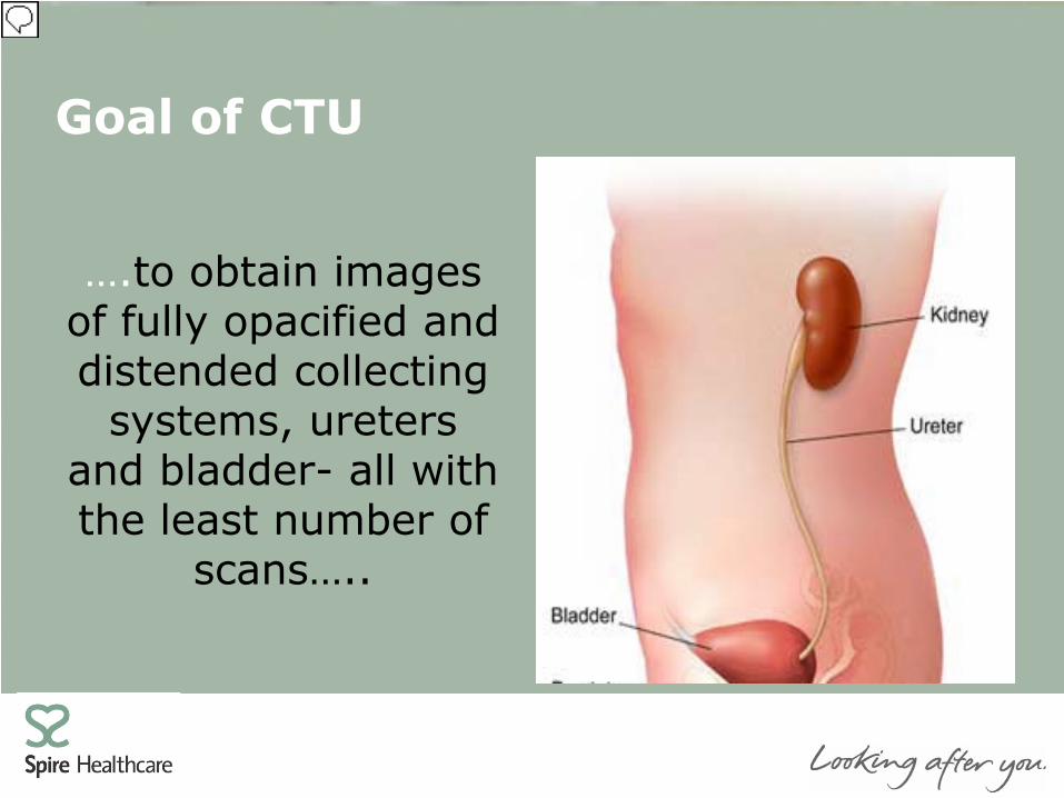
….to obtain images of fully opacified and distended collecting
systems, ureters and bladder- all with the least number of
scans…..
Presenter
Presentation Notes
Problem lies with the fact that each component of urinary tract is a destinable reservoir and in the case of the ureters is subject to intermittent peristaltic contractions. The distal ureter (particularly the distal segment) is the most difficult component to opacify and distend as it is a thin structure and runs parallel to the spine thus entering the bladder posteriorly. The requirement to achieve optimal opacification needs to be balanced against wait time for patients; effects on workflow and room utilization. Hence a plethora of literature examining how best to do this.
What Are We Looking For?
• Renal Masses: RCC & TCC • Calculi • Genitourinary trauma • Renal infection • Haematuria • ?Incidental Findings
Presenter
Presentation Notes
Song et al (2012) “Incidental Clinically Important Extraurinary Findings at MDCT Urography for Hematuria Evaluation: Prevalence in 1209 Consecutive Examinations”. 1209 CTUs, 6.8% of patients had clinically important or potentially important incidental findings requiring further investigation. Acute findings diagnosed in 0.9% Extraurinary malignancy confirmed in 0.4% Lung nodules were the most common incidental finding

What Not To Do!
Presenter
Presentation Notes
Patient presented to CT following an ultrasound scan which highlighted a possible mass in the right kidney. CT was recommended for further evaluation. The CT was carried out a few days later and was performed in the arterial and portal-venous phase. The report stated that: “The left kidney is normal apart from a simple cyst in the mid-pole. The upper pole of the right kidney is confirmed as being abnormal. It is less perfused than the lower pole. The renal “bean” is maintained. Appearances could therefore be due to an infiltrating process such as TCC. A delayed phase CT would be helpful in confirming TCC. Urine cytology may be helpful”

Our Protocols: Rad Team A
• Drink 500ml 40 minute before the scan • Pre KUB (low dose) • 50ml IV Contrast. No scan. • 600s delay • 50ml IV Contrast • Post 70s delay
Presenter
Presentation Notes
Despite the proposals by Van der Molen et al (2008) there remains no universally accepted technique for performing CTU. This is evident in our department as two “school’s of thought”
Our Protocols: Rad Team B
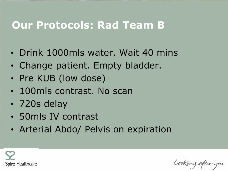
• Drink 1000mls water. Wait 40 mins • Change patient. Empty bladder. • Pre KUB (low dose) • 100mls contrast. No scan • 720s delay • 50mls IV contrast • Arterial Abdo/ Pelvis on expiration
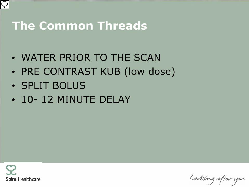
The Common Threads
• WATER PRIOR TO THE SCAN • PRE CONTRAST KUB (low dose) • SPLIT BOLUS • 10- 12 MINUTE DELAY
Presenter
Presentation Notes
Water prior to scan: Avoids dehydration; promotes diuresis; acts as a positive contrast medium for the GI tract. Recommended up to 1000ml of water 20-60 minutes before CT scan. Pre contrast KUB (low dose): To access if renal calculi Split Bolus: Compromise on getting 2 different phases- think dose. The nephrogenic phase allows detection and characterisation of renal masses (both the renal cortex and the medulla are enhanced). The excretory phase aids evaluation of the urothelium 10-12 minute delay: Allows time for the kidneys to excrete the contrast agent and for it to be visualised in the ureters.

DIFFERENCES
• Delay applied to split-bolus • Fractioned Dose • Arterial versus PV phase
Presenter
Presentation Notes
Arterial phase: Information on arterial vasculature
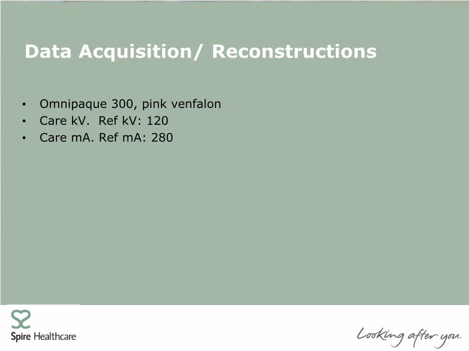
Data Acquisition/ Reconstructions
• Omnipaque 300, pink venfalon • Care kV. Ref kV: 120 • Care mA. Ref mA: 280
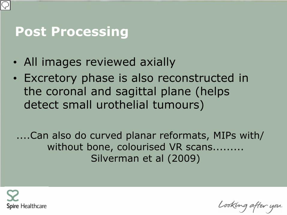
Post Processing
• All images reviewed axially • Excretory phase is also reconstructed in
the coronal and sagittal plane (helps detect small urothelial tumours)
....Can also do curved planar reformats, MIPs with/
without bone, colourised VR scans......... Silverman et al (2009)
Presenter
Presentation Notes
Silverman (2009): helpful deciphering congenital anomalies; helps surgical planning h/e/ requires additional time. Also supported by Van der Molen et al (2008). Realistically do we have time in the clinical environment?

Ancillary Maneuverers/ Techniques
• Furosemide IV (0.1mg/kg)
• Compression Higher opacification for mid and distal ureter [McNicholas et al
(1998) & Caoili et al (2002) ] May not be applied in some patients, such as those with
abdominal aortic aneurysm
Presenter
Presentation Notes
To achieve optimal opacification, McNicholas et al. [1] applied abdominal compression and reported that there was a significantly higher opacification score of the mid to distal ureter. To achieve better dissention of the renal collecting system and ureters,

Ancillary Maneuverers/ Techniques
• Patient Moving 2 topograms Kim et al (2008). Log-rolling. No difference in ureteral
opacification
• Prone Imaging
Improves ureteric distension and opacification Free intravesical/ impacted in ureterovesical junction
stones Uncomfortable and benefits disputed
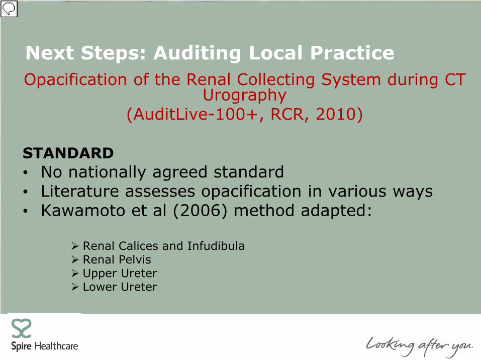
Next Steps: Auditing Local Practice Opacification of the Renal Collecting System during CT
Urography (AuditLive-100+, RCR, 2010)
STANDARD • No nationally agreed standard • Literature assesses opacification in various ways • Kawamoto et al (2006) method adapted:
Renal Calices and Infudibula Renal Pelvis Upper Ureter Lower Ureter
Presenter
Presentation Notes
What started as a “simple” presentation for the Siemens CT Users meeting has evolved into a proposed audit of local practice with a view to amend current protocols if the proposed target is not met. To do this we propose using a pre-designed audit form from the AuditLive+ section of the Royal College of Radiologists website thus maintaining creditability to our audit process and ensuring validity of our findings. This audit has been designed and adapted using a research method carried out by Kawamoto et al (2006) entitled “Opacification of the Collecting System and Ureters on Excretory Phase CT using Oral Water as a Contrast Medium”. For the propose of quantifying results the renal collecting system is divided into 4 segments with right and left renal collecting systems being assessed.

Next Steps: Auditing Local Practice TARGET • Opacification is assessed on a 1-3 likert scale
3=Complete opacification 2=Near complete opacification 1=No or poor opacification
SUGGESTIONS FOR CHANGE RESOURCES
Renal Calices & Infudibula 95% CI Renal Pelvis 95% CI Upper Ureter 85% CI Lower Ureter 75% CI
Presenter
Presentation Notes
Opacification is measured for each side using a 1-3 Likert scale with 3 representing complete opacification and 1 no/poor opacification. As highlighted by this presentation if the proposed target is not met there are a number of areas which could be addressed i.e. log-rolling, prone imaging, administration furosemide; contrast volumes. However following informal discussions with the site Radiologists the administration of Furosemide and log-rolling would be the preferred adaptions. As all injecting Radiographers at our hospital work to Patient Group Directions this would need to be set-up and agreed by the appropriate parties. Our chosen data collection method is to include an audit form for all CTUs and to gather the data using quota sampling i.e. sampling until the target number of studies are attained. In this instance 30 CTU’s which should yield data from at least 50 collecting systems. For data interpretation 1 hour is required. The audit will be carried out alongside routine reporting so there is a minimal extra time investment by the reporting Radiologist.

Finally…..
……not just how long you wait but rather what you do/ don’t do during this time
which impacts the quality of the imaging produced…….
Presenter
Presentation Notes
In conclusion the answer to our original question, “Why the wait?” is simple- to get the best possible images of the WHOLE urinary tract. However having looked at this topic in more depth we have learnt that what you do during this time can be just as important in achieving an optimised protocol. We hope our proposed audit will ascertain what this might be and help us to develop our protocols further. Thank you.