Stem cells (2)
45
Manifestation of Novel Social Challenges of the European Union in the Teaching Material of Medical Biotechnology Master’s Programmes at the University of Pécs and at the University of Debrecen Identification number: TÁMOP-4.1.2-08/1/A-2009-0011
description
Manifestation of Novel Social Challenges of the European Union in the Teaching Material of Medical Biotechnology Master’s P rogrammes at the University of Pécs and at the University of Debrecen Identification number : TÁMOP-4.1.2-08/1/A-2009-0011. - PowerPoint PPT Presentation
Transcript of Stem cells (2)

Manifestation of Novel Social Challenges of the European Unionin the Teaching Material ofMedical Biotechnology Master’s Programmesat the University of Pécs and at the University of DebrecenIdentification number: TÁMOP-4.1.2-08/1/A-2009-0011

STEM CELLS (2)
Dr. Judit PongráczThree dimensional tissue cultures and tissue engineering – Lecture 3
Manifestation of Novel Social Challenges of the European Unionin the Teaching Material ofMedical Biotechnology Master’s Programmesat the University of Pécs and at the University of DebrecenIdentification number: TÁMOP-4.1.2-08/1/A-2009-0011

TÁMOP-4.1.2-08/1/A-2009-0011
Cord blood stem cells• Approx. 130 million babies born yearly – the
umbilical cord blood is the largest potential source of stem cells for regenerative medicine
• In the past 36 yrs 10000 patients were treated for over 80 different diseases

TÁMOP-4.1.2-08/1/A-2009-0011
Cord blood stem cells and fetal stem cells
Cord blood collection from umbilical vein (after
birth)
The cord blood cells are frozen in bag or cryovials
Liquid nitrogen storage tank (-150°)
Analysis of bloodCell separation
Mix and settle Concentrate cells Freeze cells35 min 10 min
Express
Express
Add cordblood
1 2 3

TÁMOP-4.1.2-08/1/A-2009-0011
Cryopreservation• Cryopreservation of primary cells is possible
for long term (so far 20 yrs).• The low-temperature is maintained at -150-
196oC in liquid nitrogen.

TÁMOP-4.1.2-08/1/A-2009-0011
Cord blood processing1. Red cell depletion (using Ficoll, Hetastarch,
Lymphoprep, Prepacyte)2. Depletion of plasma for smaller storage size3. Testing of the final cell pool (infection,
volume, cellularity, stem cell content, CD34+)

TÁMOP-4.1.2-08/1/A-2009-0011
Cord blood processing and cryopreservation• Cord blood is primarily useful in
hematological disorders• Cord blood is collected at birth• Either processed or just simply frozen in
DMSO

TÁMOP-4.1.2-08/1/A-2009-0011
Cord blood banking• Cord blood banks should be set up in every
metropolitan city with HLY specification and linked to an international computer network
• Keeping cord blood for a considerable length of time is costly

TÁMOP-4.1.2-08/1/A-2009-0011
Pluripotenciy of cord blood stem cells
Cord Blood
Stem cellsCBE MSC
Purification
Endodermal Mesodermal EctodermalHepato-Biliary Blood Neural

TÁMOP-4.1.2-08/1/A-2009-0011
Stem cell population in cord blood
Adherent Pre-MSC MSC
Non-adherent
CBE
Lin- CD133+ CD34+
Cord Blood
Bone marrow
Peripheral Blood

TÁMOP-4.1.2-08/1/A-2009-0011
Disorders treatable with cord blood IOncologic disordersAcute lymphoblastic leukemiaAcute myeloid leukemiaAutoimmune lymphoproliferative disordersBurkitt lymphomaChronic myeloid leukemiaCytopenia related to monosomyFamilial hystocytosisHodgkin’s diseaseJuvenile myelomonocytic leukemiaLangerhans cell hystocytosisMyelodysplastic syndromesNon-Hodgkin’s lymphoma
Immune deficienciesAtaxia telangiectasiaCartilage-hair hypoplasiaChronic granulomatous diseaseDiGeorge syndromeHypogammaglobulinaemiaIKK gamma deficiencyImmune dysregulation polyendocrinophatyMucolipidosis type IIMyelokathesisSevere combined immunodeficiencyWiscott-Aldrich syndromeX-linked agammaglobulinaemia, immunodeficiency, lymphoproliferative syndrome

TÁMOP-4.1.2-08/1/A-2009-0011
Disorders treatable with cord blood IIHematological disordersAutoimmune neutropeniaCyclic neutropeniaDiamond Blackfran anemiaEvan’s syndromeRed cell aplasiaRefractory anemiaSevere aplastic anemiaSickle cell diseaseThalassaemiaFanconi’s anemiaGalnzmann’s diseaseCongenital sideroblastic anemiaJuvenile dermatomyositis and xanthogranulomas
Metabolic disordersAdrenoleukodystrophyAlpha mannosidosisType I diabetesGaucher’s diseaseGunther diseaseHermansky-Pudlak syndromeHurler syndromeHurler-Scheie syndromeKrabbe’s diseaseMaroteau-lamy syndromeMetachromatic leukodystrophyMucolipidosis Types II, IIINeimann Pick syndrome, Types A and BSandoff syndromeSanfilippo syndromeTay Sachs disease

TÁMOP-4.1.2-08/1/A-2009-0011
Fat stem cells (ASC)Fat or adipose tissue stem cells (ASC):• Easily obtainable• Consistent immunophenotype• Similar to BMSC• Multipotent• Manipulation by genetic engineering

TÁMOP-4.1.2-08/1/A-2009-0011
Types of adipose tissues
TYPE FUNCTION1 Bone marrow Fills in space no longer used for hematopoiesis
2 Brown adipose tissue (BAT) Protects vital organs in the newborn
3 Ectopic adipose tissueAbnormal fat accumulation in the liver, skeletal or cardiac muscle (e.g. in metabolic syndrome)
4 Mammary adipose tissue Lactation – energy and nutrient source
5 Mechanical adipose tissue Protection from mechanical trauma
6 White adipose tissue (WAT)Insulation, energy storage, reservoir, endocrine organ

TÁMOP-4.1.2-08/1/A-2009-0011
Isolation procedures
Digestion with collagenase at 37oC, 1hr
Stromal vascularfraction (SVF)
Wash in PBS
Aspiration of lipocytes
300g, 5 min

TÁMOP-4.1.2-08/1/A-2009-0011
Immunophenotype of ASCsPositive markers
Marker Name Marker NameCD9 Tetraspan CD55 Decay accelerating factor
CD10Common acute lymphocytic leukemia antigen
CD59 Protectin
CD13 Aminopeptidase CD71 TransferrinCD29 b1-integrin CD73 5’-ectonucleotidaseCD34 Sialomucin CD90 Thy1CD44 Hyaluronate receptor CD105 EndoglinCD49d a4-integrin CD146 Muc-18
CD54 Intracellular adhesion molecule CD166 Activated leukocyte cell
adhesion molecule
HLA-ABC Histocompatibility locus antigen-ABC a-SMA a-smooth muscle actin

TÁMOP-4.1.2-08/1/A-2009-0011
Immunophenotype of ASCsNegative markersMarker Name Marker NameCD11b ab-integrin CD50 Intracellular adhesion
molecule-3
CD14 LPS receptor CD56 Neural cell adhesion molecule
CD16 Fc receptor CD62 E-selectionCD18 b2-integrin CD104 B4-integrin
CD45 Common leukocyte antigen HLA-DR Histocompatibility locus
antigen-DR

TÁMOP-4.1.2-08/1/A-2009-0011
Cytokine profile of ASCsCYOTOKINE FUNCTION GROUP
• Adiponectin• Leptin• Plasminogen activator inhibitor-1
Adipokines
• Hepatocyte growth factor• Pigment epithelial derived factor• Vascular endothelial growth factor
Angiogenic
• Flt-3 ligand• GCSF• Leukemia inhibitory factor• IL-7• MCSF
Hematopoietic
• IL-6, IL-8, IL-11• Leukemia inhibitory factor• TNFa
Pro-inflammatory

TÁMOP-4.1.2-08/1/A-2009-0011
Immunogenecity of ASCs• Lack of immunogenicity is linked to the
absence of the major histocompatibility class II antigens (HLA-DR) on their surface.
• Their immunosuppressive properties are linked to prostaglandin E2 production.

TÁMOP-4.1.2-08/1/A-2009-0011
Differentiation potential of ASCs• Adipocyte• Cardiac myocytes• Chondrocyte• Endodermal and ectodermal lineages• Endothelial and smooth muscle cells• Hematopoietic support• Neuronal lineage• Osteoblast• Skeletal myocytes

TÁMOP-4.1.2-08/1/A-2009-0011
Differentiation into adipocytes
ORIGIN OF ACS INDUCING AGENT MARKERS INDUCED APPLICATION
WAT etc. Forskolin (AMP agonist) Neutral lipids Functional fat pads
Methylisobutylxanthine (AMP agonist)
Adiponectin Plastic surgery
Glucocorticoid receptor ligands (dexamethasone)
CAAT/enhancer binding protein-a fatty acid binding protein (aP2)
Cosmetic and reconstructive surgery
PPAR-g2 ligands (thiazolidinediones)
Leptin
Insulin Lipoprotein lipase
bFGF PPAR-g2

TÁMOP-4.1.2-08/1/A-2009-0011
Differentiation into cardiac myocytes
ORIGIN OF ACS INDUCING AGENT MARKERS INDUCED APPLICATION
BAT 5-azacytadine Sarcomeric actininRepair injured cardiac tissue after ischemic injury
WAT Cardiomyocyte extract Connexin-43
Desmin
troponin-I

TÁMOP-4.1.2-08/1/A-2009-0011
Differentiation into chondrocytes
ORIGIN OF ACS INDUCING AGENT MARKERS INDUCED APPLICATION
Bone marrow adipose tissue
Ascorbate Aggrecan Knee, hip chondroid tissue
BAT Dexamethasone Chondroitin sulfate
WAT TGF-b Collagen type II
3D structure Collagen type IV
BMP-6Chondrocyte specific proteoglycans
FGF

TÁMOP-4.1.2-08/1/A-2009-0011
Differentiation into osteocytes
ORIGIN OF ACS INDUCING AGENT MARKERS INDUCED APPLICATION
WAT Ascorbate Osteocalcin Bone implantation
Bone marrow adipose tissue
Dexamethasone DMP-1 Bone fracture repair
1,25-dihydroxy vitamin D3 Osteoadherin
B-glycerophosphate
BMP-2
BMP-7
Runx2

TÁMOP-4.1.2-08/1/A-2009-0011
Differentiation into skeletal myocytes
ORIGIN OF ACS INDUCING AGENT MARKERS INDUCED APPLICATION
Low concentration FBS myoD
Horse serum myogenin
Myosin light chain kinase

TÁMOP-4.1.2-08/1/A-2009-0011
Differentiation into neuronal cells
ORIGIN OF ACS INDUCING AGENT MARKERS INDUCED APPLICATION
IndomethacinGlial fibrillary acidic protein (GFAP)
Central nervous system injury
Insulin Nestin
methylisobutylxanthine Intermediate filament
Glutamate receptor subunits
S-100
B-III tubulin

TÁMOP-4.1.2-08/1/A-2009-0011
Differentiation into endodermal and ectodermal lineages
ORIGIN OF ACS INDUCING AGENT MARKERS INDUCED APPLICATION
Bone marrow adipoid tissue
Hepatocyte growth factor Albumin Liver
Oncostatin M A-fetoprotein
DMSO together with HGF, bFGF, nicotinamide
Urea
ATRA Cytokeratin-18Epithelial tissue repair (Crohn’s disease)

TÁMOP-4.1.2-08/1/A-2009-0011
Differentiation into endothelial and smooth muscle cells
ORIGIN OF ACS INDUCING AGENT MARKERS INDUCED APPLICATION
WAT CD31 Vascular trauma
calponin Urogenital trauma
A-smooth muscle actin

TÁMOP-4.1.2-08/1/A-2009-0011
Hematopoietic support
ORIGIN OF ACS INDUCING AGENT MARKERS INDUCED APPLICATION
ASCs secrete:IL6,IL7, IL8,IL11 SCF, TNFa, MCSF, GMCSF
CD34+ into T, NK, B markers
For patients requireng hematopoietic stem cells reconstruction following high-dose chemotherapy

STEM CELLS (3)
Dr. Judit PongráczThree dimensional tissue cultures and tissue engineering – Lecture 4
Manifestation of Novel Social Challenges of the European Unionin the Teaching Material ofMedical Biotechnology Master’s Programmesat the University of Pécs and at the University of DebrecenIdentification number: TÁMOP-4.1.2-08/1/A-2009-0011

TÁMOP-4.1.2-08/1/A-2009-0011
Application of ESCs and ASCs
Vascular lumen
Vasculogenic zone Vascular disorders
Gene therapiesGenetically modified
stem cell-based deliveryIntravenous
injection
Mesoderm
Cardiac diseases
CSCsAtria nicheApex niche
HSCEPC MSC
OsteoblastsChondrocytes
AdipocytesMyoblasts
Common lymphoidprecursor
NK-cellsT-lymphocytes
Dendritic cellsB-lymphocytes
Common myeloidprecursor
Macrophages
PlateletsErythrocytes
MonocytesGranulocytes
HSCsEndosteum surface nicheMicrovasculature nicheMSCsPerivascular surface niche
Bone
Hemopoietic and Immunesystem disorders
Endoderm
BASCsBronchioalveolarduct junction niche
Lung disorders
PSCsPancreatic ductputative niche
PancreasInsulin-secreting b-cells
Endocrine isletsof Langerhans
Exocrineacini
Diabetes
LiverHepatocytes
HDCsBile duct(canal of Hering)
Hepatic disorders
LungEctoderm
NSCsSubventricularzone nicheHippocampus niche(dentate gyrus region)
Neuron Astrocyte Oligodendrocyte
Brain and spinal cord disorders
Eye disorders
RSCsCiliary epitheliumniche
CESCsLimbus nicheRetina
Cornea
KSCsBasal layer nichebESCseNCSCsBulge nicheSKPsDermal papilla
Skin disorders
HeartCardiomyocytes
Embryonic developmentInner cell mass
ESCsBlastocyst
Pluripotent ESC
Mesodermal stem cellEndodermal stem cell
Ectodermalstem cell
Hemangioblast
HSC EPC
Vascular wall-resident stem cellsEPCs and MSCs
New endothelial cell
BloodstreamMacrophagePlatelets
Erythrocyte MonocyteEosinophil
NK-cell
T-lymphocyteDendritic cell B-lymphocyteBasophil
Neutrophil
Media
Brain
Eye
Skin

TÁMOP-4.1.2-08/1/A-2009-0011
Genetic engineering and gene delivery using ASCs• Lentiviral vectors can transduce ASCs• Other recombinant viral vectors• Nucleofection

TÁMOP-4.1.2-08/1/A-2009-0011Approaches and methods for controlling stem cell growth and differentiation
Bioreactors come in many sizes and designs and include stirred, rotary and perfused systems. All serve to improve exchange efficiency of nutrients and waste products and delivery of growth factors to enable longer term culture, helping to scale-up cell numbers or to grow larger pieces of tissue.
Scaffold can provide physical (e.g. surface roughness, porosity, etc.) and also biochemical (e.g. controlled release of doped growth factors) cues to promote attachment, recruitment, differentiation and delivery of cells.
Co-culture with the cells or tissues of interest (i.e. the target for tissue repair) can help to encourage differentiation. This can include direct physical contact and/or indirect biochemical signaling
Biochem factors added to culture medium (including serum) stimulate differentiation. Requires knowledge of factors likely to induce differentiation but is rarely, if ever, 100% effective.
Differential adhesion assays using specific ECM proteins or receptor ligands can help in encouraging selection of specific cell types based on affinity and kinetics of cell-substratum interactions. Related to this is the colony forming unit (CFU) assay.
Cell sorting techniques like MACS or FACS can positively select (or negatively deplete unwanted cell types) using cell surface antibodies or fluorescent transduced markers like GFP.
FACS MACS
Transduction with lineage specific genes can help to drive differentiation. Reporter tags like GFP aid selection (i.e. FACS) and reveal when and where genes are activated
GeneReporter(e.g. GFP)

TÁMOP-4.1.2-08/1/A-2009-0011
Reprogramming
Virus carriesreprogramming factors
into somatic cell’s nucleusPluripotent iPSC line
Culture asper hESCs
Somatic cell isreprogrammed

TÁMOP-4.1.2-08/1/A-2009-0011
Differentiation of Cells IPrecursor cell
Regulatoryprotein 2
Regulatoryprotein 3
Regulatoryprotein 1
Cell A Cell B Cell C Cell D Cell E Cell F Cell G Cell H
Regulatoryprotein 3
Regulatoryprotein 3
Regulatoryprotein 3
Regulatoryprotein 2
Cell division

TÁMOP-4.1.2-08/1/A-2009-0011
Differentiation of Cells II
Blastocyst
Zygote
Gastrula
Germ cells
Sperm Egg
Mesoderm (Middle layer)
Smooth muscleCardiac muscle Skeletal musclecells
Red blood cells Tubule cellof the kidney
Endoderm (Internal layer)
Lung cell(Alveolar cell)
Thyroid cell Pancreatic cell
Ectoderm (External layer)
Skin cells ofepidermis
Neuron of brain Pigment cell

TÁMOP-4.1.2-08/1/A-2009-0011
Mature, organ specific primary cells I
Biopsy PurificationCell culture
Cells forengineering

TÁMOP-4.1.2-08/1/A-2009-0011
Mature, organ specific primary cells II
Biopsy
Purification
Cells forengineering
Differentiated tissue cells
Tissue specific resident stem cell Cell cultures
TÁMOP-4.1.2-08/1/A-2009-0011
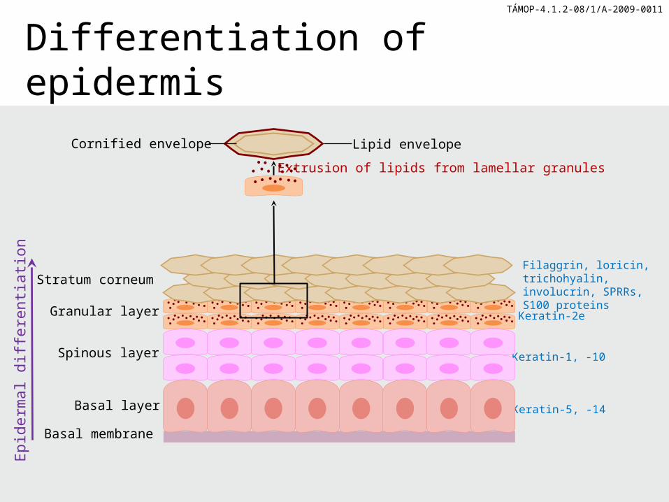
Differentiation of epidermis
Filaggrin, loricin, trichohyalin, involucrin, SPRRs, S100 proteins
Stratum corneum
Granular layer
Basal layer
Basal membrane
Keratin-2e
Keratin-1, -10
Keratin-5, -14
Epid
erm
al d
iffer
entia
tion
Spinous layer
Lipid envelopeCornified envelopeExtrusion of lipids from lamellar granules

TÁMOP-4.1.2-08/1/A-2009-0011
Mature tissue specific cells in tissue engineering• Biopsy or resection• Purification• Regaining proliferation capacity in cell culture• Re-differentiation

TÁMOP-4.1.2-08/1/A-2009-0011
Regulatory issues ICells• GLP• GMP• Permit to work on ES

TÁMOP-4.1.2-08/1/A-2009-0011
Regulatory issues IIAnimals• Permission to work on animals• UK: Home Office Licence 1986• EC 1394/2007

TÁMOP-4.1.2-08/1/A-2009-0011
Regulatory issues IIIHuman Embryonic Stem CellsEthical issues of using human embryos as sources of stem cells

TÁMOP-4.1.2-08/1/A-2009-0011
Regenerative medicine• Organ regeneration by inducing self-
regenerative biochemical and cellular processes
• Organ regeneration by addition of in vitro generated full organs or specific tissues of an organ

TÁMOP-4.1.2-08/1/A-2009-0011
Organ failure• Organ failure due to disease, accident or
aging requires full organ replacement or regeneration
• Ideally, one’s own tissues (autologue) should provide the necessary biomaterial for generation of such organs