Stem Cell Researchedoc.mdc-berlin.de/15630/1/15630oa.pdf · Lab Resource: Stem Cell Line Generation...
4
Lab Resource: Stem Cell Line Generation of human induced pluripotent stem cell line from a patient with a long QT syndrome type 2 Azra Fatima a , Dina Ivanyuk a , Stefan Herms b,c , Stefanie Heilmann-Heimbach b , Orla O'Shea d , Charlotte Chapman d , Zsuszanna Izsvák e , Martin Farr f , Jürgen Hescheler a , Tomo Šarić a, ⁎ a Center for Physiology and Pathophysiology, Institute for Neurophysiology, Medical Faculty, University of Cologne, Cologne, Germany b Institute for Human Genetics, University of Bonn, Bonn, Germany and Department of Genomics, Life & Brain Center, University of Bonn, Bonn, Germany c Research group Genomics, Medical Genetics, University Hospital Basel, Department of Biomedicine, Basel, Switzerland d UK Stem Cell Bank, Division of Advanced Therapies, National Institute for Biological Standards and Control, Hertfordshire, UK e Max-Delbrück-Center for Molecular Medicine in the Helmholtz Society, Berlin, Germany f Heart- and Diabetes Center NRW, Laboratory of the Clinic for Cardiology, University Hospital of the Ruhr-University Bochum, Bad Oeynhausen, Germany abstract article info Article history: Received 31 December 2015 Accepted 31 December 2015 Available online 6 January 2016 We report here the generation of human iPS cell line UKKi009-A from dermal fibroblasts of a patient carrying heterozygous mutation c.3035-3045delTCCCTCGATGC, p.Leu1012Pro (fs*55) in KCNH2 gene leading to long QT syndrome type 2 (LQT2). We used the Sleeping Beauty transposon-based plasmids expressing OSKM along with microRNAs 307/367 to reprogram the fibroblasts. The iPS cells possess pluripotent stem cell characteristics and differentiate to cell lineages of all three germ layers. This cell line can serve as a source for in vitro modeling of LQT2. This cell line is distributed by the European Collection of Authenticated Cell Cultures (ECACC). © 2016 The Authors. Published by Elsevier B.V. This is an open access article under the CC BY-NC-ND license (http://creativecommons.org/licenses/by-nc-nd/4.0/). Resource Table Name of stem cell line UKKi009-A Alternative name NP0011-8 Institution Institute for Neurophysiology, Medical Faculty, University of Cologne, Germany Person who created resource Dina Ivanyuk Contact person and email Tomo Saric, [email protected] Date archived/stock date 29th August 2014 Origin Dermal fibroblasts Sex Female Disease status Long QT syndrome type 2 (LQT2; OMIM entry # 613688) Mutation Heterozygous mutation c.3035-3045delTCCCTCGATGC, p.Leu1012Pro (fs*55) in KCNH2 gene Type of resource iPS cell line for disease modeling, drug and toxicity testing Sub-type hiPSC, derived from dermal fibroblast, no genetic modification Key transcription factors POU5F1, SOX2, KLF4, MYC Authentication Identity was confirmed by DNA sequencing (Fig. 1C) SNP genotyping (Table 1) and STR analy- sis of six markers (Fig. 2) Other clonal lines available UKKi009-B (NP0011-19) iPS cell line; http://hpscreg.eu/cell-line/UKKi009-B Link to related literature None Information in public databases http://hpscreg.eu/cell-line/UKKi009-A Ethics Consent from the donor of the tissue has been obtained, Ethics Review Board/competent authority approval has been obtained 1. Resource details The human iPSC line was generated by reprogramming of fibroblasts cultured from skin biopsy of a 32 year old patient suffering from LQT syndrome Type 2 (LQT2). LQT syndrome is a medical condition, either genetic or acquired, described after its characteristic electrocardiogram showing a prolonged cardiac repolarization phase resulting in a long QT interval. The clinical symptoms of LQTS include palpitations, syncope, seizures leading to sudden cardiac death. The patient carried a heterozy- gous mutation in KCNH2 gene c.3035_3045delTCCCTCGATGC leading to amino acid frame shift beginning at Leucine 1012 (p.Leu1012Pro, fs*55). Fibroblasts were reprogrammed by transposition of OCT4, SOX2, KLF4 and c-MYC (OSKM) and miRNA302/367 expression cassettes mobilized by the SB100X hyperactive transposase (Grabundzija et al., 2013). The reprogramming was performed on murine embryonic fibroblast (MEFs) in the presence of valproic acid and vitamin C for 7 days after transfection. After isolation and initial expansion, the iPS cells were maintained in a feeder-free system on vitronectin-coated plates in Stem Cell Research 16 (2016) 304–307 ⁎ Corresponding author. E-mail address: [email protected] (T. Šarić). http://dx.doi.org/10.1016/j.scr.2015.12.039 1873-5061/© 2016 The Authors. Published by Elsevier B.V. This is an open access article under the CC BY-NC-ND license (http://creativecommons.org/licenses/by-nc-nd/4.0/). Contents lists available at ScienceDirect Stem Cell Research journal homepage: www.elsevier.com/locate/scr
Transcript of Stem Cell Researchedoc.mdc-berlin.de/15630/1/15630oa.pdf · Lab Resource: Stem Cell Line Generation...
Stem Cell Research 16 (2016) 304–307
Contents lists available at ScienceDirect
Stem Cell Research
j ourna l homepage: www.e lsev ie r .com/ locate /scr
Lab Resource: Stem Cell Line
Generation of human induced pluripotent stem cell line from a patientwith a long QT syndrome type 2
Azra Fatima a, Dina Ivanyuk a, Stefan Herms b,c, Stefanie Heilmann-Heimbach b, Orla O'Shea d,Charlotte Chapman d, Zsuszanna Izsvák e, Martin Farr f, Jürgen Hescheler a, Tomo Šarić a,⁎a Center for Physiology and Pathophysiology, Institute for Neurophysiology, Medical Faculty, University of Cologne, Cologne, Germanyb Institute for Human Genetics, University of Bonn, Bonn, Germany and Department of Genomics, Life & Brain Center, University of Bonn, Bonn, Germanyc Research group Genomics, Medical Genetics, University Hospital Basel, Department of Biomedicine, Basel, Switzerlandd UK Stem Cell Bank, Division of Advanced Therapies, National Institute for Biological Standards and Control, Hertfordshire, UKe Max-Delbrück-Center for Molecular Medicine in the Helmholtz Society, Berlin, Germanyf Heart- and Diabetes Center NRW, Laboratory of the Clinic for Cardiology, University Hospital of the Ruhr-University Bochum, Bad Oeynhausen, Germany
NAIn
PCDOSD
M
T
S
KA
⁎ Corresponding author.E-mail address: [email protected] (T. Šarić).
http://dx.doi.org/10.1016/j.scr.2015.12.0391873-5061/© 2016 The Authors. Published by Elsevier B.V
a b s t r a c t
a r t i c l e i n f oArticle history:Received 31 December 2015Accepted 31 December 2015Available online 6 January 2016
We report here the generation of human iPS cell line UKKi009-A from dermal fibroblasts of a patient carryingheterozygous mutation c.3035-3045delTCCCTCGATGC, p.Leu1012Pro (fs*55) in KCNH2 gene leading to long QTsyndrome type 2 (LQT2). We used the Sleeping Beauty transposon-based plasmids expressing OSKM alongwith microRNAs 307/367 to reprogram the fibroblasts. The iPS cells possess pluripotent stem cell characteristicsand differentiate to cell lineages of all three germ layers. This cell line can serve as a source for in vitromodeling ofLQT2. This cell line is distributed by the European Collection of Authenticated Cell Cultures (ECACC).
© 2016 The Authors. Published by Elsevier B.V. This is an open access article under the CC BY-NC-ND license(http://creativecommons.org/licenses/by-nc-nd/4.0/).
O
LiIn
Resource Table
E
ame of stem cell line UKKi009-A lternative name NP0011-8 stitution Institute for Neurophysiology, Medical Faculty,University of Cologne, Germany
erson who created resource Dina Ivanyuk ontact person and email Tomo Saric, [email protected] ate archived/stock date 29th August 2014 rigin Dermal fibroblasts ex Female isease status Long QT syndrome type 2 (LQT2; OMIM entry #613688)
utation Heterozygous mutationc.3035-3045delTCCCTCGATGC, p.Leu1012Pro(fs*55) in KCNH2 gene
ype of resource
iPS cell line for disease modeling, drug and toxicitytestingub-type
hiPSC, derived from dermal fibroblast, no geneticmodificationey transcription factors
POU5F1, SOX2, KLF4, MYC uthentication Identity was confirmed by DNA sequencing(Fig. 1C) SNP genotyping (Table 1) and STR analy-sis of six markers (Fig. 2)
. This is an open access article under the
ther clonal lines available
CC BY-NC-ND license (http://
UKKi009-B (NP0011-19) iPS cell line;http://hpscreg.eu/cell-line/UKKi009-B
nk to related literature
None formation in publicdatabaseshttp://hpscreg.eu/cell-line/UKKi009-A
thics
Consent from the donor of the tissue has beenobtained, Ethics Review Board/competent authorityapproval has been obtained1. Resource details
The human iPSC linewas generated by reprogramming of fibroblastscultured from skin biopsy of a 32 year old patient suffering from LQTsyndrome Type 2 (LQT2). LQT syndrome is a medical condition, eithergenetic or acquired, described after its characteristic electrocardiogramshowing a prolonged cardiac repolarization phase resulting in a long QTinterval. The clinical symptoms of LQTS include palpitations, syncope,seizures leading to sudden cardiac death. The patient carried a heterozy-gousmutation in KCNH2gene c.3035_3045delTCCCTCGATGC leading toamino acid frame shift beginning at Leucine 1012 (p.Leu1012Pro, fs*55).Fibroblasts were reprogrammed by transposition of OCT4, SOX2, KLF4and c-MYC (OSKM) andmiRNA302/367 expression cassettes mobilizedby the SB100X hyperactive transposase (Grabundzija et al., 2013). Thereprogramming was performed on murine embryonic fibroblast(MEFs) in the presence of valproic acid and vitamin C for 7 days aftertransfection. After isolation and initial expansion, the iPS cells weremaintained in a feeder-free system on vitronectin-coated plates in
creativecommons.org/licenses/by-nc-nd/4.0/).
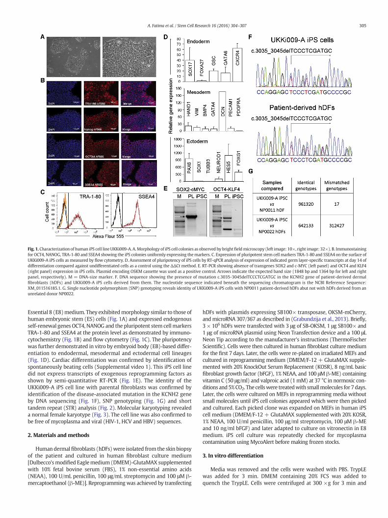
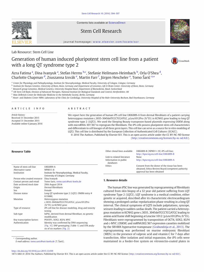
Fig. 1. Characterization of human iPS cell lineUKKi009-A. A.Morphology of iPS cell colonies as observed by brightfieldmicroscopy (left image: 10×, right image: 32×). B. Immunostainingfor OCT4, NANOG, TRA-1-80 and SSEA4 showing the iPS colonies uniformly expressing themarkers. C. Expression of pluripotent stem cell markers TRA-1-80 and SSEA4 on the surface ofUKKi009-A iPS cells as measured by flow cytometry. D. Assessment of pluripotency of iPS cells by RT-qPCR analysis of expression of indicated germ layer-specific transcripts at day 14 ofdifferentiation compared against undifferentiated cells as a control using the ΔΔCt method. E. RT-PCR showing absence of transgenes SOX2 and c-MYC (left panel) and OCT4 and KLF4(right panel) expression in iPS cells. Plasmid encoding OSKM cassette was used as a positive control. Arrows indicate the expected band size (1848 bp and 1364 bp for left and rightpanel, respectively). M = DNA-size marker. F. DNA sequence showing the presence of mutation c.3035-3045delTCCCTCGATGC in the KCNH2 gene of patient-derived dermalfibroblasts (hDFs) and UKKi009-A iPS cells derived from them. The nucleotide sequence indicated beneath the sequencing chromatogram is the NCBI Reference Sequence:XM_011516185.1. G. Single nucleotide polymorphism (SNP) genotyping reveals identity of UKKi009-A iPS cells with NP0011 patient-derived hDFs abut not with hDFs derived from anunrelated donor NP0022.
305A. Fatima et al. / Stem Cell Research 16 (2016) 304–307
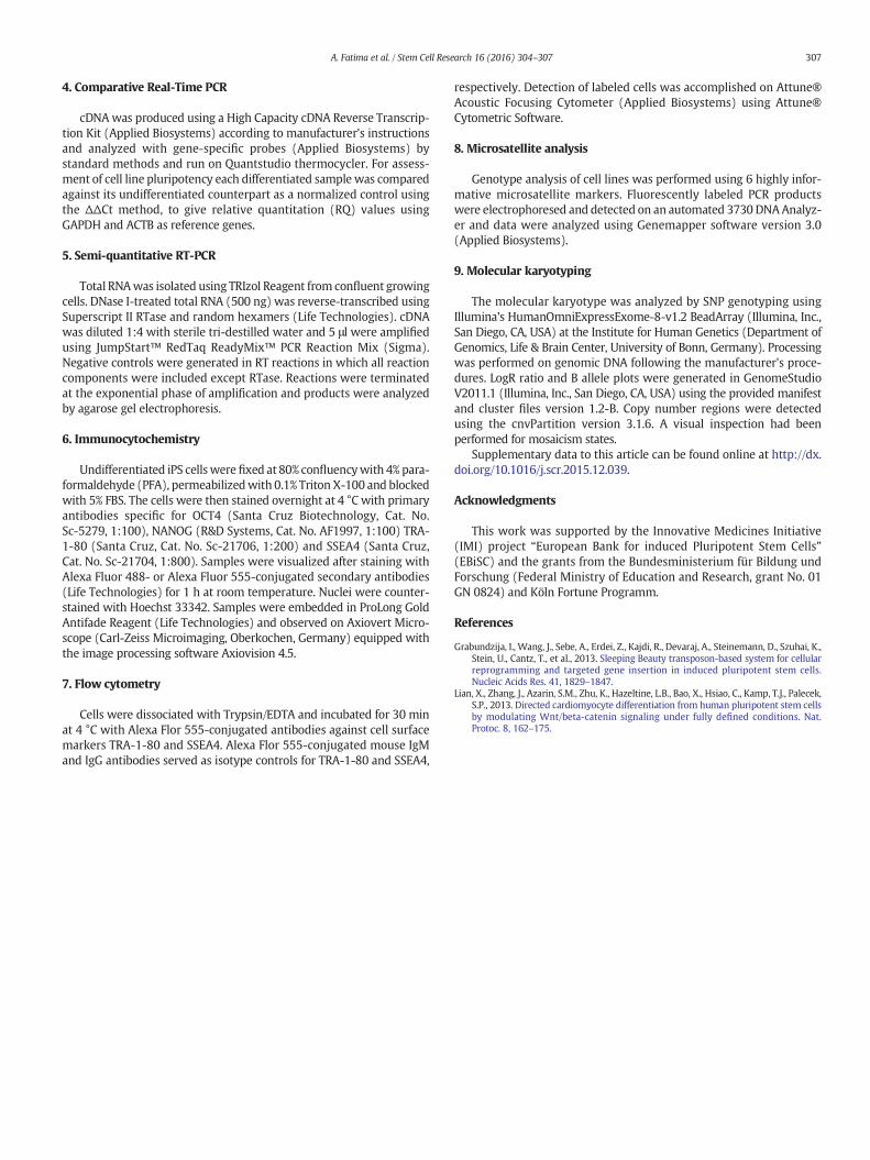
Essential 8 (E8)medium. They exhibitedmorphology similar to those ofhuman embryonic stem (ES) cells (Fig. 1A) and expressed endogenousself-renewal genes OCT4, NANOG and the pluripotent stem cell markersTRA-1-80 and SSEA4 at the protein level as demonstrated by immuno-cytochemistry (Fig. 1B) and flow cytometry (Fig. 1C). The pluripotencywas further demonstrated in vitro by embryoid body (EB)-based differ-entiation to endodermal, mesodermal and ectodermal cell lineages(Fig. 1D). Cardiac differentiation was confirmed by identification ofspontaneously beating cells (Supplemental video 1). This iPS cell linedid not express transcripts of exogenous reprogramming factors asshown by semi-quantitative RT-PCR (Fig. 1E). The identity of theUKKi009-A iPS cell line with parental fibroblasts was confirmed byidentification of the disease-associated mutation in the KCNH2 geneby DNA sequencing (Fig. 1F), SNP genotyping (Fig. 1G) and shorttandem repeat (STR) analysis (Fig. 2). Molecular karyotyping revealeda normal female karyotype (Fig. 3). The cell line was also confirmed tobe free of mycoplasma and viral (HIV-1, HCV and HBV) sequences.
2. Materials and methods
Human dermal fibroblasts (hDFs)were isolated from the skin biopsyof the patient and cultured in human fibroblast culture medium[Dulbecco'smodified Eaglemedium (DMEM)-GlutaMAX supplementedwith 10% fetal bovine serum (FBS), 1% non-essential amino acids(NEAA), 100 U/mL penicillin, 100 μg/mL streptomycin and 100 μM β-mercaptoethanol (β-ME)]. Reprogramming was achieved by transfecting
hDFs with plasmids expressing SB100× transposase, OKSM-mCherry,andmicroRNA 307/367 as described in (Grabundzija et al., 2013). Briefly,3 × 106 hDFs were transfected with 3 μg of SB-OKSM, 1 μg SB100× and1 μg of microRNA plasmid using Neon Transfection device and a 100 μLNeon Tip according to the manufacturer's instructions (ThermoFischerScientific). Cells were then cultured in human fibroblast culture mediumfor the first 7 days. Later, the cells were re-plated on irradiated MEFs andcultured in reprogramming medium (DMEM/F-12 + GlutaMAX supple-mented with 20% KnockOut Serum Replacement (KOSR), 8 ng/mL basicfibroblast growth factor (bFGF), 1% NEAA, and 100 μM β-ME) containingvitamin C (50 μg/ml) and valproic acid (1 mM) at 37 °C in normoxic con-ditions and5%CO2. The cellswere treatedwith smallmolecules for 7 days.Later, the cells were cultured on MEFs in reprogramming media withoutsmall molecules until iPS cell colonies appeared which were then pickedand cultured. Each picked clone was expanded on MEFs in human iPScell medium (DMEM/F-12 + GlutaMAX supplemented with 20% KOSR,1% NEAA, 100 U/ml penicillin, 100 μg/ml streptomycin, 100 μM β-MEand 10 ng/ml bFGF) and later adapted to culture on vitronectin in E8medium. iPS cell culture was repeatedly checked for mycoplasmacontamination using MycoAlert before making frozen stocks.
3. In vitro differentiation
Media was removed and the cells were washed with PBS. TrypLEwas added for 3 min. DMEM containing 20% FCS was added toquench the TrypLE. Cells were centrifuged at 300 ×g for 3 min and
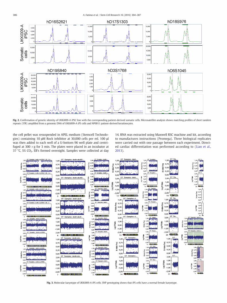
Fig. 2. Confirmation of genetic identity of UKKi009-A iPSC line with the corresponding patient-derived somatic cells. Microsatellite analysis shows matching profiles of short tandemrepeats (STR) amplified from a genomic DNA of UKKi009-A iPS cells and NP0011 patient-derived keratinocytes.
306 A. Fatima et al. / Stem Cell Research 16 (2016) 304–307
the cell pellet was resuspended in APEL medium (Stemcell Technolo-gies) containing 10 μM Rock inhibitor at 30,000 cells per ml. 100 μlwas then added to each well of a U-bottom 96 well plate and centri-fuged at 300 ×g for 3 min. The plates were placed in an incubator at37 °C, 5% CO2. EB's formed overnight. Samples were collected at day
Fig. 3.Molecular karyotype of UKKi009-A iPS cells. SNP genoty
14. RNA was extracted using Maxwell RSC machine and kit, accordingto manufactures instructions (Promega). Three biological replicateswere carried out with one passage between each experiment. Direct-ed cardiac differentiation was performed according to (Lian et al.,2013).
ping shows that iPS cells have a normal female karyotype.
307A. Fatima et al. / Stem Cell Research 16 (2016) 304–307
4. Comparative Real-Time PCR
cDNAwas produced using a High Capacity cDNA Reverse Transcrip-tion Kit (Applied Biosystems) according to manufacturer's instructionsand analyzed with gene-specific probes (Applied Biosystems) bystandard methods and run on Quantstudio thermocycler. For assess-ment of cell line pluripotency each differentiated sample was comparedagainst its undifferentiated counterpart as a normalized control usingthe ΔΔCt method, to give relative quantitation (RQ) values usingGAPDH and ACTB as reference genes.
5. Semi-quantitative RT-PCR
Total RNAwas isolated using TRIzol Reagent from confluent growingcells. DNase I-treated total RNA (500 ng) was reverse-transcribed usingSuperscript II RTase and random hexamers (Life Technologies). cDNAwas diluted 1:4 with sterile tri-destilled water and 5 μl were amplifiedusing JumpStart™ RedTaq ReadyMix™ PCR Reaction Mix (Sigma).Negative controls were generated in RT reactions in which all reactioncomponents were included except RTase. Reactions were terminatedat the exponential phase of amplification and products were analyzedby agarose gel electrophoresis.
6. Immunocytochemistry
Undifferentiated iPS cellswere fixed at 80% confluencywith 4% para-formaldehyde (PFA), permeabilizedwith 0.1% Triton X-100 and blockedwith 5% FBS. The cells were then stained overnight at 4 °C with primaryantibodies specific for OCT4 (Santa Cruz Biotechnology, Cat. No.Sc-5279, 1:100), NANOG (R&D Systems, Cat. No. AF1997, 1:100) TRA-1-80 (Santa Cruz, Cat. No. Sc-21706, 1:200) and SSEA4 (Santa Cruz,Cat. No. Sc-21704, 1:800). Samples were visualized after staining withAlexa Fluor 488- or Alexa Fluor 555-conjugated secondary antibodies(Life Technologies) for 1 h at room temperature. Nuclei were counter-stained with Hoechst 33342. Samples were embedded in ProLong GoldAntifade Reagent (Life Technologies) and observed on Axiovert Micro-scope (Carl-Zeiss Microimaging, Oberkochen, Germany) equipped withthe image processing software Axiovision 4.5.
7. Flow cytometry
Cells were dissociated with Trypsin/EDTA and incubated for 30 minat 4 °C with Alexa Flor 555-conjugated antibodies against cell surfacemarkers TRA-1-80 and SSEA4. Alexa Flor 555-conjugated mouse IgMand IgG antibodies served as isotype controls for TRA-1-80 and SSEA4,
respectively. Detection of labeled cells was accomplished on Attune®Acoustic Focusing Cytometer (Applied Biosystems) using Attune®Cytometric Software.
8. Microsatellite analysis
Genotype analysis of cell lines was performed using 6 highly infor-mative microsatellite markers. Fluorescently labeled PCR productswere electrophoresed and detected on an automated 3730 DNAAnalyz-er and data were analyzed using Genemapper software version 3.0(Applied Biosystems).
9. Molecular karyotyping
The molecular karyotype was analyzed by SNP genotyping usingIllumina's HumanOmniExpressExome-8-v1.2 BeadArray (Illumina, Inc.,San Diego, CA, USA) at the Institute for Human Genetics (Department ofGenomics, Life & Brain Center, University of Bonn, Germany). Processingwas performed on genomic DNA following the manufacturer's proce-dures. LogR ratio and B allele plots were generated in GenomeStudioV2011.1 (Illumina, Inc., San Diego, CA, USA) using the provided manifestand cluster files version 1.2-B. Copy number regions were detectedusing the cnvPartition version 3.1.6. A visual inspection had beenperformed for mosaicism states.
Supplementary data to this article can be found online at http://dx.doi.org/10.1016/j.scr.2015.12.039.
Acknowledgments
This work was supported by the Innovative Medicines Initiative(IMI) project “European Bank for induced Pluripotent Stem Cells”(EBiSC) and the grants from the Bundesministerium für Bildung undForschung (Federal Ministry of Education and Research, grant No. 01GN 0824) and Köln Fortune Programm.
References
Grabundzija, I., Wang, J., Sebe, A., Erdei, Z., Kajdi, R., Devaraj, A., Steinemann, D., Szuhai, K.,Stein, U., Cantz, T., et al., 2013. Sleeping Beauty transposon-based system for cellularreprogramming and targeted gene insertion in induced pluripotent stem cells.Nucleic Acids Res. 41, 1829–1847.
Lian, X., Zhang, J., Azarin, S.M., Zhu, K., Hazeltine, L.B., Bao, X., Hsiao, C., Kamp, T.J., Palecek,S.P., 2013. Directed cardiomyocyte differentiation from human pluripotent stem cellsby modulating Wnt/beta-catenin signaling under fully defined conditions. Nat.Protoc. 8, 162–175.